A suite of solid-state NMR experiments to utilize orphaned
magnetization for assignment of proteins using parallel high and low
gamma detection
A. Gallo
a,1, W.T. Franks
a,b,1, J.R. Lewandowski
a,⇑ aDepartment of Chemistry, University of Warwick, Gibbet Hill Road, CV4 7AL Coventry, UK
b
Department of Physics, University of Warwick, Gibbet Hill Road, CV4 7AL Coventry, UK
a r t i c l e i n f o
Article history:
Received 22 May 2019 Revised 3 July 2019 Accepted 5 July 2019 Available online 6 July 2019
Keywords:
Multiple receivers Solid-state NMR Protein
Discarded magnetization pathways Low–c
Fast magic angle spinning
a b s t r a c t
We present a suite of two-receiver solid-state NMR experiments for backbone and side chain resonance assignment. The experiments rely on either dipolar coupling or scalar coupling for polarization transfer and are devised to acquire a1H–detected 3D experiment AND a nested13C–detected 2D from a shared excitation pulse. In order to compensate for the lower sensitivity of detection on13C nucleus, 2D rows are signal averaged during 3D planes. The 3D dual receiver experiments do not suffer from any apprecia-ble signal loss compared to their single receiver versions and require no extra optimization. The resulting data is higher in information content with no additional experiment time. The approach is expected to become widespread as multiple receivers become standard for new NMR spectrometers.
Ó2019 The Authors. Published by Elsevier Inc. This is an open access article under the CC BY license (http:// creativecommons.org/licenses/by/4.0/).
1. Introduction
Solid state nuclear magnetic resonance (ssNMR) spectroscopy is a powerful method for characterizing the structure and dynamics [1–4]of a wide variety of protein preparations including micro-crystals [5–10], fibrils [11–17], sediments, membrane proteins [18–21]as well as both homomeric and heteromeric assemblies [22,23]. This approach is increasingly being used to non-destructively characterize the biomolecular machinery to better understand drug activity and cellular signaling at atomic resolu-tion, sometimes in nearly-natural conditions [19,24–28]. While NMR can provide unique data, the multidimensional experiments needed to study large biomolecules are generally slow and insen-sitive. In the recent years, there was a concerted effort to improve practicality and take advantage of high sensitivity of1H–detected biomolecular experiments by combatting the1H line broadening through fast spinning and/or dilution of the proton network by deuteration [23,29–36]. 1H–detected experiments have proven beneficial for spectral assignment[23,37–40], structure determina-tion[41,42], and quantification of dynamics [10,43–46]. On the other hand, the13C–detected experiments are typically not
mea-sured under conditions favorable for 1H–detected experiments, i.e. fast spinning in small rotors, due to poor sensitivity caused by the reduction of the sample size and/or the removal of a large portion of the high–
c
nuclei.Several approaches to collect data more efficiently without spe-cialized hardware have been invented over the years with the aim to both reduce the overall experimental time and to improve sen-sitivity and/or resolution. These data collection schemes can be generalized into four approaches: altered sampling, frequency encoded, relaxation optimized, and polarization optimized. In the first approach, altered sampling experiments, e.g. GFT NMR spectroscopy[47,48], Non-Uniform Sampling (NUS)[49–53]and Projection Reconstruction (PR)[54], do not sample a full, evenly-spaced rectangular grid of time points in the indirect dimensions, but rather a subset of points not necessarily tied to the rectangular grid. In the second approach, frequency encoding selectively excites either a portion of the sample by using gradients and imag-ing techniques (or an orientation-dependent term) as in simag-ingle scan NMR[55,56]or by exciting a portion of the spectrum using selec-tive pulses as in Hadamard NMR[57,58]. The third approach relies on relaxation optimized methods, which seek to minimize the time needed between sampling points by reducing1H T
1 and conse-quently the wait time between experiments either through clever spin manipulations as in BEST (Band-selective Excitation Short-Transient) and SO-FAST (band-Selective Optimized Flip-Angle
https://doi.org/10.1016/j.jmr.2019.07.006
1090-7807/Ó2019 The Authors. Published by Elsevier Inc.
This is an open access article under the CC BY license (http://creativecommons.org/licenses/by/4.0/). ⇑ Corresponding author.
E-mail address:[email protected](J.R. Lewandowski). 1 Authors have contributed equally.
Contents lists available atScienceDirect
Journal of Magnetic Resonance
Short-Transient)[59–61]or addition paramagnetic dopants to the sample[62–64]. In the fourth approach, ‘‘many-at-once” experi-ments aim to make the most of the initial polarization by collecting more than one experiment per excitation pulse. For example, time-shared experiments allow the polarization to follow multiple path-ways with two (or more) experiments being obtained during one acquisition taking advantage of either phase labelling, or chemical shift differences [65–72]. Alternatively, experiments such as DUMAS (DUal acquisition Magic Angle Spinning)[73], MAESTRO [74], MAESTOSO (Multiple Acquisitions via Sequential Transfer of Orphan Spin pOlarization) [75,76], Multiple Sequential Acquisi-tions [77], and UTOPIA (Unified Time-Optimized Interleaved Acquisition NMR) [78] yield multiple experiments with a small time penalty by acquiring different pathways sequentially with fraction of the magnetization being stored in a long-lived state dur-ing the encoddur-ing of the first pathway.
The frequency encoded approach is difficult to apply to biolog-ical MAS NMR; the single scan technique requires very strong, well-aligned and reproducible gradients, which are challenging for MAS probes, and there are typically no anisotropic couplings large enough for orientation selection [79,80]. In addition, the extent of inhomogeneous broadening typical for solid samples is too large for Hadamard encoding. However, the other three approaches are in current use.
Perhaps the most commonly employed approach is paramag-netic doping used with both deuterated and protonated protein samples in1H–detected[23,63,81]and13C–detected experiments [64]. Since doping also enhances transverse relaxation, this approach requires a balance between the extent of1HT1reduction and the overall line broadening[81]. The BEST and SO-FAST exper-iments rely on scalar-coupling based transfers that require 1H transverse relaxation times longer than typically observed even with 100 kHz spinning and thus, currently, do not provide a com-petitive edge over traditional approaches except for special cases. They could, however, become more practical as yet faster spinning frequencies become available.
The second most popular method is likely non-uniform sam-pling (NUS) [82,83] for arbitrarily high dimensional (nD with n3) data. NUS is a well-supported technology with several tools available through both commercial and non-commercial software. NUS is certainly an important tool to address resolution in large systems and can be applied to a broad range of experiments.
Finally, the polarization-optimized, ‘‘many-at-once” approach is attractive due to the ability to acquire multiple experiments at the same time. Polarization that is otherwise discarded, or orphaned, after the primary polarization transfer steps, and the long longitu-dinal relaxation times of certain nuclei enable the acquisition of multiple experiments either simultaneously[70]or sequentially; on one or two channels[77]. Experiments with same dimensional-ity can be recorded using multiple receivers, in general[84], but the large difference in sensitivity between nuclei makes such a solution impractical except in special cases,e.g.for small, isotopi-cally labelled molecules or when considering various quadrupolar nuclei with comparable sensitivity. In the present study, we pro-pose a practical solution on how one can utilize multiple receivers where the sensitivity on different channels is dramatically differ-ent. Towards this end, we use an approach similar to the UTOPIA scheme [78] but with a dual receiver set-up to acquire nested lower dimensionality spectra (here, a 2D) on a secondary channel while acquiring a higher dimensionality spectrum (here a 3D) on the primary nucleus. In contrast to a standard UTOPIA approach, the lower sensitivity of the low-
c
nucleus is compensated for by acquiring more transients per point for the 2D compared to the more sensitive1H–detected 3D, at the cost of an additional chem-ical shift dimension. Specifchem-ically, we describe a two-receiver approach to modify solid-state1H–detected 3D experiments forbackbone and sidechain assignments and to simultaneously obtain 13C–detected 2D spectra where the additional 2D is obtained
with-out disturbing the 3D polarization transfer pathway. We describe a suite of experiments (available on-line athttp://wrap.warwick.ac. uk/116953/) using a selection of both dipolar and scalar-coupling based carbon homonuclear mixing schemes.
The experiments presented here are designed to facilitate spec-tral assignment by increasing the information content with little additional setup or signal loss. The low–
c
detection complements the 3D data by providing direct evidence of the polarization path-way, generating an additional 2D spectrum involving frequencies not encoded in the parent 3D under identical experimental condi-tions, and maximizing the resolution for the directly detected low–c
nuclei. Our approach offers certain tangible advantages over the prior art. In relation to multiple receiver experiments developed primarily by Kupce[84], the reduced dimensionality of the low–c
detected experiments allows for adequate sensitivity without lengthening the experiment time for the high–c
detected experi-ment. Other orphaned-polarization methods[73–77]require the full optimization of all polarization transfers for claw-back, while the experiments presented here can produce useful information once the primary HNC transfer path is established without the need for extra transfers. It may be possible to merge more ‘‘parent” and ‘‘child” experiments using the other orphaned-polarization techniques, but that is beyond the scope of this work. Finally, the UTOPIA experiments utilized one receiver, and switched it between channels, while our approach had dedicated receivers for each detect channel. It is likely that our approach is more easily implemented, and expanded upon, in state-of-the-art spectrome-ters, while older spectrometers must adapt the UTOPIA channel switching implementation. Neither of the two implementations are integrated into the existing ‘‘MC” macro infrastructure.2. Materials and methods
2.1. Sample preparation
Uniformly1H/13C/15N labelled GB1 (T2Q mutant) was prepared as described previously[7]and doped with 4,4-dimethyl-4-silapen tane-1-sulfonic acid (DSS) as an internal standard. 0.5 mg of microcrystalline protein slurry was packed into a Bruker 0.7 mm zirconia solid-state NMR.
2.2. NMR experiments
All experiments were acquired on a Bruker Avance III spectrom-eter operating at 700.13 MHz1H frequency with two receivers run-ning Topspin 3.5 patch level 6. A Bruker three channel 0.7 mm MAS probe was tuned to1H–13C–15N, and the sample spinning was con-trolled to 100 kHz ± 3 Hz. The temperature of the sample was reg-ulated using 500 L/h gas flow with a nominal set-point of 278 K. The sample temperature is estimated to be300 K based on the difference between the water resonance and the DSS peak at 0.0 ppm[85,86].
Hard pulses were calibrated such that the1H 90°pulses were 2.0
l
s (m
1,H= 125 kHz), and both13C and 15N were either 2.5l
s (m
1= 100 kHz) or 3.6l
s (m
1= 70 kHz). Low-power (m
1,H10 kHz) 1H WALTZ-64 decoupling was applied during evolution,115 ppm), 320ms (bandwidth of 10.5 kHz or 60 ppm), and 760ms (bandwidth of 5.3 kHz or30 ppm)[89]. The carbon fre-quency was altered by changing the spectrometer offset frefre-quency using pre-defined constants. To avoid phase accumulation the fre-quency was changed either during theZ-filter period immediately prior to a spin-lock, or pulses were included that compensate for the phase accumulation. Each1H and 13C FID was acquired for 30 ms with a spectral width of 20 ppm and 400 ppm respectively. A relaxation delay of 2 s was used for all experiments. The States-TPPI method was employed for quadrature detection in the indi-rect dimensions[90].
1H–13C cross-polarization (CP) with average
m
1,H110 kHz and a linear 15% ramp (85%-100%, from101.5 to 119.5 kHz) and a zero-quantum (ZQ) match condition at
m
1,C10 kHz was used for carbon excitation. The contact time was 350ms and the carrier was set to 55 ppm for the initial1H–13CaCP carbon excitation, the contact time was 2 ms and the carrier was set to 40 ppm for 1H–13CaliCP, and finally, the contact time was 2.25 ms and thecar-rier was set to 175 ppm for the 1H–13CO CP.13C–15N selective transfer was optimized by 2-variable array search over a variety of possible match conditions. The optimum transfer for SPECIFIC CP [91] was observed for
m
1,N55 kHz andm
1,C45 kHz (with an 81–99% tangent ramp, from 40.5 to 49.5 kHz) with no1H decou-pling applied. The15N–1H CP was performed with a 600ms contact pulse with averagem
1,H85 kHz with a linear 15% ramp (85%-100%, from78 to 92 kHz) and a double-quantum (DQ) match con-dition atm
1,N15 kHz. The13C–13C RFDR[92]mixing was used with xy-8 phase scheme for a total of 96 rotor periods (960ms) with no frequency changes. The13CO–13Caand13Ca–13CO DREAM [93]mixing used a ±10% tangent ramp with5 ms mixing wherem
1,Cwas optimized at45 kHz and the offset was set to the source frequency (CO and Carespectively). For the13Ca–13CbDREAM, thecarrier frequency was set to 10,000 Hz from the Ca resonance (approx. 150 ppm) to avoid the preferred 13CO–13Ca transfer. The DREAM transfer was 6 ms for 13Ca–13Cb transfer, and 7 ms for13CO–13Caand13Ca–13CO. TheJ–coupling delay
s
for13CO–13Ca transfer was set to 3.0 ms for the period when Cais transverse and 3.5 ms for the period when CO is transverse, whereJCO-Ca is 55 Hz (1/(4J) = 4.7 ms). The J–coupling delays
for the half-transfer between13Ca–13Cbwas set to 3.6 ms whereJC
a
-Cbis 35 Hz (1/(4J) = 7.2 ms; 1/(8J) = 3.6 ms). For the TOCSY [94] experiments, the DIPSI-3[95]scheme was used with a 16.3 ms mixing period. For narrow-band (aliphatic) transfer, the offset was 45 ppm, and the applied field was10 kHz. For broad-band (to both carbonyl and aliphatic carbons) transfer the offset was set to 175 ppm with a field of25 kHz.
Instructions on setting up multiple receiver experiments for Bruker instruments (Topspin 3.5pl6) can be found in the supple-mentary informationand are also found in the header of the pro-vided Bruker pulse sequence files. The pulse sequences can be downloaded from http://wrap.warwick.ac.uk/116953/. The raw spectra are available online at http://wrap.warwick.ac.uk/ 120508/.
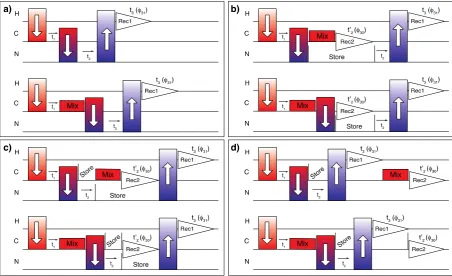
3. Pulse sequence design
[image:3.595.74.527.428.704.2]The ability to acquire high resolution1H–detected experiments has precipitated the need for triple-resonance experiments for spectral assignment. These experiments are usually based on solu-tion NMR triple-resonance assignment protocols, but there are substantive differences in the types and mechanisms of mixing which are available in solid state NMR that will be addressed in detail in later sections. It is beyond the scope of this work to compile and modify all possible proton–detected experiments.
Fig. 1.Generalized pulse sequences for embedded acquisition. Panel (a) shows the polarization transfer pathway for sequential assignment commonly used with a single1H receiver. Panels (b–d) show a selection of dual receiver configurations with an added carbon detection period. The experiments yield a1H–detected 3D C(c)NH spectrum and a 13
However, a general discussion regarding the protocol to modify 3D sequences into embedded [2D, 3D] multiple receiver experiments is warranted. To be consistent with the terminology used in the Topspin software, the higher dimensional experiment, usually acquired on the higher frequency channel (1H), is referred to as the parent 3D experiment and the lower dimensional, 2D low-
c
detected (13C) experiment is referred to as the child.A typical1H–detected triple-resonance backbone assignment experiments consist of a carbon excitation step (1H–13C CP), a13C chemical shift evolution period, possibly a carbon mixing period, followed by a13C–15N transfer, an15N chemical shift evolution per-iod, transfer from nitrogen to hydrogen (1H–15N CP), and finally acquisition on the 1H channel (Fig. 1a). In real life applications polarization transfer is rarely >50% efficient. Some signal is left
behind, or orphaned, at every transfer step during the experiment. Since we wish to observe the orphaned signal directly, an acquisi-tion can be added after any heteronuclear transfer, while the polar-ization on the primary path is preserved for later. We have chosen to observe13C since it has a higher gyromagnetic ratio than15N, and it has a higher information content due to more extensive net-work of bonded13C nuclei. In the experiments described here, the leftover carbon signal is time and phase encoded during the 3D experiment. In order to generate 2D13C–13C spectrum various car-bon mixing schemes are included.
[image:4.595.114.469.235.697.2]The13C acquisition period can be added in several different con-figurations as shown inFig. 1b–d. The acquisition on the two recei-vers could take place concurrently, but doing so restricts the ability to apply decoupling,e.g.1H decoupling could not be applied during
Fig. 2.Two-receiver 2D, 3D hC[C, NH] pulse sequences with longitudinal13 C–13
C RFDR mixing. The experiments simultaneously collect a 2D (13 C–13
C) and a 3D (13 C–15
N–1 H) spectrum. In (a) the13
C direct detection occurs between the13 C–15
N CP step and the1
H detection period. For (b) the13
C detection period is moved after the1
H detection to avoid any effects on the 3D. For (c), the 3D will also encode the sidechain resonance frequencies since the13
C–13
C mixing occurs before the13 C–15
N CP step. Narrow and broad black rectangles represent 90°and 180°hard pulses, respectively. When not shown, the phases of the pulses are x. The phase cycling is as follows: (a):/1= (x*2, x*2),/2= x, /6= (x*4, x*4),/8= (y, y),/10= (y, y, x, x),/16= ( y, y, y, y, y, y, y, y),/17= y*2, x*2), receiver 1:/31= (y, y, x, x, y, y, x, x) and receiver 2:/30= (x, x, x, x, y, y, y, y). States-TPPI is employed on/2for the shared13
C dimension of all experiments; on/7for (a) and (b), and/6for (c) for the15
13C acquisition. In the configuration presented inFig. 1b,13C
acqui-sition is placed directly after the carbon to nitrogen CP while the nitrogen signal is stored on the Z axis; the nitrogen is frequency labelled after the carbon acquisition. This arrangement is designed to limit the signal loss from spin lattice relaxation for both path-ways since the15NT
1times are usually on the order of 10–30 s, while the13C T
1 times are typically shorter, especially for side-chain carbons.Fig. 1c shows an experiment where the order of car-bon detection and nitrogen evolution is reversed fromFig. 1b. This arrangement was found to be slightly less sensitive particularly for experiments storing aliphatic carbon signal. Hence the experi-ments reported in this paper are primarily in the 1b configuration. Finally, the orphaned carbon signal can be stored until after the 3D is finished (Fig. 1d), so that the only alteration to the 3D is an addi-tional short13C storage pulse after13C–15N CP. This arrangement may be necessary if the homonuclear mixing and/or acquisition compromise the intensity of the 3D. In non-ideal cases, carbon-mixing schemes may also recouple the heteronuclear dipole when applied, which is detrimental to the overall 3D efficiency.
The modifications needed to adapt existing pulse sequences into an embedded multiple receiver experiment are straightfor-ward (Fig. 1, seeSI for the pulse programs in the Bruker format and instructions). The receiver phase for the embedded (child) experiment must be determined separately from the primary experiment. For example, the receiver phase of the ‘‘child” experi-ment is not altered by any change to the spin-lock phases during the 13C–15N CP, while the ‘‘parent” receiver phase must follow the changes to the spin lock phases on both channels. The phase cycling of the pulses after the13C–15N CP step on the parent path-way are on an independent pathpath-way to the ‘‘child” acquisition and cannot affect the ‘‘child” (which may have already been observed). To illustrate, in the experiment shown inFig. 2a, changing/5does not affect the13C receiver phase, since there is no change to the
coherence order for the orphaned spins, but such a change is affected in the1H receiver. Since a 2D13C–13C correlation spectrum is the desired child experiment, the 15N dimension must be the inner loop. The inner (fast) loop of the 3D is used to collect one row of the child while a full plane of the parent experiment is being collected. The number of coadded transients for the child 2D is the number of transients multiplied by the number of rows in the unshared chemical shift evolution period, i.e. number of transients in 2D = (number of transients in 3D) * (number of rows = real + imaginary data in the indirect dimension),e.g.16 transients * 38 TD1 = 608 transients for the child 2D. The parent data is written to disk after the specified number of scans, while the child is only written after the completion of a 3D plane (at the time of the child indirect dimension evolution; the outer loop). Such construction ensures that much larger number of transients is recorded for the13C–detected 2D child experiment compared to the 3D parent experiment thus partially compensating for the lower sensitivity of13C detection. Storage and recall pulses are added as needed.
Since the dynamics can differ greatly between proteins, and even different segments within a protein, different mixing schemes are better for different samples. Consequently, we have created and tested experiments with dipolar coupling based mixing schemes including RFDR [92] and DREAM [93] and J–coupling based schemes including COSY [37,38,94]and TOCSY [42]. As a general rule, dipolar transfers are better for sites with short coher-ence lifetimes, while scalar-coupling based schemes are better for sites with long coherence lifetimes.
4. Pulse sequences
[image:5.595.74.528.436.722.2]At moderate (<20 kHz) spinning frequencies proton driven spin diffusion (PDSD) and dipolar assisted rotational recoupling (DARR) are commonly employed for longitudinal 13C–13C mixing. The
Fig. 3.Cube representation of the parent 3D13Cb/13Ca-15N-1H and child 2D13Cb-13Cacorrelation spectra on crystalline [U–13C,15N]GB1 acquired with (a) RFDR, (b) DREAM, and (c) COSY coupling based homonuclear13
C–13
prevalence of these mixing schemes is in large part due to the easy set up rather than their superior properties. The mechanism of transfer in these experiments is deceptively complicated and is dependent on the spinning rate and magnetic field. At spinning fre-quencies >60 kHz and high magnetic fields proton driven spin dif-fusion is greatly attenuated[95]leading to poor performance for PDSD and DARR. It is thus challenging to select for one-bond 13C–13C polarization transfer exclusively and efficiently. Improved
spin diffusion schemes for application at fast spinning frequencies have been developed but not widely adapted [96]. After spin-diffusion based13C–13C longitudinal mixing, RFDR is probably the next easiest to calibrate longitudinal mixing scheme that works at very fast spinning. RFDR will be used throughout as an example of a longitudinal mixing scheme, though any other longitudinal mixing scheme can be substituted in its place in the described experiments.13C–13C RFDR (and13C–13C TOCSY) are better options for experiments where broad-band 13C–13C mixing is desirable. The DREAM and COSY schemes are more suitable for selective 13C–13C transfer but are typically more challenging to optimize
compared to RFDR or TOCSY.
We inserted an RFDR13C–13C mixing period into a 3D hCNH fin-gerprint experiment following the heteronuclear polarization transfer to minimize the disturbance to the 3D experiment. Fig. 2a shows a pulse sequence for a [2D, 3D] hC[C, NH] experiment where the carbon mixing and acquisition is before the nitrogen evolution, as in top ofFig. 1b (SIfile war.hCA[C, NH]_RFDR_AFTER).
An alternative pulse sequence (Fig. 2b) moves the13C acquisition after the 3D experiment is finished avoiding any disturbance to the 3D. The additional storage pulses are not usually phase-cycled but their presence means that a different pulse must be used for indirect dimension quadrature detection. This class of experiments (RFDR mixing) produces a 3D experiment with a ser-ies of nominally one-bond polarization transfers where a13C–13C 2D is acquired at the same time, as shown inFig. 3a. The 3D hCANH obtained on GB1 using this approach is of high quality and is com-parable to the traditionally acquired single receiver experiment in terms of sensitivity and resolution. The biggest difference between the multiple receiver (hCA[C, NH]_RFDR_AFTER) and single recei-ver (hCANH) is that the spectral width for the indirect carbon dimension must be increased to fit the full aliphatic region instead of the CA region.
DREAM[93]can be used for transverse13C–13C mixing using the pulse programs shown schematically inFig. 4. These experi-ments employ a soft-hard
p
-pulse pair to provide frequency dis-crimination to ensure the correct polarization pathway is chosen during optimization, with the bonus feature that the13C–13C sca-lar couplings are decoupled. A Z-filter is implemented before the DREAM mixing to improve the quadrature detection, provide a time for frequency switching if needed, and to ensure a constant duty cycle for the entire experiment. In this, and all following pulse schemes, we focused our effort on experiments with 13C–13C mixing before the13C-15N transfer since these carry the
[image:6.595.110.479.353.716.2]relevant backbone walk information for solution NMR-style assignments[97,98].
The h(CBCA)NH 3D with embedded 2D is shown in Fig. 3b. Because DREAM is double quantum mixing, the Cb peaks in the 3D, and the cross peaks of the 2D have the opposite sign
[image:7.595.101.501.144.712.2]of their transfer partners. This is the same as in solution NMR HNCACB experiments and can be a helpful feature during the backbone walk. In the 13C–13C 2D, there are positive cross-peaks in the Thr Ca to C
c
2 region, which indicates that some 2-bond, relayed transfers occurred. The digital resolution in theFig. 5.Pulse sequence diagrams for [2D, 3D] assignment experiments with COSY mixing. Modifying the 3D COSY–based experiments for assignments results in (a) hcaCBca[C, NH], (b) hCAco[C, NH], and (c) hCOca[C, NH]. Rounded pulses represent 180°selective shaped pulses. Unless otherwise noted, the phase is x. Forathe phase cycling is/0= (y, y),/2= (y, y, y, y),/5= (y*4, y*4),/6= (x*8, x*8),/7= y,/12= y,/13= y,/14= x, receiver 1 is/31= (x, x, x, x, x, x, x, x, x, x, x, x, x, x, x, x) and receiver 2 is /30= (x, x, x, x). Forbandcthe phase cycling is/0= y/1= (x, x),/3= y,/6= (x*8, x*8),/7= y,/13= (x, x, y, y),/18= (x*4, y*4); receiver 1 is/31= ( y, y, y, y, y, y,
indirect 13C dimension was reduced for the experiment in 3b compared to 3a, which explains some of the apparent change in linewidth between the two experiments. The sample had also partially dehydrated between the collection of the datasets. In the 13C–13C 2D DREAM, the signals of the Ca nuclei are more intense than the other aliphatic carbons. This difference in signal intensity might be due to the selectivity of the DREAM mixing within the full aliphatic bandwidth, or to the reduced T1q of the sidechains during the 13Ca–15N CP, and DREAM spin locks. Still, the DREAM spectrum contains information about the side-chain contained in the 2D that is lost in the 3D dataset. The13 -Ca–13CO and 13CO–13Ca2D experiments are of high quality, and show a very efficient DREAM transfer, and are much less chal-lenging to optimize compared to 13Cb–13Ca transfer. The 13 -CO–13Ca and 13Ca–13CO DREAM transfer efficiencies (50%) are higher than RFDR (15%).
The scalar coupling based sequences, COSY (hCOcaNH, hCA-coNH, hcaCBcaNH, hcaCBcacoNH) or TOCSY (hCCaNH TOCSY, and hCCONH TOCSY), are also amenable to use with parallel acquisi-tion. The COSY–based pulse sequences, shown inFig. 5, utilize free precession and selective echoes to transfer the polarization via the scalar coupling through the chemical bonds, in close analogy to solution NMR experiments. TOCSY experiments are useful to iden-tify the type of amino acid sidechain when the Cbresonance is not sufficient. This information is needed when there are many long chain amino acids with nearly degenerate chemical shifts. TOCSY correlations can be obtained using the DIPSI3 pulse sequence as shown inFig. 6 [99,100].
The COSY–based sequence for backbone correlations (Fig. 5a) produced a high-quality 3D, but the13C–13C 2D (Fig. 3c) had some-what poor sensitivity. As is common practice in solution NMR, the scalar coupling was half-evolved, to produce both13Ca and13Cb
resonances (where they have opposite signs). In this case the 3D was of sufficient quality for assignments, but the13C–13C 2D was not. The possible explanation for the poor quality of13C–13C 2D might be sample dehydration or too long J–coupling evolution periods based on the typical coupling magnitudes without taking relaxation into account, or insufficient decoupling during 13C detection, but the true reason for the discrepancy is not known.
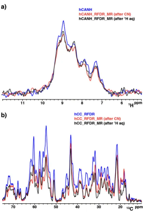
The primary consideration for the application of multiple recei-ver experiments, apart from hardware availability, is the potential loss in sensitivity of the 3D. We find that the first row of the single receiver 3D experiment is <10% higher than when in a dual receiver configuration, as shown inFig. 7a., but we note that there is a vari-ance of3% in consecutively acquired first row experiments due to noise and water suppression. We also find that the placement of the13C acquisition is not a large factor in the 3D’s sensitivity, since the 3D pathway is stored on15N, and the15NT
[image:8.595.112.479.387.723.2]1times are very long (20 s) in GB1 (and most other solid proteins). Comparing13C sen-sitivity between single and double receiver configuration, as shown in Fig. 7b, we found that 50–60% of the 13C intensity is depleted by13C–15N transfer. The placement of the13C acquisition period effects the sensitivity in a complicated, but minor way. Despite this loss, the multiple receiver versions of the experiment still result in a more efficient use of spectrometer time since a 13C–13C 2D would not otherwise be acquired. There is only a very
Fig. 6.Pulse sequence diagrams for [2D, 3D] assignment experiments with TOCSY mixing. 3D TOCSY experiments for (a)inter-residue hCalico[C, NH] and (b)intra-residue hCali
small effect on the 3D spectrum intensity, and a13C–13C 2D with a large number of scans is acquired for free with magnetization that would be discarded otherwise.
In summary, we use two receivers to simultaneously record complementary a1H–detected parent 3D experiment and13 C–de-tected child 2D experiment. The1H–detected parent 3D spectra correlate1H,15N, and adjacent13CO/13Ca-13Cb, and aliphatic13C using a heteronuclear 1H/13C, 15N/13C, and finally 15N/1H cross-polarization steps, possibly with a homonuclear13C–13C transfer using either an RFDR, DREAM, COSY or TOCSY schemes. The intra– and inter–residue correlations obtained in such experiments are sufficient to perform a backbone walk. A ‘‘child” 2D13C–13C spec-trum is acquired using a second receiver to give Cali, Cb, Caand/ or CO shifts which are not observed in the parent 3D (seeFigs. 8 and 9). One row in the 2D is acquired for each 3D plane, which results in a larger number of scans per point compared to 3D and thus partially offsets the lower sensitivity of13C detection com-pared to 1H detection. The result is an efficient exploitation of the discarded13C carbon magnetization and more effective use of spectrometer time.
These experiments are based on the hC(c)NH polarization path-way commonly used for 1H–detected assignment experiments, which is currently the best performing polarization transfer scheme for extensively deuterium labelled proteins. Any triply-labelled and back-exchanged sample that gives quality spectra should be amenable once polarization transfer conditions are found, whatever the desired spinning rate, although no such exper-iments were performed in this study. It should also be possible to adapt experiments that excite the initial polarization in different ways, and on all relevant nuclei[102]. While the goal of this study was to productively utilize the second receiver, it may be possible to adapt this implementation for use with one receiver, as in the UTOPIA suite of solution NMR pulse sequences. Additionally, the pulse sequences should be combatable with sparse sampling approaches, such as NUS, if care is taken such that the sampling results in the same number of scans in each row of the 2D.
However, fast spinning presents the possibility to explore alter-native polarization pathways especially where there is good reso-lution for the sidechain protons. Out-and-stay or out-and-back schemes (hN(c)CH, hn(c)CNH) or sidechain only (hCCH) pathways should provide a way to create additional useful correlation spec-tra from the initial excitation. Combining multiple polarization pathways using simultaneous transfers and time-sharing with any number of sequential acquisitions on any number of receivers should be possible as shown by Gopinath and Veglia[73–75]and Sharma et al.[77].
The 3D experiments with 13C–13C transfer could possibly be expanded into four dimensions by adding in an evolution time for the ‘‘silent” nucleus. However, it is not clear which approach to reduce the dimensionality for the low-
c
acquired spectrum is best. The most straightforward approach is to reduce the dimen-sion by one to produce, for example, a 4D hCACONH and a 3D CACOCX. However, it might be better to utilize a separate polariza-tion pathway to acquire a 2D (or 3D) N(co)CX. It may turn out that neither approach is tenable, and that one of the many multiple receiver experiments from solution NMR which could be adapted by applying this to approach or those designed for small molecule solution NMR, like PANACEA [84]. It should also be possible to design new experiments to measure recoupled interactions or relaxation on multiple nuclei, or to combine more experiments into this framework to improve both the data acquisition rate and to more efficiently utilize all of the initial polarization, and fur-ther development is warranted.The resonance assignment of the micro-crystalline protein GB1 is easily confirmed from the application of the double receiver experiments (Figs. 8 and 9). While it might be unnecessary for GB1 crystals, connecting multiple nuclei to the fingerprint experi-ment (HN, or CC) improves confidence for the backbone assign-ment. The silent Cb, Ca, and CO correlations from the carbon– detected experiments essentially provide a 4th dimension of infor-mation at high resolution and should help resolve ambiguities.1H, 13
C, and15N resonances assignments were manually picked and interactively analyzed for all the acquired spectra. GB1 backbone cross-peaks were manually assigned corresponding to 100% of the observed backbone1H–15N resonances in the CP-based1 H–de-tected 2D1H–15N correlation spectra. Full backbone and side chain assignment confirms those found in literature for 111 kHz (BMRB accession code 30088)[42].
5. Conclusion
[image:9.595.49.286.311.662.2]In line with other techniques available in solution NMR, our solid-state approach relies on the parallel acquisition of several spectra using a single relaxation recycle delay. Generally, as with most multidimensional NMR experiments, the majority of the Fig. 7.Sensitivity comparison of single receiver vs dual receiver acquisition
experiments. (a) 1D overlay of single receiver hCANH (blue) vs dual receiver experiments with different position of the RFDR mixing,e.g.RDFR after CN transfer (red) and RFDR after the 3D acquisition (black). All these experiments were acquired and processed in the same way (ns 128). (b) 1D overlay of single receiver hCC RFDR (blue) vs dual receiver experiments with different position of the RFDR mixing,e.g.RDFR after CN transfer (red) and RFDR after the 3D acquisition (black). All these experiments were acquired and processed in the same way. The 4864 ns is determined by the number of scans for the 3D (128) times the number of rows in the15
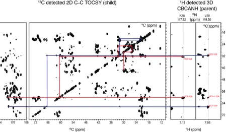
Fig. 8.Schematic representation of the intra–residue assignment process using the simultaneous and parallel acquisition. Intra-residue assignment process for the K28 (red lines) and V29 (blue lines) using the TOCSY (CBCANH 3D + C–C TOCSY 2D). (For interpretation of the references to colour in this figure legend, the reader is referred to the web version of this article.)
[image:10.595.45.543.404.688.2]experimental time needed to acquire a spectrum is taken waiting for the recycle delay. It is also desirable that all available magneti-zation is used effectively. Indeed, in solid state NMR experiments much of the available magnetization is orphaned on the low–
c
nuclei during polarization transfer. We demonstrate that the dis-carded magnetization from the low–c
nuclei can be effectively exploited to give useful correlation spectra. The additional low–c
acquisition can be arranged in a way that it does not compromise the efficiency of the higher dimension parent experiment, which means that the additional acquisition comes at negligible cost. Ide-ally such an approach provides pseudo-4D information expanding the information content of the parent 3Ds. However, the 4D may still be needed for the improved resolution that these experiments cannot wholly address. This approach could also be powerful for monitoring sample integrity in higher dimensional experiments. There is also a need to bridge a gap between datasets collected on the same biological system but on different (typically larger vol-ume and slower spinning) MAS probes, where13C is the preferred acquisition nucleus. Additionally, portions of the protein may be incompatible with proton spectroscopy, perhaps there is motional averaging or PRE-broadening which more strongly effect the pro-ton experiments but are not as detrimental to carbon detection [101,102].Thus far, there have been few applications for multiple receiver spectrometers, and most have not become widespread. All Bruker spectrometers moving forward will have a receiver for each chan-nel as standard equipment. Our approach suggests one practical way to exploit this new norm for hardware. The experiments pre-sented here are applicable at slower MAS for substantially deuter-ated systems (e.g.3.2 mm rotors with 20% back exchanged 1H; samples produced for1H detection) given appropriate CP transfers are accessible. The modifications to the pulse sequence do not neg-atively affect the quality of the final spectrum in this approach, nor do they significantly increase the difficulty of the experimental optimizations, and thus we expect wide adoption.
Acknowledgements
The research leading to these results has received funding from the European Research Council under the European Union’s Seventh Framework Programme (FP/2007-2013)/ERC Grant Agree-ment 639907. J.R.L. also acknowledges funding from BBSRC Grants BB/L022761/1 and BB/R010218/1. We also thank Dr Peter Gierth (Bruker UK) for helping in setting up the double receiver and Jac-queline Tognetti for reading the manuscript.
Appendix A. Supplementary data
Supplementary data to this article can be found online at https://doi.org/10.1016/j.jmr.2019.07.006.
References
[1]J.R. Lewandowski, Advances in solid-state relaxation methodology for probing site-specific protein dynamics, Acc. Chem. Res. 46 (2013) 2018–2027. [2]V.S. Mandala, J.K. Williams, M. Hong, Structure and dynamics of membrane
proteins from solid-state NMR, Annu. Rev. Biophys. 47 (2018) 201–222. [3]P. Schanda, M. Ernst, Studying dynamics by magic-angle spinning solid-state
NMR spectroscopy: principles and applications to biomolecules, Prog. Nucl. Magn. Reson. Spectrosc. 96 (2016) 1–46.
[4]P.C.A. van der Wel, New applications of solid-state NMR in structural biology, Emerg. Top. Life Sci. 2 (2018) 57–67.
[5]F. Castellani, B. van Rossum, A. Diehl, M. Schubert, K. Rehbein, H. Oschkinat, Structure of a protein determined by solid-state magic-angle-spinning NMR spectroscopy, Nature 420 (2002) 98–102.
[6]S.G. Zech, A.J. Wand, A.E. McDermott, Protein structure determination by high-resolution solid-state NMR spectroscopy: application to microcrystalline ubiquitin, J. Am. Chem. Soc. 127 (2005) 8618–8626.
[7]W.T. Franks, D.H. Zhou, B.J. Wylie, B.G. Money, D.T. Graesser, H.L. Frericks, G. Sahota, C.M. Rienstra, Magic-angle spinning solid-state NMR spectroscopy of the beta1 immunoglobulin binding domain of protein G (GB1): 15N and 13C chemical shift assignments and conformational analysis, J. Am. Chem. Soc. 127 (2005) 12291–12305.
[8]W.T. Franks, B.J. Wylie, H.L. Schmidt, A.J. Nieuwkoop, R.M. Mayrhofer, G.J. Shah, D.T. Graesser, C.M. Rienstra, Dipole tensor-based atomic-resolution structure determination of a nanocrystalline protein by solid-state NMR, Proc. Natl. Acad. Sci. U. S. A. 105 (2008) 4621–4626.
[9]I. Bertini, A. Bhaumik, G. De Paepe, R.G. Griffin, M. Lelli, J.R. Lewandowski, C. Luchinat, High-resolution solid-state NMR structure of a 17.6 kDa protein, J. Am. Chem. Soc. 132 (2010) 1032–1040.
[10]M.J. Knight, A.J. Pell, I. Bertini, I.C. Felli, L. Gonnelli, R. Pierattelli, T. Herrmann, L. Emsley, G. Pintacuda, Structure and backbone dynamics of a microcrystalline metalloprotein by solid-state NMR, Proc. Natl. Acad. Sci. U. S. A. 109 (2012) 11095–11100.
[11]C. Wasmer, A. Lange, H. Van Melckebeke, A.B. Siemer, R. Riek, B.H. Meier, Amyloid fibrils of the HET-s(218–289) prion form a beta solenoid with a triangular hydrophobic core, Science 319 (2008) 1523–1526.
[12]M.D. Tuttle, G. Comellas, A.J. Nieuwkoop, D.J. Covell, D.A. Berthold, K.D. Kloepper, J.M. Courtney, J.K. Kim, A.M. Barclay, A. Kendall, W. Wan, G. Stubbs, C.D. Schwieters, V.M. Lee, J.M. George, C.M. Rienstra, Solid-state NMR structure of a pathogenic fibril of full-length human alpha-synuclein, Nat. Struct. Mol. Biol. 23 (2016) 409–415.
[13]M.T. Colvin, R. Silvers, Q.Z. Ni, T.V. Can, I. Sergeyev, M. Rosay, K.J. Donovan, B. Michael, J. Wall, S. Linse, R.G. Griffin, Atomic resolution structure of monomorphic Abeta42 amyloid fibrils, J. Am. Chem. Soc. 138 (2016) 9663– 9674.
[14]Y. Xiao, B. Ma, D. McElheny, S. Parthasarathy, F. Long, M. Hoshi, R. Nussinov, Y. Ishii, Abeta(1–42) fibril structure illuminates self-recognition and replication of amyloid in Alzheimer’s disease, Nat. Struct. Mol. Biol. 22 (2015) 499–505. [15]M.A. Walti, F. Ravotti, H. Arai, C.G. Glabe, J.S. Wall, A. Bockmann, P. Guntert, B. H. Meier, R. Riek, Atomic-resolution structure of a disease-relevant Abeta(1– 42) amyloid fibril, Proc. Natl. Acad. Sci. U. S. A. 113 (2016) E4976–E4984. [16]C.L. Hoop, H.K. Lin, K. Kar, G. Magyarfalvi, J.M. Lamley, J.C. Boatz, A. Mandal, J.
R. Lewandowski, R. Wetzel, P.C. van der Wel, Huntingtin exon 1 fibrils feature an interdigitated beta-hairpin-based polyglutamine core, Proc. Natl. Acad. Sci. U. S. A. 113 (2016) 1546–1551.
[17]J.J. Helmus, K. Surewicz, W.K. Surewicz, C.P. Jaroniec, Conformational flexibility of Y145Stop human prion protein amyloid fibrils probed by solid-state nuclear magnetic resonance spectroscopy, J. Am. Chem. Soc. 132 (2010) 2393–2403.
[18]S.H. Park, B.B. Das, F. Casagrande, Y. Tian, H.J. Nothnagel, M. Chu, H. Kiefer, K. Maier, A.A. De Angelis, F.M. Marassi, S.J. Opella, Structure of the chemokine receptor CXCR1 in phospholipid bilayers, Nature 491 (2012) 779–783. [19]J.S. Retel, A.J. Nieuwkoop, M. Hiller, V.A. Higman, E. Barbet-Massin, J. Stanek,
L.B. Andreas, W.T. Franks, B.J. van Rossum, K.R. Vinothkumar, L. Handel, G.G. de Palma, B. Bardiaux, G. Pintacuda, L. Emsley, W. Kuhlbrandt, H. Oschkinat, Structure of outer membrane protein G in lipid bilayers, Nat. Commun. 8 (2017) 2073.
[20]B. Bersch, J.M. Dorr, A. Hessel, J.A. Killian, P. Schanda, Proton-detected solid-state NMR spectroscopy of a zinc diffusion facilitator protein in native nanodiscs, Angew. Chem. Int. Ed. Engl. 56 (2017) 2508–2512.
[21]D. Good, C. Pham, J. Jagas, J.R. Lewandowski, V. Ladizhansky, Solid-state NMR provides evidence for small-amplitude slow domain motions in a multispanning transmembrane alpha-helical protein, J. Am. Chem. Soc. 139 (2017) 9246–9258.
[22]A. Loquet, N.G. Sgourakis, R. Gupta, K. Giller, D. Riedel, C. Goosmann, C. Griesinger, M. Kolbe, D. Baker, S. Becker, A. Lange, Atomic model of the type III secretion system needle, Nature 486 (2012) 276–279.
[23]J.M. Lamley, D. Iuga, C. Oster, H.J. Sass, M. Rogowski, A. Oss, J. Past, A. Reinhold, S. Grzesiek, A. Samoson, J.R. Lewandowski, Solid-state NMR of a protein in a precipitated complex with a full-length antibody, J. Am. Chem. Soc. 136 (2014) 16800–16806.
[24]L. Krabben, B.J. van Rossum, S. Jehle, E. Bocharov, E.N. Lyukmanova, A.A. Schulga, A. Arseniev, F. Hucho, H. Oschkinat, Loop 3 of short neurotoxin II is an additional interaction site with membrane-bound nicotinic acetylcholine receptor as detected by solid-state NMR spectroscopy, J. Mol. Biol. 390 (2009) 662–671.
[25]A.H. Linden, S. Lange, W.T. Franks, U. Akbey, E. Specker, B.J. van Rossum, H. Oschkinat, Neurotoxin II bound to acetylcholine receptors in native membranes studied by dynamic nuclear polarization NMR, J. Am. Chem. Soc. 133 (2011) 19266–19269.
[26]T. Jacso, W.T. Franks, H. Rose, U. Fink, J. Broecker, S. Keller, H. Oschkinat, B. Reif, Characterization of membrane proteins in isolated native cellular membranes by dynamic nuclear polarization solid-state NMR spectroscopy without purification and reconstitution, Angew. Chem. Int. Ed. Engl. 51 (2012) 432–435.
[27]M. Renault, R. Tommassen-van Boxtel, M.P. Bos, J.A. Post, J. Tommassen, M. Baldus, Cellular solid-state nuclear magnetic resonance spectroscopy, Proc. Natl. Acad. Sci. U. S. A. 109 (2012) 4863–4868.
[29]B. Reif, C.P. Jaroniec, C.M. Rienstra, M. Hohwy, R.G. Griffin, 1H–1H MAS correlation spectroscopy and distance measurements in a deuterated peptide, J. Magn. Reson. 151 (2001) 320–327.
[30]U. Sternberg, R. Witter, I. Kuprov, J.M. Lamley, A. Oss, J.R. Lewandowski, A. Samoson, (1)H line width dependence on MAS speed in solid state NMR – comparison of experiment and simulation, J. Magn. Reson. 291 (2018) 32–39. [31]D.H. Zhou, D.T. Graesser, W.T. Franks, C.M. Rienstra, Sensitivity and resolution in proton solid-state NMR at intermediate deuteration levels: quantitative linewidth characterization and applications to correlation spectroscopy, J. Magn. Reson. 178 (2006) 297–307.
[32]J.R. Lewandowski, J.-N. Dumez, Ü. Akbey, S. Lange, L. Emsley, H. Oschkinat, Enhanced resolution and coherence lifetimes in the solid-state NMR spectroscopy of perdeuterated proteins under ultrafast magic-angle spinning, J. Phys. Chem. Lett. 2 (2011) 2205–2211.
[33]A. Samoson, T. Tuherm, J. Past, A. Reinhold, T. Anupold, I. Heinmaa, New horizons for magic-angle spinning NMR, Top. Curr. Chem. 246 (2005) 15–31.
[34]V. Chevelkov, B.J. van Rossum, F. Castellani, K. Rehbein, A. Diehl, M. Hohwy, S. Steuernagel, F. Engelke, H. Oschkinat, B. Reif, 1H detection in MAS solid-state NMR spectroscopy of biomacromolecules employing pulsed field gradients for residual solvent suppression, J. Am. Chem. Soc. 125 (2003) 7788–7789. [35]A.J. Nieuwkoop, W.T. Franks, K. Rehbein, A. Diehl, U. Akbey, F. Engelke, L.
Emsley, G. Pintacuda, H. Oschkinat, Sensitivity and resolution of proton detected spectra of a deuterated protein at 40 and 60 kHz magic-angle-spinning, J. Biomol. NMR 61 (2015) 161–171.
[36]J. Stanek, L.B. Andreas, K. Jaudzems, D. Cala, D. Lalli, A. Bertarello, T. Schubeis, I. Akopjana, S. Kotelovica, K. Tars, A. Pica, S. Leone, D. Picone, Z.Q. Xu, N.E. Dixon, D. Martinez, M. Berbon, N. El Mammeri, A. Noubhani, S. Saupe, B. Habenstein, A. Loquet, G. Pintacuda, NMR spectroscopic assignment of backbone and side-chain protons in fully protonated proteins: microcrystals, sedimented assemblies, and amyloid fibrils, Angew. Chem. Int. Ed. Engl. 55 (2016) 15504–15509.
[37]M.J. Knight, A.L. Webber, A.J. Pell, P. Guerry, E. Barbet-Massin, I. Bertini, I.C. Felli, L. Gonnelli, R. Pierattelli, L. Emsley, A. Lesage, T. Herrmann, G. Pintacuda, Fast resonance assignment and fold determination of human superoxide dismutase by high-resolution proton-detected solid-state MAS NMR spectroscopy, Angew. Chem. Int. Ed. Engl. 50 (2011) 11697–11701. [38]E. Barbet-Massin, A.J. Pell, J.S. Retel, L.B. Andreas, K. Jaudzems, W.T. Franks, A.
J. Nieuwkoop, M. Hiller, V. Higman, P. Guerry, A. Bertarello, M.J. Knight, M. Felletti, T. Le Marchand, S. Kotelovica, I. Akopjana, K. Tars, M. Stoppini, V. Bellotti, M. Bolognesi, S. Ricagno, J.J. Chou, R.G. Griffin, H. Oschkinat, A. Lesage, L. Emsley, T. Herrmann, G. Pintacuda, Rapid proton-detected NMR assignment for proteins with fast magic angle spinning, J. Am. Chem. Soc. 136 (2014) 12489–12497.
[39]P. Fricke, V. Chevelkov, M. Zinke, K. Giller, S. Becker, A. Lange, Backbone assignment of perdeuterated proteins by solid-state NMR using proton detection and ultrafast magic-angle spinning, Nat. Protoc. 12 (2017) 764– 782.
[40]C. Oster, G.P. Walkowiak, D.E. Hughes, A.L. Spoering, A.J. Peoples, A.C. Catherwood, J.A. Tod, A.J. Lloyd, T. Herrmann, K. Lewis, C.G. Dowson, J.R. Lewandowski, Structural studies suggest aggregation as one of the modes of action for teixobactin, Chem. Sci. 9 (2018) 8850–8859.
[41]D.H. Zhou, J.J. Shea, A.J. Nieuwkoop, W.T. Franks, B.J. Wylie, C. Mullen, D. Sandoz, C.M. Rienstra, Solid-state protein-structure determination with proton-detected triple-resonance 3D magic-angle-spinning NMR spectroscopy, Angew. Chem. Int. Ed. Engl. 46 (2007) 8380–8383.
[42]L.B. Andreas, K. Jaudzems, J. Stanek, D. Lalli, A. Bertarello, T. Le Marchand, D. Cala-De Paepe, S. Kotelovica, I. Akopjana, B. Knott, S. Wegner, F. Engelke, A. Lesage, L. Emsley, K. Tars, T. Herrmann, G. Pintacuda, Structure of fully protonated proteins by proton-detected magic-angle spinning NMR, Proc. Natl. Acad. Sci. U. S. A. 113 (2016) 9187–9192.
[43]J.M. Lamley, C. Oster, R.A. Stevens, J.R. Lewandowski, Intermolecular interactions and protein dynamics by solid-state NMR spectroscopy, Angew. Chem. Int. Ed. Engl. 54 (2015) 15374–15378.
[44]V. Kurauskas, S.A. Izmailov, O.N. Rogacheva, A. Hessel, I. Ayala, J. Woodhouse, A. Shilova, Y. Xue, T. Yuwen, N. Coquelle, J.P. Colletier, N.R. Skrynnikov, P. Schanda, Slow conformational exchange and overall rocking motion in ubiquitin protein crystals, Nat. Commun. 8 (2017) 145.
[45]P. Ma, J.D. Haller, J. Zajakala, P. Macek, A.C. Sivertsen, D. Willbold, J. Boisbouvier, P. Schanda, Probing transient conformational states of proteins by solid-state R(1rho) relaxation-dispersion NMR spectroscopy, Angew. Chem. Int. Ed. Engl. 53 (2014) 4312–4317.
[46]P. Rovo, R. Linser, Microsecond timescale protein dynamics: a combined solid-state NMR approach, ChemPhysChem 19 (2018) 34–39.
[47]S. Kim, T. Szyperski, GFT NMR, a new approach to rapidly obtain precise high-dimensional NMR spectral information, J. Am. Chem. Soc. 125 (2003) 1385– 1393.
[48]W.T. Franks, H.S. Atreya, T. Szyperski, C.M. Rienstra, GFT projection NMR spectroscopy for proteins in the solid state, J. Biomol. NMR 48 (2010) 213– 223.
[49]P. Schmieder, A.S. Stern, G. Wagner, J.C. Hoch, Improved resolution in triple-resonance spectra by nonlinear sampling in the constant-time domain, J. Biomol. NMR 4 (1994) 483–490.
[50]V. Jaravine, I. Ibraghimov, V.Y. Orekhov, Removal of a time barrier for high-resolution multidimensional NMR spectroscopy, Nat. Methods 3 (2006) 605–607.
[51]S.G. Hyberts, K. Takeuchi, G. Wagner, Poisson-gap sampling and forward maximum entropy reconstruction for enhancing the resolution and sensitivity of protein NMR data, J. Am. Chem. Soc. 132 (2010) 2145–2147. [52]V.Y. Orekhov, V.A. Jaravine, Analysis of non-uniformly sampled spectra with
multi-dimensional decomposition, Prog. Nucl. Magn. Reson. Spectrosc. 59 (2011) 271–292.
[53]K. Kazimierczuk, V. Orekhov, Non-uniform sampling: post-Fourier era of NMR data collection and processing, Magn. Reson. Chem. 53 (2015) 921–926. [54]E. Kupce, R. Freeman, Projection-reconstruction technique for speeding up
multidimensional NMR spectroscopy, J. Am. Chem. Soc. 126 (2004) 6429– 6440.
[55]L. Frydman, J. Peng, Non-Cartesian sampling schemes and the acquisition of 2D NMR correlation spectra from single-scan experiments, Chem. Phys. Lett. 222 (1994) 371–377.
[56]L. Frydman, T. Scherf, A. Lupulescu, The acquisition of multidimensional NMR spectra within a single scan, Proc. Natl. Acad. Sci. U. S. A. 99 (2002) 15858– 15862.
[57]E. Kupce, R. Freeman, Fast multi-dimensional Hadamard spectroscopy, J. Magn. Reson. 163 (2003) 56–63.
[58]E. Kupce, R. Freeman, Two-dimensional Hadamard spectroscopy, J. Magn. Reson. 162 (2003) 300–310.
[59]K. Pervushin, B. Vogeli, A. Eletsky, Longitudinal (1)H relaxation optimization in TROSY NMR spectroscopy, J. Am. Chem. Soc. 124 (2002) 12898–12902. [60] P. Schanda, B. Brutscher, Very fast two-dimensional NMR spectroscopy for
real-time investigation of dynamic events in proteins on the time scale of seconds, J. Am. Chem. Soc. 127 (2005) 8014–8015.
[61]P. Schanda, H. Van Melckebeke, B. Brutscher, Speeding up three-dimensional protein NMR experiments to a few minutes, J. Am. Chem. Soc. 128 (2006) 9042–9043.
[62]S. Ganapathy, A. Naito, C.A. McDowell, Paramagnetic doping as an aid in obtaining high-resolution carbon-13 NMR spectra of biomolecules in the solid state, J. Am. Chem. Soc. 103 (1981) 6011–6015.
[63]R. Linser, V. Chevelkov, A. Diehl, B. Reif, Sensitivity enhancement using paramagnetic relaxation in MAS solid-state NMR of perdeuterated proteins, J. Magn. Reson. 189 (2007) 209–216.
[64]N.P. Wickramasinghe, M. Kotecha, A. Samoson, J. Past, Y. Ishii, Sensitivity enhancement in (13)C solid-state NMR of protein microcrystals by use of paramagnetic metal ions for optimizing (1)H T(1) relaxation, J. Magn. Reson. 184 (2007) 350–356.
[65]O. Sørensen, Aspects and prospects of multidimensional time-domain spectroscopy, J. Magn. Reson. 89 (1990) (1969) 210–216.
[66]R. Boelens, M. Burgering, R.H. Fogh, R. Kaptein, Time-saving methods for heteronuclear multidimensional NMR of ((13)C, (15)N) doubly labeled proteins, J. Biomol. NMR 4 (1994) 201–213.
[67]D.P. Frueh, H. Arthanari, G. Wagner, Unambiguous assignment of NMR protein backbone signals with a time-shared triple-resonance experiment, J. Biomol. NMR 33 (2005) 187–196.
[68]T. Parella, P. Nolis, Time-shared NMR experiments, Concepts Magn. Reson. Part A 36A (2010) 1–23.
[69]J.M. Lamley, J.R. Lewandowski, Simultaneous acquisition of homonuclear and heteronuclear long-distance contacts with time-shared third spin assisted recoupling, J. Magn. Reson. 218 (2012) 30–34.
[70] B.B. Das, S.J. Opella, Simultaneous cross polarization to (13)C and (15)N with (1)H detection at 60kHz MAS solid-state NMR, J. Magn. Reson. 262 (2016) 20– 26.
[71]A.B. Nielsen, K. Szekely, J. Gath, M. Ernst, N.C. Nielsen, B.H. Meier, Simultaneous acquisition of PAR and PAIN spectra, J. Biomol. NMR 52 (2012) 283–288.
[72]R. Linser, B. Bardiaux, V. Higman, U. Fink, B. Reif, Structure calculation from unambiguous long-range amide and methyl 1H–1H distance restraints for a microcrystalline protein with MAS solid-state NMR spectroscopy, J. Am. Chem. Soc. 133 (2011) 5905–5912.
[73]T. Gopinath, G. Veglia, Dual acquisition magic-angle spinning solid-state NMR-spectroscopy: simultaneous acquisition of multidimensional spectra of biomacromolecules, Angew. Chem. Int. Ed. Engl. 51 (2012) 2731–2735. [74]T. Gopinath, G. Veglia, Orphan spin operators enable the acquisition of
multiple 2D and 3D magic angle spinning solid-state NMR spectra, J. Chem. Phys. 138 (2013) 184201.
[75]T. Gopinath, G. Veglia, Multiple acquisitions via sequential transfer of orphan spin polarization (MAeSTOSO): How far can we push residual spin polarization in solid-state NMR?, J Magn. Reson. 267 (2016) 1–8.
[76]T. Gopinath, G. Veglia, Experimental aspects of polarization optimized experiments (POE) for magic angle spinning solid-state NMR of microcrystalline and membrane-bound proteins, Methods Mol. Biol. 1688 (2018) 37–53.
[77]K. Sharma, P.K. Madhu, K.R. Mote, A suite of pulse sequences based on multiple sequential acquisitions at one and two radiofrequency channels for solid-state magic-angle spinning NMR studies of proteins, J. Biomol. NMR 65 (2016) 127–141.
[78]A. Viegas, T. Viennet, T.Y. Yu, F. Schumann, W. Bermel, G. Wagner, M. Etzkorn, UTOPIA NMR: activating unexploited magnetization using interleaved low-gamma detection, J. Biomol. NMR 64 (2016) 9–15.
[79]R. Bhattacharyya, L. Frydman, Ultrafast solid-state 2D NMR experiments via orientational encoding, J. Am. Chem. Soc. 128 (2006) 16014–16015. [80] M. Gal, C. Melian, D.E. Demco, B. Blümich, L. Frydman, Solid-state single-scan
[81]C. Oster, S. Kosol, C. Hartlmuller, J.M. Lamley, D. Iuga, A. Oss, M.L. Org, K. Vanatalu, A. Samoson, T. Madl, J.R. Lewandowski, Characterization of protein-protein interfaces in large complexes by solid-state NMR solvent paramagnetic relaxation enhancements, J. Am. Chem. Soc. 139 (2017) 12165–12174.
[82]R. Linser, B. Bardiaux, L.B. Andreas, S.G. Hyberts, V.K. Morris, G. Pintacuda, M. Sunde, A.H. Kwan, G. Wagner, Solid-state NMR structure determination from diagonal-compensated, sparsely nonuniform-sampled 4D proton-proton restraints, J. Am. Chem. Soc. 136 (2014) 11002–11010.
[83]M. Zinke, P. Fricke, C. Samson, S. Hwang, J.S. Wall, S. Lange, S. Zinn-Justin, A. Lange, Bacteriophage tail-tube assembly studied by proton-detected 4D solid-state NMR, Angew. Chem. Int. Ed. Engl. 56 (2017) 9497–9501. [84]E. Kupce, K.R. Mote, P.K. Madhu, Experiments with direct detection of
multiple FIDs, J. Magn. Reson. 304 (2019) 16–34.
[85]Y.T. van den Hoogen, S.J. Treurniet, H.C.P.F. Roelen, E. de Vroom, G.A. van der Marel, J.H. van Boom, C. Altona, Conformational analysis of the tetranucleotides m62A–m62A-U-m62A m62A =N6-dimethyladenosine) and U-m62A-U-m62A and of the hybrid dA-r(U-A), Eur. J. Biochem. 171 (1988) 155–162.
[86]D.S. Wishart, C.G. Bigam, J. Yao, F. Abildgaard, H.J. Dyson, E. Oldfield, J.L. Markley, B.D. Sykes, 1
H, 13
C and 15
N chemical shift referencing in biomolecular NMR, J. Biomol. NMR 6 (1995) 135–140.
[87]A.J. Shaka, J. Keeler, T. Frenkiel, R. Freeman, An improved sequence for broadband decoupling: WALTZ-16, J. Magn. Reson. 52 (1983) (1969) 335– 338.
[88]D.H. Zhou, C.M. Rienstra, High-performance solvent suppression for proton detected solid-state NMR, J. Magn. Reson. 192 (2008) 167–172.
[89]L. Emsley, G. Bodenhausen, Gaussian pulse cascades: New analytical functions for rectangular selective inversion and in-phase excitation in NMR, Chem. Phys. Lett. 165 (1990) 469–476.
[90]D. Marion, M. Ikura, R. Tschudin, A. Bax, Rapid recording of 2D NMR spectra without phase cycling. Application to the study of hydrogen exchange in proteins, J. Magn. Reson. 85 (1989) (1969) 393–399.
[91]M. Baldus, A.T. Petkova, J. Herzfeld, R.G. Griffin, Cross polarization in the tilted frame: assignment and spectral simplification in heteronuclear spin systems, Mol. Phys. 95 (1998) 1197–1207.
[92]A.E. Bennett, R.G. Griffin, J.H. Ok, S. Vega, Chemical shift correlation spectroscopy in rotating solids: radio frequency-driven dipolar recoupling and longitudinal exchange, J. Chem. Phys. 96 (1992) 8624–8627.
[93]R. Verel, M. Ernst, B.H. Meier, Adiabatic dipolar recoupling in solid-state NMR: the DREAM scheme, J. Magn. Reson. 150 (2001) 81–99.
[94]E. Barbet-Massin, A.J. Pell, K. Jaudzems, W.T. Franks, J.S. Retel, S. Kotelovica, I. Akopjana, K. Tars, L. Emsley, H. Oschkinat, A. Lesage, G. Pintacuda, Out-and-back 13C–13C scalar transfers in protein resonance assignment by proton-detected solid-state NMR under ultra-fast MAS, J. Biomol. NMR 56 (2013) 379–386.
[95]J.R. Lewandowski, J. Sein, H.J. Sass, S. Grzesiek, M. Blackledge, L. Emsley, Measurement of site-specific 13
C spin-lattice relaxation in a crystalline protein, J. Am. Chem. Soc. 132 (2010) 8252–8254.
[96]I. Scholz, M. Huber, T. Manolikas, B.H. Meier, M. Ernst, MIRROR recoupling and its application to spin diffusion under fast magic-angle spinning, Chem. Phys. Lett. 460 (2008) 278–283.
[97]M. Ikura, L.E. Kay, A. Bax, A novel approach for sequential assignment of proton, carbon-13, and nitrogen-15 spectra of larger proteins: heteronuclear triple-resonance three-dimensional NMR spectroscopy. Application to calmodulin, Biochemistry 29 (1990) 4659–4667.
[98]L.E. Kay, M. Ikura, R. Tschudin, A. Bax, Three-dimensional triple-resonance NMR Spectroscopy of isotopically enriched proteins, J. Magn. Reson. 213 (2011) 423–441.
[99]A. Liu, R. Riek, G. Wider, C. von Schroetter, R. Zahn, K. Wuthrich, NMR experiments for resonance assignments of13C,15N doubly-labeled flexible
polypeptides: application to the human prion protein hPrP(23–230), J. Biomol. NMR 16 (2000) 127–138.
[100]A.J. Shaka, C.J. Lee, A. Pines, Iterative schemes for bilinear operators; application to spin decoupling, J. Magn. Reson. 77 (1988) (1969) 274–293. [101]S. Ciofi-Baffoni, A. Gallo, R. Muzzioli, M. Piccioli, The IR-(1)(5)N-HSQC-AP
experiment: a new tool for NMR spectroscopy of paramagnetic molecules, J. Biomol. NMR 58 (2014) 123–128.
![Fig. 2. Two-receiver 2D, 3D hC[C, NH] pulse sequences with longitudinal 13C–13C RFDR mixing](https://thumb-us.123doks.com/thumbv2/123dok_us/9423534.445808/4.595.114.469.235.697/fig-receiver-nh-pulse-sequences-longitudinal-rfdr-mixing.webp)
![Fig. 3. Cube representation of the parent 3D 13Cb/13Ca -15N-1H and child 2D 13Cb-13Ca correlation spectra on crystalline [U–13C, 15N]GB1 acquired with (a) RFDR, (b) DREAM,and (c) COSY coupling based homonuclear 13C–13C mixing](https://thumb-us.123doks.com/thumbv2/123dok_us/9423534.445808/5.595.74.528.436.722/representation-correlation-spectra-crystalline-acquired-coupling-homonuclear-mixing.webp)
![Fig. 2a shows a pulse sequence for a [2D, 3D] hC[C, NH] experiment](https://thumb-us.123doks.com/thumbv2/123dok_us/9423534.445808/6.595.110.479.353.716/fig-shows-pulse-sequence-d-d-nh-experiment.webp)
![Fig. 5. Pulse sequence diagrams for [2D, 3D] assignment experiments with COSY mixing. Modifying the 3D COSY–based experiments for assignments results in (a) hcaCBca[C,NH], (b) hCAco[C, NH], and (c) hCOca[C, NH]](https://thumb-us.123doks.com/thumbv2/123dok_us/9423534.445808/7.595.101.501.144.712/sequence-diagrams-assignment-experiments-modifying-experiments-assignments-results.webp)
![Fig. 6. Pulse sequence diagrams for [2D, 3D] assignment experiments with TOCSY mixing](https://thumb-us.123doks.com/thumbv2/123dok_us/9423534.445808/8.595.112.479.387.723/fig-pulse-sequence-diagrams-assignment-experiments-tocsy-mixing.webp)