warwick.ac.uk/lib-publications
Original citation:Brodel, Andreas K., Jaramillo, Alfonso and Isalan, Mark. (2017) Intracellular directed evolution of proteins from combinatorial libraries based on conditional phage replication. Nature Protocols, 12 (9). pp. 1830-1843.
Permanent WRAP URL:
http://wrap.warwick.ac.uk/91282
Copyright and reuse:
The Warwick Research Archive Portal (WRAP) makes this work by researchers of the University of Warwick available open access under the following conditions. Copyright © and all moral rights to the version of the paper presented here belong to the individual author(s) and/or other copyright owners. To the extent reasonable and practicable the material made available in WRAP has been checked for eligibility before being made available.
Copies of full items can be used for personal research or study, educational, or not-for-profit purposes without prior permission or charge. Provided that the authors, title and full
bibliographic details are credited, a hyperlink and/or URL is given for the original metadata page and the content is not changed in any way.
Publisher’s statement:
http://dx.doi.org/10.1038/nprot.2017.084
A note on versions:
The version presented here may differ from the published version or, version of record, if you wish to cite this item you are advised to consult the publisher’s version. Please see the ‘permanent WRAP URL’ above for details on accessing the published version and note that access may require a subscription.
1
FULL TITLE
1Intracellular directed evolution of proteins from combinatorial libraries based on conditional
2
phage replication
3 4
5
AUTHORS
6Andreas K. Brödel1, Alfonso Jaramillo2,3,4, Mark Isalan1* 7
8
9
AFFILIATIONS
101 Department of Life Sciences, Imperial College London, London SW7 2AZ, UK. 11
2 Warwick Integrative Synthetic Biology Centre and School of Life Sciences, University of 12
Warwick, Coventry CV4 7AL, UK.
13
3 CNRS-UMR8030, Laboratoire iSSB and Université Paris-Saclay and Université d’Évry and 14
CEA, DRF, IG, Genoscope, Évry 91000, France.
15
4 Institute for Integrative Systems Biology (I2SysBio), University of Valencia-CSIC, 46980 16
Paterna, Spain.
17
18
19
*To whom correspondence should be addressed: Mark Isalan, Department of Life Sciences,
20
South Kensington Campus, Imperial College London, London SW7 2AZ, UK,
21
[email protected], Tel: +44 (0)20 7594 6482
22
2
ABSTRACT
25Directed evolution is a powerful tool to improve the characteristics of biomolecules. Here we
26
present a protocol for the intracellular evolution of proteins with distinct differences and
27
advantages to established techniques. These include the ability to select for a particular
28
function from a library of protein variants inside cells, minimizing undesired co-evolution and
29
propagation of non-functional library members, as well as allowing positive and negative
30
selection logics using basally-active promoters. A typical evolution experiment comprises the
31
following steps: (i) Preparation of a combinatorial M13 phagemid library expressing variants
32
of the gene of interest and the E. coli host cells; (ii) Multiple rounds of an intracellular
33
selection process towards a desired activity; (iii) The characterization of the evolved target
34
proteins. The system has been developed for the selection of new orthogonal transcription
35
factors1 (TFs) but is capable of evolving any gene – or gene circuit function – that can be
36
linked to conditional M13 phage replication. Here we demonstrate our approach by the
37
directed evolution of TFs based on λ cI against two synthetic bidirectional promoters. The
38
evolved TF variants enable simultaneous activation and repression against their engineered
39
promoters and do not cross-react with the wild-type promoter, thus ensuring orthogonality.
40
This protocol requires no special equipment, allowing synthetic biologists and general users
41
to evolve improved biomolecules within ~7 weeks.
42 43
Keywords: Directed evolution, Gene circuit engineering, Synthetic biology, Protein
44
engineering, M13 bacteriophage, Gene networks, Logic gates, Artificial transcription factors
45 46
EDITORIAL SUMMARY This protocol describes a phagemid-based intracellular evolution
47
approach to generate and select for proteins with improved biological characteristics.
48
TWEET Improving biomolecule function using an intracellular directed evolution approach.
49
COVER TEASER Improving biomolecules by intracellular directed evolution.
50 51
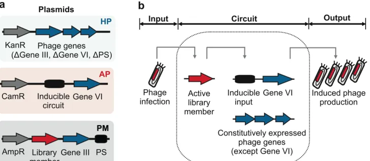
Key references:
52
Brödel, A.K., Jaramillo, A. & Isalan, M. Engineering orthogonal dual transcription factors for
53
multi-input synthetic promoters. Nature Communications 7, 13858 (2016).
54
Schaerli, Y., Munteanu, A., Gili, M.L., Sharpe, J. & Isalan M. A unified design space of
55
synthetic stripe-forming networks. Nature Communications 5, 4905 (2014).
56
Isalan, M., Klug, A. & Choo, Y. A rapid, generally applicable method to engineer zinc fingers
57
illustrated by targeting the HIV-1 promoter. Nat Biotech 19, 656-660 (2001).
3
INTRODUCTION
59Directed evolution has emerged as a powerful tool to improve the characteristics of
60
biomolecules2-4. The approach mimics natural selection to evolve biomolecules towards a 61
desired activity5. One efficient and commonly-used strategy to achieve this in a laboratory
62
environment is to employ filamentous bacteriophages such as M13, to link a mutable
63
genotype to a selectable phenotype. In this way, a number of M13 phage-assisted methods,
64
such as the widely-used phage display technology6, have been developed and applied to 65
improve a wide variety of proteins, including antibodies7-9, DNA-binding proteins10,11, and
66
enzymes12,13. These systems are characterized by an extracellular (in vitro) or intracellular (in 67
vivo) mode of operation. In vitro systems are generally easier to engineer in terms of
68
selection stringency adjustments14, but possess certain limitations that can only be overcome 69
by applying intracellular processes. For example, selection from combinatorial libraries in 70
vivo ensures compatibility with the host cell machinery. This facilitates the optimization of
71
synthetic proteins and gene circuits15-17, which ultimately have to function in a host cell
72
context. In vivo methods promote selection for orthogonality18,19,– a lack of cross-reactions – 73
by intrinsically counter-selecting against adverse effects inside the cell. To further broaden
74
the applications of in vivo directed evolution, we recently developed an M13 phage-based
75
method1 for the intracellular selection of proteins from combinatorial libraries with distinct 76
differences and advantages to established techniques.
77
78
Overview of the protocol
79
This protocol describes a general approach for the directed evolution of proteins from
80
combinatorial libraries on phagemids (Fig. 1). The selection process takes place inside
81
E. coli cells by linking the target protein’s activity to conditional phage production, thus
82
allowing enrichment of functional library members. This is exemplified here by the directed
83
evolution of orthogonal dual transcription factors (TFs) based on bacteriophage λ cI variants1, 84
selecting against synthetic promoters. However, the method can be readily adapted for other
85
target biomolecules (see Applications of the method). A typical evolution experiment
86
consists of: 1) The preparation of a combinatorial M13 phage library (steps 1-38) and E. coli 87
host cells (steps 39-47); 2) The selection process towards a desired activity (steps 48-61);
88
and 3) The characterization of the selected target proteins (steps 62-68) (Fig. 2).
89
90
The system is based on E. coli cultures and three compatible plasmids (available from
91
Addgene; see MATERIALS). Together, these conditionally produce phage (containing the
92
evolving gene) in correlation to the activity of a library member. A selection experiment
4
always begins with an E. coli culture that contains the first two plasmids: a modified helper
94
phage plasmid (HP) and an accessory plasmid (AP) (Fig. 3a). The HP provides almost all
95
that is needed for phage propagation, except for two essential genes (gIII and gVI).
96
Furthermore, the weak M13 packaging signal (PS) is removed from the original M13KO7 HP
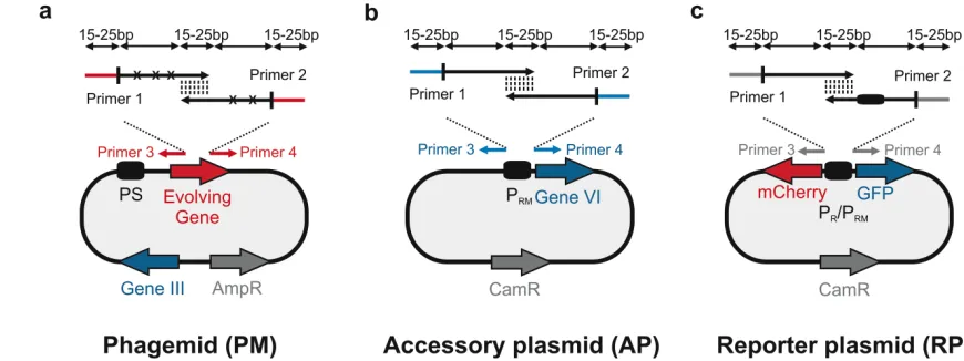
97
to obtain the final M13KO7-ΔPS-ΔgIII-ΔgVI HP. The second plasmid, AP, contains a
98
conditional gene circuit that links an inducible input (e.g. a promoter with a novel operator) to
99
gVI expression. The evolving gene or gene circuit is placed on the third plasmid, termed a
100
phagemid (PM), which is packaged into an infectious phage particle only when all phage
101
genes are expressed. The PM contains the second missing gene (gIII) and a
combinatorially-102
randomized gene of interest (GOI) and is provided to the E. coli culture in the form of an
103
infectious phage library (Fig. 3b). Crucially, our system moves Gene III onto the phagemid
104
so that phage replication occurs only after initial infection, thus circumventing infection
105
resistance20,21, and decreasing the chances of propagating non-functional library members 106
due to multiple infections. A GOI with the desired characteristics upregulates gene VI
107
expression on the AP, completing the phage life cycle. For example, a randomized TF library
108
member that activates an artificial promoter upstream of gVI will increase its own phage
109
production (Fig. 4a). In this way, a protein with novel desired properties can be selected after
110
several rounds of reinfection.
111 112
Applications of the method
113
The method has been used to evolve a set of dual activator-repressor switches for
114
orthogonal logic gates, based on bacteriophage λ cI variants, and multi-input promoter
115
architectures, and these switches have been successfully applied in downstream synthetic
116
gene circuits1. In general, the method is capable of evolving any gene – or gene circuit 117
function – on the phagemid that can be linked to pVI production. This is analogous to
118
previous uses of phage-assisted continuous evolution (PACE)22 (Fig. 4). With PACE, a wide 119
range of medically and biotechnologically relevant biomolecules, including polymerases22,
120
proteases23, genome-editing proteins11 as well as protein-protein interactions24 were linked to 121
conditional M13 phage propagation. In principle, any application where directed evolution
122
approaches have been proposed (e.g. biosensors25 or hybrids with chemical evolution26) can 123
be adapted to this method if the target protein’s activity can be linked to conditional M13
124
phage production. Although certain applications (e.g. membrane proteins) would be harder to
125
adapt, which is why other methods such as liposome display27 have been developed.
126
5 Comparison with other methods
129
Several bacterial directed evolution methods have been developed based on phage
130
replication22, display technologies6,27-29, genome engineering30, as well as conditional cell 131
growth31,32. Linking a target protein’s activity to cell growth is a widely-used strategy and is 132
particularly suitable when the evolving gene directly improves cellular fitness33,34. The use of
133
bacteriophage offers a convenient way to uncouple the fitness function of a cell with target
134
protein activity. This is achieved by linking a target gene’s activity to phage replication using
135
a conditional gene circuit. The main advantage of conditional phage production over display
136
technologies is the compatibility of target genes or gene circuits with the host cell machinery
137
as these have to function in a host cell context. In contrast to PACE (which uses gIII as the
138
sole conditional gene), our phagemid-based approach facilitates the selection of large
139
combinatorial libraries and enables positive and negative selection logics using promoters
140
with basal gene expression. Our system also minimizes the undesired co-evolution of phage
141
genes as only the packaged phagemid is evolving and not the helper phage itself. In
142
comparison to PACE, the protocol is performed in batch mode and therefore requires no
143
special equipment for reactor assembly but instead relies on a daily researcher intervention
144
during selections. Moreover, the batch process facilitates the performance of multiple
145
selections in parallel, enabling the scalability of each individual selection and easy handling.
146
Continuous culture evolution systems can suffer from 'phage washout' (loss of phage) when
147
conditional phage production rates are not compatible with the flow rates. By contrast, batch
148
modes are not as sensitive to loss of phage. On the other hand, dozens of rounds of
149
reinfections occur in a single day of PACE whereas our system is currently limited to one
150
round per overnight cycle. In addition, combinatorial libraries have to be designed and cloned
151
because, unlike PACE, our system does not include a random mutagenesis plasmid35. This 152
means that structural information or a partial understanding of how a set of amino acid
153
changes will affect the target protein’s activity is required to run our system.
154
155
Limitations of the phagemid-based system
156
The main limitation of the system is the combinatorial size of the library which is linked to
157
transformation efficiency (106-1010 variants)36. The selection process itself is not limited to a 158
certain number of gene variants but it has to be noted that the use of larger libraries comes
159
with the cost of prolonged experiment times. Another limitation can be the linkage of the
160
target protein’s activity to conditional M13 phage replication as this depends on the individual
161
protein’s characteristics. This is certainly more complicated for complex proteins such as
162
membrane proteins than it is for cytosolic proteins. Furthermore, general limitations of
6
bacterial expression over mammalian expression (e.g. protein solubility, disulfide bonds,
164
posttranslational modifications) need to be considered for individual target proteins. For
165
instance, our system would need to be adapted to enable the selection of proteins that
166
require disulfide bonds for proper folding in bacterial cells37. 167
168
Experimental Design
169
Combinatorial library cloning on phagemid (PM). Choosing which positions to randomize
170
in the protein of interest is a critical step as this affects the library size, the cloning strategy,
171
and ultimately the overall selection results. Small libraries with only one or two randomized
172
positions can easily be obtained by round-the-world PCR whereas bigger libraries require
173
overlap extension PCR or end-to-end ligation36. Round-the-world PCR means in this context, 174
that single base pair mutations are inserted into the target region by amplification of the
175
whole plasmid DNA with randomized primers so that no additional step for plasmid ligation is
176
required. For round-the-world PCR, both randomized primers must contain the mutations and
177
bind to the same DNA sequence on opposite strands of the plasmid. Primers are generally
178
30-60 nucleotides long (N) and contain mutations in the middle of the randomized primers,
179
flanked with 15-20 bases of correct sequence on both sides. These primers should ideally
180
have a minimum GC content of 40% (%GC), end with one or more C or G bases and are
181
purified by polyacrylamide gel electrophoresis (PAGE). The annealing region should have a
182
melting temperature (Tm) of ≥78°C using the following formula: Tm = 81.5 + 0.41(%GC) - 183
675/N - %mismatch. In this protocol, we focus on an overlap PCR approach prior to Gibson
184
Assembly38 as this has been our method of choice for building λ cIopt libraries with a 185
combinatorial space of >106 variants (Fig. 5a). These libraries are based on a λ cI optimized 186
mutant (cIopt) with a strong activation region39. The protocol presented here is optimized for 187
the construction of combinatorial libraries using Gibson Assembly. It is our method of choice
188
because it bypasses the need for restriction sites inside target genes which makes it much
189
easier to construct sequence-targeted libraries. However, the selection system itself is
190
compatible with any other library generation method36,40 as long as our phagemid vector
191
backbone is used. The design of randomized oligonucleotides for overlap PCR is similar to
192
conventional Gibson primer design38. Briefly, PCR primers for insert amplification require a 193
15-25 bp overlap with each other, as well as a 15-25 bp overlap with the amplified PM vector
194
backbone. Randomized positions should be avoided in the annealing regions and primers
195
should ideally have a Tm of 50-60°C using the following formula: Tm = 4(G + C) + 2(A + T) 196
(where A, C, G and T are the numbers of each base in the primer). The temperature
197
difference of the primer pairs should be matched and lie within a 5°C range. The maximum
198
insert size is limited by oligonucleotide synthesis (currently about 120 bp; desalted
7
oligonucleotides are sufficiently pure). For evolving a novel protein, the user should ideally
200
start with a crystal structure of the target molecule (if available) and randomize positions
201
known to affect the desired activity (e.g. change positions of the binding interface in order to
202
alter the protein binding interaction). In other cases, biochemical information might also be
203
sufficient to guide library construction.
204
Accessory plasmid (AP) design. The conditional gene circuit that links an inducible input to
205
gVI expression has to be adapted to individual needs. This is achieved by replacing the λ
206
PRM promoter (pJPC12-ΔPS-PRM-B0034-gVI) with a different promoter or inducible input 207
depending on the desired application (Fig. 5b). The bidirectional promoter PR/PRM consists of 208
three operator sites (O1-O2-O3) where λ cI binding to O1-O2 leads to PRM activation41. 209
Counterselection via repression is achieved by putting a specific DNA sequence at operator
210
position O3 which is located between the -35 and -10 regions. For example, the O3site of
211
the PRM promoter can be replaced with the consensus wild-type (WT) sequence called OCS. 212
Thus, binding of a cIopt library member to O1-O2 of an engineered PM promoter activates 213
gene VI expression (and so promotes selection) while simultaneous binding to WT O3
214
represses gene VI, enabling counterselection against unwanted WT activity. Positive and
215
negative selections against the synthetic promoters PM,5G6G and PM,5T6T are depicted as 216
examples for this protocol (Supplementary Fig. 1). The engineered promoters are
217
designated according to the positions of the base substitutions in the consensus half-site of
218
O1 and O2.
219
Reporter plasmid (RP) design. This protocol describes the downstream functional
220
characterization of evolved TFs by fluorescence analysis. It has to be noted that a suitable
221
reporter assay needs to be adapted to the target protein’s properties according to the user’s
222
needs. To achieve this, the bidirectional PR/PRM promoter on the RP plasmid (pJPC12-Δ PS-223
mCherry-PR/PRM-GFP) has to be replaced by the same inducible input used on the AP for 224
selection (Fig. 5c). The insertion of the bidirectional promoters P/PM,5G6G and P/PM,5T6T into 225
the RP is depicted as examples for this protocol (Supplementary Fig. 2). For other target
226
proteins, it might be sufficient to use one of the two reporters to analyse activity of the
227
selected proteins.
228
Control selections. Enrichment assays can be performed to test the efficiency of the
229
selection process. Mix plasmids containing λ cIopt (Addgene plasmid ID: 80852) and one of 230
the orthogonal cI variants (e.g. cI5G6G,P; Addgene plasmid ID: 80861) in different ratios (e.g. 231
10-3 and 10-6). Then transform these into TOP10 cells with the modified helper phage
232
M13KO7-ΔPS-ΔgeneIII-ΔgeneVI and the accessory plasmid pJPC12-ΔPS-PRM -B0034-233
8
23-38). Use the obtained phage population and run a batch selection using the accessory
235
plasmid pJPC12-ΔPS-PRM-B0034-geneVI (steps 41-61). Enrichment of λ cIopt can be 236
monitored by infecting TG1 cells (containing the plasmid pJPC12-ΔPS-mCherry-PR/PRM -237
GFP; Addgene plasmid ID: 80859) with the phage titer obtained after each round of
238
selection. Streak out infected cells on agar plates supplemented with chloramphenicol and
239
ampicillin and grow overnight at 37°C. The next day, analyse plates under the UV light of a
240
gel documentation system. The ratio of green to red colonies should increase over time
241
because the non-active TF cI5G6G,P results in red colonies while the enriched active cIopt leads 242
to green colonies due to GFP activation and mCherry repression. As an alternative control
243
selection, the transcription factor cI5G6G,P can be replaced by a reporter (e.g. a red fluorescent 244
protein, RFP) on the PM and the selection process can be monitored by infecting TG1 cells
245
and counting the ratio of red to white colonies after each round of selection1.
246
247
MATERIALS
248REAGENTS
249
Cloning and plasmid construction
250
• Plasmids: M13KO7-ΔPS-ΔgeneIII-ΔgeneVI (Addgene plasmid ID: 80840), pLITMUS-251
rpoN-cIopt-J23106-geneIII (Addgene plasmid ID: 80852), pJPC12-ΔPS-PRM-B0034-geneVI 252
(Addgene plasmid ID: 80858), optional: pJPC12-ΔPS-mCherry-PR/PRM-GFP (Addgene 253
plasmid ID: 80859), pLITMUS-rpoN-cI5G6G,P-J23106-geneIII (Addgene plasmid ID: 80861) 254
(Supplementary Fig. 3, Supplementary Table 1). Sequences of all plasmids are listed in
255
Supplementary Data 1-5.
256
• Oligonucleotides (Sigma). Primers used for cloning are listed in Supplementary Table 2. 257
• KOD Hot Start DNA Polymerase (Merck Millipore, cat. no. 71086). PCR reaction 258
components are listed in the Equipment Setup.
259
• Diethyl pyrocarbonate (DEPC)-treated and sterile filtered water (Sigma, cat. no. 95284) 260
• Gibson Assembly Master Mix (New England BioLabs, cat. no. E2611) 261
• DpnI endonuclease (New England BioLabs, cat. no. R0176) 262
• Super Optimal broth with Catabolite repression (S.O.C.) medium (Sigma, cat. no. 263
15544034)
264
• DNA Gel Loading Dye, 6× (Thermo Scientific, cat. no. R0611) 265
• 1 kb Plus DNA Ladder (Thermo Scientific, cat. no. 10787026) 266
• SYBR Safe DNA Gel Stain (Life Technologies, cat. no. S33102
)
267• Tris-borate-ethylenediaminetetraacetic acid (TBE) Buffer, 10× (Sigma, cat. no. T4415) 268
9
• QIAquick Gel Extraction Kit (Qiagen, cat. no. 28704) 270
• QIAquick PCR Purification Kit (Qiagen, cat. no. 28104) 271
• MinElute PCR Purification Kit (Qiagen, cat. no. 28004) 272
• QIAprep Spin Miniprep Kit (Qiagen, cat. no. 27104) 273
• HiSpeed Plasmid Maxi Kit (Qiagen, cat. no. 12663) 274
275
Strains, buffers and media
276
• One Shot Chemically Competent TOP10 E. coli (Fisher Scientific, cat. no. C404010) 277
• Mix & Go Competent Cells - Strain TG1 (Zymo Research, cat. no. T3017) 278
• 5-alpha Electrocompetent E. coli, optional (New England BioLabs, cat. no. C2989K) 279
• Lysogeny broth (LB) with agar (Sigma, cat. no. L2897) 280
• Ampicillin (Sigma, cat. no. A0166), chloramphenicol (Sigma, cat. no. C0378), kanamycin 281
(Sigma, cat. no. K4000), carbenicillin disodium salt (Sigma, cat. no. C1389)
282
• 2× tryptone yeast extract (2×TY): NaCl (Sigma, cat. no. S9888), yeast extract (Sigma, cat. 283
no. Y1625), tryptone (Sigma, cat. no. T7293)
284
• Glycerol (Sigma, cat. no. G5516) 285
• Ethanol (≥ 99.8%) for molecular biology (Merck Millipore, cat. no. 1085430250) 286
• M9 Minimal Salts, 5× (Sigma, cat. no. M6030) 287
• M9 plates: bacteriological agar (Sigma, cat. no. A5306), MgSO4 (Sigma, cat. no. M7506), 288
D-(+)-glucose (Sigma, cat. no. G8270), CaCl2 (Sigma, cat. no. C1016), thiamine-HCl 289
(Sigma, cat. no. T1270)
290 291
EQUIPMENT
292
• Polymerase chain reaction (PCR) tubes (VWR, cat. no. 732-0545) 293
• Microcentrifuge tubes (1.5 ml; Thermo Scientific, cat. no. 05-408-129) 294
• Conical centrifuge tube, polypropylene, 15 ml (BD Falcon, cat. no. 352097) 295
• Conical centrifuge tubes, polypropylene, 50 ml (Corning, cat. no. 430829) 296
• Schott culture flasks, 250 ml (Sigma, cat. no. Z620033) 297
• Nunc CryoTubes (Thermo Scientific, cat. no. 366656) 298
• Serological pipettes (5 ml, 10 ml, and 25 ml; Fisher Scientific, cat. nos. 678-11D, 13-299
678-11E and 13-678-11)
300
• Sterile filters (0.22 µm pore size, Millex-GV, cat. no. SLGV033RS) 301
• L-shaped cell spreaders (Fisher Scientific, cat. no. 14-665-231) 302
• Cell culture centrifuge Avanti J-26XP (Beckman Coulter, cat. no. 393124) 303
10
• Dri-block heater (Techne, DB100/2) 305
• Eppendorf Thermomixer Compact (Sigma, cat. no. T1317) 306
• Balance Sartorius Excellence (Sartorius) 307
• NanoDrop Lite Spectrophotometer (Thermo Scientific) 308
• Biophotometer (Eppendorf) 309
• Biophotometer cuvettes (Sigma, cat. no. Z605050) 310
• Horizontal gel electrophoresis systems (Bio-Rad) 311
• Gel documentation system (InGenius 3, Syngene) 312
• Gene Pulser Cuvette, 0.1 cm electrode (Bio-Rad, cat. no. 165-2089) 313
• Gene Pulser Xcell Microbial System (Bio-Rad, cat. no. 1652662) 314
• PCR thermocycler (Bio-Rad S1000, cat. no. 1852196) 315
• Petri dishes, 57 cm2 (Sigma, cat. no. P7741) 316
• Mini Incubator (Labnet International, I5110A) 317
• Nunc Square BioAssay Dishes, 24.1 cm × 24.1 cm (Thermo Scientific, cat. no. 10570502) 318
• Shaking Incubator SI500 (Stuart) 319
• Cell culture microplate, 96 well, optional (Greiner Bio-One, cat. no. 655090) 320
• Infinite M200 plate reader, optional (Tecan) 321
• Research pipettes: 10 µl, 100 µl, 1000 µl (Sigma, cat. no. Z683884) 322
• Tips: 10 µl, 200 µl, 1000 µl (Starlab, cat. nos. 3700-C, S1113-1700-C, S1111-323
6701-C)
324 325
REAGENT SETUP
326
Antibiotic stocks Prepare 100 mg ml-1 ampicillin in H
2O (sterile filtered), 100 mg ml-1 327
kanamycin in H2O (sterile filtered) and 100 mg ml-1 chloramphenicol in ethanol. Aliquot stocks 328
into sterile 1.5 ml tubes and store at -20°C for up to 6 months. The final concentrations, if not
329
stated otherwise, are 100 µg ml-1 ampicillin (1:1000), 50 µg ml-1 kanamycin (1:2000), and
330
25 µg ml-1 chloramphenicol (1:4000). 331
Glycerol Prepare a sterile 10% (v/v) glycerol solution in H2O for making electrocompetent 332
cells. Glycerol stocks are obtained by preparing a sterile 50% (v/v) glycerol solution in H2O 333
and adding glycerol to the cell culture (f.c. 20% (v/v)) prior to freezing at -80°C. Store glycerol
334
stock at 4°C for up to 3 months.
335
TBE electrophoresis bufferDilute TBE buffer in distilled water to a 1× working solution and
336
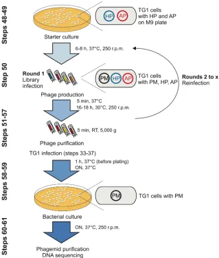
store at room temperature (15-25°C) for up to 6 months.
11
Culture medium Autoclave 2×TY medium (5 g l-1 NaCl, 10 g l-1 yeast extract, 16 g l-1 338
tryptone) and add antibiotics where appropriate before use. Store medium at 4°C for up to
339
several months.
340
LB plates Add 35 g LB powder in 1 l water and autoclave. Add antibiotics where appropriate,
341
pour into petri dishes and allow to solidify. Store plates at 4°C for up to several weeks. Note
342
that antibiotics degrade over time which might affect the concentration when stored for
343
prolonged times.
344
M9 minimal medium plates Autoclave 7 g bacteriological agar in 500 ml 1x M9 medium.
345
Add 1 ml 1M MgSO4 (autoclaved), 5 ml 20% (w/v) D-(+)-glucose (sterile filtered), 50 µl 1M 346
CaCl2 (autoclaved) and 500 µl 1M thiamine-HCl (sterile filtered) to M9 agar just before use. 347
Add antibiotics where appropriate. Store plates at 4°C for up to several months.
348 349
EQUIPMENT SETUP
350
PCR thermocycler The PCR reaction components are listed below.
351
Component Volume [µl] Final concentration
10× Buffer 5 1×
25 mM MgSO4 3 1.5 mM
dNTPs (2 mM each) 5 0.2 mM (each)
H2O varies
Forward primer (5 µM) 3 0.3 µM
Reverse primer (5 µM) 3 0.3 µM
Template DNA varies 0.02-0.2 ng/µl
KOD DNA Polymerase (1 Unit/µl) 1 0.02 U/µl
Total reaction volume 50
CRITICAL: For targets greater than 2 kb, final Mg2+ concentrations are adjusted to 2mM.
352 353
The following conditions are used for all PCR reactions:
354
Step Conditions
1. Polymerase activation 95°C, 2 min
2. Denature 95°C, 30 s
3. Annealing temperature varies, 30 s
4. Extension 70°C, time varies
Repeat steps 2.-4. Number of cycles vary
Final extension 70°C, 10 min
Infinite hold 4°C 355
See Supplementary Table 3 for PCR conditions specific for individual reactions.
12
Infinite M200 plate reader Temperature: 37°C, duration: 10 h, shaking: 281 r.p.m.,
357
absorbance: 600 nm +/- 9 nm, fluorescence mCherry: excitation 585 nm +/- 9 nm; emission
358
625 nm +/- 20 nm; gain value 70, fluorescence GFP: excitation 485 nm +/- 9 nm; emission
359
520 nm +/- 20 nm; gain value 40.
360 361
PROCEDURE
362Phagemid construction by Gibson Assembly TIMING 2 weeks
363
1. Design and order generic forward and reverse primers (e.g. F and
pLITMUS-364
R; Supplementary Table 2) for the amplification of the PM vector backbone
(pLITMUS-365
rpoN-cIopt-J23106-geneIII) upstream of cIopt and downstream the terminator BBa_B0015. 366
Note that the terminator BBa_B0015 occurs twice in the parental plasmid. The "medium
367
strength" rpoN promoter is used to express the evolving gene to achieve a balance
368
between functional expression and any potential metabolic load. The levels of the
369
expressed target gene may need to be adjusted to the function in other cases.
370
2. Design and order user-specific primers (e.g. cI-F and cI-R; Supplementary Table 2) for
371
the gene of interest plus terminator of choice (e.g. BBa_B0015) with a 15-25 bp overlap
372
to the PM vector backbone.
373
3. Amplify the gene of interest and vector backbone by PCR (See Equipment Setup) and
374
purify the samples using the QIAquick PCR Purification Kit. pCRITICAL STEP If the
375
PCR reactions contain unwanted by-products, gel extraction should be performed
376
throughout the protocol using the QIAquick Gel Extraction Kit. Use a DNA polymerase
377
with proof-reading activity (e.g. KOD DNA Polymerase) for all PCR reactions throughout
378
the protocol.
379
4. Remove the parental plasmid by adding 1 µl DpnI per 50 µl PCR reaction product and
380
incubating for 1-2 h at 37°C and 400 r.p.m. (Thermomixer Compact).
381
5. Fuse the two fragments by Gibson Assembly38 according to the manufacturer’s
382
instructions. Note that Gibson reactions can be downscaled to 5 µl per reaction.
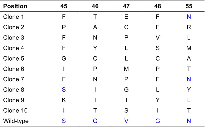
383
6. Dilute the assembled products 4-fold with H2O, add 2 µl of the diluted product to 50 µl 384
chemically competent Top10 cells and transform the cells according to the
385
manufacturer’s instructions.
386
7. Incubate the cells for 1 h at 37°C, 220 r.p.m. (incubator SI500) and spread them onto LB
387
plates supplemented with 100 µg ml-1 ampicillin. 388
8. Grow the cells overnight at 37°C.
389
9. The next day, pick single colonies and grow them in 5 ml 2×TY supplemented with
390
100 µg ml-1 ampicillin overnight at 37°C and 220 r.p.m. (incubator SI500).
13
10. Extract the phagemid DNA (QIAprep Spin Miniprep Kit) according to the manufacturer’s
392
instructions and confirm the nucleotide sequences by DNA sequencing using the primers
393
pLITMUS-F and pLITMUS-R (Table 1).
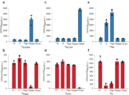
394 395
Combinatorial library cloning on phagemids TIMING 2 weeks
396
11. Design and order user-specific forward and reverse primers for the amplification of the
397
PM vector backbone (e.g. pLITMUS-Lib-F and pLITMUS-Lib-R; Supplementary Table
398
2) and the insertion of the randomized target sequence (e.g. Library 1-F, Library 1-R;
399
Supplementary Table 2). PCR primers for insert amplification require a 15-25 bp
400
overlap with each other as well as a 15-25 bp overlap with the amplified vector
401
backbone. pCRITICAL STEP Avoid randomized library positions within the primer
402
overlap regions.
403
12. Amplify the PCR fragments (see Equipment Setup) and purify the samples using the
404
QIAquick PCR Purification Kit or the MinElute PCR Purification Kit (for samples
405
<100 bp). pCRITICAL STEP The PCR product concentration affects the efficiency of
406
the assembly reaction. Optimized cloning efficiency requires at least 20 ng µl-1 of the PM 407
vector backbone.
408
13. Add 1 µl DpnI per 50 µl PCR reaction product and incubate for 1-2 h at 37°C and
409
400 r.p.m. (Thermomixer Compact).
410
14. Fuse the DNA fragments by Gibson Assembly38. Upscale Gibson reactions (e.g. 4 ×
411
20 µl) to increase the total plasmid concentration.
412
15. Pool the Gibson reactions, purify the assembled plasmid using the QIAquick PCR
413
Purification Kit and elute in 30 µl H2O. pCRITICAL STEPNote that purification is 414
important to decrease the salt concentration and to decrease the Gibson reaction
415
components as these are toxic to the cells at high concentrations.
416
16. Measure the plasmid concentration with a spectrophotometer (NanoDrop Lite).
417
pCRITICAL STEP DNA concentrations should be >10 ng µl-1 for high transformation
418
efficiency.
419
17. Transform 1-2 µl of DNA into 50 µl electrocompetent cells (DH5-alpha or TG1) and add
420
950 µl S.O.C medium. pCRITICAL STEP Use electroporation as the method of choice
421
for transformation as it allows much larger library sizes.
422
18. Incubate for 1 h at 37°C, 220 r.p.m. (incubator SI500).
423
19. Plate the transformation reaction on Nunc Square BioAssay Dishes (24.1 cm × 24.1 cm)
424
supplemented with 100 µg ml-1 ampicillin and incubate overnight at 37°C. 425
20. The next day, harvest the cells with a cell spreader. pCRITICAL STEP Only use plates
426
14
serial dilutions (10-2 and 10-4 in 2×TY) on additional petri dishes (57 cm2) supplemented 428
with 100 µg ml-1 ampicillin and colony counting the following day. Ideally, to cover the 429
whole library space, at least a 3-fold excess of colonies relative to the theoretical library
430
size is desired. ? TROUBLESHOOTING 431
21. Purify the combinatorial DNA library using the HiSpeed Plasmid Maxi Kit and elute in
432
0.5 ml TE buffer. Measure the plasmid concentration with a spectrophotometer
433
(NanoDrop Lite). The obtained plasmid concentration should ideally be >50 ng ml-1.
434
22. Pick individual colonies (10-100 clones of a library depending on the library size and
435
quality control desired) from the petri dishes which were used to estimate the
436
transformation efficiency (see Step 20) and culture each in 5 ml 2×TY supplemented
437
with 100 µg ml-1 ampicillin overnight at 37°C, 220 r.p.m. (incubator SI500). The next day, 438
extract phagemid DNA (QIAprep Spin Miniprep Kit) and sequence the gene of interest
439
using the primers pLITMUS-F and/or pLITMUS-R to confirm library diversity (Table 1).
440
? TROUBLESHOOTING 441
442
Production of M13 phage from a combinatorial phagemid library TIMING 1 week
443
23. Transform 50 µl chemically competent Top10 cells with equal moles of HP
(M13KO7-444
ΔPS-ΔgeneIII-ΔgeneVI) and AP (pJPC12-ΔPS-PRM-B0034-geneVI) (10-20 fmol per 445
plasmid, typically 1-2 µl in total). Note that the PRM promoter can be replaced by an 446
alternative promoter (e.g. T7) to obtain higher phage titers in the absence of the activator
447
λ cI.
448
24. Add 250 µl S.O.C. medium to the samples and incubate for 1 h at 37°C, 220 r.p.m.
449
(incubator SI500).
450
25. Spread the cells on LB plates supplemented with 25 µg ml-1 chloramphenicol and 50 µg 451
ml-1 kanamycin. Grow overnight at 37°C. 452
26. The next day, pick a single colony, grow in 2×TY supplemented with 12.5 µg ml-1 453
chloramphenicol and 25 µg ml-1 kanamycin at 37°C, 250 r.p.m. (incubator SI500) until
454
the OD600 reaches 0.4-0.6 (mid-exponential phase) and make cells electrocompetent as 455
described in Gonzales et al.42.
456
nPAUSE POINT Stored electrocompetent cells can be used for the construction of any
457
phage library.
458
27. Transfer 50 µl of the electrocompetent cells to a prechilled 1.5 ml tube on ice and add
1-459
2 µl of the cloned combinatorial phagemid library.
460
28. Electroporate cells, add immediately 950 µl S.O.C. medium and incubate for 1 h at 37°C,
461
220 r.p.m. (incubator SI500).
15
29. Estimate the actual phage library by colony counting of serial dilutions (10-2 and 10-4 in 463
2×TY) on LB plates supplemented with ampicillin (see step 20). pCRITICAL STEP 464
Make sure not to lose any library members through low transformation efficiencies.
465
? TROUBLESHOOTING 466
30. Add 3 ml 2×TY supplemented with 12.5 µg ml-1 chloramphenicol, 25 µg ml-1 kanamycin,
467
and 50 µg ml-1 ampicillin to the transformation reaction and grow for 18-20 h at 30°C, 468
250 r.p.m. (incubator SI500). Note that the volume can be adjusted depending on the
469
desired volume of the phage titer.
470
31. The next day, centrifuge sample for 5 min at 5,000 g.
471
32. Sterile-filter the phage supernatant (0.22 µm pore size). nPAUSE POINT The phage
472
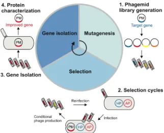
library can be kept at 4°C for short-term storage (weeks) or at -20°C for the long-term
473
(years).
474 475
Phage titer analysis TIMING 3 d
476
33. Streak out TG1 cells from a glycerol stock (~1-5 µl) on an M9 minimal medium plate and
477
incubate overnight at 37°C. Note that TG1 plates can be used for a minimum of two
478
weeks when stored at 4°C. pCRITICAL STEP Use M9 minimal medium plates to select
479
F-pilus positive TG1 cells.
480
34. The next day, pick 1-4 single isolated colonies from the M9 minimal medium plate and
481
inoculate in 10 ml 2×TY medium in a 50 ml conical centrifuge tube.
482
35. Incubate at 37°C and 250 r.p.m. (incubator SI500) until the OD600 reaches 0.4-0.6 (mid-483
exponential phase). It typically takes 4-6 hours for the culture to reach the desired OD600. 484
pCRITICAL STEP Do not let the cells grow into stationary phase as TG1 cells tend to
485
lose the F’ episome and this lowers the overall infection rate.
486
36. In the meantime, prepare serial dilutions (10-2, 10-4, 10-6, 10-8 in 2×TY) of the phage 487
library in sterile 1.5 ml tubes. Phage stocks are diluted before infection to ensure that
488
each cell is only infected by one phage particle (number of colonies on plates equal
489
number of phage particles).
490
37. Add 100 µl of the phage dilutions to 900 µl TG1 cells in a sterile microcentrifuge tube.
491
Mix gently and incubate the samples for 1 h at 37°C with no shaking. Plate 100 µl cell
492
suspension on prewarmed LB plates supplemented with 100 µg ml-1 ampicillin and
493
incubate overnight at 37°C.
494
38. The next day, count the number of colonies and calculate the phage titer (Equation 1).
495
Ideally, use the plates containing 20 to 400 colonies. Note that the 100-fold dilution
496
(step 37) has to be taken into consideration.
497
Phage titer per ml = dilution factor x 100 x number of colonies on plate (Equation 1)
16
pCRITICAL STEP The phage titer should lie between 108-1013 colony-forming units (cfu) 499
per ml.
500 501
Construction of accessory and reporter plasmids TIMING 2 weeks
502
39. Order user-specific forward and reverse primers to replace the PRM promoter on the AP 503
plasmid (pJPC12-ΔPS-PRM-B0034-geneVI) with a different promoter or inducible input. 504
For vector amplification, use primers B0034-gVI-F and gVI-R that bind upstream and
505
downstream of PRM (Supplementary Table 2). For insert amplification make sure to add 506
a 15-25 bp overlap for assembly. Optional: Clone the same inducible input into
pJPC12-507
ΔPS-mCherry-PR/PRM-GFP to obtain a reporter for the functional characterization of 508
selected proteins. Use primers GFP-F and mCherry-R for vector amplification
509
(Supplementary Table 2).
510
40. Clone the accessory and reporter plasmids as described in steps 3-10. Use sequencing
511
primers pJPC12-F and/or pJPC12-R for gene VI constructs and pJPC12-F2 for reporters
512
(Table 1).
513 514
Preparation of host cells for directed evolution TIMING 3 d
515
41. Transform 50 µl competent TG1 cells with equal moles of HP M13KO7-ΔPS-Δ
geneIII-516
ΔgeneVI and the cloned accessory plasmid (AP) (10-20 fmol per plasmid, typically 1-2 µl
517
in total). pCRITICAL STEP Always use an E. coli strain that contains the F-factor
518
needed for M13 phage infection.
519
42. Add 250 µl S.O.C. medium to the sample and incubate for 1 h at 37°C, 220 r.p.m.
520
(incubator SI500).
521
43. Spread the cells on LB plates supplemented with 25 µg ml-1 chloramphenicol and 50 µg 522
ml-1 kanamycin and grow cells overnight at 37°C. 523
44. The next day, pick a single colony and grow cells in 2 ml 2×TY supplemented with
524
kanamycin and chloramphenicol for 4-6 h at 37°C, 250 r.p.m. (incubator SI500) until the
525
cells reach the late-exponential phase.
526
45. (Optional) Make glycerol stock and store at -80°C (see Reagent Setup).
527
46. Make serial dilutions of the cell suspension (e.g. 10-6 or 10-8 in M9 medium) and spread 528
the diluted cells on an M9 minimal medium plate supplemented with 25 µg ml-1
529
chloramphenicol and 50 µg ml-1 kanamycin. These conditions promote phage 530
infectability by maintaining F’ pili.
531
47. Incubate plates for 30-48 h at 37°C. Note that bacteria grow much slower on minimal
532
media than on rich media. This plate is used as a source of fresh colonies for selection
533
experiments and can be used for up to two weeks when stored at 4°C.
17 Phage-assisted batch selection TIMING 2 weeks
535
48. Inoculate 10-20 ml 2×TY containing 12.5 µg ml-1 chloramphenicol and 12.5 µg ml-1 536
kanamycin with 1-4 colonies from prepared M9 plate (step 47) in a 50 ml tube (Fig. 6).
537
49. Grow the starter culture for 6-8 h at 37°C and 250 r.p.m. (incubator SI500) until the
538
OD600 reaches 0.4-0.6. 539
50. Infect 10 ml of the starter culture with the combinatorial phage library at a multiplicity of
540
infection (MOI) of 0.5-5. An excess of cell culture can be chilled on ice and then be
541
stored at 4°C for up to one week. This culture may be used for the next rounds of
542
selection. Note that the selection volume can be easily up- or downscaled according to
543
the user’s need.
544
51. Incubate the infected cells at 37°C without stirring for 5 min.
545
52. Incubate the sample for 18-20 h at 30°C and 250 r.p.m. (incubator SI500).
546
53. The next day, centrifuge the culture for 5 min at 5,000 g and transfer 1 ml of the
547
supernatant into a sterile microfuge tube. This sample is used to start a new round of
548
selection.
549
nPAUSE POINT Phage supernatants for each round of selection can be stored at 4°C
550
for short-term storage, or at -20°C for the long-term, to continue selection at a later time.
551
54. Infect the starter culture (step 49) at a ratio of 10-3-10-1 (e.g. 10-1000 µl phage
552
supernatant in 10 ml culture) for the next round of selection.
553
55. Run selection cycle (steps 51-54) for several rounds until the target protein(s) are
554
enriched. This usually takes four to eight rounds depending on the target protein’s
555
activity and thus the conditional gene VI expression.pCRITICAL STEP The phage titer
556
should ideally stay between 106-1012 cfu ml-1 after each round of the selection (see 557
step 56). Very high infection rates (MOI >10) lead to multiple infections and thus
558
propagation of non-functional library members (‘cheaters’) whereas very low rates (MOI
559
<0.1) decrease the performance of the system. ? TROUBLESHOOTING 560
56. (Optional) During the selection process, monitor the phage titer for each round by phage
561
titer analysis (steps 33-38). ? TROUBLESHOOTING 562
57. (Optional) Monitor the selection process by infecting reporter cells (TG1 with a suitable
563
reporter plasmid, e.g. pJPC12-ΔPS-mCherry-P/PM,5G6G-GFP) with the obtained phage 564
titer for each round (analogous to steps 33-38). Streak out infected cells on LB plates
565
supplemented with 25 µg ml-1 chloramphenicol and 100 µg ml-1 ampicillin and grow 566
overnight at 37°C. The next day, analyse the plates under the UV light of a gel
567
documentation system. Non-active library member result in red colonies while active
568
library members lead to green colonies due to GFP activation and mCherry repression.
569
Store plates at 4°C overnight for improved mCherry signals.
18
58. After selection, sterile-filter the phage supernatant (0.22 µm pore size) and serial dilute
571
the sample with 2×TY medium before infecting TG1 cells with an OD600 of 0.4-0.6. 572
Incubate the infected cells for 1 h at 37°C before plating (see step 37).
573
59. Select infected cells on 100 µg ml-1 ampicillin plates overnight at 37°C. 574
60. The next day, pick at least three colonies per selection and grow each colony in 5 ml
575
2×TY supplemented with ampicillin overnight at 37°C and 250 r.p.m. (incubator SI500).
576
61. The next day, extract phagemid DNA (QIAprep Spin Miniprep Kit) and sequence the
577
gene of interest using the primers pLITMUS-F and/or pLITMUS-R (Table 1).
578 579
Characterization of evolved proteins (optional) TIMING 3 d
580
62. Transform 50 µl competent TG1 cells with equal moles of a selected phagemid and a
581
suitable reporter plasmid (e.g. pJPC12-ΔPS-mCherry-P/PM,5G6G-GFP) (10-20 fmol per 582
plasmid, typically 1-2 µl in total). Transform the reporter plasmid into TG1 cells and use
583
as a control. (Optional) Delete the expression cassette rpoN-cIopt-B0015 from the 584
phagemid (e.g. pLITMUS-ΔcIopt-F, pLITMUS-ΔcIopt-R) and transform the obtained 585
plasmid (pLITMUS-J23106-geneIII) together with the reporter to compensate for growth
586
effects between control and selected phagemids.
587
63. Spread the cells on LB plates supplemented with 25 µg ml-1 chloramphenicol and 100 µg
588
ml-1 ampicillin and grow the cells overnight at 37°C. 589
64. The next day, pick single colonies and grow in 1 ml 2×TY supplemented with 5 µg ml-1
590
chloramphenicol and 5 µg ml-1 carbenicillin for 4-6 h at 37°C, 250 r.p.m. (incubator 591
SI500). Analyse at least three replicates per transformation.
592
65. Measure OD600 of each replicate (150 µl) using the Tecan Infinite M200 plate reader. 593
66. Dilute cultures in 2×TY supplemented with 5 µg ml-1 chloramphenicol and 5 µg ml-1 594
carbenicillin to a final OD600 of 0.01 (150 µl) in a 96-well microplate. 595
67. Measure the absorbance at 600 nm, green fluorescence (excitation: 485 nm, emission:
596
520 nm), and red fluorescence (excitation: 585 nm, emission: 625 nm) every 10 min with
597
the Infinite M200 plate reader (37°C, shaking between readings) until the cells reach
598
stationary phase.
599
68. For data analysis use fluorescence readings in the mid-exponential phase (OD600 of 0.2) 600
and correct absorbance and fluorescence against readings of a TG1 culture. Normalize
601
the fluorescence for the number of cells by dividing by the absorbance.
602
603
? TROUBLESHOOTING
604
Troubleshooting advice can be found in Table 2.
19
TIMING
606
Phagemid construction by Gibson Assembly
607
Steps 1-2, design of primers and oligo synthesis by supplier: 1 week
608
Steps 3-10, cloning of phagemid: 1 week
609
Combinatorial library cloning on phagemids
610
Step 11, design of primers and oligo synthesis by supplier: 1 week
611
Steps 12-22, cloning of combinatorial library: 1 week
612
Production of M13 phage from a combinatorial phagemid library
613
Steps 23-32, transfer from plasmid library to phage library: 1 week
614
Phage titer analysis
615
Steps 33-38, analysis of phage concentration: 3 d
616
Construction of accessory and reporter plasmids (can be done in parallel with phage
617
library cloning)
618
Step 39, design of primers and oligo synthesis by supplier: 1 week
619
Step 40, cloning of accessory plasmid and reporter plasmid: 1 week
620
Preparation of host cells for directed evolution (can be done in parallel after
621
successful AP cloning)
622
Steps 41-47, transformation and plating of cells: 3 d
623
Phage-assisted batch selection
624
Steps 48-57, batch selections: 1 week
625
Steps 58-61, extraction and sequencing of selected genes: 1 week
626
Characterization of evolved proteins (optional)
627
Steps 62-68, functional characterization by reporter assay: 3 d
628 629
ANTICIPATED RESULTS
630The first section of this protocol describes the construction of combinatorial libraries used for
631
subsequent directed evolution experiments. As examples, we describe the construction of
632
two cIopt libraries which contain five randomized positions: Library 1 (45S, 46G, 47V, 48G, 633
55N); Library 2 (45S, 46G, 48G, 49A, 55N). Quality control sequencing of 10-100 clones of a
634
library may be performed to confirm diversity, depending on the library size and quality
635
control desired. For example, ten individual clones of a constructed library should ideally
636
result in ten different variants (Table 3).
637 638
The second section illustrates the directed evolution of proteins based on conditional M13
639
phage propagation. Libraries 1 and 2 are selected against engineered promoters for six to
640
eight rounds leading to enrichment of TFs with binding activations against their novel
20
promoters (Table 4). We frequently obtain amino acid substitutions that occur spontaneously
642
at certain positions not covered by the combinatorial space of the library. These mutations
643
can originate either from mutations during library cloning or from the spontaneous error rate
644
of M13 phage replication which is ~0.0046 mutation rate per genome per replication43. Such 645
mutations can provide function1and contribute towards directed evolution.
646 647
The last section of the protocol describes the characterization of selected TFs. The reporter
648
assay is designed in a way that TF binding to the bidirectional promoter results in GFP
649
activation and mCherry repression. For baseline comparison, GFP and mCherry expression
650
is measured for each promoter in the absence of a TF. The evolved TF variants enable
651
simultaneous activation and repression against their engineered bidirectional promoters. For
652
the selected cI variant (cI5G6G,P) against the bidirectional promoter P/PM,5G6G, GFP production 653
is upregulated 10-fold and 94% of mCherry is repressed (Fig. 7a,b). The evolved cI variant
654
(cI5T6T,P) against the bidirectional promoter P/PM,5T6T results in a 9-fold activation and 98% 655
mCherry repression (Fig. 7c,d). This protocol further shows a method to analyse
cross-656
reactivities for DNA-binding proteins. WT cIand cIopt activate GFP 6-fold and 9-fold and 657
simultaneously repress 90% and 82% of mCherry production on the WT PR/PRM promoter 658
whereas this effect is not observed for any of the engineered promoter variants (Fig. 7e,f).
659
The selected TFs also do not cross-react with each other, thus ensuring orthogonality.
660
661
AUTHOR CONTRIBUTIONS
662AKB, AJ and MI developed the protocol. AKB performed the experiments. AKB and MI wrote
663
the manuscript. MI and AJ supervised the project and contributed reagents, materials and
664
analysis tools.
665
ACKNOWLEDGMENTS
666This research was supported by the European Commission grant FP7-ICT-2013-10 (no
667
610730, EVOPROG). AJ was funded by FP7-KBBE (no 613745, PROMYS), H2020 Marie
668
Sklodowska-Curie (no 642738, MetaRNA) and EPSRC-BBSRC (no BB/M017982/1, WISB
669
center). MI is funded by New Investigator award no WT102944 from the Wellcome Trust U.K.
670
The authors like to thank Tina Bartels and Marko Storch for their critical reading of the
671
manuscript.
672
COMPETING FINANCIAL INTERESTS
673The authors declare that they have no competing financial interests.
21
REFERENCES
6751. Brödel, A.K., Jaramillo, A. & Isalan, M. Engineering orthogonal dual transcription
676
factors for multi-input synthetic promoters. Nature Communications 7, 13858 (2016).
677
2. Packer, M.S. & Liu, D.R. Methods for the directed evolution of proteins. Nature 678
Reviews Genetics 16, 379-394 (2015).
679
3. Jäckel, C., Kast, P. & Hilvert, D. Protein Design by Directed Evolution. Annual Review 680
of Biophysics 37, 153-173 (2008).
681
4. Cobb, R.E., Sun, N. & Zhao, H. Directed Evolution as a Powerful Synthetic Biology
682
Tool. Methods (San Diego, Calif.) 60, 81-90 (2013).
683
5. Lutz, S. Beyond directed evolution - semi-rational protein engineering and design.
684
Current opinion in biotechnology 21, 734-743 (2010).
685
6. Smith, G.P. Filamentous fusion phage: novel expression vectors that display cloned
686
antigens on the virion surface. Science 228, 1315 (1985).
687
7. Lee, C.M.Y., Iorno, N., Sierro, F. & Christ, D. Selection of human antibody fragments
688
by phage display. Nat. Protocols 2, 3001-3008 (2007).
689
8. McCafferty, J., Griffiths, A.D., Winter, G. & Chiswell, D.J. Phage antibodies:
690
filamentous phage displaying antibody variable domains. Nature 348, 552-554
691
(1990).
692
9. Hoogenboom, H.R. Overview of Antibody Phage-Display Technology and Its
693
Applications. in Antibody Phage Display: Methods and Protocols (eds. O’Brien, P.M.
694
& Aitken, R.) 1-37 (Humana Press, Totowa, NJ, 2002).
695
10. Isalan, M., Klug, A. & Choo, Y. A rapid, generally applicable method to engineer zinc
696
fingers illustrated by targeting the HIV-1 promoter. Nat Biotech 19, 656-660 (2001).
697
11. Hubbard, B.P. et al. Continuous directed evolution of DNA-binding proteins to
698
improve TALEN specificity. Nature Methods (2015).
699
12. Fernandez-Gacio, A., Uguen, M. & Fastrez, J. Phage display as a tool for the directed
700
evolution of enzymes. Trends in Biotechnology 21, 408-414 (2003).
701
13. Demartis, S. et al. A strategy for the isolation of catalytic activities from repertoires of
702
enzymes displayed on phage1. Journal of Molecular Biology 286, 617-633 (1999).
703
14. Badran, A.H. & Liu, D.R. In vivo continuous directed evolution. Current Opinion in 704
Chemical Biology 24, 1-10 (2015).
705
15. Hasty, J., Dolnik, M., Rottschäfer, V. & Collins, J.J. Synthetic Gene Network for
706
Entraining and Amplifying Cellular Oscillations. Physical Review Letters 88, 148101
707
(2002).
708
16. Hasty, J., Isaacs, F., Dolnik, M., McMillen, D. & Collins, J. Designer gene networks:
709
Towards fundamental cellular control. Chaos: An Interdisciplinary Journal of 710
Nonlinear Science 11, 207-220 (2001).
22
17. Guet, C.C., Elowitz, M.B., Hsing, W. & Leibler, S. Combinatorial Synthesis of Genetic
712
Networks. Science 296, 1466-1470 (2002).
713
18. Stanton, B.C. et al. Genomic mining of prokaryotic repressors for orthogonal logic
714
gates. Nat Chem Biol 10, 99-105 (2014).
715
19. Rhodius, V.A. et al. Design of orthogonal genetic switches based on a crosstalk map
716
of σs, anti-σs, and promoters. Molecular Systems Biology 9, 702-702 (2013).
717
20. Boeke, J., Model, P. & Zinder, N. Effects of bacteriophage f1 gene III protein on the
718
host cell membrane. Molecular and General Genetics MGG 186, 185-192 (1982).
719
21. Rakonjac, J. & Model, P. Roles of pIII in filamentous phage assembly. Journal of 720
Molecular Biology 282, 25-41 (1998).
721
22. Esvelt, K.M., Carlson, J.C. & Liu, D.R. A system for the continuous directed evolution
722
of biomolecules. Nature 472, 499-503 (2011).
723
23. Dickinson, B.C., Packer, M.S., Badran, A.H. & Liu, D.R. A system for the continuous
724
directed evolution of proteases rapidly reveals drug-resistance mutations. Nat 725
Commun 5(2014).
726
24. Badran, A.H. et al. Continuous evolution of Bacillus thuringiensis toxins overcomes
727
insect resistance. Nature 533, 58-63 (2016).
728
25. Galvão, T.C. & de Lorenzo, V. Transcriptional regulators à la carte: engineering new
729
effector specificities in bacterial regulatory proteins. Current Opinion in Biotechnology 730
17, 34-42 (2006).
731
26. Gutierrez, J.M.P., Hinkley, T., Taylor, J.W., Yanev, K. & Cronin, L. Evolution of oil
732
droplets in a chemorobotic platform. Nature Communications 5, 5571 (2014).
733
27. Fujii, S. et al. Liposome display for in vitro selection and evolution of membrane
734
proteins. Nat. Protocols 9, 1578-1591 (2014).
735
28. Wilson, D.S., Keefe, A.D. & Szostak, J.W. The use of mRNA display to select
high-736
affinity protein-binding peptides. Proceedings of the National Academy of Sciences of 737
the United States of America 98, 3750-3755 (2001).
738
29. Seelig, B. mRNA display for the selection and evolution of enzymes from in
vitro-739
translated protein libraries. Nat. Protocols 6, 540-552 (2011).
740
30. Wang, H.H. et al. Programming cells by multiplex genome engineering and
741
accelerated evolution. Nature 460, 894-898 (2009).
742
31. Kleinstiver, B.P. et al. Engineered CRISPR-Cas9 nucleases with altered PAM
743
specificities. Nature 523, 481-485 (2015).
744
32. Digianantonio, K.M. & Hecht, M.H. A protein constructed de novo enables cell growth
745
by altering gene regulation. Proceedings of the National Academy of Sciences of the 746
United States of America 113, 2400-2405 (2016).
23
33. Fisher, M.A., McKinley, K.L., Bradley, L.H., Viola, S.R. & Hecht, M.H. De Novo
748
Designed Proteins from a Library of Artificial Sequences Function in Escherichia Coli
749
and Enable Cell Growth. PLOS ONE 6, e15364 (2011).
750
34. Kim, Y.-S., Jung, H.-C. & Pan, J.-G. Bacterial Cell Surface Display of an Enzyme
751
Library for Selective Screening of Improved Cellulase Variants. Applied and 752
Environmental Microbiology 66, 788-793 (2000).
753
35. Badran, A.H. & Liu, D.R. Development of potent in vivo mutagenesis plasmids with
754
broad mutational spectra. Nat Commun 6(2015).
755
36. Isalan, M. Construction of semi-randomized gene libraries with weighted
756
oligonucleotide synthesis and PCR. Nat. Protocols 1, 468-475 (2006).
757
37. de Marco, A. Strategies for successful recombinant expression of disulfide
bond-758
dependent proteins in Escherichia coli. Microbial Cell Factories 8, 26 (2009).
759
38. Gibson, D.G. et al. Enzymatic assembly of DNA molecules up to several hundred
760
kilobases. Nat Meth 6, 343-345 (2009).
761
39. Bushman, F.D., Shang, C. & Ptashne, M. A single glutamic acid residue plays a key
762
role in the transcriptional activation function of lambda repressor. Cell 58, 1163-1171
763
(1989).
764
40. Weiss, G.A., Watanabe, C.K., Zhong, A., Goddard, A. & Sidhu, S.S. Rapid mapping
765
of protein functional epitopes by combinatorial alanine scanning. Proceedings of the 766
National Academy of Sciences 97, 8950-8954 (2000).
767
41. Hochschild, A. & Lewis, M. The bacteriophage λ CI protein finds an asymmetric
768
solution. Current Opinion in Structural Biology 19, 79-86 (2009).
769
42. Gonzales, M.F., Brooks, T., Pukatzki, S.U. & Provenzano, D. Rapid Protocol for
770
Preparation of Electrocompetent Escherichia coli and Vibrio cholerae. Journal of 771
Visualized Experiments : JoVE, 50684 (2013).
772
43. Drake, J.W., Charlesworth, B., Charlesworth, D. & Crow, J.F. Rates of spontaneous
773
mutation. Genetics 148, 1667-1686 (1998).
774
44. Stano, N.M. & Patel, S.S. T7 Lysozyme Represses T7 RNA Polymerase
775
Transcription by Destabilizing the Open Complex during Initiation. Journal of 776
Biological Chemistry 279, 16136-16143 (2004).
777
45. Albright, R.A. & Matthews, B.W. How Cro and λ-repressor distinguish between
778
operators: The structural basis underlying a genetic switch. Proceedings of the 779
National Academy of Sciences of the United States of America 95, 3431-3436 (1998).
780
46. Beamer, L.J. & Pabo, C.O. Refined 1.8 Å crystal structure of the λ repressor-operator
781
complex. Journal of Molecular Biology 227, 177-196 (1992).
782
47. Stayrook, S., Jaru-Ampornpan, P., Ni, J., Hochschild, A. & Lewis, M. Crystal structure
783
of the lambda repressor and a model for pairwise cooperative operator binding.
784
Nature 452, 1022-1025 (2008).
24
FIGURES
786787
Figure 1. Intracellular directed evolution of proteins from combinatorial libraries based on 788
conditional phage replication. 1) Phagemid library generation: A combinatorial DNA library is
789
generated from the target gene on a phagemid (PM), which also contains conditionally expressed M13
790
gene III and the M13 packaging signal. The DNA library members are then packaged into phage
791
particles which are the starting point for selection. 2) Selection cycles: TG1 cells containing a modified
792
helper phage (HP) and an accessory plasmid (AP) are infected with the constructed phage library. The
793
HP provides all that is required for phage propagation, except for two essential genes (gIII and gVI).
794
The AP contains a conditional gene circuit that links the target protein’s activity to conditional phage
795
production (gene VI expression). Enrichment for a particular protein function occurs after several
796
rounds of selection. 3) Gene isolation: Cells are infected with selected phages and the phagemid DNA
797
is amplified and purified. 4) Protein characterization: The target protein’s activity needs to be analysed
798
using a suitable reporter assay.