Production, characterization, and epitope mapping of a
1monoclonal antibody against genotype VII Newcastle
2disease virus V protein
34
Li J1, Meng C1, Ren T1, Wang W1, Zhang Y1, Yuan W1, Xu S1, Sun Y1, Tan 5
L1, Song C1, Liao Y1, Nair V2, Munir M2, Ding Z3, Liu X4, Qiu X5, Ding C6 6
7 8
1 Shanghai Veterinary Research Institute, Chinese Academy of Agricultural
9
Sciences, Shanghai 200241, PR China.
10
2 The Pirbright Institute, United Kingdom.
11
3 Laboratory of Infectious Diseases, College of Veterinary Medicine, Jilin University,
12
Changchun 130062, PR China.
13
4 Key Laboratory of Animal Infectious Diseases, Yangzhou University, Yangzhou
14
225009, PR China; Jiangsu Co-innovation Center for Prevention and Control of
15
Important Animal Infectious Diseases and Zoonoses, Yangzhou 225009, PR China.
16
5 Shanghai Veterinary Research Institute, Chinese Academy of Agricultural
17
Sciences, Shanghai 200241, PR China. Electronic address: xsqiu1981@shvri.ac.cn.
18
6 Shanghai Veterinary Research Institute, Chinese Academy of Agricultural
19
Sciences, Shanghai 200241, PR China; Jiangsu Co-innovation Center for Prevention
20
and Control of Important Animal Infectious Diseases and Zoonoses, Yangzhou
21
225009, PR China.
22 23
Electronic address: shoveldeen@shvri.ac.cn. 24
Abstract 50
51
Newcastle disease virus (NDV) V protein is crucial for viral interferon (IFN) 52
antagonism and virulence, determining its host range restriction. However, little 53
information is available on the B cell epitopes of V protein and the subcellular 54
movement of V protein in the process of NDV infection. In this study, the monoclonal 55
antibody (mAb) clone 3D7 against genotype VII NDV V protein was generated by 56
immunizing mice with a purified recombinant His-tagged carboxyl-terminal domain 57
(CTD) region of V protein. Fine epitope mapping analysis and B-cell epitope 58
prediction indicated that mAb 3D7 recognized a linear epitope 152RGPAELWK159, 59
which is located in the V protein CTD region. Sequence alignment showed that the 60
mAb clone 3D7-recognized epitope is highly conserved among Class II genotype VII 61
NDV strains, but not among other genotypes, suggesting it could serve as a genetic 62
marker to differentiate NDV genotypes. Furthermore, the movement of V protein 63
during NDV replication in infected cells were determined by using this mAb. It was 64
found that V protein localized around the nucleus during virus replication. The 65
establishment of V protein-specific mAb and identification of its epitope extend our 66
understanding of the antigenic characteristics of V protein and provide a basis for the 67
development of epitope-based diagnostic assays. 68
69
1. Introduction 70
Newcastle disease (ND) is one of the most serious infectious diseases of birds 71
causing major economic losses to the poultry industry (Aldous and Alexander, 2001; 72
Dimitrov et al., 2016). Its causative agent, virulent Newcastle disease virus (NDV), 73
belongs to the genus Avulavirus, in the subfamily Paramyxovirinae, family 74
Paramyxoviridae, order Mononegaviriales (de Leeuw and Peeters, 1999). 75
Phylogenetically, NDVs have been classified into two major categories, class I and 76
class II (Czegledi et al., 2006; Gould et al., 2003). Class I NDVs are occasionally 77
isolated from wild aquatic birds and domestic poultry and all but one are nonvirulent 78
(Liu et al., 2009; Mia Kim et al., 2007). Class II NDVs, which were recently 79
subcategorized into 18 genotypes, are genetically and phenotypically more diverse, 80
and exhibit a wider range of virulence (Diel et al., 2012; Dimitrov et al., 2016; Miller 81
et al., 2010; Ramey et al., 2013). 82
83
NDV has a negative-sense, single-stranded continuous RNA genome of 15,186, 84
15,192 or 15,198 nucleotides (nt) that contains six genes in the order 3’-NP-P-M-F-85
HN-L-5’, encoding the six viral proteins nucleoprotein, phosphoprotein, matrix 86
protein, fusion protein, hemagglutinin-neuraminidase and large protein (Yusoff and 87
Tan, 2001). Two additional proteins, V and W, are encoded by mRNAs derived from 88
the P gene via RNA editing (Qiu et al., 2016a; Steward et al., 1993). In the process 89
of P gene transcription, some transcripts have inserts of one or more pseudo-90
template G nucleotides behind the RNA-editing site, leading to open reading frame 91
(ORF) frameshift. The reported proportions of protein-encoding mRNAs in NDV-92
infected cells are about 68% for P, 29% for V, and 2% for W (Mebatsion et al., 2001; 93
Qiu et al., 2016a). P, V and W protein shared a common N-terminal moiety of ORF 94
and contained unique C-terminal moiety (Huang et al., 2003; Park et al., 2003a). 95
96
The V protein of paramyxoviruses is characterized by a unique cysteine-rich 97
carboxyl-terminal domain (CTD), which binds two zinc atoms (Paterson et al., 1995; 98
variety of ways (Horvath, 2004b). The V protein of parainfluenza virus 5 (PIV5) and 100
mumps virus (MuV) target signal transducer and activator of transcription 1 (STAT1) 101
for proteasome-mediated degradation through assembly of a degradation complex 102
containing signal transducer and activator of transcription 2 (STAT2), damaged DNA 103
binding protein 1, and cullin 4 A (Didcock et al., 1999; Kubota et al., 2001). The V 104
proteins of Nipah virus and Hendra virus inhibit cellular responses to IFN through 105
binding and cytoplasmic sequestration of both STAT1 and STAT2 (Rodriguez et al., 106
2002, 2003). Measles virus (MV) V protein inhibits host IFN-induced transcriptional 107
responses by preventing IFN-induced STAT1 and STAT2 nuclear import (Palosaari 108
et al., 2003). 109
110
Similar to PIV5, NDV V protein is a structural component of virions and considered 111
an effector for IFN antagonism (Paterson et al., 1995; Steward et al., 1995); 112
however, its underlying mechanism is unknown. Based on reverse genetics, several 113
V-deficient NDV mutants have been recovered, which were much more sensitive to 114
the antiviral effects of IFN (Alamares et al., 2010; Mebatsion et al., 2001; Park et al., 115
2003a; Qiu et al., 2016b); resistance to IFN is restored when V protein is re-116
expressed in infected cells (Park et al., 2003a). NDV inhibits IFN through the C-117
terminal region of the V protein, which promotes degradation of phosphorylated 118
STAT1 and blocks IFN signaling (Huang et al., 2003; Park et al., 2003b; Qiu et al., 119
2016b). 120
121
Due to lack of commercial antibodies against V protein, little is known about 122
structural and antigenic differences of V protein between different NDV strains, nor 123
the detailed IFN antagonism mechanism of NDV V protein. In this study, a 124
recombinant protein containing the CTD domain of NDV V protein was used as an 125
antigen for production of mouse hybridomas that secreted an anti-V protein 126
monoclonal antibody (mAb). Sensitivity and specificity of the anti-V mAb was 127
examined by enzyme-linked immunosorbent assay (ELISA), Western blot (WB) and 128
indirect immunofluorescence assay (IFA). The epitope recognized by the mAb were 129
also identified by WB and ELISA assay. Our study indicated that the mAb we 130
developed could be a useful tool for investigating the antigenic structure and function 131
of NDV V protein. 132
133
2. Materials and methods 134
2.1. Virus, cells and plasmids 135
NDV strains used in this study, La Sota/46, Mukteswar, Queensland V4, Herts/33, 136
F48E8 and Hitchner B1, were from China Institute of Veterinary Drug Control. 137
Pi/China/SD/2012/132 (designated ND132 in this study), Pi/China/SD/2012/167 138
(ND167), CN/ZJ-1/00 (ZJ-1), JSD0812 and JS-7-05-Ch (HM) were previously 139
isolated on mainland China (Dai et al., 2014; Qiu et al., 2011). All viruses were 140
maintained in our laboratory (Detailed information on NDV strains is in Table 1). All 141
viruses were propagated in 9- to 11-day-old specific-pathogen-free chicken 142
embryonated eggs as previously described (Gough et al., 1988). Fresh allantoic fluid 143
was harvested from embryonated eggs dead between 24 and 120 h after inoculation 144
and kept at -80 °C. DF-1, HeLa and SP2/0 cells were from the American Type 145
Culture Collection and cultured in Dulbecco's modified Eagle's medium (DMEM; 146
Gibco, Grand Island, NY) or RPMI 1640 medium (Gibco) containing 10% fetal bovine 147
serum (FBS, Gibco) at 37 °C and 5% CO2. Prokaryotic expression plasmid pET-28a-148
pFLAG-ZJ1-V encoding the complete V protein of ZJ1 were constructed previously 150
(Qiu et al., 2016b). 151
152
2.2. Expression and purification of recombinant protein 153
The His-tagged CTD region of V protein (VCD) from strain ZJ1 was prepared, 154
purified and quantified according to previous reports (Qiu et al., 2016b). The pET-155
28a-ZJ1/VCD was transformed into Escherichia coli. (E. coli.) BL2l and induced at 156
37 °C for 8 h with 1 mM isopropyl-β-D-thiogalactoside. Bacteria were harvested by 157
centrifugation at 5000g and washed for three times in phosphate buffered saline 158
(PBS). Pellets were resuspended in buffer A (50 mM Tris-HCl pH 8.0, 300 mM NaCl, 159
20 mM imidazole, 1 mM phenylmethylsulfonyl fluoride) supplemented with lysozyme 160
(0.4 mg/mL) and DNase I (10 μg/mL). After 30 min with gentle shaking, bacteria were 161
sonicated on ice. After centrifugation at 13,000 g for 20 min at 4 °C, pellets were 162
collected and solubilized overnight with gentle shaking at 4 °C in 50 mM Tris-HCl (pH 163
8.0) containing 8 M urea, 0.5 M NaCl, 5 mM 2-mercaptoethanol and 5 mM imidazole. 164
Supernatant containing solubilized inclusion bodies was collected after centrifugation 165
at 12,000 g for 15 min. Purified recombinant protein was harvested from 166
supernatants using Ni-NTA His•Bind Resin (Novagen, Madison, WI, USA) according 167
to the manufacturer’s instruction. Recombinant His-tagged VCD (His-VCD) protein 168
was confirmed by sodium dodecyl sulfate polyacrylamide gel electrophoresis (SDS-169
PAGE) and WB with anti-His (Sigma-Aldrich, St. Louis, MO). Protein concentration 170
was determined using Bio-Rad protein assay reagent (Bio-Rad, Hercules, CA). 171
172
2.3. Immunization of mice and establishment of hybridomas 173
Six-week-old female BALB/c mice (SPF grade) were purchased from the Shanghai 174
SLAC Laboratory Animal Co., Ltd. (China). The His-VCD protein was emulsified with 175
an equal amount of Freund’s complete adjuvant (Sigma-Aldrich, USA) and 176
subcutaneously immunized the mice in the abdomen with 50 μg His-VCD protein for 177
each mouse. Four weeks after priming, mice were boosted four times in 2-week 178
intervals by intraperitoneal injection of 100 μg His-VCD protein per mouse. Five days 179
after last injection, spleen cells were collected from immunized mice, washed twice 180
with RPMI 1640 medium, and mixed and fused with logarithmically growing SP2/0 181
myeloma cells in a ratio of 5:1 in the presence of polyethylene glycol 4000 (Sigma– 182
Aldrich, St. Louis, MO, USA). Treated cells were suspended in HAT (RPMI 1640 183
medium containing 20% FBS, 100 mg/mL streptomycin, 100 IU/mL penicillin, 184
100 mM hypoxanthine, 16 mM thymidine, and 400 mM aminopterin), and plated into 185
96-well tissue culture plates at 1 × 10⁵ cells per well in 200 μL media. After 186
cultivation at 37 °C at 5% CO2 for 10 days, medium was detected for anti-VCD 187
antibodies by indirect ELISA with His-VCD protein. Positive hybridoma cells were 188
subcloned though a limited dilution method several times until monoclonal hybridoma 189
cells were established following standard procedures. Hybridoma cells were cultured 190
in the abdominal cavity of liquid-paraffin-primed BALB/c mice to obtain ascitic fluid. 191
Globulin fractions were precipitated with 2 M (NH4)2SO4 and purified by gel filtration 192
with Sephacryl S-200 HR (GE Healthcare UK Ltd.). MAb clones were tested for 193
immunoglobulin class/subclass using the SBA Clonotyping System 194
(SouthernBiotech, USA). 195
196
2.4. Indirect enzyme-linked immunosorbent assays 197
Purified ZJ1-VCD protein or fresh allantoic fluid for distinct NDV strains treated with 198
Microtiter ELISA plates with 96 wells were coated with 1:200 dilutions of prepared 200
allantoic fluid or 1 ng/μL purified ZJ1-VCD protein in 100 μL carbonate buffer solution 201
(CBS) per well. After cultivation at 4 °C overnight, ELISA plates were washed three 202
times with PBS containing 0.05% Tween-20 (PBST) and blocked with 300 μL/well 203
5% skim milk powder in PBST for 2 h at 37 °C. Hybridoma cultured medium or 204
antibodies were plated into coated ELISA plates, 100 μL per well, and incubated at 205
37 °C for 1 h. After washing three times with PBST, plates were incubated with 206
1:6000 diluted horseradish peroxidase-conjugated goat anti-mouse IgG (Santa Cruz, 207
CA, USA) for 1 h at 37 °C. To each well, 100 μL 3,3′,5,5′-tetramethylbenzidine 208
(Sigma-Aldrich, USA) was added and absorbance was measured at 450 nm in a 209
microplate reader (Synergy 2, BioTek). 210
211
2.5. Specificity of mAbs for NDV genotypes 212
DF-1 cells were cultured in 6-well plates, washed 3 times with PBS, and incubated 213
with NDV strains at multiplicity of infection (MOI) 5 in 600 μL DMEM per well at 37 °C 214
for 30 min. Supernatants were discarded and cells cultured in DMEM containing 2% 215
FBS (Gibco). At indicated time points, cells were washed thoroughly and subjected 216
to IFA and WB assays with anti-VCD mAbs. 217
218
2.6. Western blots 219
Cells harvested at indicated time points were washed three times with PBS and 220
lysed with RIPA buffer (50 mM Tris-HCl pH 7.6, 150 mM NaCl, 1% v/v NP-40, 1% 221
w/v sodium deoxycholate, 0.1% w/v sodium dodecyl sulfate, 1 mM 222
phenylmethanesulfonyl fluoride, 1 mM Na3VO4, 1 mM NaF, 0.15 μM aprotinin, 1 μM 223
leupeptin, and 1 μM pepstatin). Lysates were incubated at 100 °C for 10 min after 224
addition of 5× SDS-PAGE loading buffer (250 mM Tris-HCl, pH 6.8, 10% SDS, 0.5% 225
bromophenol blue, 50% glycerol, 5% β-mercaptoethanol), and proteins separated by 226
SDS-PAGE on 10% polyacrylamide gels before transferring to nitrocellulose 227
membranes (Millipore, Billerica, MA, USA). Membranes were blocked overnight at 228
4 °C in Tris-HCl buffer solution (TBS) containing 5% skim milk, then for 2 h with anti-229
VCD mAb, anti-NP mAb(Zhan et al., 2014) or anti-FLAG (positive control). After 230
incubation with 1:6000 diluted HRP-goat anti-mouse IgG (Santa Cruz) in TBS for 1 h 231
at room temperature, blots were visualized using an enhanced chemiluminescence 232
detection system (Thermo Fisher Scientific, Waltham, MA, USA). 233
234
2.7. Indirect immunofluorescence assays 235
MAbs were detected in IFA according to previously described procedures (Sun et al., 236
2017). DF1 cells cultured on glass coverslips were infected with ZJ1 virus at a MOI 237
of 5. Cells were fixed with 4% formaldehyde solution for 10 min at 6, 8, 10, 12, 18, 238
and 24 h post infection (hpi), and permeabilized with 0.25% Triton X-100 for 10 min 239
at ambient temperature. After blocking with PBS containing 3% bovine serum 240
albumin (BSA), cells were incubated with mAb for 1 h at 37 °C. Unbound antibodies 241
were removed with PBST three times and cells were incubated with anti-mouse IgG 242
Alexa Fluor 488 (Zymed-Invitrogen, CA, USA) for 1 h. After staining with 4ʹ,6-243
diamidino-2-phenylindole (DAPI) for 10 min, coverslips were examined using a Zeiss 244
Laser confocal fluorescence microscope (Nikon, JPN). 245
246
2.8. Plasmid construction for epitope mapping 247
To map the epitopes of generated mAbs, a series of eukaryotic plasmids expressing 248
the V protein ORF of pFLAG-ZJ1-V by overlapping PCR using PfuUltra II Fusion HS 250
DNA polymerase (Stratagene, Agilent, US). Primers and locations of truncated 251
sequences are in Table 2, Table 3. PCR was 30 cycles of 98 °C 10 s, 58 °C 20 s and 252
72 °C 8 min. After purification by PCR purification kits (Axygen), PCR products were 253
digested with DpnI (Fermentas) at 1 U/μL at 37 °C for 1 h with inactivation at 80 °C 254
for 10 min. Digested PCR products were directly transformed into E. coli DH5α. All 255
plasmids were identified by sequencing (Sangon Biotechnology, Shanghai, China). 256
257
2.9. Plasmid transfection 258
HeLa cells at 70% confluence, seeded in 6-well plates, were transfected with 259
pFLAG-ZJ1-V and derived recombinant plasmids using FuGENE HD transfection 260
reagent (Promega, Madison, WI, USA) according to the manufacturer's instructions. 261
Transfected cells were harvested at 48 h post transfection and subjected to FLAG-262
tagged V protein detection by WB. 263
264
2.10. Polypeptide design and detection 265
To fine-map epitopes recognized by mAb 3D7, three polypeptides spanning amino 266
acid (aa) 144–167 of V protein were synthesized by GL Biochem (Shanghai, China) 267
were 144SPTSGPTTRGPAELWK159, 147SGPTTRGPAELWKQPGK163 and 268
152RGPAELWKQPGKTAAS167. A panel of polypeptide mutants was synthesized 269
based on 147SGPTTRGPAELWKQPGK163, in which certain aa residues were 270
replaced by alanine (A) and named S147 A, G148 A, P149 A, T150 A, T151 A, 271
R152 A, G153 A, P154 A, E156 A, L157 A, W158 A, K159 A and Q160 A. Purified 272
His-VCD was the positive control and an irrelevant peptide (aa 40PQGKTKALSTA50 273
from ZJ1 V protein) was the negative control. Reactivity of mAb 3D7 with each 274
polypeptide was determined by ELISA and WB. 275
276
2.11. Bioinformatics analysis 277
Prediction of aa sequences, alignment of sequences and phylogenetic analysis used 278
the MegAlign program in the Lasergene package (DNASTAR Inc., Madison, WI, 279
USA). V protein sequences of 27 reference NDV strains of different genotypes were 280
from EMBL/GenBank (Table 1). BepiPred-2.0 online software was used to predict 281
sequential B-cell epitopes of NDV V protein 282
(http://www.cbs.dtu.dk/services/BepiPred/). Amino acid sequences of NDV V 283
proteins were sent to Swiss Model (http://swissmodel.expasy.org/) for modelling of a 284
three-dimensional (3D) structure of NDV V protein, which was subjected to 285
DiscoTope 2.0 Server for discontinuous B-cell epitope analysis (Kringelum et al., 286
2012). 287
288
3. Results 289
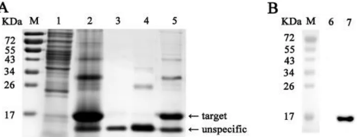
3.1. Expression and purification of recombinant NDV ZJ1-VCD protein 290
A His-tagged form of NDV V protein CTD polypeptide was highly expressed by pET-291
28a-ZJ1/VCD in E. coli and had a molecular weight of 15 kDa as determined by 292
SDS-PAGE, as predicted (Fig. 1A). Recombinant protein His-VCD was purified 293
through Ni-chelating affinity chromatography under denaturing conditions and 294
confirmed by WB with anti-His, in which a single band with the expected molecular 295
weight of approximately 15 kDa was observed (Fig. 1B). The recombinant protein 296
was harvested and used as the antigen for immunization and detection. 297
299 300
Fig. 1. SDS-PAGE and Western blot assays for the recombinant CTD polypeptide of
301
NDV V protein expressed from pET-28a-ZJ1/VCD in E. coli BL21. (A) SDS-PAGE
302
assay of His-tagged form of the ZJ1 V protein CTD region expressed in E. coli. (B)
303
WB assay of the purified recombinant protein His-VCD using anti-His. M, PageRuler
304
prestained protein ladder; 1 and 6, E. coli BL21 lysate (negative control); 2, total
His-305
VCD expressed from pET28a-VCD; 3, His-VCD expressed in the supernatant; 4 and
306
7, His-VCD expressed in inclusion bodies; 5, His-VCD purified from inclusion bodies.
307 308
3.2. Generation of the mAb 3D7 against NDV V protein 309
Five hybridoma cell lines were acquired and only one stably produced antibodies 310
that reacted strongly with His-VCD in indirect ELISA and IFA (data not shown). This 311
mAb clone was designated as 3D7. Using a commercially available isotyping kit 312
(Roche), the mAb 3D7 heavy chain was determined to be IgG1 and the light chain 313
was к. The ascites fluid of mAb 3D7 was produced and purified to the final 314
concentration of 1.5 mg/mL. 315
316
3.3. Specificity of the mAb 3D7 for different NDV genotypes 317
As shown in Fig. 2A, the mAb 3D7 reacted with the recombinant His-tagged V 318
protein expressed by pFLAG-ZJ1-V in DF1 cells. A single band of about 35 kDa was 319
observed, the same as the result with anti-His antibodies. Further, the purified ZJ-1 320
(class II, genotype VII), JS10 (class I), La Sota/46 (class II, genotype II), Herts/33 321
(class II, genotype IV) and ND167 (class II, genotype VI) viruses were detected in 322
WB to determine the reactivity of mAb 3D7 with distinct NDV virions. Both P and V 323
protein were detectible in all of the NDV virions using antiserum anti-PNT; however, 324
mAb 3D7 reacted only with the V protein of ZJ1, but not any other strains. 325
327 328
Fig. 2. Reactivity and specificity assay of mAb clone 3D7 by Western blot. (A) WB
329
assay of the Histagged V proteins expressed by the recombinant plasmid pFLAG
-330
ZJ1-V in DF1 cells using anti-His or mAb 3D7 antibodies. (B) WB assay of V and P
331
proteins contained in different NDV virions using the mAb 3D7 and antiserum
anti-332
PNT. (C) WB assays of the V and P protein expressed in NDV-infected DF1 cells.
333 334
This result was confirmed in NDV-infected DF1 cells. Using mAb 3D7, V protein with 335
a molecular weight of 35 kDa was detected only in ZJ1-infected DF1 cells (Fig. 2C), 336
but not any other NDV-infected cells, involving JS10 (class I), V4 (class II, genotype 337
I), La Sota/46 (class II, genotype II), Mukteswar, HM (class II, genotype III), Herts/33 338
(class II, genotype IV), ND132, ND167 (class II, genotype VI) and F48E8 (class II, 339
genotype IX). As a comparison, P protein were detected in those NDV strains-340
infected cells, with varying molecular weights of around 55 kDa. This result was 341
probably due to different phosphorylation levels of P protein in different strains (Qiu 342
et al., 2016c). 343
344
Indirect ELISA assay was performed and the titer of mAb 3D7 against purified His-345
VCD protein was 1:3200. The mAb 3D7 never react with all the virus detected except 346
for ZJ1. The titer was 1:800. 347
348
3.4. Identification of B cell epitopes recognized by the mAb 3D7 349
The epitopes recognized by mAb 3D7 was mapped in WBs with NDV V protein and 350
its derived protein mutants. As shown in Fig. 3A, the mAb 3D7 did not react with V 351
protein when aa 132-161 or 152-181 were truncated; by contrast, the truncation of 352
172-201, 192-221, 212-231 from V protein never influenced the reactivity of 3D7. 353
Subsequent experiments showed that truncated V protein mutants without peptides 354
spanning aa 140-154, 141-155, 142-156, 143-157, 144-158, 145-159, 146-160, 147-355
161, 148-162, 149-163, 150-164, 151-165, 152-166, 153-167, 154-168, 155-169, 356
156-170 or 157-171 were not recognized by mAb 3D7 (Table 3 and Fig. 3B). These 357
peptides contain 154PAEL157 as the common aa, suggesting that mAb 3D7 358
recognized epitope was between aa 140 and 171 and peptide 154PAEL157 was 359
363
Fig. 3. Mapping of the epitope recognized by mAb 3D7. (A) WB detection of a panel
364
of recombinant V protein, in which 30 aa were consecutively truncated in the CTD
365
region. All the V protein mutants and their deleted regions are listed in Table 2. (B)
366
WB detection of a panel of recombinant V protein, in which 15 aa were consecutively
367
truncated in the ORF spanning from aa 138 to 174. All the V protein mutants and
368
their deleted regions are listed in Table 3. (C) Dot blots detection for the mAb 3D7
369
using synthesized peptides. The labels 144-159, 147-163 and 152-167 indicate the
370
peptides 144SPTSGPTTRGPAELWK159, 147SGPTTRGPAELWKQPGK163 and
371
152RGPAELWKQPGKTAAS167, respectively. S147 A, G148 A, P149 A, T150 A,
372
T151 A, R152 A, G153 A, P154 A, E156 A, L157 A, W158 A, K159 A and Q160 A
373
indicate the polypeptide mutants based on 147SGPTTRGPAELWKQPGK163, in
374
which certain aa was replaced by alanine. All the positive results are labeled with △
375
under the blot. (D) Dot ELISA detection for the mAb 3D7 using synthesized peptides.
376
Error bars represent standard deviation. The OD450 value of synthesized peptides
377
were compared with the positive control by using the Student’s t-test and the ELISA
378
readings that were significantly different from the positive control are labelled *
379
(P < 0.05).
380 381
Fine mapping of the epitope was performed by dot blots and dot ELISAs using three 382
synthesized peptides spanning aa 144-159, 147-163 and 152-167 of V protein. 383
Reactivity of 3D7 with peptide 152-167 in dot blots was impaired compared to 384
peptides 144-159 and 147-163 (Fig. 3C). In dot ELISAs, all three peptides were 385
recognized by 3D7. The OD450 of peptide 152-167 was slightly lower than other 386
peptides (Fig. 3D). This result suggested that aa region 147-159 was involved in 387
formation of the epitope, but only the common aa sequence 152RGPAELWK159 of 388
these peptides was essential. A panel of point mutations was introduced into the 389
synthesized peptides. Removal of residues at R152, G153, E156, W158 and K159 390
blocked recognition by 3D7 in dot blots; while the residue removal at S147, G148, 391
P149, T150 and T151 did not influence the reactivity with 3D7. Besides, the mutation 392
of P154 and L157 compromised mAb reactivity. Similar results were observed in dot 393
ELISA results, the ELISA readings of 3D7 with the peptide mutant R152 A, G153 A, 394
E156 A, W158 A and K159 A were significantly lower than the positive control. In 395
addition, residue K159 was not recognized by the mAb in dot blot assays but was 396
detectible in dot ELISA assays (Fig. 3C and D). 397
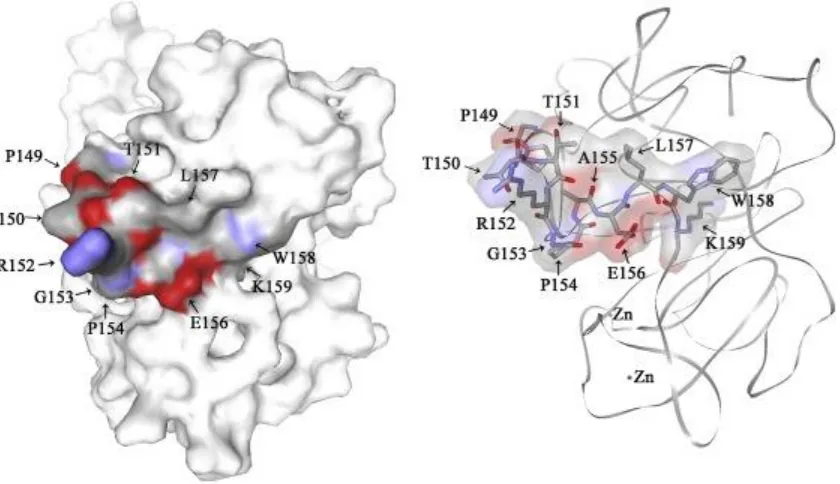
398
3.5. Protein modelling and B-cell epitope analysis of NDV V protein 399
The complete 3D structure of the NDV V protein was modelled according to the 400
crystal structure of its counterpart of PIV5 (Fig. 4). The 3D7-recognized epitope was 401
in the region spanning aa 140–171, all of which was exposed on the surface of V 402
protein. The region 147-159, determined to be recognized by 3D7 were marked in 403
the 3D structure of V protein. The V protein structure displayed that the crucial 404
peptide 154PAEL157 of the 3D7 recognized epitope was not in a same plane with aa 405
147-151, suggesting that aa 147-151 was not a crucial element for direct mAb 406
binding. The linear B-cell epitopes of the NDV V protein was predicted from the 407
primary protein sequences and the discontinuous B-cell epitopes was predicted 408
based on the 3D structure, all of which covered the identified 3D7-recognized 409
peptide 152RGPAELWK159 (Table 4). 410
412 413
Fig. 4. Relative localization of the identified epitopes in the predicted 3D structure of
414
the NDV V protein. The three-dimensional structure of the NDV V protein was
415
modelled by the online services Swiss Model. The identified mAb 3D7-recognized
416
peptide 152RGPAELWK159 and its structurally supporting part is labeled blue and red
417
in the figure. The red areas represent oxygen and the blue areas indicate nitrogen.
418
The model on the left is the predicted V protein structure with the calculated surface
419
structure; while the model on the right displays the atoms of the mAb
3D7-420
recognized peptide in a NDV V protein backbone without showing the surface
421
structure. (For interpretation of the references to colour in this figure legend, the
422
reader is referred to the web version of this article.)
423 424
3.6. Specificity and conservation of the epitope among NDV strains 425
V proteins from distinct NDV genotypes (Table1) were aligned for analysis. The mAb 426
clone 3D7-recognized epitope 147SGPTTRGPAELWK159 and its surrounding 427
region aa 140–146 and 160–171 were conserved among genotype VII NDV strains; 428
however, the epitope shared low identity among other genotypes (Fig. 5), indicating 429
that these sequences were only conserved in specific genotypes. The epitope 430
recognized by mAb clone 3D7 was not in the zinc finger structure of V protein and 431
was likely not a crucial element for NDV V protein, suggesting it as a potential target 432
for NDV genotype and subgenotype differentiation. 433
435 436
Fig. 5. Alignment of the identified epitope in the V proteins of NDV strains from
437
diverse genotypes. V protein sequences from the 27 reference NDV strains of
438
different genotypes were from the EMBL/GenBank (Table 1). Prediction of amino
439
acid (aa) sequences, alignment of sequences and phylogenetic analysis were
440
performed using the MegAlign program in the Lasergene package (DNASTAR Inc.
441
Madison, WI, USA). The aa sequence that resembled the consensus sequence is
442
indicated by a “.”. The region of the mAb 3D7-recognized peptide
443
152RGPAELWK159 identified in the different NDV strains is boxed in the figure.
444 445
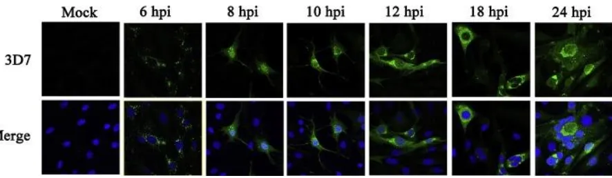
3.7. Expression of V protein during genotype VII NDV infection 446
To determine if mAb clone 3D7 could be used as a tool for immune-detection, 447
dynamic expression of the V protein in NDV-infected cells was surveyed by IFAs and 448
WBs. Firstly, the WB assay of NP protein showed that NDV was propagated in the 449
infected cells. The mAb clone 3D7 reacted with V protein in ZJ-1-infected cells (Fig. 450
6). V protein was detectable at 6 hpi as early as NP of NDV, suggesting it was likely 451
expressed early during the virus’s replicative cycle. Similar results were observed by 452
IFAs (Fig. 7). V protein was initially detected at 6 hpi, scattered in the cytoplasm with 453
dotted distribution. From 8 hpi to 12 hpi, V protein accumulated in the cytoplasm and 454
moved towards the periphery of the host cell nucleus. At 18 hpi, some nuclei of 455
infected cells were surrounded by V protein. A large area of syncytia was observed 456
at 24 hpi, and V protein was observed in the middle of the syncytia surrounded by 457
several host nuclei and starting to dissipate. 458
460 461
Fig. 6. Dynamic expression of V protein in DF-1 cells infected with the NDV strain
462
ZJ1 by WB. DF-1 cells were infected with NDV ZJ1 at MOI 5 and collected at 6, 12,
463
18, 24 hpi. Lysates from cells were immunoblotted with the indicated 3D7, anti-NP
464
and anti-actin monoclonal antibodies.
465 466
467 468
Fig. 7. Dynamic expression of V protein in DF-1 cells infected with the NDV strain 469
ZJ1 by IFA. DF-1 cells were infected with NDV ZJ1 at a MOI of 5 and harvested at 6, 470
8, 10, 12, 18, 24 hpi. Cells were double stained with the mAb3D7 for V protein 471
and 4′,6′-diamidino- 2-phenylindole (DAPI) for nuclei. The upper panel of figures 472
show the V protein (green), while the lower figures show overlapping of V protein 473
(green) and DAPI (blue). (For interpretation of the references to colour in this figure 474
legend, the reader is referred to the web version of this article.) 475
476
4. Discussion 477
It has been reported that NDV V protein plays an important role in facilitating virus 478
replication in infected cells via antagonizing cellular IFN signaling (Huang et al., 479
2003; Park et al., 2003b; Qiu et al., 2016b). The V proteins from distinct NDV strains 480
showed different interferon antagonistic activities (Alamares et al., 2010), albeit the 481
molecular mechanism is unclear since there is little information about the structure 482
and functional domains of NDV V protein. Mapping mAbs binding peptides may shed 483
light on the V protein structure analysis. In this study, a NDV V protein-reactive Mab 484
3D7 was generated. The reactivity of this mAb was limited to genotype VII strain and 485
could be genotype specific (Fig. 2). To analyze the specificity of mAb 3D7, epitope 486
mapping was performed based on detection of consecutive truncated His-tagged V 487
proteins and synthesized peptides. The results showed that the epitope recognized 488
by mAb 3D7 was located in aa 147–159, and in which peptide 152RGPAELWK159 489
was essential (Fig. 3). 490
491
The epitope (152RGPAELWK159) that we identified in NDV V protein was located at 492
a region after the RNA editing site, which was flanked by two important functional 493
[image:14.595.79.522.260.389.2]2016c) and CTD (Horvath, 2004b); nevertheless, there is rare information regarding 495
this region of NDV V protein at present. Based on the crystal structure of SV5 V 496
protein (Li et al., 2006), the 3D structure of NDV V protein was established using the 497
primary sequence. The predicted 3D structure provides potential useful structural 498
information about the function and antigenic characteristics of the V protein. The 499
predicted V protein structure displayed that the aa 147-159 of V protein was exposed 500
on the protein surface and the core part peptide 152RGPAELWK159 formed a 501
pocket, which would be recognized by the mAb. Furthermore, the reactivity of 502
peptide 152-167 with 3D7 was weaker than peptide 144-159 and 148-163, 503
suggesting that the region of aa 147-151 structurally contributed to epitope 504
presentation on the V protein surface although it was not indispensable for mAb-505
epitope interaction, which was supported by the 3D structure of V protein. 506
507
One purpose of our study was to determine if V protein could be used as a tool for 508
quick differentiation of genotypes and subgenotypes. Different from their 509
counterparts in other paramyxoviruses, V protein NDVs are reported to be a 510
structural component of virions (Lamb and Kolakofsky, 2002; Steward et al., 1995), 511
which is confirmed by our results (Fig. 2). It suggested that V protein can be used as 512
a detection target for NDV virion. Bioinformatics analysis of the NDV V protein 513
revealed that aa 147-159 of V protein, especially the core peptide 514
152RGPAELWK159 was exposed on the protein surface and displayed strong 515
antigenicity for B-cell recognition based on the online analysis (Table 4), making it a 516
good target epitope for detection. 517
518
The identified and predicted B-cell epitopes were compared (Fig. 5). The identified 519
epitope region aa 147–159 overlapped with predicted epitopes aa 141-148 and aa 520
150-173. Furthermore, the core peptide 152RGPAELWK159 was totally included in 521
the predicted epitopes aa 150-173. Not only linear epitope but the predicted 522
discontinuous epitope aa 141–163 contained all the region recognized by mAb 3D7, 523
suggesting the peptide would contribute to the formation of conformational epitopes. 524
All the above results indicated that the region of NDV V protein recognized by mAb 525
3D7 was highly immunogenic. 526
527
The mAb only react with genotype VII strain and the sequence alignment indicated 528
that the sequence of aa 140-171 of NDV V protein varied among genotypes but was 529
conserved among NDV strains belonging to the same genotype. Importantly, the 530
152RGPAELWK159 was conserved in genotype VII NDV strains, suggesting that the 531
mAb clone 3D7 recognized an epitope specific for genotype VII or VIId. Defining 532
conserved epitopes can contribute to the development of epitope-based diagnosis 533
methods. It is widely known that most of the prevalent virulent NDV isolates belong 534
to class II, genotype VII; meanwhile, the class I and class II non-virulent strains are 535
spread worldwide due to live vaccine administration and natural infection (Kim et al., 536
2007; Ramey et al., 2013). The limited genetic and antigenic diversity of NDV 537
genotypes makes quick diagnosis complicated and difficult (Miller et al., 2010). Since 538
the epitope recognized by mAb clone 3D7 was conserved in the genotype VII NDV 539
strains, it could be a potential targeting site for NDV genotype and subgenotype 540
differentiation. However, the NDV isolates used for detection in this study is limited, 541
one cannot rule out the possibility that there would be some NDV variants with 542
different reactivity with 3D7. More detection is required before it can be used for 543
conservation, it can be used as an immunogen to establish more genotype-specific 545
mAbs, or directly used for epitope-based genotype differentiation. 546
547
It is found in this study that the V protein-specific mAb clone 3D7 could be applied to 548
various assays. The expression levels and cellular movement of V protein during 549
viral replication were determined, since the subcellular localization of V protein 550
during NDV replication has not been previously reported. Dynamic expression of V 551
protein in NDV-infected cells was seen in IFAs and WBs (Fig. 6, Fig. 7). Scattering of 552
V protein in the cytoplasm was initially detectible at 6 hpi, and the protein moved to 553
the periphery of the host cell nucleus in the process of infection. At late stage of 554
infection, a mass of V protein was observed around the nuclei of infected cells. This 555
result showed the subcellular movement of NDV V protein in the process of IFN 556
antagonism. In response to NDV infection, latent cytoplasmic STAT proteins are 557
phosphorylated on tyrosine by the Janus family of tyrosine kinase (JAK) enzymes 558
and form a heterotrimer of phosphorylated STAT-1, STAT-2 and IRF-9. 559
Subsequently, this heterotrimer translocates to the nucleus and binds to cis-acting 560
DNA elements to activate the IFN-I-stimulated antiviral genes (Horvath, 2004a, b; 561
Samuel, 2001). V protein has IFN-antagonist activity in the CTD, which promotes 562
degradation of STAT1 and blocks IFN signaling (Alamares et al., 2010; Park et al., 563
2003b). Our results showed that V protein tend to accumulate around the nuclei of 564
infected cells, suggesting it might act on STAT-1 protein in the course of nuclear 565
import of phosphorylated STAT-1. 566
567
5. Conclusion 568
The mAb clone 3D7 against NDV V protein was isolated and the mAb recognized 569
epitope was identified to be 152RGPAELWK159. This peptide was located in a 570
region which was varied in sequence among genotypes but conserved in sequence 571
and structure among NDV strains in the same genotype. The generated V protein-572
specific mAb clone 3D7 could be applied to various assays and helped us to 573
determine the location of V protein during NDV replication in infected cells. These 574
results extend our understanding of the antigenic structure of V protein and the 575
function of V protein during NDV infection. They also provide a foundation for 576
development of novel, epitope-based genotype differentiation of NDV genotypes. 577
578
Conflict of interest 579
The authors declare that they have no competing interests. 580
581
Author contributions 582
CD and XQ conceived and designed the research. JL, WW, TR, CM, and YZ 583
performed the experiments. XQ, CM, CS, ZD, XL and YS analyzed the data. LT, SX, 584
WY, XL, VN, MM and YL contributed reagents/materials/analysis tools. XQ and CD 585
wrote the paper. 586
587
Ethical approval 588
The Animal experiment protocol was approved by the Institutional Animal Care and 589
Use Committee (IACUC) of Shanghai Veterinary Research Institute (SHVRI), 590
Chinese Academy of Agricultural Sciences (CAAS), and the Permit Number is shvri-591
mo-0124. The Animal experiment was carried out in agreement with the IACUC 592
guidelines set by SHVRI, CAAS. 593
Informed consent 595
Informed consent was obtained from all individual participants included in the study. 596
597
Data availability 598
All data generated or analyzed during this study are included in this published article. 599
600
Acknowledgements 601
This work was funded by the National Key Research and Development Program of 602
China (2016YFD0501603) and Chinese Special Fund for Agro-scientific Research in 603
the Public Interest (201303033). 604
605
References 606
J.G. Alamares, S. Elankumaran, S.K. Samal, R.M. Iorio 607
The interferon antagonistic activities of the V proteins from two strains of Newcastle 608
disease virus correlate with their known virulence properties Virus Res., 147 (2010), 609
pp. 153-157 610
611
E.W. Aldous, D.J. Alexander Detection and differentiation of Newcastle disease virus 612
(avian paramyxovirus type 1) Avian Pathol., 30 (2001), pp. 117-128 613
614
A. Czegledi, D. Ujvari, E. Somogyi, E. Wehmann, O. Werner, B. Lomniczi 615
Third genome size category of avian paramyxovirus serotype 1 (Newcastle disease 616
virus) and evolutionary implications 617
Virus Res., 120 (2006), pp. 36-48 618
619
Y. Dai, X. Cheng, M. Liu, X. Shen, J. Li, S. Yu, J. Zou, C. Ding 620
Experimental infection of duck origin virulent Newcastle disease virus strain in ducks 621
BMC Vet. Res., 10 (2014), p. 164 622
623
O. de Leeuw, B. Peeters 624
Complete nucleotide sequence of Newcastle disease virus: evidence for the 625
existence of a new genus within the subfamily Paramyxovirinae 626
J. Gen. Virol., 80 (Pt. 1) (1999), pp. 131-136 627
628 629
L. Didcock, D.F. Young, S. Goodbourn, R.E. Randall 630
The V protein of simian virus 5 inhibits interferon signalling by targeting STAT1 for 631
proteasome-mediated degradation 632
J. Virol., 73 (1999), pp. 9928-9933 633
634 635
D.G. Diel, L.H. da Silva, H. Liu, Z. Wang, P.J. Miller, C.L. Afonso 636
Genetic diversity of avian paramyxovirus type 1: proposal for a unified nomenclature 637
and classification system of Newcastle disease virus genotypes 638
Infect. Genet. Evol., 12 (2012), pp. 1770-1779 639
640
K.M. Dimitrov, A.M. Ramey, X. Qiu, J. Bahl, C.L. Afonso 641
Temporal, geographic, and host distribution of avian paramyxovirus 1 (Newcastle 642
disease virus) 643
645
R.E. Gough, D.J. Alexander, M.S. Collins, S.A. Lister, W.J. Cox 646
Routine virus isolation or detection in the diagnosis of diseases in birds 647
Avian Pathol., 17 (1988), pp. 893-907 648
649
A.R. Gould, E. Hansson, K. Selleck, J.A. Kattenbelt, M. Mackenzie, A.J. Della-Porta 650
Newcastle disease virus fusion and haemagglutinin-neuraminidase gene motifs as 651
markers for viral lineage 652
Avian Pathol., 32 (2003), pp. 361-373 653
654
C.M. Horvath 655
Silencing STATs: lessons from paramyxovirus interferon evasion 656
Cytokine Growth Factor Rev., 15 (2004), pp. 117-127 657
658
C.M. Horvath 659
Weapons of STAT destruction. Interferon evasion by paramyxovirus V protein 660
Eur. J. Biochem., 271 (2004), pp. 4621-4628 661
662
Z. Huang, S. Krishnamurthy, A. Panda, S.K. Samal 663
Newcastle disease virus V protein is associated with viral pathogenesis and 664
functions as an alpha interferon antagonist 665
J. Virol., 77 (2003), pp. 8676-8685 666
667
D. Karlin, F. Ferron, B. Canard, S. Longhi 668
Structural disorder and modular organization in Paramyxovirinae N and P 669
J. Gen. Virol., 84 (2003), pp. 3239-3252 670
671
L.M. Kim, D.J. King, D.L. Suarez, C.W. Wong, C.L. Afonso 672
Characterization of class I Newcastle disease virus isolates from Hong Kong live bird 673
markets and detection using real-time reverse transcription-PCR 674
J. Clin. Microbiol., 45 (2007), pp. 1310-1314 675
676
J.V. Kringelum, C. Lundegaard, O. Lund, M. Nielsen 677
Reliable B cell epitope predictions: impacts of method development and improved 678
benchmarking 679
PLoS Comput. Biol., 8 (2012), p. e1002829 680
681
T. Kubota, N. Yokosawa, S. Yokota, N. Fujii 682
C terminal CYS-RICH region of mumps virus structural V protein correlates with 683
block of interferon alpha and gamma signal transduction pathway through decrease 684
of STAT 1-alpha 685
Biochem. Biophys. Res. Commun., 283 (2001), pp. 255-259 686
687
R.A. Lamb, D. Kolakofsky 688
Fundamental virology 689
B.B. Fields, D.M. Kniepe, P.M. Howley (Eds.), Paramyxoviridae: The Viruses and 690
Their Replication, Lippincott Williams & Wilkins, Philadelphia (2002) 691
pp. xi, 1395 p 692
693
Structure of DDB1 in complex with a paramyxovirus V protein: viral hijack of a 695
propeller cluster in ubiquitin ligase 696
Cell, 124 (2006), pp. 105-117 697
698
X. Liu, X. Wang, S. Wu, S. Hu, Y. Peng, F. Xue 699
Surveillance for avirulent Newcastle disease viruses in domestic ducks (Anas 700
platyrhynchos and Cairina moschata) at live bird markets in Eastern China and 701
characterization of the viruses isolated 702
Avian Pathol., 38 (2009), pp. 377-391 703
704
T. Mebatsion, S. Verstegen, L.T. De Vaan, A. Romer-Oberdorfer, C.C. Schrier 705
A recombinant newcastle disease virus with low-level V protein expression is 706
immunogenic and lacks pathogenicity for chicken embryos 707
J. Virol., 75 (2001), pp. 420-428 708
709
L. Mia Kim, D.J. King, D.L. Suarez, C.W. Wong, C.L. Afonso 710
Characterization of class I Newcastle disease virus isolates from Hong Kong live bird 711
markets and detection using real-time reverse transcription-PCR 712
J. Clin. Microbiol., 45 (2007), pp. 1310-1314 713
714 715
P.J. Miller, E.L. Decanini, C.L. Afonso 716
Newcastle disease: evolution of genotypes and the related diagnostic challenges 717
Infect. Genet. Evol., 10 (2010), pp. 26-35 718
719
H. Palosaari, J.P. Parisien, J.J. Rodriguez, C.M. Ulane, C.M. Horvath 720
STAT protein interference and suppression of cytokine signal transduction by 721
measles virus V protein 722
J. Virol., 77 (2003), pp. 7635-7644 723
724 725
M.S. Park, A. Garcia-Sastre, J.F. Cros, C.F. Basler, P. Palese 726
Newcastle disease virus V protein is a determinant of host range restriction 727
J. Virol., 77 (2003), pp. 9522-9532 728
729
M.S. Park, M.L. Shaw, J. Munoz-Jordan, J.F. Cros, T. Nakaya, N. Bouvier, P. 730
Palese, A. Garcia-Sastre, C.F. Basler 731
Newcastle disease virus (NDV)-based assay demonstrates interferon-antagonist 732
activity for the NDV V protein and the Nipah virus V, W, and C proteins 733
J. Virol., 77 (2003), pp. 1501-1511 734
735 736
R.G. Paterson, G.P. Leser, M.A. Shaughnessy, R.A. Lamb 737
The paramyxovirus SV5 V protein binds two atoms of zinc and is a structural 738
component of virions 739
Virology, 208 (1995), pp. 121-131 740
741
X. Qiu, Q. Sun, S. Wu, L. Dong, S. Hu, C. Meng, Y. Wu, X. Liu 742
Entire genome sequence analysis of genotype IX Newcastle disease viruses reveals 743
Virol. J., 8 (2011), p. 117 745
746 747
X. Qiu, Q. Fu, C. Meng, S. Yu, Y. Zhan, L. Dong, T. Ren, Y. Sun, L. Tan, C. Song, X. 748
Han, C. Ding 749
Kinetic analysis of RNA editing of Newcastle disease virus P gene in the early period 750
of infection 751
Acta Virol., 60 (2016), pp. 71-77 752
753 754
X. Qiu, Q. Fu, C. Meng, S. Yu, Y. Zhan, L. Dong, C. Song, Y. Sun, L. Tan, S. Hu, X. 755
Wang, X. Liu, D. Peng, X. Liu, C. Ding 756
Newcastle disease virus V protein targets phosphorylated STAT1 to block IFN-I 757
signaling 758
PloS one, 11 (2016), p. e0148560 759
760
X. Qiu, Y. Zhan, C. Meng, J. Wang, L. Dong, Y. Sun, L. Tan, C. Song, S. Yu, C. Ding 761
Identification and functional analysis of phosphorylation in Newcastle disease virus 762
phosphoprotein 763
Arch. Virol, 161 (2016), pp. 2103-2116 764