DIFFERENTIATION OF BONE CELLS IN VITRO
Cathy Wigzell
A Thesis Submitted for the Degree of PhD
at the
University of St Andrews
1990
Full metadata for this item is available in
St Andrews Research Repository
at:
http://research-repository.st-andrews.ac.uk/
Please use this identifier to cite or link to this item:
http://hdl.handle.net/10023/14070
DIFFERENTIATION OF BONE CELLS IN VITRO
A Thesis
submitted to the University of St Andrews for the Degree of Doctor of Philosophy
by Cathy Wigzell
Department of Biology and Preclinical Medicine University of St Andrews
ProQuest Number: 10166583
All rights reserved INFORMATION TO ALL USERS
The quality of this reproduction is dependent upon the quality of the copy submitted. In the unlikely event that the author did not send a com plete manuscript and there are missing pages, these will be noted. Also, if material had to be removed,
a note will indicate the deletion.
uest
ProQuest 10166583
Published by ProQuest LLO (2017). Copyright of the Dissertation is held by the Author. All rights reserved.
This work is protected against unauthorized copying under Title 17, United States C ode Microform Edition © ProQuest LLO.
ProQuest LLO.
789 East Eisenhower Parkway P.Q. Box 1346
* ® ï
LIST OF CONTENTS
page
Abstract (i)
Declaration (n)
Copyright (iii)
Acknowledgements (iv)
Abbreviations (v)
Figures (vii)
Tables (vni)
Summary (X)
Introduction 1
Materials and Methods 39
Results 65
Discussion 106
ABSTRACT
Osteoblastic differentiation was studied in vitro using primary cultures of bone cells derived from neonatal mouse calvaria.
Using alkaline phosphatase as a marker, maintenance of the osteoblastic phenotype was found to be dependent upon the presence of ascorbic acid. No toxic effect due to ascorbic acid was seen. Insulin and dexamethasone were found to stimulate alkaline phosphatase expression, the former only in the absence of ascorbic acid. Two growth factors, epidermal growth factor and platelet-derived growth factor, were found to inhibit alkaline phosphatase expression in the presence of ascorbic acid.
Osteogenesis was most pronounced in cultures supplemented with ascorbic acid. The osteoblasts formed multilayers of cells and secreted an organic extracellular matrix composed mainly of type I collagen. Matrix vesicles were found among the collagen fibres. In the presence of 6-glycerophosphate, calcium phosphate crystals were deposited in discrete patches forming a mineralisation front which progressively engulfed osteoblasts. The type of matrix formed and the pattern of mineralisation resembled those of lamellar bone.
Insulin at 5000ng/ml stimulated matrix calcification in the absence of ascorbic acid. Dexamethasone, EOF and PDGF inhibited calcification. The extent of calcification was dependent upon the concentration of 6-glycerophosphate in the culture medium.
DECLARATION
I hereby declare that the research reported in this thesis in fulfilment of the requirements governing candidates for the degree of Doctor of Philosophy is my own composition and is the result of work done by me during the period of matriculation for the above degree. No part of this work has been submitted previously for a higher degree.
The research was conducted in the Department of Biology and Preclinical Medicine, United College of St Salvator and St Leonard, University of St Andrews, under the supervision of Dr. C. W. Evans.
Signed Date n
I was admitted to the Faculty of Science of the University of St Andrews under Ordinance General No. 12 in September 1985, and as a candidate for the Degree of PhD in September 1985.
Signed Date
I hereby certify that Cathy Wigzell has spent nine terms engaged in research under my direction, and that she has fulfilled the conditions of General Ordinance No. 12 (Resolution of the University Court No. 1 1967), and that she is qualified to submit the accompanying thesis for the Degree of Doctor of Philosophy.
COPYRIGHT
Unrestricted
ACKNOWLEDGEMENTS
1
ABBREVIATIONS
1 ,25(0H)2D3 1,25-dihydroxyvitamin D3
aFGF Acidic Fibroblast Growth Factor
ALP Alkaline Phosphatase
BDGF Bone-derived Growth Factor
bFGF Basic Fibroblast Growth Factor
BGP Bone Gla Protein
BMP Bone Morphogenetic Protein
BSA Bovine Serum Albumin
cAMP Cyclic Adenosine Monophosphate
CFC Colony Forming Cell
CFU-O Colony Forming Cell - Osteogenic
CM Conditioned Medium
CMF-PBS Calcium/Magnesium Free Phosphate Buffered Saline
CSF Colony Stimulating Factor
CT Calcitonin
DNA Deoxyribonucleic Acid
DOPC Determined Osteogenic Precursor Cell
EDTA Ethylene-Diamine-Tetra-Acetic Acid
EGF Epidermal Growth Factor
FCS Fetal Calf Serum
IB MX 3-Isobutyl- 1-Methyl-Xanthine
IGF Insulin-like Growth Factor
lOPC Inducible Osteogenic Precursor Cell
MEM Minimum Essential Medium
MGP Matrix Gla Protein
mRNA Messenger Ribonucleic Acid
PDGF Platelet-derived Growth Factor
PTH Parathyroid Hormone
SGF Skeletal Growth Factor
TGF Transforming Growth Factor
LIST OF FIGURES
Figure 1. Lineage diagram showing the progression by proliferation and differentiation from a few, undifferentiated cells to the much larger population of terminally differentiated end cells.
4 i
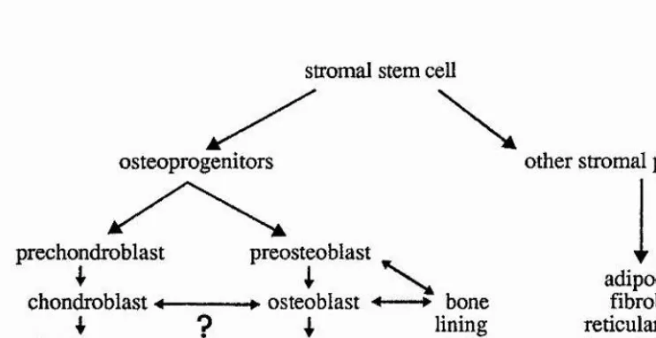
i Figure 2. Lineage diagram showing the inter-relationships between the various
members of the stromal compartment associated with marrow. 5
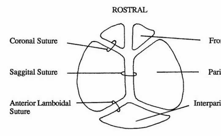
Figure 3. Diagram describing the anatomy of the calvarial plates. 8
Figure 4, Graph to show the effect of ascorbic acid upon alkaline phosphatase
content. 72
I
Figure 5. Graph to show the effect of 6-glycerophosphate upon alkaline
phosphatase content. 73
Figure 6. Graph to show the relationship between p-nitrophenol against alkaline
phosphatase. 76 £
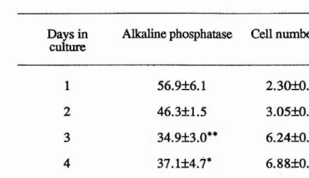
Figure 7. Graph to show the change in alkaline phosphatase activity with time in
culture. 79 %1
i
■H Figure 8. Graph to show the effect of EGF upon alkaline phosphatase activity. 83
Figure 9. Speckle picture of the localisation of calcium in an osteogenic culture. 8 6 J'
Figure 10. X-ray emission analysis of a mineralised nodule. 87 A
Figure 11. TEM from a 7 day old osteogenic culture. 8 8
Figure 12. TEM from a 10 day old osteogenic culture. 8 8
Figure 13. TEM of matrix vesicles amongst collagen fibrils in the extracellular
space. 89 Î
Figure 14. TEM of banded collagen and nascent mineral spicules. 90
1
Figure 15. TEM of an osteoblast in close proximity to mineral. 91 Î
Figure 16. TEM of an osteocyte in the centre of an osteogenic nodule. 92
Figure 17. TEM of a rounded cell on the surface of a nodule. 93 ■■
Figure 18. Phase contrast light micrograph of polygonal osteoblasts in vitro. 94 Figure 19a. Light micrograph of an unmineralised osteogenic culture. 95 Figure 19b. Light micrograph of a mineralised osteogenic culture. 95 Figure 20. Graph to show the effect of insulin upon the extent of
mineralisation. 97
i
Figure 21. Graph to show the effect of epidermal growth factor upon the
extent of calcification. 99 J
Figure 22. Graph to show the effect of culture age upon the ability of the
LIST OF TABLES
Table 1. Summary of the main molecules associated with the bone matrix. 18 Table 2. Summary of the characteristics of the main growth factors found 25
within the bone matrix.
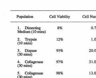
Table 3. Viability and number of cells produced during method II for cell 6 6 preparation.
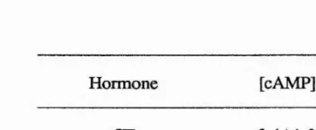
Table 4. Stimulation of cAMP production in bone cells by calcitonin and 67 parathyroid hormone.
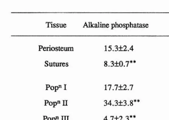
Table 5. Percentage primary cells positive for alkaline phosphatase extracte 6 8 following method I.
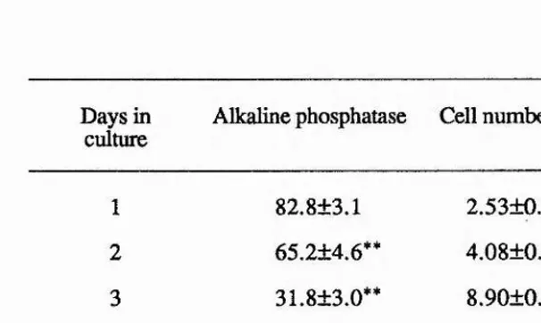
Table 6a. Table showing the alkaline phosphatase content and cell numbers 69 of cultures of cells of the early population prepared
following method II.
Table 6b. Table showing the alkaline phosphatase content and ceU numbers 70 of cultures of cells of the late population prepared
following method II.
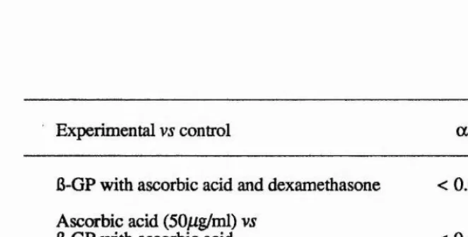
Table 7. Comparison using two way analysis of variance of supplemented 71 cultures, in terms of alkaline phosphatase.
Table 8. Comparison using two way analysis of variance of supplemented 74 cultures, in terms of final cell number.
Table 9. Alkaline phosphatase content and cell number following 14 days 75 in culture with various concentrations of ascorbic acid.
Table 10a. Effect of varying concentrations of ascorbic acid upon cell numbers 77 and alkaline phosphatase activity following 7 days in culture.
Table 10b. Effect of varying concentrations of ascorbic acid upon cell numbers 78 and alkaline phosphatase activity following 14 days in culture.
Table 11. Effect of organic phosphate upon cell numbers and alkaline 78 phosphatase activity.
Table 12. Effect of insulin upon cell numbers and alkaline phosphatase activity. 80 Table 13. Effect of dexamethasone upon cell numbers and alkaline phosphatase 81
activity.
Table 14. Effect of EGF upon cell numbers and alkaline phosphatase activity. 82 Table 15. Effect of PDGF upon cell numbers and alkaline phosphatase activity. 82 Table 16. Data showing the effect of dexamethasone upon the extent of 96
calcification.
Table 18. Effect of seeding density on calcium deposition. 101 Table 19. Proportion of GM-CFC colony types following stimulation with 102
bone cell conditioned medium
SUMMARY
1. The differentiation of mouse osteoblasts was studied in vitro. Bone formation and the expression of alkaline phosphatase were used as markers of the differentiated phenotype. The effects of osteoblastic products upon haemopoietic and mesenchymal cells were also studied.
2. Primary cultures of osteoblasts were established following enzymatic digestion of neonatal mouse calvaria with dispase and collagenase. Three similar methods are compared.
3. Osteoblasts were found to be most abundant in neonatal mouse calvaria, using the expression of alkaline phosphatase as a marker. The periostea and sutures contained few osteoblasts. An initial method of sequential digestion of the calvaria revealed an increased release of osteoblasts from the bones with increased digestion time.
4. Post-confluent cultures (7 days old) of bone cells produced a strong response to parathyroid hormone in terms of cAMP production (140.5 ± 33.2 pmoles/10^ cells). The response to calcitonin was negligible (3.1 ±1.2 pmoles/10^ cells).
5. Ascorbic acid is essential for the maintenance of alkaline phosphatase expression, up to 14 days in culture. In the absence of ascorbic acid there was a general decline in alkaline
Î
phosphatase expression with time in culture. There was no observable toxic action | resulting from the addition of ascorbic acid. This was possibly due to the creation of
hypoxic conditions as the cultures formed multilayers of cells.
6. Following the formation of multilayers, the cultures of osteoblasts developed osteogenic
vesicles were observed. Initial mineral deposition occurred in discrete patches in close association with collagen fibres. In the centre of the nodule, there were cells (osteocytes) embedded within the mineralised matrix and separated from this by a thin layer of osteoid.
7. In the absence of ascorbic acid, 6-glycerophosphate caused a significant increase in alkaline phosphatase expression. However, in the presence of ascorbic acid, 6- glycerophosphate caused a significant (but transient) decrease. 6-glycerophosphate was found to be necessary for the mineralisation of osteogenic nodules. The deposition of calcium increased in a dose-dependent manner with increasing concentrations of 6- glycerophosphate.
10. Media conditioned by osteogenic cultures were found to contain a factor(s) capable of stimulating the formation of colonies by GM-CFC in agar cultures. The activity was secreted constitutively and was probably GM-CSF.
•J
8. Only in the absence of ascorbic acid, did insulin cause a significant increase in alkaline
phosphatase expression and matrix calcification with a concomitant decrease in cell proliferation. No changes in these parameters were seen in the presence of ascorbic acid. However, dexamethasone in the presence of ascorbic acid caused a transient increase in alkaline phosphatase expression over the 14 day culture period but was without effect on cell numbers. By day 14 the stimulation of alkaline phosphatase was no longer apparent and cell proliferation and the extent of calcification were both inhibited. In the absence of ascorbic acid, dexamethasone stimulated alkaline phosphatase expression at day 14.
9. In this system, EGF and PEK3F were both found to lack mitogenic activity and to inhibit | calcification. In the presence of ascorbic acid, both growth factors inhibited alkaline
I
LIST OF CONTENTS
A. INTRODUCTION ... ... 2
B. STRUCTURE OF BONE 1. Cells of bone ... 2
i) Osteoblasts ... 2
a) In vivo ... 2
b) In vitro ... 7
ii) Osteoclasts ... 11
a) In vivo ... 1 1 b) In vitro ... 15
2. The extracellular matrix ... 18
i) The organic extracellular matrix ... 19
a) Collagen ... 19
b) Glycoproteins ... 20
c) Gla-containing proteins ... 2 1 d) Proteoglycans ... 22
e) Plasma proteins ... 23
f) Growth factors ... 23
ii) The inorganic extracellular matrix ... 26
a) The mineral phase ... 26
b) Mineralisation mechanisms ... 27
C. BONE FORMATION 1. Developmental bone formation ... 28
i) Endochondral osteogenesis ... 28
ii) Intramembranous osteogenesis ... 29
2. Bone repair ... 30
3. Experimental bone formation ... 30
i) 7/1 vivo bone formation ... 30
ii) In vitro bone formation ... 32
a) Organ culture ... 32
b) Cell culture ... 33
A. INTRODUCTION
The differentiation of bone cells in vitro is characterised ultimately by the ability to form bone in a controlled manner. This study was undertaken to establish osteogenic cultures in vitro and to determine the influence of various factors upon bone differentiation, namely: ascorbic acid, organic phosphate, insulin, dexamethasone (a glucocorticoid) and growth factors. Since in vivo bone does not exist as an isolated tissue it was also of interest to study the interplay between bone cells and haemopoietic and pluripotent mesenchymal cells.
B. STRUCTURE OF BONE
The two main components of bone are the cellular compartment and the extracellular matrix. Bone is continuously being turned over; matrix is first resorbed by osteoclasts and then replaced by osteoblasts. This dynamic nature is coupled with a highly organised architecture indicating the existence of a complex regulatory mechanism. Bone tissue is responsive to systemic and local factors as well as mechanical stress and as such presents a very complicated model to study.
1. Cells of bone
There are two main cell types associated with bone, osteoblasts and osteoclasts. The sequential activity of these cells results in the steady and orchestrated turnover of the matrix while the terminal differentiation of osteoblasts (to osteocytes) provides an intricate cellular network within the bone matrix.
i) Osteoblasts a) In vivo
reticulum for protein synthesis and a Golgi complex for protein packaging and secretion. Osteoblastic protein products include type I collagen, alkaline phosphatase, osteocalcin and osteonectin (Martin et al.y 1988).
Osteoblasts are found towards the end of a lineage derived from a stem cell which is part of the stromal compartment of bone marrow (Friedenstein et aL, 1987). Cells derived from colonies of bone marrow fibroblasts in vitro are capable of forming bone and cartilage following implantation in vivo in diffusion chambers, however, this has proved unrepeatable with similar cultures of human bone and marrow cells (Ashton et at., 1985). Uncloned spleen stromal fibroblasts transplanted in diffusion chambers following culture in vitro will not normally form bone, the tissue formed being alkaline phosphatase negative and consisting of fibroblasts surrounded by collagen fibres (Friedenstein et al., 1970). Stromal cells with spontaneous osteogenic ability are therefore to be found in marrow stroma, and not in stroma of non-marrow organs.
Having stated that non-marrow stromal cells are incapable of spontaneous osteogenesis, it should be noted that these cells can be induced to differentiate along the osteogenic pathway (Friedenstein et al., 1970). Potent inductive stimuli have been found to be,
i) transitional epithelium, and ii) decalcified bone matrix.
Stem cell ---> committed ---- > differentiated
progenitors end cells
Figure 1. Lineage diagram, showing the progression by proliferation and differentiation from a few, undifferentiated stem cells to the much larger population of terminally differentiated end cells.
Transplantation of marrow fragments results in osteogenesis at the new site followed by infiltration by haemopoietic cells of host origin (Friedenstein, 1976). The type of haemopoietic tissue formed is dependent upon the source of the transplant, ie red or yellow marrow (Patt et al., 1982). Osteogenesis occurs in a similar manner, regardless of the marrow origin so the haemopoietic regulation is transferred by a set of marrow stromal cells. Retransplantation of the marrow component of the graft results in the formation of new osteogenic tissue. However, the number of possible retransplantations is very limited. There is therefore some particular element in bone marrow stroma that is capable of osteogenesis which is not found in the stroma of other organs. This indicates a finite ability of the stromal cells to self-maintain in an obvious absence of replenishment by host haemopoietic cells. Marrow transplanted from male to female mice survives and forms a new organ composed ostensibly of donor stroma and host marrow. Retransplantation of this organ to a secondary female host pre-immunised against male antigens results in resorption of the graft. If the transplant in the prirnary host had been repopulated with any host (female) stroma cells then the organ would survive transplantation to the secondary host (female). However, this does not happen and consequently all the stroma cells are male and therefore rejected by the pre-immunised secondary host (Friedenstein and Kuralesova, 1971). Therefore, despite the close structural and functional interplay between the osteogenic and haemopoietic compartments in vivo, the two lineages appear to self- maintain independently in the adult animal.
progenitors and differentiating end cells (Owen, 1985). It is proposed that there is a pluripotent stromal stem cell capable of giving rise to committed progenitors for reticular, fibroblastic, adipocytic and osteogenic lineages (Owen and Ashton, 1986). Similar specialised stromal stem cells may exist in other systems (Figure 2). Following proliferation, the osteoprogenitor gives rise to the preosteoblast, an alkaline phosphatase positive, fibroblastic cell still capable of mitosis (Owen, 1980). The preosteoblast is the immediate precursor to the osteoblast, which has very little, if any, mitotic potential.
stromal stem cell
osteoprogenitors
prechondroblast
chondroblast 4
---4 ?
chondrocyte 4 ---hypertrophied
chondrocyte
preosteoblast
osteoblast 4---► bone
j lining
-► osteocyte cell
other stromal progenitors
adipocyte fibroblast reticular cell endothelial cell
Figure 2. Lineage diagram showing the inter-relationships of the various members of the stromal compartment associated with marrow (after Marks and Popoff, 1988; Martin et ai, 1988).
[image:23.612.110.481.275.466.2]lining cells. They form a cellular membrane physically separating the bone fluids from the interstitial fluids and could therefore contribute to the regulation of mineral homeostasis by maintaining and regulating ion gradients (Bushinsky et al,, 1989). The bone lining cells form gap junctions between each other and also with osteocytes, raising the possibility of the existence of a 'functional syncytium' composed of bone lining cells and osteocytes; this may support the metabolism of osteocytes within the matrix. The bone lining cell may also be involved in the initiation of remodeling by;
i) attracting osteoclast precursors,
ii) retracting and therefore exposing the bone surface, and
iii) removing osteoid, ie the covering layer of unmineralised bone matrix.
It is also believed that the bone lining cell may represent an inactive osteoblast (Peck and Woods, 1988).
osteoid-osteocyte (Palumbo, 1986). This cell type is still capable of synthesising matrix proteins and is completely surrounded by osteoid.
b) In vitro
The heterogeneity of bone tissue poses problems concerning the interpretation of results. A method to simplify any system is to break it down into its component parts and to examine these individually. The culture of osteoblastic cells can be used to answer questions concerning, for example, the regulation and mechanisms of osteoblastic behaviour. The results from such studies, however, may not bear a direct correlation with the in vivo situation, although they will be an approximation and will aid the interpretation of in vivo data.
ROSTRAL
Frontal Bone Coronal Suture
Saggital Suture Parietal Bone
Anterior Lamboidal
Suture Interpaiietal Bone
CAUDAL
Figure 3. Diagram describing the anatomy of the calvarial plates.
The use of organ culture provides a system which maintains the inter-cell spatial arrangements and contacts and as such is very like the in vivo situation. However, these methods provide scant information concerning the function and responses of individual cell types. To do this it is necessary to isolate the bone cell types and study cultures derived from purified cell fractions. There are various methods for doing this and they can be broadly divided into mechanical and enzymatic.
[image:26.613.114.475.67.290.2]to be passaged before being used, which could result in non-mitotic osteoblasts being over run by more prolific cell types, eg fibroblasts.
Enzymatic isolation techniques produce a high yield of primary cells and can be used to establish cultures derived from cell populations with different properties eg response to parathyroid hormone and calcitonin. Peck and colleagues (1964) prepared a single cell suspension of bone cells by incubating fetal and neonatal rat calvaria with collagenase. All the cells released exhibited detectable alkaline phosphatase and half showed intense cytoplasmic reaction. Other enzymes were also tried but these caused either too much cell damage (pronase) or released too few cells (trypsin). Another early attempt to isolate bone cells used a cocktail of enzymes (Hekkelman and Moskalewski, 1969). Collagenase, trypsin and DNA-ase dissolved in phosphate buffered saline were found to release alkaline phosphatase positive cells from fetal rat calvaria. Tryspin alone has also been used on periosteum-free fetal rat calvaria (Binderman et al., 1974). Alkaline phosphatase activity was found in the bone cells isolated, and cultures established with these cells produced a calcified extracellular matrix.
A major step forward in the production of bone cell suspensions was the development of sequential digestion by Wong and Cohn (1974). They digested neonatal mouse calvaria with an enzyme solution of trypsin and collagenase for a total of one hour. However, at twenty minute intervals the enzymes were aspirated and replaced with fresh solution. This method produced three different aspirates, each containing a different population of bone cells. These populations were then characterised in terms of cAMP response to parathyroid hormone and/or calcitonin. The last population was the most responsive and also exhibited an additive effect of the hormones, thus showing;
i) the existence of a sub-population of bone cells acting as a target for parathyroid hormone and calcitonin, and also
Cultures established following five digestions showed the early populations ( 2 and 3) to respond to calcitonin and the late populations (3, 4 and 5) to respond to parathyroid hormone (Wong and Cohn, 1975). Populations of bone cells with different properties can therefore be isolated from bone using sequential digestion.
The methodology of sequential digestion has the advantage of producing functionally different bone cell populations. However, large numbers (> 50) of calvaria are required to achieve a good cell yield (Boonekamp et ai, 1981). Careful dissection of the bones before digestion can greatly reduce contamination by non-osteoblastic cells. Yagiela and Woodbury (1977) dissected out the frontal and parietal bones of fetal and neonatal rats, ensuring that the bone segments were free of both suture tissue and immature bone. The periosteum was then peeled off the bones and the fragments incubated with Worthington collagenase class II for two hours. They found that the highest numbers of osteoblastic cells (as judged by morphological criteria) were present in the 18 to 19 day old rat fetus. Cells with osteoblastic properties can also be isolated from fetal chick calvaria using a similar method (Nijweide et al., 1981).
Contamination by non-osteoblastic cell types can be avoided, not only by careful dissection, but also by discarding the supernatant of early digests (Heath et ai, 1984). Neonatal mouse calvaria were stripped of periostea and sutures and then incubated with trypsin (10 minutes) and dispase (30 minutes). The cell suspensions produced were discarded and a final two incubations with Worthington collagenase class II (30 minutes each) were pooled. The cell population was enriched for osteoblasts, as assessed by alkaline phosphatase content, collagen type I synthesis and cAMP response to parathyroid hormone.
pronounced cAMP response to parathyroid hormone and no response to calcitonin and they also retained an osteogenic ability, forming osteogenic tumours following transplantation in vivo. MC3T3 lines were established from neonatal mouse calvaria (Kodama et al., 1981). Primary cultures were produced by allowing cellular outgrowth from periosteum-free bone fragments in vitro. The cells were passaged using trypsin and selection of a suitable clone, MC3T3-E1, was based upon alkaline phosphatase activity.
ii) Osteoclasts a) In vivo
The osteoclast is a large multinucleated cell found associated with bone surfaces and it is the major cell type involved in bone resorption (Marks and Popoff, 1988). The cell membrane of the active osteoclast in contact with the bone is divided into two functional areas. The inner, central area of the membrane forms a ruffled border and is the site of secretion of enzymes. The extracellular space bounded by the ruffled membrane is also acidified due to the catalytic action of carbonic anhydrase on metabolic products (Hall and Kenny, 1986);
CO2 + H2O "> H+ + HCO3
The outer ring of membrane is flattened and is in close contact with the bone surface to form a clear zone functioning as a tight lateral seal limiting the area of bone resorption and thus allowing the formation of a microenvironment suitable for osteolysis (Sakamoto and Sakamoto, 1986). Inactive osteoclasts are not in contact with the bone surface and are kept away by the covering of osteoblasts and bone lining cells (Sterrett, 1986). Inactive osteoclasts do not possess a ruffled membrane.
of cell types; local mononucleated preosteoclasts, osteocytes, preosteoblasts and blood- borne cells (Hanaoka et al., 1989).
The distinctness of osteoclasts from the osteogenic lineage has been shown by studying the quail nuclear marker during endochondral ossification (Kahn and Simmons, 1975; Jotereau and Le Douarin, 1978). On the basis that chick chromatin is evenly distributed and quail chromatin is found in clumps it is possible to follow cell lineages in chimeric bones. Transplantation of quail limb bud onto chick embryo chorio-allantoic membrane resulted in endochondral ossification of the transplanted tissue and the formation of a bone organ with associated marrow. All the stroma cells were of donor (quail) origin and all the haemopoietic cells of host (chick) origin. It was also found that none of the osteoclasts exhibited quail chromatin, ie they were of host origin, indicating a blood borne (as opposed to osteal) and therefore haemopoietic derivation. Transplantation from chick to quail produced similar results, though not as conclusively as only two thirds of quail cells possess clumped chromatin.
The preosteoclast is a mononuclear precursor with the morphology of a mononuclear leukocyte (Bonucci, 1981). Osteoclasts have been shown to develop in initially osteoclast-free explants of fetal mouse calvaria following co-culture with previously cultured mononuclear cells from adult marrow (Ko and Bernard, 1981). Osteoclasts were not found in control cultures, showing that the osteoclastic precursors were amongst the mononuclear population.
As has been seen, the haemopoietic and osteogenic lineages in the adult animal self- maintain independently (Friedenstein and Kuralesova, 1971). Therefore the osteoclast is ultimately derived from a haemopoietic stem cell (Schneider et al., 1986). Developmentally, osteoclast stem cells and/or progenitors migrate from the yolk sac and are initially widely disseminated throughout the fetus (Thesingh, 1986).
The major role of the osteoclast is bone resorption. In vivo, resorption is followed by bone formation and the two complementary processes are coupled together to maintain a steady bone mass, despite variable turnover. This mechanism is referred to as coupling and it occurs during bone remodeling (Peck and Woods, 1988).
The remodeling cycle is characterised by the orderly progression through the sequence;
i) quiescence,
ii) initiation and activation, iii) resorption,
iv) reversal, v) formation,
vi) return to quiescence,
(Kahn et al., 1983; Marcus, 1987; Martin et al., 1988; Parfitt, 1988; Peck and Woods, 1988; Raisz, 1988).
collagenase (Heath et al., 1984; Puzas and Brand, 1979; S live et al., 1982). The action of parathyroid hormone upon osteogenic cells results in the digestion of the unmineralised layer of matrix thus exposing the bone mineral. The release of parathyroid hormone may represent an initiation event causing the activation of osteoblasts and the subsequent chemotactic response of osteoclasts, possibly to the exposed bone mineral (Osdoby et al.,
1987). Monocytes can respond chemotactically to bone proteins (osteocalcin, a2HS glycoprotein and collagen type I) (Malone et al., 1982). Not only do hormones which stimulate bone resorption, for example parathyroid hormone and prostaglandin E^, promote collagenase release, they also cause shape changes in osteoblastic cells, thus uncovering the matrix (Rodan and Martin, 1981). Osteoblasts may also play an important role in the differentiation of mononucleate osteoclast precursors by fusion to form functional osteoclasts (Takahashi et al, 1988).
possibly by removing the exposed collagen fibres. These cells may also deposit the cement line, demarcating the furthest extent of resorption. The stage between the end of resorption and the beginning of bone formation is known as reversal and may last for one to two weeks.
Bone formation is initiated by the recruitment of osteoblasts and the subsequent secretion of matrix. Osteoblasts in vitro respond chemotactically to a protein component of bone matrix (Somerman et al., 1983). Resorbing bone is also a good source of mitogenic activity which may act on osteoprogenitor cells to produce an increase in osteoblast number (Farley et al., 1987). The attraction of osteoblasts to the site of bone remodeling may therefore be controlled by the release of chemoattractants and mitogens from secondary lysosomes of osteoclasts following resorption from the matrix.
Initially, the matrix formed by the osteoblast is unmineralised but as formation progresses the matrix calcifies and a mineralisation front forms, above which lies an osteoid seam. The most recently formed collagen fibres in the osteoid are disorganised but become increasingly aligned due to cross-linking before the matrix is mineralised. As formation progresses the osteoblast becomes smaller and there is a probable corresponding decrease in the rate of matrix synthesis. Eventually the osteoid seam disappears as the mineralisation front advances and the bone surface returns to the quiescent state.
b) In vitro
Cultures of isolated osteoclasts can give more information concerning, for instance, the specific cellular response to bone hormones. To establish such cultures, the femoral and tibial shafts of neonatal rats are split longitudinally and the cut surface is scraped to release bone fragments into culture medium (Chambers and Magnus, 1982). The fragments are agitated by pipetting to release cells and aliquots of the medium transferred to tissue culture grade Petri dishes. Following a 30 minute incubation at 37°C, the dishes are washed and the medium replaced. The adherent cells remaining in the Petri dishes are predominantly osteoclasts; they are large multinucleated cells with a membranous 'skirt' giving a 'fried egg' appearance (Arnett and Dempster, 1986; Chambers and Magnus,
1982).
The regulation of osteoclastic behaviour is complex and not always direct. Two potent stimulators of bone resorption are parathyroid hormone (PTH) and a metabolite of vitamin D, 1,25-dihydroxyvitamin D3 (l,25(OH)2D3). In vivo, a drop in blood calcium levels stimulates the parathyroid gland to release PTH (Dickson, 1987). The hormone can then influence bone resorption via two prominent mechanisms.
PTH can act directly upon bone cells (Chambers et at., 1984). Added to isolated osteoclasts, PTH has no discernible effect, however, co-culture of osteoclasts with osteoblasts results in increased osteoclastic spreading and bone resorption (McSheehy and Chambers, 1986a). PTH stimulates the osteoblastic cells to produce a factor that directly affects osteoclasts and stimulates bone resorption (McSheehy and Chambers, 1986b). PTH has also been shown to stimulate osteoblastic cells to secrete prostaglandin E (MacDonald et al., 1984). Prostaglandins can either inhibit osteoclast motility, or can stimulate the release of osteoclast activation factor (OAF) from phytohemaglutin-stimulated lymphocytes (Chambers and Ali, 1983; Sterrett, 1986).
Alternatively, PTH can influence the production of l,25(OH)2D3 (Dickson, 1987). Vitamin D is converted to 25-hydroxyvitamin D3 in the liver. This metabolite is acted upon by a PTH-regulated kidney hydroxylase to produce l,25(OH)2D3. The systemic effect of
i) stimulating the synthesis of calcium-binding protein by the gut and thus calcium absorption, and
Ü) stimulating bone resorption.
In bone, the cellular site of action of l,25(OH)2D3 is very likely to be the osteoblast. Two cell lines have been shown to possess cytosolic receptors for 1,25(0H)2D3 (Partridge et al., 1980; Walters et aL, 1982). The indirect effect of 1,25(0H)2D3 upon bone resorption may be mediated through osteoblastic cells, in a manner similar to that of PTH.
Vitamin D is also, paradoxically, involved in mineralisation (Wong et al., 1977). However, there are many metabolites of this vitamin and it is probably the concerted action of 1,25(0H)2D3 raising the serum calcium levels (by increased absorption from the intestine) with 24,25-dihydroxy vitamin D3 that enhances mineralisation (Ornoy et al., 1978).
Calcitonin, produced in the thyroid, acts directly via an osteoclastic receptor to cause inactivation and inhibition of bone resorption (Nicholson et al., 1986). Exposure of isolated osteoclasts to calcitonin results in a decrease in motility and retraction of lamellipodia (Chambers and Magnus, 1982). The transformation caused by calcitonin occurs at very low concentrations (> 3pg/ml), is reversible and can be prevented by pre incubation with trypsin (Chambers and Moore, 1983).
2. The extracellular matrix
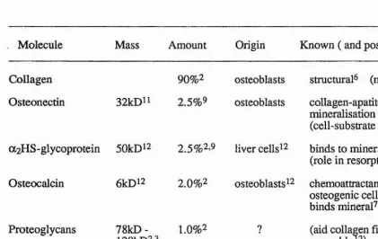
The extracellular matrix of bone consists of a mineralised framework mainly of type I collagen, and some non-collagenous proteins. The organic component is synthesised and secreted largely by osteoblasts but it does contain some plasma-derived proteins. The matrix is mineralised by the deposition of hydroxy apatite (a form of calcium phosphate). The non-collagenous proteins are probably involved in the general maintenance and regulation of the matrix secretion and mineralisation (Table 1.)
Molecule Mass Amount Origin Known ( and possible) roles
Collagen 90%2 osteoblasts structural^ (mitogen®)
Osteonectin 32kD" 2.5%9 osteoblasts collagen-apatite bridge2
mineralisation regulator^ (cell-substrate bridge^®) a2HS-glycoprotein 50kDi2 2.5%2.9 liver cells ^2 binds to mineraP2
(role in resorption^2) Osteocalcin 6kDi2 2.0 % 2 osteoblasts ^2 chemoattractant for
osteogenic cells binds mineral^
Proteoglycans
78kD-120kD2.3 1.0 % 2 7 (aid collagen fibrilassembly42) Bone
sialoprotein 23kDi2 01..82%i- 2
? cell adhesion^
binds to calcium^^
Table 1. Summary of the main molecules associated with the bone matrix 1. D oiera/., 1989
2. Gehron Robey et al., 1988 3. Goldberg era/., 1988 4. Lucas et al., 1988 5. Oldberg et al, 1986 6. Piez, 1987
1. Price, 1983
8. Rath and Reddi, 1979 9. Rodan and Rodan, 1984
[image:36.618.77.498.255.520.2]i) The organic extracellular matrix a) Collagen
Type I collagen is the major constituent of bone matrix, of which it comprises roughly 90% of the organic part (Rodan and Rodan, 1984). Collagen is formed from three collagenous propeptide chains wound round each other to produce a triple helix structure (Gehron Robey et al., 1988). The polypeptide chains of type I collagen are two a 1(1) chains and one a2(I) chain. There are at least ten different collagen types whose functions are predominantly structural (Piez, 1987). It may also act as a local mitogen (Rath and Reddi, 1979).
Triple helical procollagen is released from secretory vesicles into the extracellular space where the molecule is converted to collagen by the enzymatic removal of the amino- and carboxyl-terminal propeptides. The collagen molecules are then arranged into bundles to form collagen fibrils which are characterised by a regular banding pattern. Cross-linking then occurs between the fibrils to produce a very stable structure and to protect the molecule from degradation (Laurent, 1987). Nevertheless, collagen molecules undergo degradation both intra- and extracellularly, possibly to preserve collagen homeostasis.
The secretion of normal collagen is dependent upon the presence of ascorbic acid (de Clerck and Jones, 1980). It is necessary for the normal synthesis of hydroxyproline (which stabilises the helical structure) and hydroxylysine (which forms the cross-links) (Murad et al., 1981). Ascorbic acid functions as a co-factor in the hydroxylation of prolyl and lysyl residues (Pinnell et al., 1987). Ascorbic acid has also been shown to cause an increase in the levels of mRNA for the procollagen polypeptide chains.
likely that the development of the fibril orientation is controlled by osteoblasts (Jones and Boyde, 1976).
b) Glycoproteins
The bone matrix contains several different glycoproteins. Bone sialoprotein accounts for 8% to 12% of the non-collagenous protein (Triffitt, 1987). Bone sialoprotein contains roughly 20% sialic acid (Butler, 1987). Two other proteins, sialoprotein I and sialoprotein
n,
contain 5% and 13% sialic acid respectively. Sialoprotein I (or osteopontin) and sialoprotein II share sequences with fibronectin and vitronectin and can cause the adhesion and spreading of ROS cells (Oldberg et al., 1986; Oldberg et al., 1988b). Indeed, the receptor for osteopontin is identical to that for vitronectin, Sialoprotein mRNA is found in bone, but not in ROS cells, suggesting that osteoblasts may not necessarily be the source of this glycoprotein (Oldberg et al., 1988a). Despite the marked presence of sialoproteins within the bone matrix, their function is unknown. Apart from the cell- binding regions they exhibit strong calcium binding properties and may act as organic- inorganic bridges (Vaughan, 1984).Osteonectin is synthesised in vitro by osteoblastic cells isolated by enzymatic digestion of fetal porcine calvaria. It is also found within platelets and may be released during coagulation. This possibility has given rise to the idea that osteonectin may also be
5
1
involved in the adhesion of cells to substrates (Otsuka et al., 1984; Stenner et al., 1986;
Tracy et al., 1988). Osteonectin shows some sequence homology with two glycoproteins | not associated with bone, namely a 43K protein and SPARC (secreted protein which is
acidic and rich in cysteine) (Tracy et al., 1988).
Thrombospondin is an adhesive glycoprotein synthesised by osteoblasts and is incorporated into the matrix, although this may not be the sole source of bone thrombospondin (Gehron Robey et al., 1989). The molecule is endowed with several binding sites for Ca2+ as well as for collagen and has been found to form complexes with osteonectin; this may reflect its bone-related function (Clezardin et al., 1988).
c) Gla-containing proteins
Two y-carboxyglutamic acid (Gla)-containing proteins are found in the bone matrix. The presence of Gla in clotting factors is important during coagulation (Triffitt, 1987). Glutamic acid is converted to Gla by a vitamin K-dependent process.
osteogenic cells. It is likely that osteocalcin is synthesised and secreted by a cell belonging to the osteogenic lineage. Its production is stimulated by l,2 5(OH>2D3 and inhibited by TGFB (Lian et a l, 1985; Noda, 1989; Price, 1983). This response to l,25(OH)2D3 is very specific and provides a method to identify osteoblasts in vivo (Gallagher et al,
1986).
As the synthesis of osteocalcin is dependent upon vitamin K, the function of osteocalcin can be studied in animals treated with the vitamin K antagonist, Warfarin (Price, 1983). However, Warfarin-treated animals appear to have normally mineralised bones, however, although there are some abnormalities associated with the mineralisation of cartilaige. These effects are due to the inhibition of synthesis of another Gla-containing protein, matrix Gla-protein (MGP) (Price, 1988).
MGP is found within the matrices of bone and cartilage, and in bone it is associated with BMP (Triffitt, 1987). It is a larger molecule than osteocalcin but they share homologous sequences, suggesting a common genetic ancestor.
d) Proteoglycans
Proteoglycans are characteristically composed of a protein core with glycosaminoglycan side chains. In mineralised matrix they constitute roughly 10% of the non-collagenous protein (Gehron Robey et al, 1988). The function of proteoglycans may be to aid the assembly of collagen fibrils (Triffitt, 1987). Proteoglycans have been found to be localised to the developing bone matrix and osteogenic cells (Termine, 1983).
hydroxyapatite-4
associated proteoglycans (HAPGl, 2 and 3) have been recorded (Goldberg et al., 1988). HAPGl has a molecular weight of 110 - 120kD and a protein core of 45kD. HAPG2 and HAPG3, though distinct, have a total weight of 100 - llOkD and protein cores of 37 - 38kD.
e) Plasma proteins
The bone matrix contains proteins concentrated from plasma. a2HS glycoprotein accounts for approximately 25% of the non-collagenous protein (Rodan and Rodan, 1984). a2HS glycoprotein is synthesised in the liver and has a high affinity for bone mineral and may play a role in resorption (Triffitt, 1987). Serum albumin is also found in bone (Gehron Robey etal., 1988).
f) Growth factors
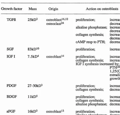
Growth factors are usually small polypeptides with hormone-like characteristics. They function as growth regulators by acting as mitogens (Canalis, 1988; Maclean and Hall, 1987). Growth factors associated with the bone matrix may play an important role in the coupling of resorption and formation as well as acting as mediators for systemic hormones. Growth factors found within the matrix include fibroblast growth factor (both acidic and basic), transforming growth factor (6i and 6 2). insulin-like growth factor (I and II), bone-derived growth factor, platelet-derived growth factor and skeletal growth factor (Table 2) (CanaHs, 1988; Lau etal., 1988).
Transforming growth factor (TGF) can induce the formation of anchorage- independent colonies by non-neoplastic cells. There are two classes of TGF; a and 6. TG Fa acting alone can induce colony formation, and it is not found in bone. TGFB induces colony formation in the presence of epidermal growth factor (EGF) and is found in three forms; Bi, B2 and B1 .2 (Canalis, 1988). TGFB is probably identical in structure and function to cartilage-inducing factor A (Ellingsworth et al., 1986).
al., 1987; Sandberg et al., 1988). During endochondral osteogenesis TGFB is localised within the developing mineralised matrix (Carrington et al., 1988). In vitro, TGFB affects the differentiation and proliferation of bone cells. Cell growth is inhibited in two cell lines, ROS and MC3T3-E1, by TGFB in the range 2 to lOng/ml (Elford et al., 1987b; Noda and Rodan, 1986; Noda and Rodan, 1987). However, the phenotypic expression of alkaline phosphatase is stimulated in ROS cells and inhibited in MC3T3-E1 cells. Primary bone cell cultures show stimulated proliferation and collagen synthesis in response to TGFB and inhibition of alkaline phosphatase activity (Centrella eta l, 1987a; Centrella eta l, 1987b; Rosen eta l, 1988; Wranaera/., 1988).
TGFB may act as a coupling factor during the cycle of resorption and formation. Osteoclasts are capable of synthesising TGFB, which is activated by an acidic environment, such as that found associated with the ruffled border of activated osteoclasts. The activated TGFB could then act upon osteoblastic cells consequently stimulating both collagen synthesis and proliferation in direct relation to osteoclast action.
Bone-derived growth factor (BDGF) is synthesised, though not exclusively, by bone cells (Canalis, 1988; Canalis et al, 1988b). However, to call BDGF a growth factor may be a misnomer, as it is 6 2 microglobulin, a small globular peptide associated with a major histocompatability complex antigen. Nevertheless, BDGF has been found to stimulate collagen and DNA synthesis in bone, as well as to act as a mitogen for fibroblasts (Centrella and Canalis, 1985; Canalis and Centrella, 1986). Six other distinct BDGFs have been extracted from fetal calf bone (Hauschka et al, 1986).
Growth factor Mass Origin Action on osteoblasts
TGFB 25kD3 osteoblast^ ^ '^ 2
osteoclast24 proliferation;
alkaline phosphatase; collagen synthesis; cAMP resp to PTH;
increase^'^dz
decrease®’^^’2 1 ,2 2 increase2 2
decrease^’®’i^’2i increase^’^ b2 2 decrease^^ decrease®’ SGF 83kDio proliferation; increase^^’^®
IGF I 7.5kD4 osteoblast^4 proliferation; increase^*!^
collagen synthesis; increase4 4 7,i9 IGF I synthesis increased by;
PTH2 0,
l,25(OH)2D3i4, estradioP4^ growth hormone^
PDGF 27-30kD® proliferation;
collagen synthesis; increase^ decrease1
BDGF HkD5 proliferation;
collagen synthesis; alkaline phosphatase;
increase^ increase® decrease2®
aFGF 16kD3 osteoblast^® proliferation;
alkaline phosphatase; increase ^2.13,23decrease2® bFGF 17kD3 osteoblast^® proliferation;
collagen synthesis; increase2’i2,i3decrease2
Table 2. Summary of the characteristics of the main growth factors found within the bone matrix.
1. Canalis, 1981 2. Canalisera/., 1988a 3. Canalis et a/., 1988b 4. Canalis et al, 1988c
5. Canalis and Centrella, 1986 6. Centrella er a/., 1987a 7. Centrella era/., 1988 8. Elford era/., 1987 9. Ernst and Froesch, 1988
10. Farley and Bay link, 1982 11. Gehron Robey et al, 1987 1 2. Globus eta l, 1988
13. Globus era/., 1989 14. Gray et al, 1989 15. Guenther er a/., 1988 16. Gutierrez er a/., 1987 17. Hock era/., 1988 18. Lau e ta l, 1988
[image:43.616.80.498.95.498.2]Platelet-derived growth factor (PDGF) is synthesised by bone cells and is found in the matrix (Canalis, 1988; Canalis et al., 1988b). PDGF has been shown to stimulate proliferation and protein synthesis. However, it can also stimulate resorption via a prostaglandin-dependent pathway. Acidic and basic fibroblast growth factors (aFGF and bFGF) are both synthesised by osteoblastic cells and are found in the bone matrix (Globus et al., 1989). They both stimulate bone cell proliferation causing a subsequent increase in collagen synthesis. Skeletal growth factor (SGF) is also found in bone matrix (Farley and Bay link, 1982). SGF has been shown to stimulate proliferation in primary cultures of chicken bone cells, however, no such stimulation occurs in the MC3T3-E1 cell line. MC3T3-E1 cells respond to SGF with an increase in collagen synthesis (Lau etal., 1988; Linkhart et al., 1986). The primary bone cells may represent a more immature cell type and SGF may, therefore, cause different effects in different osteogenic populations.
Bone morphogenetic protein (BMP) isolated from demineralised bone is capable of inducing osteogenic differentiation in connective tissue cells and was thought to be a glycoprotein (Urist et al., 1979). However, BMP has been found to be composed of the combined action of several factors (Wozney et al., 1988). At least three components of BMP are related to TGFB in sequence structure and another may act as a protease activator for TGFBi.
ii) The inorganic extracellular matrix a) The mineral phase
b) Mineralisation mechanisms
Mineralisation is initiated by the nucleating action of collagen fibrils or matrix vesicles, depending on the type of bone.
Woven bone (primary or immature) is found in fetal tissues and in areas of rapid bone growth. It is covered by a thin layer of osteoid and the collagen fibrils are randomly orientated. Mineral crystals first appear in association with matrix vesicles. These are membrane-bound extracellular vesicles containing glycoproteins, lipids, alkaline phosphatase and mineral ions, particularly Ca2+ and Pi (McLean et al., 1987). The vesicles are probably derived by 'blebbing' from the cell membrane (Anderson, 1989).
Electron microscopy has shown that the earliest crystals form within matrix vesicles. As the crystals grow the vesicle is obliterated and the surrounding matrix is calcified (Bonucci, 1987). The role of alkaline phosphatase during crystal formation is unclear. Matrix vesicles do not always require alkaline phosphatase substrates to initiate mineralisation (Wuthier, 1986). It may be the Ca2+-binding properties of alkaline phosphatase that are important during mineralisation (de Bernard et al., 1986).
Lamellar bone (secondary or mature) is usually found in adult tissues. It is covered by a thick layer of osteoid and is characterised by the arrangement of the collagen fibrils into regular lamellae (Gehron Robey et al, 1988). Very few matrix vesicles are found in lamellar bone and calcification is first seen in association with the collagen fibrils. As mineralisation progresses, the banding pattern is obliterated as the volume of crystal expands. The packing arrangement of collagen results in the formation of regular holes and grooves along the fibre (Mann, 1988). These spaces act as nucleators for the deposition of calcium phosphate. The hole zones may function as nucleators by providing the necessary environment for non-collagenous proteins to act as bridges between the collagen and mineral. It is possible that these molecules encourage mineral deposition by electrostatic accumulation of Ca2+ and phosphate ions. The local organisation of the ionic charge could result in the alignment of crystals parallel to the long axis of the collagen fibre.
matrix of randomly orientated collagen interspersed with matrix vesicles. The matrix vesicles serve as the initiators of calcification and it is possible that the disorganisation of collagen precludes any nucleating capability. The differences between lamellar and woven bone may be due to either the presence of different osteogenic cells, although members of the same lineage, or the existence of differential behaviour of the osteogenic cells in response to local and/or systemic control.
C. BONE FORMATION
The formation of bone in vivo is, normally, a very controlled process both spatially and temporally. Osteogenesis occurs initially during skeletal development and then continues during growth. Osteogenesis also takes place in the adult animal as part of the normal remodelling process.
1. Developmental bone formation
There are two methods of developmental bone formation;
i) endochondral osteogenesis, characterised by the formation of a cartilage model and its subsequent replacement with bone, and
ii) intramembranous osteogenesis, characterised by the direct transition of mesenchyme to osteogenic cells without chondrogenic involvement.
i) Endochondral osteogenesis
Embryonic bone is formed from mesenchymal cells derived from the mesoderm. The initial formation of cartilage cells by the differentiation of mesenchyme is induced by short-range (matrix-mediated) interactions with surrounding epithelia (Hall, 1988). The cartilage model is then replaced by bone cells possibly derived by;
i) direct transformation of the cartilage cells,
The appearance of osteoblasts precedes that of osteoclasts, possibly due to a chemoattractic response by osteoclast progenitor cells to osteoblastic products eg collagen and osteocalcin (Parry, 1985).
Mineralisation first occurs in the collar surrounding the middle of the shaft forming a ring of bone around a core of calcified cartilage. The shaft is then invaded by capillaries carrying in haemopoietic cells as well as undifferentiated mesenchymal cells from the perichondrium (Vaughan, 1981). The calcified cartilage core is then resorbed and replaced by bony trabeculae. Vascular invasion of the cartilaginous ends (epiphyses) occurs separately resulting in the formation of the epiphyseal growth plate, thus allowing longitudinal growth (Reddi, 1985).
It has also been suggested that instead of the cartilage rudiment forming a model for the bone, it may provide a model for the marrow cavity (Caplan, 1987; Caplan and Pechak, 1987). All bone formation occurs outside the first ring of diaphyseal bone while the cartilage model is replaced by marrow and vasculature.
Endochondral osteogenesis occurs during the development of the long bones, backbone, ribs and parts of the facial skeleton. Bone formed in this manner mineralises more rapidly than that formed by intramembranous osteogenesis (Dziedzic-Goclawska et al., 1988). This may reflect the functional differences between weight bearing and non weight bearing bones.
ii) Intramembranous osteogenesis
occur in the absence of epithelial interactions, although they may be necessary at earlier stages.
Intramembranous osteogenesis occurs during the development of the skull, most of the facial skeleton and the clavicle.
2. Bone repair
Following a fracture the first response is acute inflammation bringing in phagocytic cells and systemic factors eg growth factors (Caplan, 1987). The fracture gap is then plugged by a blastema of mesenchymal cells, possibly derived from the pool of bone cells, or by chemoattraction from a blood-borne source, or from an inducible osteogenic reserve of perivascular undifferentiated connective tissue cells (Mikhailova and Pal'tsyn, 1986). The mesenchymal cells then differentiate into chondrocytes, possibly due to the action of a soluble factor present in bone. The chondrocytes hypertrophy and the cartilage mineralises before osteogenesis occurs, probably in a manner similar to that seen in endochondral osteogenesis.
3. Experimental bone formation i) In vivo bone formation
Bone development can be induced following implantation of demineralised bone into extra-skeletal sites in vivo (Bemick et al., 1989; Harakas, 1984; Muthukumaran and Reddi, 1985; Urist, 1965). Induced osteogenesis by HCl-demineralised bone is initiated by the infiltration of connective tissue macrophages, lymphocytes and fibroblasts and the implant becomes covered in vascularised connective tissue (Bernick et al., 1989; Urist, 1965). Vascular channels are then enlarged by the action of acid-phosphatase positive cells prior to bone formation. Following osteogenesis the implant can provide a suitable microenvironment for the establishment of a marrow organ by invading host haemopoeitic cells (Harakas, 1984).
osteogenesis. Implants exhibiting chondrogenic differentiation and subsequent osteogenesis provide a model system for induced endochondral osteogenesis (Muthukumaran and Reddi, 1985).
There are two important events occurring here;
i) the initial attraction of mesenchymal cells and blood vessels, ii) the subsequent induction of osteogenic differentiation.
The location of the implantation site affects the extent of bone formation. Bone and bone marrow yield the greatest amounts, followed by skeletal muscle, whereas testes, pancreas and ovary are inefficient osteoinductors (Harakas, 1984).
The inductive ability of demineralised bone is due to the presence of BMP (Urist et al., 1979). Other osteogenic factors can be isolated from demineralised bone matrix, as well as cartilage-inducing factor (Muthukumaran and Reddi, 1985). The action of BMP can be potentiated by interleukin-1 (IL-1) (Mahy and Urist, 1988). IL-1 may act as a mitogen for osteoprogenitors or it may modulate the response to BMP.
Bone can also form in vivo following the intra-rauscular transplantation of freshly isolated bone cells from fetal rat calvaria (Groot et al., 1983; Moskalewski et al., 1983). The cells start to synthesise collagen by day 2 or 3, forming islands of matrix surrounded by alkaline phosphatase positive cells. The first bone laid down is woven bone but by 1 to 2 months lamellar bone predominates. Very few osteoclasts are observed and the lamellar bone consequently contains cores of woven bone and marrow cavities do not form. The lack of osteoclasts may be due to the absence of an appropriate chemoattractant in the transplant.
case, it is as if the isolated osteoblastic cells are behaving as though they are within a sealed chamber, exhibiting no cellular interaction with the host.
There is, therefore, a very basic difference between bone formation following implantation of demineralised bone matrix and that following implantation of osteoblastic cells. Induction of bone formation involves the attraction, proliferation and differentiation of unspecialised mesenchymal cells to establish a bone organ from first principles. Implanted cells are obviously more differentiated initially and may have, in situ, passed through the stage capable of directly influencing mesenchymal cells.
ii) In vitro bone formation a) Organ culture
Endo (1960) initially reported osteogenesis in vitro using organ culture. Embryonic chick femurs increased in length following culture in medium supplemented with chick embryo extract. However, abnormal calcification sequences were observed. Fetal rat radii and ulnae in organ culture are capable of collagen synthesis and both show increases in length (Raisz et al., 1976). Collagen synthesis was stimulated by phosphate and ascorbic acid and inhibited by PTH. However, fetal rat calvaria, in terms of amount of tissue present, are far more amenable to organ culture.
In this system, the amount of collagen synthesised is used as a measure to enable the quantification of bone formation. Using this criterion, it has been shown that PTH, EGF and tumour necrosis factor inhibit bone formation and insulin, insulin-like growth factor (somatomedin) and cortisol all stimulate bone formation (Canalis, 1987; Canalis and Raisz, 1979; Raisz et al., 1976).
organic phosphate to produce a mineralised matrix surrounded by unmineralised osteoid (Tenenbaum, 1981). Unfolded periostea exhibit osteoblastic differentiation only if they are cultured with the osteogenic surface facing the air, indicating a possible development of a microenvironment due to either apposition or restricted diffusion (Tenenbaum and Heersche, 1986).
b) Cell culture
Bone formation in cultures of isolated osteoblastic cells provides models to answer questions about the regulation and mechanisms of osteogenesis at the cellular level. A very early report of bone formation in vitro by isolated osteoblastic cells used cultures established by enzymatic digestion of periosteum-ffee fetal rat calvaria (Binderman et al.,
1974). The cells secreted a collagenous matrix that was progressively mineralised. However, the appearance of mineralisation was slow, the mineral crystals bore no spatial relation to the collagen fibres and the pattern of mineralisation was diffuse and presented no mineralisation front.
A later study used cultures established by migration of osteogenic cells away from bone fragments (Ecarot-Charrier et al., 1983). Following an initial period of outgrowth the cells were replated by scraping and thus at no point did the cells come into contact with exogenous digestive enzymes. The cultures secreted an extracellular collagenous matrix (primarily of type I collagen) which showed mineral deposits in the presence of organic phosphate. The calcified matrix contained embedded cells, each surrounded by a layer of osteoid, resembling the in vivo situation. Matrix vesicles were also seen within these cultures and these structures may have acted as nucleators although mineral crystals were also seen in association with collagen fibrils.
addition of organic phosphate to the culture medium (Nefussi et ai, 1985). Periosteum- free fetal rat calvaria were digested using collagenase. The cells released were plated in medium supplemented with both ascorbic acid and 6-glycerophosphate. The cultures were characterised by the early formation (day 4) of multilayered nodules of cells. By day 11 the cultures started to deposit mineral in discrete sites within the nodules. The formation of nodules may be an in vitro representation of the developmental appearance of bony nodules during intramembranous osteogenesis.
Structurally, the nodules are formed of cells and extracellular matrix and bear a close correlation to bone in vivo (Bhargava et al., 1988). The upper surface of the nodule is covered in a layer of cuboidal osteoblastic cells joined by adherens type junctions (desmosomal). These cells contain abundant cytoplasmic rough endoplasmic reticulum, Golgi complexes and mitochondria. Within the nodule there are osteocytic cells, completely surrounded by matrix. Gap junctions are present between the cell processes which extend into the matrix. The matrix collagen is highly organised and exhibits a closely packed orthogonal arrangement. Mineral crystals are found in association with the collagen fibres, although some matrix vesicles are also present. An unmineralised seam of osteoid lies between the embedded osteocytes and the mineralised matrix.
Thus, bone formed in cell culture exhibits regulated mineralisation associated with organised collagen fibrils and some matrix vesicles, as well as an intricate network of interconnected osteocytic cells within a mineralised matrix overlain by cuboidal osteoblasts.
Undifferentiated chick limb mesenchymal cells can form bone, as well as other connective tissues, following culture in vitro (Osdoby and Caplan, 1979). The phenotypic expression by the mesenchymal cells is dependent upon the initial cell density at plating (Osdoby and Caplan, 1980). Chondrogenesis occurs in high density cultures, whereas osteogenesis and mineral deposition occurs at lower densities.
The clonal osteoblastic cell line, MC3T3-E1, is capable of secreting an extracellular matrix that can mineralise in vitro (Sudo et al., 1983). By day 18, the banding pattern of the extracellular collagen fibres became apparent and small nodules of osteoblastic cells began to develop by day 21. Matrix vesicles are found at day 24 and these provide nucléation sites for crystal formation. By day 30 mineral crystal starts to align along the collagen fibrils, and osteocytes within the nodules became enveloped in the mineralised matrix. X-ray emmission analysis shows the mineral to be composed of calcium and phosphorous. Addition of 6-glycerophosphate causes a marked increase in the rate of mineral deposition (Kodama et al., 1986).
MC3T3-E1 cells can also form a mineralised tissue following growth and differentiation within a three-dimensional type 1 collagen gel matrix (Sudo et al., 1986). Crystal-containing matrix vesicles appears at day 15 with subsequent mineralisation of the matrix. Mineralisation is advanced by the addition of 6-glycerophosphate.
c) Marrow culture
The osteogenic properties of marrow stromal cells and the possibility of an osteogenic stem cell are discussed in a recent review by Beresford (1989).
development (Mardon et al., 1987). Measurement of alkaline phosphatase activity and the content of calcium and phosphorous provides a method of quantifying osteogenesis (Bab et al., 1984). Inclusion of demineralised bone matrix stimulates osteogenesis either by the action of bone morphogenetic protein upon a possible lOPC fraction of marrow or by the action of other matrix components upon osteoblastic proliferation and synthetic ability (Green et al., 1986).
Marrow stromal cells maintain their osteogenic potential in diffusion chambers in vivo following a period of in vitro growth (Friedenstein et al., 1987). Fibroblast colonies can be established in vitro from bone marrow suspensions. Transplantation of cells from either one or several colonies results in the formation of bone and cartilage in diffusion chambers in vivo. This shows that within the original population of fibroblast colony- forming cells there exists an osteogenic potential indicating the presence of osteogenic stem cells. These stem cells are not distributed evenly throughout the marrow cavity. The efficiency of the marrow cells to form fibroblastic colonies in vitro increases through the sequence core and intermediate marrow towards the endosteal surface, where it is highest (Ashton et al., 1984). Correspondingly, osteogenic marrow stroma cells are more abundant towards the bone surface.