FINE NEEDLE ASPIRATION OF CERVICAL LYMPH NODES
Dr. Bolde, S. A.,
*Dr. Pudale,
Assistant Professor, Dr. V. M. G. M. C. Solapur
ARTICLE INFO ABSTRACT
Introduction
aspiration cytology (FNAC) is found to be simple, safe, cheap, OPD based procedure which is very useful in diagnosing cervical lymphadenitis, which are superficial and easily accessible, and have very little cosmetic damage.
Aims: To diagnose various cervical lymph node lesions by FNAC and to correlate FNAC findings clinically and histopathologically.
Material and method:
tertiary care center. A total of 234 FNAC were included in this study. Detailed physical examination and investigations were recorded.
Results
category, followed by malignant lesions in 20 cases. Tuberculous lymphadenitis was the m common non neoplastic lesion encountered in 102 cases (
20 cases (8.54%), in which metastatic squamous cell carcinoma accounted for 12 cases, followed by adenocarcinoma (2 cases) and lymphoma (6 cases). Thus,
inflammatory lesions from cystic and neoplastic lesions in which no surgical excision is required. Early specific diagnosis allows prompt and appropriate treatment.
Copyright©2016, Dr. Bolde et al. This is an open access article distributed under the Creative Commons Att distribution, and reproduction in any medium, provided the original work is properly cited.
INTRODUCTION
FNAC is valuable investigatory tool for the diagnosis of neoplastic and non- neoplastic lesions of lymph nodes. It is quick and inexpensive method which is used to sample cervical lymph node (Orell et al., 2012; Singh, 1989
traumatic diagnostic procedure and advocated its routine use before subjecting the patient to open biopsy
1997). It helps in separating inflammatory lesions from neoplastic lesions and makes surgical excision unnecessary.
MATERIALS AND METHODS
This two years prospective study was done in department of Pathology in tertiary care center. The consent was taken from ethical committee of the institute. Detailed clinical history, physical examination and investigations were recorded. This study included 234 cases with cervical lymphadenopath were detected clinically.
*Corresponding author: Dr. Pudale, S, S.,
Assistant Professor, Dr. V. M. G. M. C. Solapur, India.
ISSN: 0975-833X
Article History:
Received 23rd June, 2016
Received in revised form 29th July, 2016
Accepted 26th August, 2016
Published online 20th September,2016
Key words:
FNAC,
Cervical Lymphadenopathy, Tuberculous Lymphadenitis.
Citation: Dr. Bolde, S. A., Dr. Pudale, S, S., Dr. Nikhar, C. K., Dr. Pandit, G. A. and Dr. Kamble, S. nodes”, International Journal of Current Research, 8, (09),
RESEARCH ARTICLE
FINE NEEDLE ASPIRATION OF CERVICAL LYMPH NODES
, S, S., Dr. Nikhar, C. K., Dr. Pandit, G. A. and
Assistant Professor, Dr. V. M. G. M. C. Solapur, India
ABSTRACT
Introduction: Lump in the neck is the most likely clinical problem to be encountered. Fine needle aspiration cytology (FNAC) is found to be simple, safe, cheap, OPD based procedure which is very useful in diagnosing cervical lymphadenitis, which are superficial and easily accessible, and have very little cosmetic damage.
: To diagnose various cervical lymph node lesions by FNAC and to correlate FNAC findings clinically and histopathologically.
Material and method: This two years prospective study was done in department of Pathology in tertiary care center. A total of 234 FNAC were included in this study. Detailed
physical examination and investigations were recorded.
Results: Out of 234 cases of cervical lymph node aspiration, maximum belonged to nonneoplastic category, followed by malignant lesions in 20 cases. Tuberculous lymphadenitis was the m common non neoplastic lesion encountered in 102 cases (48.11%). Malignant lesion
20 cases (8.54%), in which metastatic squamous cell carcinoma accounted for 12 cases, followed by adenocarcinoma (2 cases) and lymphoma (6 cases). Thus, FNAC plays an important role in separating inflammatory lesions from cystic and neoplastic lesions in which no surgical excision is required. Early specific diagnosis allows prompt and appropriate treatment.
open access article distributed under the Creative Commons Attribution License, which distribution, and reproduction in any medium, provided the original work is properly cited.
valuable investigatory tool for the diagnosis of neoplastic lesions of lymph nodes. It is quick and inexpensive method which is used to sample cervical 1989). It is minimally rocedure and advocated its routine use (Ansari and Deria, . It helps in separating inflammatory lesions from neoplastic lesions and makes surgical excision unnecessary.
years prospective study was done in department of Pathology in tertiary care center. The consent was taken from ethical committee of the institute. Detailed clinical history, physical examination and investigations were recorded. This ses with cervical lymphadenopath which
sor, Dr. V. M. G. M. C. Solapur, India.
FNAC was done in allcases, smears
papanicoulaou and Haematoxylin and Eosin stain. Special stains like Ziehl Nelson were used wherever required. Under light microscopy, lesions were reported and categorized as inflammatory, benign and malignant.
RESULTS
The present study included 234 cases
lymphadenopathy which were detected clinically. The age of patients ranged from 3 years to 75 years. Male to female ratio was 1.4: 1. Out of 234 cases of cervical lymph node aspirations, 212 (90.60%) belonged to non
category, followed by malignan
Tuberculous lymphadenitis was the most common non neoplastic lesion encountered in 102 cases (48.11), followed by reactive lymphadenitis 80 cases (37.74%), suppurative lymphadenitis 22 cases (10.38%) and granulomatous lymphadenitis 08 cases (3.77%). Maximum cases of tuberculous lymphadenitis were having AFB and HIV positive status. Malignant lesions were detected in 20 cases
of which 12 cases showed metastatic squamous cell carcinoma, followed by adenocarcinoma (2 ca
cases).
Available online at http://www.journalcra.com
International Journal of Current Research
Vol. 8, Issue, 09, pp.38030-38034, September, 2016
INTERNATIONAL
Dr. Nikhar, C. K., Dr. Pandit, G. A. and Dr. Kamble, S. 2016. “Fine needle aspiration of cervical lymph , 8, (09), 38030-38034.
z
FINE NEEDLE ASPIRATION OF CERVICAL LYMPH NODES
G. A. and Dr. Kamble, S.
most likely clinical problem to be encountered. Fine needle aspiration cytology (FNAC) is found to be simple, safe, cheap, OPD based procedure which is very useful in diagnosing cervical lymphadenitis, which are superficial and easily accessible, and have
: To diagnose various cervical lymph node lesions by FNAC and to correlate FNAC findings
This two years prospective study was done in department of Pathology in tertiary care center. A total of 234 FNAC were included in this study. Detailed clinical history,
: Out of 234 cases of cervical lymph node aspiration, maximum belonged to nonneoplastic category, followed by malignant lesions in 20 cases. Tuberculous lymphadenitis was the most 48.11%). Malignant lesion were detected in 20 cases (8.54%), in which metastatic squamous cell carcinoma accounted for 12 cases, followed by NAC plays an important role in separating inflammatory lesions from cystic and neoplastic lesions in which no surgical excision is required.
ribution License, which permits unrestricted use,
smears were stained with rapid Haematoxylin and Eosin stain. Special stains like Ziehl Nelson were used wherever required. Under light microscopy, lesions were reported and categorized as inflammatory, benign and malignant.
The present study included 234 cases with cervical which were detected clinically. The age of patients ranged from 3 years to 75 years. Male to female ratio : 1. Out of 234 cases of cervical lymph node aspirations, 212 (90.60%) belonged to non- neoplastic category, followed by malignant lesions in 20 cases (8.55%). Tuberculous lymphadenitis was the most common non neoplastic lesion encountered in 102 cases (48.11), followed by reactive lymphadenitis 80 cases (37.74%), suppurative lymphadenitis 22 cases (10.38%) and granulomatous enitis 08 cases (3.77%). Maximum cases of tuberculous lymphadenitis were having AFB and HIV positive status. Malignant lesions were detected in 20 cases (8.54%), of which 12 cases showed metastatic squamous cell carcinoma, followed by adenocarcinoma (2 cases) and lymphoma (6
INTERNATIONAL JOURNAL OF CURRENT RESEARCH
38031 Dr. Bolde et al. Fine needle aspiration of cervical lymph nodes
Table 1. Cytohistological correlation of lymph node lesions
CYTODIAGNOSIS No. OF CASES(T=234) No. OF BIOPSIES HISTOPATHOLOGICAL DIAGNOSIS
CORRECT ON HPR REACTIVE LN TB LN
Reactive lymphadenitis 80 (37.74%) 08 07 01
TB lymphadenitis 102(48.11%) 30 29 01
Suppurative lymphadenitis 22(10.38%) 04 03 01
Granulomatous lymphadenitis 08(3.77%) 02 0 02
Metastatic SCC 12(5.13%) 03 03
Metastatic adenocarcinoma 02(0.85%) 02 02
NHL 04(1.71%) 02 02
Hodgkin’s disease 02(0.85%) 02 02
Inadequate 02(0.85%) 02 NA 02
TOTAL 234 (100%) 53 48 03 04
No- Number TB – Tuberculosis
HPR – Histopathological report NHL – Non – Hodgkin’s lymphoma LN – Lymph node SCC – Squamous cell carcinoma
[image:2.595.63.531.319.536.2]NA – Not applicable
Table 2. comparison of present study with other studies for distribution of lesions of lymph node
LESIO
N
Fr
ab
le&Fra
b
le (1
9
7
4)
5
Bo
rge
sA
M(1986)
6
Shaha a
t
al(19
86
)
7
Bandy
opadh
y
ay
at al (
1
996)
8
Hag
et
al
(2
0
0
3
)
4
MazarIq
b
al
et al
(2
0
1
0
)
9
Pr
esent
serie
s
BENIGN/ NONNEOPLASTIC 43 % 14.96% 53% 64% 84% - 90.60%
a)Reactive - 6.58% - 20% 45% 13.63 % 34.19%
b)Tuberculosis - 8.38% - 44% 30% 70.45% 43.59%
c)Others - - - - 9% - 12.82%
MALIGNANT 57% 66.95% 44% 30% 16% - 8.54%
a)Metastatic carcinoma - 65% 33% 24% 11% 11.36 % 5.98%
i)SCC - 50.56% 22% 14% - - 5.13%
ii) Adenocarcinoma - 4.51% 11% 4% - - 0.85%
iii) Others - 9.93% - 6% - - -
b)Lymphomas - 1.95% 11% 6% 5% 4.54 % 2.56%
INCONCLUSIVE - 5.41% - - - - -
INADEQUATE - 12.68% 3 6 - - 0.85%
[image:2.595.165.434.561.765.2]SCC- Squamous cell carcinoma
38032 International Journal of Current Research,
[image:3.595.161.435.286.493.2]Fig. 2. TB Lymphadenitis (H &
Fig. 3. TB Lymphadenitis (H & E, High Power)
Fig. 4. Cervicle Lymphnode
International Journal of Current Research, Vol. 08, Issue, 09, pp.38030-38034, September
TB Lymphadenitis (H & E, High Power) –Epithelioid granuloma with necrosis
TB Lymphadenitis (H & E, High Power) – Multi-nucleated giant cell
Cervicle Lymphnode (H & E, Low Power) - Epithelial metastases (SCC)
September, 2016
Epithelioid granuloma with necrosis
nucleated giant cell
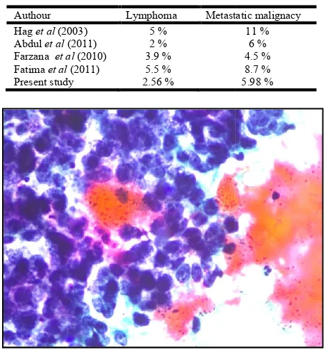
[image:3.595.159.440.521.735.2]Table 3. comparison of distribution of malignant lymph node lesions with other studies
Authour Lymphoma Metastatic malignacy
Hag et al (2003) 5 %
Abdul et al (2011) 2 %
Farzana et al (2010) 3.9 %
Fatima et al (2011) 5.5 %
Present study 2.56 %
Fig. 5. Cervicle Lymphnode (Pap stain, High Power) metastases (SCC)
The larynx was the most common site of primary metastasizing to cervical lymph node. The cytologic diagnosis of lymph node FNA cases was confirmed on histopathology in 48 (90.57%) out of 53 lymph node biopsies.
DISCUSSION
In the present study, age group of patients ranged from 3 years to 75 years with. Male to female ratio was 1.4:1. With respect
to this, Hag et al. (2003)found wide age range of 7 months to
90 years. In the present study, 234 cervical lymph nodes aspirates were studied. The benign and non
formed the largest group in the present series (90.60%) and this is consistent with the observations as described in table number 2. Among the non- neoplastic group, tuberculous lymphadenitis was the most frequent lesion observed. Similar observations were reported by Bandopadhyay et al. (1996)
(1991).High prevalence of tuberculosis in our country explains
high incidence of tuberculosis in the present study 2007). In maximum number of cases of tuberculous lymphadenitis, epitheliod cell granulomas with necrosis was observed. Gupta et al. (1992), Raguveer et al
et al. (1999)have also observed similar results.
node aspirates were positive for malignancies in the present much more frequently than lymphoma. With respect to this, all of the authors enlisted in table number 3 found similar observations.
Acknowledgement
Patients and their families who had given consent for FNAC.
38033 Dr. Bolde et al. Fine needle aspiration of cervical lymph nodes
Table 3. comparison of distribution of malignant lymph node
Metastatic malignacy 11 % 6 % 4.5 % 8.7 % 5.98 %
Cervicle Lymphnode (Pap stain, High Power) - Epithelial
The larynx was the most common site of primary metastasizing to cervical lymph node. The cytologic diagnosis of lymph node FNA cases was confirmed on histopathology in
In the present study, age group of patients ranged from 3 years to 75 years with. Male to female ratio was 1.4:1. With respect found wide age range of 7 months to 90 years. In the present study, 234 cervical lymph nodes aspirates were studied. The benign and non-neoplastic lesions formed the largest group in the present series (90.60%) and this observations as described in table number neoplastic group, tuberculous lymphadenitis was the most frequent lesion observed. Similar observations
. (1996) and Borger,
sis in our country explains high incidence of tuberculosis in the present study (Park, In maximum number of cases of tuberculous lymphadenitis, epitheliod cell granulomas with necrosis was
et al. (1998) and Sen
have also observed similar results. Twenty lymph node aspirates were positive for malignancies in the present much more frequently than lymphoma. With respect to this, all of the authors enlisted in table number 3 found similar
Patients and their families who had given consent for FNAC.
Conclusion
FNAC is found to be a simple, safe, inexpensive OPD based procedure which is very useful in diagnosing superficial and easily accessible cervical lymph node
separating inflammatory lesions from neoplastic lesions and makes surgical excision unnecessary. Early specific diagnosis such as tuberculosis allows prompt and appropriate treatment. Thus, FNAC plays an important role in diagnosing va cervical lymph node lesions and helps in avoiding unnecessary surgical intervention.
REFERENCES
Ansari, M. and Deria, N. 1997.
Aspiration Cytology”, journal clinical pathology 543.
Bandopadhayay, S.N. et al. 1996.
Aspiration Cytology in the diagnosis of cervical lymphadenopathy”, WO & HNS; vol.48(4): 289
Borges, A.M. 1991. Aspiration cytology in the management of head and neck masses. In: Krishnamurthy SC, editor. Aspiration cytology for clinicians and pathologists. 1 Bombay: TATA Press Limited
Farzana, S., Mirza, T., Mustafa
2010. An experimental status of fine needle aspiration cytology of head and neck lesions in a tertiary care scenario. Journal of Basic and Applied Sciences,
162.
Fatima, S., Arshad, S., Ahmad
of Cytological findings in patients with Neck
Lymphadenopathy – Experience in a tertiary Care Hospial in Pakistan. Asia Pacific journal of Cancer Preventi 12.
Frable WJ, Frable MA. Thin needle aspiration biopsy in the diagnosis of head and neck tumors. The laryngoscope 1974;84: 1069-1077
Gupta, A.K., Nayar, M., Chandra
of fine needle aspiration cytology in tuberculous lymphadenitis. Acta Cytologica,
Hag, I.A.E., Chiedozi, L.C., Reyees
2003. Fine needle aspiration cytology of head and neck masses : seven years experience in tertiary care hospital.
Actacytologica, 47:387-389.
Khan, A.H., Hayat, A.S., Ghulam
Role of Fine Needle aspiration Cytology in Cervical lymphadenopathy. World Applied Sciences Journal. (11):1951-1954.
Mazar, I., Anis, S., Asadullah tuberculosis in cervical lymphade
Surgery Pakistan, 15(2):107
Orell, S.R., Sterret, G.F., Walters
D. 2012. Ed., Manual and atlas of fine needle aspiration
cytology, 5th ed. London, Churchill Livingstone,
Park, K. 2007. Textbook of preventive and social medicine.
19thed: Jabalpur; M/S BanarsidasBhanot Publications
Raghuveer, C.V., Bhattacharya
needle aspiration cytological study.
Pathology and Microbiology,
Sen, R. 1999. Cytomorphological pattern in tuberculous lymphadenitis. Indian Journal of Tuberculosis
Dr. Bolde et al. Fine needle aspiration of cervical lymph nodes
FNAC is found to be a simple, safe, inexpensive OPD based procedure which is very useful in diagnosing superficial and easily accessible cervical lymph node lesions. It helps in separating inflammatory lesions from neoplastic lesions and makes surgical excision unnecessary. Early specific diagnosis such as tuberculosis allows prompt and appropriate treatment. Thus, FNAC plays an important role in diagnosing various cervical lymph node lesions and helps in avoiding unnecessary
. 1997. “Origins of Fine Needle
journal clinical pathology, 10;
541-. 1996541-. “Role of Fine Needle Aspiration Cytology in the diagnosis of cervical
”, WO & HNS; vol.48(4): 289-294. Aspiration cytology in the management of head and neck masses. In: Krishnamurthy SC, editor.
linicians and pathologists. 1st ed.
Bombay: TATA Press Limited.
, Mustafa, S., Saima, S., Sharafat, S. An experimental status of fine needle aspiration cytology of head and neck lesions in a tertiary care
Basic and Applied Sciences,
6(2):159-, Ahmad6(2):159-, Z.6(2):159-, Hasan6(2):159-, S. 2011. Spectrum
of Cytological findings in patients with Neck
Experience in a tertiary Care Hospial
Asia Pacific journal of Cancer Prevention, vol
Frable WJ, Frable MA. Thin needle aspiration biopsy in the diagnosis of head and neck tumors. The laryngoscope
, Chandra, M. 1992. Critical appraisal of fine needle aspiration cytology in tuberculous
Cytologica, 36(3): 391-394.
, Reyees, F.A.A., Kollur, S.M. Fine needle aspiration cytology of head and neck masses : seven years experience in tertiary care hospital.
389.
, Ghulam, H.B. et al. 2011. Study on Role of Fine Needle aspiration Cytology in Cervical
World Applied Sciences Journal. 12
, Asadullah, A. 2010. Frequency of tuberculosis in cervical lymphadenopathy. Journal of
15(2):107-109.
, Walters, Max, N.I., Darrel Whitaker, Ed., Manual and atlas of fine needle aspiration
London, Churchill Livingstone, 1-3. preventive and social medicine. S BanarsidasBhanot Publications. , Bhattacharya, S., Pai, M.R. 1998. Fine needle aspiration cytological study. Indian Journal of
Pathology and Microbiology, 41:206.
Cytomorphological pattern in tuberculous
Shaha, A. Webber, C. and Marty, J. 1986. Fine needle aspiration in the diagnosis of cervical lymphadenopathy.
The American journal of Surgery, Oct:152:420-423.
Singh, J.P. et al. 1989. ”Role of Fine Needle Aspiration Cytology in diagnosis of Tuberculuos lymphadenitis”,
Indian journal Pathology & Microbiology, vol.32(2):
101-104, 1989.