25 - HYDROXY VITAMIN
1
Dr. Roua Hameed
2
Department of Pediatrics
ARTICLE INFO ABSTRACT
Background:
differentiation throughout adverse pro-convulsant
Aim: A
parameters antiepileptic
Methods
neurological of July Information and type and systemic applied phosphatase
Results:
Two third 50% of more than serum level 2.13) respectively higher level respectively alanine >0.05).
Conclusion:
therapy
Copyright©2017, Dr. Roua Hameed Kadhum and Dr.
License, which permits unrestricted use, distribution, and
INTRODUCTION
Epilepsy describes a condition of susceptibility
seizures. (Friedman and Sharieff, 2006)It is
present when 2 or more unprovoked seizures interval greater than 24 hr apart. The clinical epilepsy usually requires the occurrence unprovoked epileptic seizure with either a enough EEG and clinical information predisposition to develop recurrences. (Mikati antiepileptic drugs (AED) remain the mainstay
epilepsy around the world. (Razazizan et al
shown that epileptic patients can exhibit
vitamin D. (Baek et al., 2014) Twenty to 60%
*Corresponding author: Dr. Sawsan Issa Habeeb,
Department of Pediatrics, College of Medicine, University of Basra, Iraq.
ISSN: 0975-833X
Article History: Received 13th June, 2017
Received in revised form 26th July, 2017
Accepted 18th August, 2017
Published online 30th September, 2017
Citation: Dr. Roua Hameed Kadhum and Dr. Sawsan
Journal of Current Research, 9, (09), 57788-57792. Key words:
25-hydroxy vitamin D, Epilepsy,
Children.
RESEARCH ARTICLE
VITAMIN D LEVEL IN CHILDREN WITH EPILEPSY
Hameed Kadhum and
2,*Dr. Sawsan Issa Habeeb
1
Basra General Hospital, Iraq
Pediatrics, College of Medicine, University of Basra
ABSTRACT
Background: The importance of vitamin D was recently emphasized when
differentiation, proliferation, immune function and important for optimal throughout the body, including the central nervous system.This vitamin may adverse effect of anticonvulsant drugs so its supplementation is associated
convulsant and anticonvulsant factors.
A case-control study had been carried out to determine serum vitamin parameters in epileptic children and study it's relation to some selected antiepileptic drugs for at least 6 months.
Methods: Thirty seven epileptic patients who had normal age - appropriate
neurological clinic in Basra General Hospital, their ages ranged from 2-14 2014. Forty four apparently healthy children and adolescent; were Information regarding history of epilepsy; age of diagnosis, type and frequency
type of antiepileptic drugs were recorded. All patients were under went systemic examination. Growth parameters were assessed and body to appropriate growth charts. A list of investigation was measured phosphatase and serum 25-Hydroxy vitamin D.
Results: Mean age of epileptic patients and control group was (7.67 ± 3.23)
third of patients were younger than 10 years, as well as (86.5%) have of patients received carbamazepine treatment for epilepsy and the
than 24 months and more than 80% of patients were on monotherapy level of vitamin D was significantly low in epileptic patients than respectively as well as in children treated with polytherapy than those
level of alkaline phosphatase in epileptic patients than control group respectively (P value 0.000). On the other hand; levels of Calcium and phosphorus
aminotransferase (ALT) were not significantly differ among epileptic
Conclusion: Children and adolescents treated with anti-epileptic drugs especially
therapy should given supplements of vitamin D to satisfy their bodies requirement.
Sawsan Issa Habeeb. This is an open access article distributed under and reproduction in any medium, provided the original work is properly
susceptibility to recurrent is considered to be seizures occur at an clinical diagnosis of occurrence of at least 1 second seizure or to demonstrate
Mikati, 2011)Although,
mainstay of treatment for
et al., 2013) It has exhibit a deficiency of
60% of AED users
Department of Pediatrics, College of Medicine, University of Basra, Iraq.
can develop rickets or osteomalasia oxidase activity throughout cytochrome (P450), there promoting
D and its derivatives. (Borba et al
of the AED involves a direct bone resorption and formation. influence bone turnover. Both with a reduction in bone mineral 2003) Vitamin D supplementation seizures. As it regulates proconvulsant factors. More specifically, this
regulation of cytokine IL-6
(Bozzetto et al., 2012)Vitamin
the expression of genes associated acid (GABA); It is one of the main
in the brain. (Féron et al., 2005
International Journal of Current Research
Vol. 9, Issue, 09, pp.57788-57792, September, 2017
Sawsan Issa Habeeb, 2017. “25 - hydroxy vitamin d level in children
EPILEPSY IN BASRA
Habeeb
Basra, Iraq
when it was reported to be involved in cell optimal function of many organs and tissues may reduce seizure frequency and treating associated with decreased seizures as it regulates
vitamin D level and other biochemical selected variables who were treated with
appropriate development; visited the out patients 14 years from the1stof February to the end
age and sex matched as a control group. frequency of seizures over the past 3 months went physical examination including general mass index (BMI) was calculated and measured as; serum calcium, phosphorus, alkaline
3.23) and (7.68 ± 3.5) year respectively. have normal body mass index, approximately duration of anti-epileptic drugs' therapy monotherapy and were well controlled. The mean than control group (21.41 ± 2.90), (67.59 ± those with monotherapy for epilepsy with group (299.09 ± 67.67) (122.21 ± 29.59) phosphorus, serum level of blood urea and epileptic patient and control group (p value
especially those patients on polytherapy requirement.
under the Creative Commons Attribution properly cited.
osteomalasia due to induce hepatic throughout the microsomal enzymes promoting the catabolism of vitamin
et al., 2004) The other mechanism action on bone cells, increasing formation. In this way, the AED may Both mechanisms can be associated
mineral density (BMD).(Pack et al.,
supplementation associated with decreased proconvulsant and anticonvulsant vitamin is involved in the down 6, which is a proconvulsant. Vitamin D has also been found to affect associated with Gamma-amino butyric main inhibitory neurotransmitters 2005) Also it has been shown to
INTERNATIONAL JOURNAL OF CURRENT RESEARCH
promote the expression of calcium binding proteins that are
known to possess antiepileptic properties.(Evatt, 2012)
MATERIALS AND METHODS
A Case-control study has been carried out to determine serum level of vitamin D in 37 epileptic children and adolescent who were treated with antiepileptic drugs for at least 6 months. All the children had normal age - appropriate development; visited the out patients neurological clinic in Basra General Hospital
from the 1st of February 2014 to the end of July 2014; their ages
range from 2-14 years. Forty four healthy children and adolescent were aged and sex matched, selected from children visiting primary health care center for routine checkup or mild illnesses, have normal growth parameters were consider as control group.
Exclusion criteria
1. Calcium or vitamin D supplements for the last one
month (Razazizan et al., 2013)
2. Neurological deficit as cerebral palsy or mental retardation
3. Chronic disease as history of chronic kidney disease, diabetes mellitus and bone deformities
4. Presence of conditions known to affect bone metabolism such as hepatic, hematological (e.g. sickle cell disease), rheumatologic, hyper-parathyroidism, hyperthyroidism, gastrointestinal disorders e.g., malabsorption as celiac
disease. (Lee et al., 2007; Trunz et al., 2008)
5. Medications known to affect bone turnover e.g. steroid, bisphosphonate, thiazide and anticoagulant drugs.
(Borba et al., 2004)
Data collection
A special questionnaire was designed for the purpose of the study including: Identity; name, date of birth, sex and residence. History of epilepsy; age of diagnosis, type and frequency of seizures over the past 3 months, well controlled is
defined as seizures free for the last 3months. (Friedman and
Sharieff, 2006) Type of antiepileptic drugs; doses and duration of antiepileptic drugs (AED), if monotherapy (single AED) or
polytherapy (more than one AED) used.(Bozzetto et al., 2012)
Informed consent had been taken from parents to be involved in the study. All patients were under went physical examination including general and systemic examination. Growth parameters were assessed, weight-for-height Z score, height-for-age Z score and weight-height-for-age Z score were estimated according to CDC/WHO normalized references. (Bikle, 2005) Body mass index (BMI) was calculated by weight in kilograms
(kg) divided by the height in square meters (m)2.(Wortsman et
al., 2000) According to BMI percentiles growth charts
underweight is < 50 %; normal weight 50 - 85 % and over
weight is > 85 %. (Razazizan et al., 2013)
Laboratory procedures
A list of investigations was measured by spectrophotometer
(Cobas C 111), Serum calcium (Ca+), serum phosphorus (Ph),
serum Alkaline phosphates (ALPh), blood urea and alanine aminotransferase (ALT). Serum 25-Hydroxy vitamin D (25OH); was measured by ELISA test. Very severe vitamin D deficiency is <5 ng/ml; deficiency level <15 ng/ml; insufficiency level (15-20 ng/ml); and consider as sufficient if
its level 20-30 ng/ml. Optimal level is 30-50 ng/ml and the upper normal located between 50-70 ng/ml. (Hollick, 2007)
Statistical analysis
Data were analyzed using SPSS software version 18. Data were expressed by mean ± Standard Deviation (SD) and Standard Error (SE) Comparison was performed by using Chi-Square test. The t-test was used for quantitative comparison and between two mean of different samples. Comparisons between groups were made by using the one way analysis of variance (ANOVA) test .Logistic regression analysis was done for the analysis of different marker, by using Odd Ratio (OR) and 95% Confidence Interval (CI). For all tests p-value of <0.05 was considered as statistically significant.
RESULTS
Selected demographic characteristics of epileptic patients and control
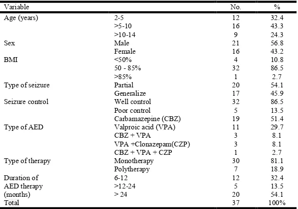
A total of 37 epileptic patients and 44 children and adolescent as control group were included in the study, their ages ranged from 2-14 years and mean age was (7.67± 3.23); (7.68 ± 3.50) years respectively. Twenty eight (75.7%) of epileptic children were younger than 10 years, As well as (86.5 %) had normal weight; their BMI was 50-85%. Table 1 shows that (51.4%) of patients received carbamazepine treatment for epilepsy. As well as more than 80% patients were well controlled on monotherapy and 54.1% of patients were on AED therapy for more than 24 months.
Biochemical Parameters among epileptic Patients and control group
It has been found that vitamin D level was significantly lower among epileptic patient compared to healthy control group (p
value <0.05), as well as alkaline phosphatase significantly
higher among epileptic patients. P value <0.000 (Table 2)
Vitamin D level and selected variable in epileptic patients
Table 3 shows that; (13.5% and 24.3%) of epileptic patients had (severe deficiency level < 5ng/dl and deficient level< 15ng/dl) respectively with higher frequency in patients with generalized seizure (77.8%), receiving poly therapy (55.6%); with duration of anti-epileptic drugs >24 months (66.7%). On the other hand (39.7%) of epileptic patients had sufficient level (20-30ng/dl) mainly those with partial seizure (81.8%); treated with carbamazepine (72.7%) and receiving mono therapy (100%)
Mean vitamin D in relation to selected characteristics of epileptic children
Table 1. Selected characteristics of epileptic patients % No. Variable 32.4 12 2-5 Age (years) 43.3 16 >5-10 24.3 9 >10-14 56.8 21 Male Sex 43.2 16 Female 10.8 4 <50% BMI 86.5 32
50 - 85%
2.7 1 >85% 54.1 20 Partial Type of seizure
45.9 17 Generalize 86.5 32 Well control Seizure control 13.5 5 Poor control 51.4 19 Carbamazepine (CBZ)
Type of AED Valproic acid (VPA) 11 29.7
8.1 3
CBZ + VPA
8.1 3
VPA +Clonazepam(CZP)
2.7 1
CBZ + VPA + CZP
81.1 30
Monotherapy Type of therapy
[image:3.595.79.518.305.372.2]18.9 7 Polytherapy 32.4 12 6-12 Duration of AED therapy (months) 13.5 5 >12-24 54.1 20 > 24 100% 37 Total
Table 2. Biochemical Parameters
P-value Mean ± SD
Variables
Control group Epileptic patients
0.000 67.59 ± 2.13
21.41 ± 2.90 Vitamin D (ng/ mL)
0.620 8.81 ± 1.63
8.09 ± 0.97 Calcium (mg/dL)
0.812 1.48 ± 0.36
0.81 ± 0.42 Phosphorus (mg/dL)
0.000 122.21 ± 29.59
299.09 ± 67.67 Alkaline phosphatase (IU/L)
0.427 10.89 ± 1.58
11.09 ± 1.13 ALT (IU/L)
Table 3. Vitamin D level in relation to selected variables of epileptic patients
Optimal >30 Sufficiency 20-30
Insufficiency 15-20 Deficiency < 15
Severe deficiency< 5 Variables % No % No % No % No % No 2-5 Age (years)
0 0 18.2 2 28.6 2 44.4 4 20 1
3 60 63.6 7 14.3 1 44.4 4 40 2 >5-10
2 40 18.2 2 57.1 4 11.1 1 40 2 >10-14
3 60 54.5 6 71.4 5 66.7 6 20 1 Male
Sex Female 4 80 3 33.3 2 28.6 5 45.5 2 40
0 0 18.2 2 0 0 11.1 1 20 1 <50%
BMI 50-85% 4 80 8 88.9 7 100 8 72.7 4 80
1 20 9.1 1 0 0 0 0 0 0 >85%
3 60 81.8 9 57.1 4 22.2 2 60 3 Partial Type of
seizure Generalize 2 40 7 77.8 3 42.9 2 18.2 2 40
3 60 72.7 8 42.9 3 22.2 2 60 3 CBZ AED
2 40 27.3 3 42.9 3 22.2 2 20 1 VPA
0 0 0 0 0 0 22.2 2 20 1 CBZ+VPA
0 0 0 0 14.3 1 22.2 2 0 0 VPA+CZP
0 0 0 0 0 0 11.1 1 0 0 VPA+CBZ+CZP
5 100 100 11 85.7 6 44.4 4 80 4 Mono Type of
therapy Poly 1 20 5 55.6 1 14.3 0 0 0 0
2 40 45.5 5 0 0 33.3 3 0 0 <24
Duration of AED (months)
3 60 54.5 6 100 7 66.7 6 100 5 >24
5 13.5 39.7 11 18.9 7 24.3 9 13.5 5 Total
Table 4. Mean vitamin D level in relation to selected characteristic of epileptic patients
P- value Mean Vitamin D level ± (SE)
Variable
0.921* 20.86 ± 5.21
2-5
Age(years) >5-10 22.60 ± 3.90
20.02 ± 3.65 >10-14
0.980 22.02 ± 3.77
Male Sex
20.61 ± 4.69 Female
0.060* 2o.15 ± 8.20
<50%
BMI 50-85% 20.42 ± 3.12
58.10 ± 0.00 >85%
0.073 25.71 ± 4.32
Partial Type of seizure
16.36 ± 3.81 Generalize
0.062* 25.64 ± 4.42
CBZ
Types AED VPA 21.89 ± 3.54
9.30 ± 6.85 Other medication
0.023 24.24 ± 3.23
Monotherapy Type of therapy
9.30 ± 2.63 Polytherapy
0.001 23.11 ± 3.17
Well Seizure control
10.56 ± 3.58 Poor
0.032 30.50 ± 3.78
<24 Duration of AED (months)
18.04 ± 3.11 > 24
[image:3.595.37.556.399.592.2] [image:3.595.80.516.616.795.2]seizure and vitamin D level <15 ng /ml (p-value 0.02) and significant differences also found between epileptic children who taken AED more than 24 months and vitamin D level < 15ng/ml. (Table 4)
Logistic regression analysis of selected variable with low level of vitamin D in epileptic patients
Table 5 shows that patients on poly therapy is significantly have low level of vitamin D (p-value <0.05), Odd ratio (6.991). But there is no statistically significant correlation of other variable (age, BMI, type of AED, seizure control and duration of therapy) with low level of vitamin D in Epileptic children.
DISCUSSION
In this study the mean level of vitamin D is low among epileptic patients compared with healthy children. Similar
result was concluded by Alison et al in New Yourk and Silvana
et al in Brazil. (Alison, 2003; Silvana et al., 2000) The mean level of calcium for epileptic patients was in the normal limit
this is in agreement to that reported by Razazizan et al in Iran,
(Razazizan et al., 2013) which could be due to secondary
hyperparathyroidism to maintain normocalcimia. (Misra, 2010)
and in contrast to Babyigit et al in Turkey (Babayigit et al.,
2006)whom concluded that epileptic children have low level of
calcium than control; possibly due to direct impact of AED on bone mineral density and intestinal transport and the effect of hypovitaminosis D which lead to decreased absorption of the
calcium in intestine. (Babayigit et al., 2006; Okesina et al.,
1991)Alkaline phosphatase is an important biochemical marker
of bone metabolism in epileptic patients. (Razazizan et al.,
2013) In this study, the mean level of alkaline phosphatase is significantly higher in epileptic children than control group,
this result is similar to that reported by Razazizan et al in Iran,
Babyigit et al in Turkey and Okesina in Nigeria. (Razazizan et
al., 2013; Babayigit et al., 2006; Okesina et al., 1991) Because
AED are inducers of the hepatic cytochrome P450 system which
promote the metabolism of 25-hydroxyvitamin D (25-OHD) to less biologically active analogues, resulting in decreased bone mineralization, decreased intestinal calcium absorption, increased calcium mobilization from the skeleton to maintain
eucalcemia, and decreased bone density. (Babayigit et al.,
2006) Also this study revealed that more than one third of patients had sub optimal vitamin D level while less frequent of patients who have optimal vitamin D level, this result is in
agreement with Menton et al in India. (Menon, 2010)There is
higher frequency of vitamin D deficiency among young children with epilepsy in Basra than older children, this is in
agreement with Baek et al in Korea. (Baek et al., 2014) Which
could be related to age- biological processes or less physical activities with increasing age which lead decrease sunlight exposure. BMI of epileptic patients in Basra are not significantly associated with decrease level of vitamin D, This
is in agreement with Misra et al in India. (Alison, 2003) But in
contrast to a study carried out by Baek et al in Korea and
Lagunova et al in Norway (Baek et al., 2014; Lagunova et al.,
2009)whom show; that the level of vitamin D decrease with
increasing BMI which can be explained by the fact that peoples with high BMI usually have a high content of body fat, acting as a reservoir for lipid-soluble vitamin D. Current study reveals that no significant association between type of AED (CBZ, VPA or other AED) and vitamin D level . This is in agreement
with a study conducted by Menon et al in India. (Menon, 2010)
Which shows that the level of vitamin D decrease irrespective
to the type of AED used. This is in contrast to Valsamis et al in
USA and Kim et al in Korea. (Valsamis et al., 2006; Kim et al.,
2007) They found that bone turnover is higher with hepatic enzyme inducing AED such as (CBZ) than non–inducing AED
as (VPA). (Valsamis et al., 2006) That can be explained by
enzyme inducing AED (PHT, PB, CBZ) and enzyme inhibiting AED (VPA) affected the 25(OH)D metabolism at sub-therapeutic doses of AED. Patients on polytherapy demonstrated significantly lower 25(OH) D levels than patients on monotherapy, This result is in agreement with a study by
Sina et al in Germany. (Kim et al., 2007) And Menon et al in
India.(Okesina et al., 1991) It can be explained by effected of
AED on vitamin D which more increase by using more than
one AED (polytherapy). (Okesina et al., 1991)In contrast to a
study done by Beak et al in Korea. (Baek et al., 2014)Who
found no significant difference in level of vitamin D among patient in monotherapy and polytherapy. In Basra the effect of un controlled seizure had been found to be associated with low vitamin D level. This is similar to the result concluded by
Behar et al in Hungary. (Behav, 2012) Possible explanation
that normal level of vitamin D in serum has anticonvulsive role, As well as any poorly controlled patient already kept on many AED to control seizures. (Behav, 2012) Duration of AED therapy is significantly correlated with vitamin D level, lower level of vitamin D for epileptic children who received AED for more than 24 months than those who received AED for less
than 24 months. Similar result was reported by Misra et al in
India, Back et al in Korea and Menon et al in India. (Baek et
al., 2014; Misra, 2010; Menon, 2010) Whom concluded that
there is significant impaction of AED on level of vitamin D and this effect increase with longer duration of therapy and low dietary intake of vitamin D. (Menon, 2010) In contrast with a
study of Yuan Guo in Canada (Guo et al., 2001) and Razazizen
et al in Iran (Razazizan et al., 2013) whom found; no relation between vitamin D level and duration of AED, that can be possibly explained by using a single drug and children recruited in the study have adequate nutritious diet. (Razazizan
et al., 2013)
Recommendation
Vitamin D3 supplement should be given for all epileptic
[image:4.595.150.448.78.158.2]children even before initiation of anti-epileptic therapy and children with epilepsy should follow well balanced diet and
Table 5. Risk factor for low serum level of vitamin D
95% confidence interval Odd ratio
p-value Variable
Upper value Lower value
13.720 0.068
1.547 0.136
Age
11.463 0.075
1.352 0.831
BMI
11.23 0.028
1.178 0.060
Type of AED
0.258 0.062
6.991 0.020
Type of therapy
13.621 0.075
1.497 0.128
Seizure control
11.142 0.048
1.341 0.928
good nutritional habits and healthy lifestyle to optimize seizure control.
REFERENCES
Alison P. 2003. The association between antiepileptic drugs
and bone Disease. Epilepsy Curr., 3(3):91-95
Babayigit A, Dirik E, Bober E, Cakmakci H. 2006. Adverse effects antiepileptic drugs on bone mineral density.
Pediatr Neurol., 35:177-181.
Baek J, Seo Y, Kim G. 2014. Vitamin D Levels in Children
and Adolescents with Antiepileptic Drug Treatment. Yonsei
Med J., 55(2):417-421.,
Behav E. 2012. Correction of vitamin D deficiency improves seizure control in epilepsy: apilot study. Epilepsy Behav., 24(1) 131-133.
Bikle D. 2005. Vitamin D: role in skin and hair. AP, (1) :609-630.
Borba V, Bilezikian J, Silvado C, Kulak C. 2004. Bone mineral densityand serum levels of 25 OH vitamin D in
chronic users ofantiepileptic drugs. Arq Neuropsiquiatr.,
62(4):940-948.
Bozzetto S, Carraro S, Giordano G, Boner A, Baraldi E. 2012. Asthma, allergy, and respiratory infections: the vitamin D
hypothesis. Allerg., 67:10-17
Evatt L, Interview by T.N. Smith. 2012. Vitamin d deficiency and associated neurological conditions. Atlanta, Ga, 25:100-105.
Féron F, Burne TH, Brown J, Smith E, Eyles DW et al. 2005.
Developmental vitamin D3deficiency alters the adult rat
brain. Brain Res Bull., 65:148-141.
Friedman M. and Sharieff G. 2006. Seizures in Children.
Pediatr Clin N Am., 53:257-277.
Guo CY, Ronen GM, Atkinson SA. 2001. Long-term valproate and lamotrigine treatment may be a marker for reduced growth and bone mass in children with epilepsy. Epilepsia 42:1141-1147.
Hollick. MF. 2007. Vitamin D deficiency. N Engl J.,
357:266-281.
Kim S, Lee J, Chung H, Lee H. 2007. A 6-month longitudinal study of bone mineral density with antiepileptic drug monotherapy. E & B.10 (2) 213-348.
Lagunova Z, Moan J, Carmen A, Hexebreg S. 2009. The Dependency of Vitamin D Status on Body Mass Index, Gender, Age an Season I.I.A.R, 29 (9) 3713-3720.
Lee JW, Choi KG, Chung HW. and Lee HW. 2007. A 6-month longitudinal study of bone mineral density with
antiepileptic drug monotherapy. Epilepsy Behav., 10(2):
291-295.
Menon B. The effect of anti-epileptic drug therapy on serum 25 hydroxyl vitamin D and parameters of calcium and
bone metabolism a longitudinal study. Seizure
2010;19:153-158.
Mikati M. 2011. Seizures in childhood. in: Behrman ER, Kliegmanl RM, Jenson HB (eds). Nelson Text Book of
Pediatrics. 19th Philadelphia. Elsevier Saunders Co.
2013-2039.
Misra A. 2010. Effect of Carbamazepine Therapy on Vitamin D and Parathormone in Epileptic Children. PN 43(5):320-324.
Nettekoven S, Ströhle A, Trunz B, Wolters M, Hoffmann S,
Horn R et al. 2008. Effects of antiepileptic drug therapy on
vitamin D status and biochemical markers of bone turnover
in children with epilepsy. Eur J Pediatr., 167:1369-1377
Okesina AB, Donaldson D, Lascelles PT. 1991. Effect of
antiepileptic drugs on alkaline phosphatase. J Clin Pathol.,
44(6): 480–482.
Pack AM, Olarte LS, Morrel MM, Flasher E, Stanley RR, Shane E. 2003. Bone mineral density in an outpatient population receiving enzyme- inducing antiepileptic drugs.
Epilepsy Behav., 4:169-174.
Razazizan N, Mirmoeini M, Daeichin S, Ghadiri K. 2013. Comparison of 25- Hydroxy Vitamin D, Calcium and Alkaline Phosphatase Levels in Epileptic and
Non-Epileptic Children. Acta Neurol Taiwan., 22 :112-116.
Silvana F, Guerreiro CA, Magna LA, 2000. Marques Neto Bone mineral density, vitamin D and anticonvulsant thera-py. Arq Neuropsiquiatr., 58:616-620.
Trunz B, Wolters M, Hoffmann S, Horn R, Steinert M, et al.
2008.Effects of antiepileptic drug therapy on vitamin D
status and biochemical marker of bone turnover in children
with epilepsy. Eur J Pediatr., 167(12):1369-1377
Valsamis H, Arora S, Labban B, Farlane S. 2006. Antiepileptic drugs and bone metabolism. NM, 3(36) 1743-7075
Wortsman J, ChenT, Holick M. 2000. Decreased
bioavailability of vitamin D in obesity. ASN, 72:690-693