ISSN Online: 2158-2882 ISSN Print: 2158-284X
DOI: 10.4236/ijcm.2019.103010 Mar. 5, 2019 101 International Journal of Clinical Medicine
The Application and Operation-Effect Analysis
for Complex Tibial Plateau Fractures with
3D Printing Technique
Changjin Guo
1, Yubo Zhang
2, Li Yang
1, Qiaofeng Zhu
1, Sanming Zou
2*1Medical College, Wuhan University of Science and Technology, Wuhan, China
2Department of Orthopaedics, Xiaogan Central Hospital, Wuhan University of Science and Technology, Xiaogan, China
Abstract
Objective: To investigate the value of 3D printing techniques in the treat-ment of complex tibial plateau fractures. Methods: From September 2016 to September 2018, 28 patients with complex tibial plateau fractures were treated in our hospital. According to the odevity of hospitalized order, the patients were divided into two groups. Group A used 3D reconstruction, virtually reduction, 3D printing and demonstration of individual fracture model before operation while group B only received conventional process by use X-rays or CT image. Comparison between the two groups was made in operation time, operative blood loss, radiation frequency, surgery in-strument cost and knee function score. Results: The follow-up was 14.4 months on average (ranged 6 to 22 months). There was no statistical dif-ference of the surgery instrument cost between the 2 groups (P > 0.05). The operation time of group A was significantly shorter than that of group B (P < 0.05). Group A also performed better than group B in comparison of operative blood loss and radiation frequency. The excellent good rate of HSS score in group A was 92.86%; it was higher than that 85.71% in group B. There was no statistical difference between the 2 groups (χ2 = 0.373, P =
0.54). Conclusion: 3D printing techniques can improve surgery effect in complex tibial plateau fractures.
Keywords
Tibial Plateau Fracture, 3D Printing Technique, Individualized Treatment
1. Introduction
Complex tibial plateau fracture is one of the most common and complex
frac-How to cite this paper: Guo, C.J., Zhang, Y.B., Yang, L., Zhu, Q.F. and Zou, S.M. (2019) The Application and Operation- Effect Analysis for Complex Tibial Plateau Fractures with 3D Printing Technique. International Journal of Clinical Medicine, 10, 101-108.
https://doi.org/10.4236/ijcm.2019.103010
Received: January 30, 2019 Accepted: March 2, 2019 Published: March 5, 2019
Copyright © 2019 by author(s) and Scientific Research Publishing Inc. This work is licensed under the Creative Commons Attribution International License (CC BY 4.0).
DOI: 10.4236/ijcm.2019.103010 102 International Journal of Clinical Medicine
tures in limb trauma, in which the knee joint suffers from inversion or vertical compression violence, resulting in unicondylar or bicondylar fractures of the medial and lateral tibial plateau condyles. Large data analysis shows that tibial plateau fractures account for 1% - 2% of the total body fractures and 8% of the elderly fractures [1]. With the increasing incidence of accidents and traffic acci-dents in recent years, the incidence of various complex injuries has gradually in-creased, and the incidence of tibial plateau fractures has also increased. Surgical treatment of complex tibial plateau fractures requires a high standard of surge-ons. Since it is a complex intra-articular fracture with high energy, it is often ac-companied by severe articular surface collapse, ligament injury around the knee joint and poor soft tissue condition. Therefore, the incidence of early complica-tions such as skin ischemic necrosis, infection, poor wound healing, bone and internal fixation exposure, as well as late complications such as long-term pain, postoperative dysfunction, and traumatic osteoarthritis is higher than other fractures. The way to restore the smooth joint surface as much as possible, and at the same time to achieve the minimum damage to the soft tissue, is the primary problem in front of the surgeon. In recent years, the rapid development of digital software and 3D printing technique in China has provided a rare opportunity to solve the above problems. Orthopaedics surgeons can design and simulate the operation on the 3D printing model in advance, so as to improve the surgical treatment effect. In 2014, the team of Professor Wang Liao and Professor Dai Kerong had introduced the individualized orthopaedic treatment and 3D print-ing technique in detail [2], bringing digital orthopaedic surgery into the vision of domestic orthopaedic physicians. Therefore, the application of 3D printing tech-nique in complex tibial plateau fracture surgery is an effective means to realize the individualization and precision of orthopaedic surgery.
2. Materials and Methods
2.1. General Materials
DOI: 10.4236/ijcm.2019.103010 103 International Journal of Clinical Medicine
with even number were classified into group B. There were 14 cases in group A, 9 males and 5 females; aged from 19 to 58 years, with an average age of 38.13 years. For the fracture classification: 5 cases of type II, 3 cases of type IV, 5 cases of type V and 1 case of type VI. There were 14 cases in group B, 8 males and 6 females; aged from 21 to 62 years, with an average age of 38.47 years. For the fracture classification: 6 cases of type II, 4 cases of type IV, 3 cases of type V and 1 case of type VI. Inclusion criteria: patients meeting the diagnostic criteria for complex tibial plateau fractures; patients with severe complications and open fractures were excluded. All the patients are able to tolerate surgery and anesthe-sia, and informed consent to the treatment.
2.2. Grouping Data Processing and 3D
Reconstruction Software
Application Analysis
All patients in group A underwent anteroposterior and lateral films of knee joint, CT plain scan of knee joint and 3D reconstruction before surgery. Profes-sionals imported CT data into Mimics software and convert them into 3D mod-els. The situation of fracture blocks were observed, the pixel set of each bone block was separated to establish a 3D model of each bone block, and each bone block was marked with different colors for fracture classification. All patients in group B underwent anteroposterior and lateral films of knee joint, CT plain scan of knee joint and 3D reconstruction before surgery.
2.3. Preoperative Virtually Reduction and 3D
Printing Model
For the reconstructed 3D model of bone, according to the actual needs of frac-ture surgery, the virtually reduction of the main fracfrac-ture directly related to sur-gery was completed, and the articular surface of tibial plateau was reconstructed to obtain the 3D model after reduction. The virtual reduction process was ob-served to fully understand the size and shape of the fracture block, displacement, reduction mode, movement distance and rotation angle, and determine the in-dividualized surgical plan: the choice of operative approach, the position of the steel plate and the length and direction of the screw. After that, the models of each bone block were integrated and the 1:1 model was printed out hierarchical-ly. After observing the printed fracture model, the surgical team carried out pre-operation process, including understanding the match of the fracture block, simulating the placement of anatomical steel plate, understanding the relation-ship between the fracture line and the position of the steel plate after reduction, and the design of the position and angle of the cancellous bone screw on the in-ner and outer platform.
2.4. Evaluation of efficacy
DOI: 10.4236/ijcm.2019.103010 104 International Journal of Clinical Medicine
recovery of joint function before and after surgery, especially in the evaluation of early postoperative period, which can comprehensively evaluate the movement of the patellofemoral joint and the femoral condyle.
2.5. Statistical Method
Data were analyzed by the statistical software of SPSS 13.0. The operation time and operative blood loss, radiation frequency, surgery instrument cost were ex-pressed by mean (+ standard deviation) (x ± s) and t-test was adopted. While the knee function score after operation was tested by χ2 test, the difference was
con-sidered statistically significant with P < 0.05.
3. Results
3.1. Comparison of Surgical Effects
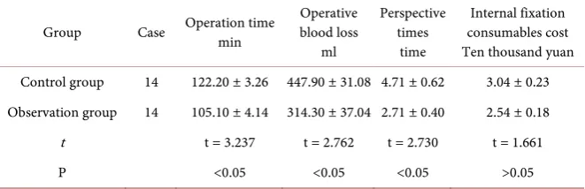
[image:4.595.208.539.436.543.2]There was no significant difference in the surgery instrument cost between group A and group B (t = 1.661, P > 0.05), indicating that the application of 3D printing technique had no significant impact on the use of surgery instrument in traditional surgery. While, there were statistically significant differences in oper-ation time, operative blood loss and radioper-ation frequency (P < 0.05), indicating that compared with traditional surgery, the application of 3D printing technique reduced the operation time and intraoperative fluoroscopy times, thus improv-ing the operation efficiency and safety. See Table 1.
Table 1. Comparison of key intraoperative parameters between group A and group B for
complex tibial plateau fractures.
Group Case Operation time min blood loss Operative ml
Perspective times
time
Internal fixation consumables cost Ten thousand yuan Control group 14 122.20 ± 3.26 447.90 ± 31.08 4.71 ± 0.62 3.04 ± 0.23 Observation group 14 105.10 ± 4.14 314.30 ± 37.04 2.71 ± 0.40 2.54 ± 0.18
t t = 3.237 t = 2.762 t = 2.730 t = 1.661
P <0.05 <0.05 <0.05 >0.05
3.2. Comparison of Functional Recovery after
Operation
Six months after operation, the HSS score was used. In group A, 11 cases were excellent, 2 cases were good, 1 case was fair, the excellent and good rate was 92.86%. In group B, 5 cases were excellent, 7 cases were good, 2 cases were fair, the excellent and good rate was 85.71%. There was no significant difference be-tween the two groups (χ2 = 0.373, P = 0.54) for the excellent and good rate, while
the difference of the excellent rate between the two groups was statistically sig-nificant (χ2 = 5.250, P < 0.05), indicating that the application of 3D printing
DOI: 10.4236/ijcm.2019.103010 105 International Journal of Clinical Medicine
Table 2. Comparison of functional recovery of knee joint between group A and group B
after operation for complex tibial plateau fractures.
Group Excellent (case) Good (case) Fair (case) Total cases Excellent and good rate (%) Excellent rate (%)
Control
group 5 7 2 14 85.71 35.71
Observation
group 11 2 1 14 92.86 78.57
χ2 χ2 = 0.373 χ2 = 5.250
[image:5.595.210.535.394.569.2]P 0.54 <0.05
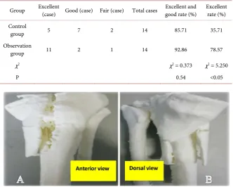
Figure 1. The Anterior view and Dorsal view of 3D printing model.
Figure 2. X-rays was reexamined one month after surgery.
4. Discussion
4.1. The Characteristics of Tibial Plateau Fractures and the
Commonly Used Treatment Methods in China and Abroad
DOI: 10.4236/ijcm.2019.103010 106 International Journal of Clinical Medicine
column Classification of tibial plateau fractures based on CT scan results in 2012. The tibial plateau was divided into lateral column, medial column and posterior column. Cortical rupture was defined as column fracture, and the col-lapse of tibial plateau articular surface without cortical rupture was defined as zero column fracture. Three-column Classification can better guide orthopaedics to choose surgical approaches and internal fixation methods, so as to obtain bet-ter reduction and more stable fixation, which is conducive to the recovery of knee joint function, and significantly improve the therapeutic effect. In recent years, Zhang Yingze et al. [6] have proposed that the current classification can-not include the special type of tibial plateau fracture combined with fibular head fracture, so a new 6-type comprehensive classification method is proposed to make the surgical treatment of tibial plateau fracture more integrated. According to different types of fractures, the treatment methods are also different, and in-dividualized surgical plans should be made according to different types of frac-tures. With the development of modern orthopaedics and the deepening of re-search on tibial plateau fractures, the concept of treatment is constantly updated, and the treatment methods are gradually improved and perfected. Limited inci-sion, minimally invasive technique, indirect reduction by prying technique, ap-plication of external fixator and biological fixation are the future development directions of tibial plateau fracture treatment [7].
4.2. Advantages of 3D Printing Technique over Other Methods
4.2.1. Preoperative Planning of Sham Operated
Preoperative design of complex tibial plateau fracture is formulated by using 3D printing technique to simulate the operation of virtually reduction and internal fixation of complex tibial plateau fracture before operation [8]. In this way the fracture can be accurately and effectively reduced and fixed during operation, the operative blood loss can be reduced, the success rate of one-time placement of implants and devices can be increased, and the accuracy of surgery can be improved, it is considered as a relatively simple and practical clinical tool [9].
4.2.2. Accuracy of Intraoperative Navigation
The application of navigation template for 3D printing technique can help to accurately insert the bone fracture plate and screw, reduce the penetration rate and direction error rate of screw placement [10] [11], and avoid iatrogenic os-teoporosis and internal fixation loosening caused by repeated screw placement.
4.2.3. Clinical Teaching and Surgical Training
Through the intuitive understanding of 3D printing model, medical students and young doctors can not only diagnose and classify accurately, but also im-prove the understanding of complex fractures, which is conducive to the growth of young doctors and shorten the learning curve [12] [13].
DOI: 10.4236/ijcm.2019.103010 107 International Journal of Clinical Medicine
tient communication, in conversation with the patient, using 3D printing tech-nique to make the model vividly and describe the disease condition to patients and their families and inform them of the process during the operation, so that patients can truly feel “clear” treatment, effectively alleviate the tension between patients, with significant clinical benefits [14]. Therefore, it is an effective tool to help doctors and patients communicate [15].
4.3. Problems in the Promotion and Application of
3D Printing Technique
Although preliminary studies have shown that 3D printing technique can im-prove the therapeutic effect of fracture surgery and functional recovery after surgery. However, in clinic, there are still many difficulties in making 3D print-ing model and applyprint-ing and popularizprint-ing virtually reduction technology [16]. 1) Increase the total cost of surgery, increase the financial burden of patients seek-ing medical treatment. 2) Printseek-ing of individualized prostheses is still in the scientific research stage, and commercial promotion is still limited by the appli-cation of biomaterials, whether it can adapt to long-term high-intensity use is still unknown. 3) The relevant clinical guidelines and laws and regulations are faultiness: 3D printing technique involves many fields such as intellectual pro- perty rights, human ethics, dangerous goods manufacturing and so on. At present, there is no clear related policies and laws to complement with it. 4) Low timeliness: From the establishment of imaging data to the printing of physical models and the manufacture of individualized prostheses and internal plants, the whole process takes hours to days, so it is difficult to be used in emergency sur-gery.
5. Prospect of 3D Printing for Tibial Plateau Fractures
The development and application of 3D printing technique has indeed improved the therapeutic effect and functional recovery of traditional tibial plateau frac-ture surgery, and made individualized and precise medical treatment possible. However, how to further control the cost, research and development of biomate-rials and gradually popularize the application are still worth considering. With the rapid development of digital medicine and the deepening of research, the application of computer software-assisted surgery, surgical robots, complex tibi-al plateau fractures and other complex trauma diagnosis and treatment will be more rapid and effective.
Conflicts of Interest
The authors declare no conflicts of interest regarding the publication of this pa-per.
References
DOI: 10.4236/ijcm.2019.103010 108 International Journal of Clinical Medicine [2] Wang, L. and Dai, K.R. (2014) Individualized Treatment of Orthopaedics and 3D
Printing Technology. Journal of Medical Biomechanics, 29, 193-199.
[3] Cho, W.S., Park, S.S., Kim, D.H., et al. (2000) The Reliability of HSS Knee Rating System.
[4] Schatzker, J., Mcbroom, R., Bruce, D., et al. (1979) The Tibial Plateau Fracture. The Toronto experience 1968-1975. Clinical Orthopaedics, 138, 94-104.
[5] Luo, C.F., Hu, C.F., Gao, H., et al. (2009) Three-Column Classification for Tibial Plateau Fractures. Chinese Journal of Orthopaedic Trauma, 11, 201-205.
[6] Chang, Z.L., Chang, H.R., Zhang, Y.Z., et al. (2018) Preliminary Study on Compre-hensive Classification of Tibial Platform Fractures. Journal of Hebei Medical Uni-versity, 39, 1354-1355.
[7] Editorial Committee of Chin J Orthop Trauma (2015) Expert Consensus on Diag-nosis and Treatment of Tibial Plateau Fractures. Chinese Journal of Orthopaedic Trauma, 17, 1671-7600.
[8] Meng, G.L., Liu, J., Hu, Y.Y., et al. (2011) Rapid Prototyping Used in the Operation Design for Complex Fractures of Tibial Plateau. Chinese Journal of Orthopaedic Trauma, 13, 1135-1138.
[9] Dong, L.L., Guo, P.M., Zuo, Q., et al. (2016) Application of 3D Printed Fracture Model in Schatzker Classification of Tibial Platform Fractures. Chinese Journal of Bone and Joint Injury, 31, 860-861.
[10] Lu, S., Zhang, Y.Z., Wang, Z., et al. (2012) Accuracy and Efficacy of Thoracic Pe-dicle Screws in Scoliosis with Patient-Specific Drill Template. Medical & Biological Engineering & Computing, 7, 751-758. https://doi.org/10.1007/s11517-012-0900-1 [11] Kawaguchi, Y., Nakano, M., Yasuda, T., et al. (2012) Development of a New Tech-nique for Pedicle Screw and Magerl Screw Insertion Using a 3-Dimensional Image Guide. Spine, 23, 1983-1988.
[12] Sodian, R., Schmauss, D., Schmitz, C., et al. (2009) 3-Dimensianal Printing of Me-dels to Create Custom-Made Devices for Coil Embolization of an Anastomotic Leak after Aortic Arch Replacement. The Annals of Thoracic Surgery, 88, 974-978.
https://doi.org/10.1016/j.athoracsur.2009.03.014
[13] Kim, G.B., Lee, S., Kim, H., et al. (2016) Three-Dimensional Printing: Basic Prin-ciples and Applications in Medicine and Radiology. Korean Journal of Radiology, 17, 182-197.
[14] Yi, C.R., Luo, J.S., Wang, W.J., et al. (2015) Progress of 3D Printing Technology in Individualized Treatment of Bone Tissue Repair. Medical Science Journal of Central South China, 43, 330-333.
[15] Yang, L., Shang, X.W., Fan, J.N., et al. (2016) Application of 3D Printing in the Sur-gical Planning of Trimalleolar Fracture and Doctor-Patient Communication. Bio-Med Research International, 2016, 2482086.