1071-412X/05/$08.00⫹0 doi:10.1128/CDLI.12.8.959–969.2005
Copyright © 2005, American Society for Microbiology. All Rights Reserved.
Optimization and Validation of a Multiplexed Luminex Assay To
Quantify Antibodies to Neutralizing Epitopes on Human
Papillomaviruses 6, 11, 16, and 18
Dennis Dias,
1†‡ Jeff Van Doren,
1† Sonela Schlottmann,
1Sheri Kelly,
1Derek Puchalski,
1Wanda Ruiz,
1Patricia Boerckel,
1Joseph Kessler,
1Joseph M. Antonello,
2Tina Green,
2Martha Brown,
3Judith Smith,
3Narendra Chirmule,
1Eliav Barr,
4Kathrin U. Jansen,
3§
and Mark T. Esser
1*
Vaccine and Biologics Research, Merck Research Laboratories, 466 Devon Park Dr., Wayne, Pennsylvania 19087-86301;
Vaccine Biometrics Research2and Vaccine and Biologics Research,3Merck Research Laboratories, West Point,
Pennsylvania 19486; and Vaccine and Biologics Clinical Research, Merck Research Laboratories, Blue Bell, Pennsylvania 194224
Received 16 January 2005/Returned for modification 23 February 2005/Accepted 29 April 2005
A human papillomavirus (HPV) multiplexed competitive Luminex immunoassay first described by Opalka et al. (D. Opalka, C. E. Lachman, S. A. MacMullen, K. U. Jansen, J. F. Smith, N. Chirmule, and M. T. Esser, Clin. Diagn. Lab. Immunol. 10:108–115, 2003) was optimized and validated for use in epidemiology studies and vaccine clinical trials. Optimization increased both the analytical sensitivity and the clinical specificity of the assay to more effectively discriminate the low-titer antibody response of HPV-infected persons from nonin-fected individuals. The characteristics of the assay that were optimized included monoclonal antibody (MAb) specificity, scaling up the conjugation of virus-like particles (VLPs) to microspheres, VLP concentration, MAb concentration, sample matrix, sample dilution, incubation time, heat inactivation of sample sera, and detergent effects on assay buffer. The assay was automated by use of a TECAN Genesis Workstation, thus improving assay throughput, reproducibility, and operator safety. Following optimization, the assay was validated using several distinct serum panels from individuals determined to be at low and high risk for HPV infection. The validated assay was then used to determine the clinical serostatus cutoff. This high-throughput assay has proven useful for performing epidemiology studies and evaluating the efficacy of prophylactic HPV vaccines.
Cervical cancer is the second most common cancer in women worldwide. Every year, 450,000 women are diagnosed with cervical cancer and 220,000 succumb to this disease (27). Current approaches to cervical cancer control involve lifelong screening using the Papanicolau (Pap) test (13). The goal of screening is to detect precancerous lesions so that they can be removed prior to the development of cancer. Despite wide-spread Pap testing, there were an estimated 10,520 new cases of cervical cancer and nearly 4,000 cervical cancer-related deaths in the United States in 2004 (1). The national health care burden of current screening systems combined with direct costs of treating precancerous and cancerous lesions is in ex-cess of 3.5 billion U.S. dollars per annum (7).
Infection with human papillomavirus (HPV) is the first and obligate step in the development of cervical cancer (3, 4). Infection of the cervical epithelium with HPV results in ex-pression of the E6 and E7 proteins, which have been shown to be potent oncogenes. More than 35 different HPV types are
capable of infecting the human genital tract (2, 4, 28). Of these, four types cause the majority of the HPV-related cervical pa-thology. HPV 16 and 18 together account for 74.6% of all cervical cancers (23), whereas HPV6 and -11 cause a signifi-cant fraction of precancerous lesions which rarely develop into cervical cancer but morphologically are indistinguishable from lesions from more dangerous HPV types (37). HVP 6 and 11 are responsible for approximately 90% of all genital wart cases (37).
The HPV LI capsid protein, when expressed recombinantly, assembles into empty viral capsids or “virus like particles” (VLPs) (12, 15, 16, 29). Several prophylactic vaccines based on HPV LI VLPs are currently in phases II and III clinical devel-opment (14, 17, 36). The VLPs in the vaccine present the immune system with the conformational, neutralizing epitopes found on the natural virus and prime the immune system to generate antibodies that neutralize the virus and prevent in-fection upon future exposure. Recently, we have shown that a prototype HPV 16 vaccine was 100% efficacious in preventing acquisition of HPV 16 infection and cervical disease among women who were HPV 16 naı¨ve at baseline (19). These results have led to phase II and III studies of a quadrivalent vaccine to HPV 6, 11, 16, and 18. Early results from a randomized, pla-cebo-controlled, phase II trial have shown this quadrivalent vaccine to be 90% efficacious against HPV 6-, 11-, 16-, and 18-related infection or disease (36).
* Corresponding author. Mailing address: Vaccine and Biologics Research, Merck Research Laboratories, 466 Devon Park Dr., Wayne, PA 19087-8630. Phone: (215) 652-0373. Fax: (215) 993-1320. E-mail: [email protected].
† These authors contributed equally to this work.
‡ Present address: Medical College of Virginia, Richmond, VA 23220.
§ VaxGen Inc., Brisbane, CA 94005.
959
on August 17, 2020 by guest
http://cvi.asm.org/
The vaccines under development in Merck’s HPV vaccine program are designed to be effective when given to subjects who are naı¨ve for at least one of the vaccine HPV types. However, these studies do not include a screening phase. Therefore, it is expected that a small cohort of subjects who are positive to at least one of the vaccine HPV types at day 1 will be enrolled. Thus, in order to fully assess the vaccines clinical impact, the serology assays must be able to reliably distinguish “positive” from “negative” samples, and the serostatus cutoff must be defined. Furthermore, as several phase II clinical studies have shown that vaccine-induced anti-HPV antibody levels are protective against HPV infection and disease, a re-liable measure of the duration of the immune response, and hence the duration of efficacy, requires an assay to measure a broad range of antibody titers within diverse clinical popula-tions. These vaccine-induced immune responses must be accu-rately measured across an extensive range of HPV types with-out interference from the immune response to other HPV types. Using the multiplexed, competitive Luminex Immuno-assay (cLIA) described by Opalka et al. (26), antibody titers to neutralizing epitopes on HPV 6, 11, 16, and 18 virions can be simultaneously measured in the serum of vaccinated or natu-rally infected subjects. While the original version of the assay generated accurate results, we modified and formally validated the assay to ensure it was sensitive, specific, and robust enough to meet the needs of testing samples from our large epidemi-ology studies and phase II and III vaccine clinical trials. To this end, we evaluated the analytical sensitivity, specificity, accu-racy, precision, and reproducibility of the assay. Lastly, we determined the clinical sensitivity and specificity of the assay and set serostatus cutoffs for HPV 6, 11, 16, and 18 using serum samples from individuals of diverse geographical regions and age groups with low or high probability of having HPV anti-bodies.
MATERIALS AND METHODS
Virus-like particles (VLPs).The L1 genes of HPV types 6, 11, 16, and 18 were
expressed in the yeastSaccharomyces cerevisiae. The products were then purified
from lysates of the yeast as previously described (15, 16, 24, 29) with modifica-tions (11). The VLPs used in the development of the assay were from consistency lots used in phase III vaccine clinical trials.
Covalent conjugation of HPV VLPs to Luminex microspheres.The HPV VLPs for types 6, 11, 16, and 18 were each covalently conjugated to the free carboxyl groups on xMAP Multi-Analyte COOH Microspheres (Luminex Corporation,
Austin, TX) 6, 11, 16, and 18, respectively, using anN-hydroxysulfosuccinimide
(sulfo-NHS)-enhanced carbodiimide-mediated conjugation chemistry (31) as previously described (26). Carboxylated microspheres were brought to room temperature (RT) and sonicated followed by vortexing for approximately 1 min
to obtain a homogeneous distribution of microspheres. An aliquot of 1.25⫻107
microspheres from the stock vial was added to a clear 1.5-ml copolymer micro-centrifuge tube (USA Scientific, Ocala, FL) and pelleted by centrifugation.
Microspheres were then washed with 500l of 0.1 M NaH2PO4(phosphate
buffer) pH 6.2 resuspended by sonication and then repelleted. To activate the
carboxyl sites on the microspheres, 50l of a 50-mg/ml solution ofN
-hydroxy-sulfosuccinimide (Pierce, Rockford, IL) and 50l of a 50-mg/ml solution of
1-ethyl-3-(3-dimethylaminopropyl)-carbodiimide hydrochloride (EDC; Pierce,
Rockford, IL) solution were added to the microspheres and 400l of
phosphate-buffered saline (PBS). The tubes were sonicated for 1 min and then incubated for 20 min at RT in the dark. Following activation, the microspheres were washed
with 500l of either 50 mM 2-(N-morpholino)ethane sulfonic acid buffer, pH 6.0
(MES; Sigma, St. Louis, MO) or PBS, pH 7.4, to remove residual EDC and sulfo-NHS.
The HPV VLPs for types 6, 11, 16, and 18 were diluted in PBS or MES
coupling buffer at concentrations ranging from 12.5 to 300g/ml. Upon dilution,
500l of each VLP solution was added to the corresponding microsphere (i.e.,
VLP 6, 11, 16, and 18 to microsphere 6, 11, 16, and 18, respectively). Each tube of microspheres was then sonicated and vortexed for approximately 1 min, covered in foil, and placed on a rotator overnight at RT.
Following the overnight incubation, VLP-microspheres were washed twice with 1 ml of wash buffer, in either PBS, pH 7.4, containing 0.05% Tween-20 or PBS, pH 7.4, containing 1% Triton X-100 (TX100). After washing, VLP-micro-spheres were resuspended in 1.0 ml of histidine buffer (20 mM histidine, 0.5 M NaCl, pH 6.2) containing 1% bovine serum albumin (histidine-BSA) to block any remaining activated carboxyl sites on the microspheres. VLP-microspheres were
diluted to 1.0⫻106
beads/ml in histidine-BSA buffer and stored at 4°C in the dark.
Monoclonal antibodies.The H6.B10.5 (11), H6.M48 (11), H11.B2, monoclo-nal antibody (MAb) 8740 (Chemicon, Temecula, CA) (10), K11.B2, H16.V5 (11), and H18.J4 (11) were used to detect neutralizing epitopes on VLPs 6, 11, 16, and 18. The monoclonal antibodies were directly conjugated to R-phyco-erythrin (PE) via succinyl esters containing either a thiol or maleimide reactive group (21) by Chromaprobe Inc. (Maryland Heights, MO). The use of these heterobifunctional cross-linkers results in a nonreducible, thioether linkage.
HPV competitive Luminex Immunoassay (cLIA).The basic procedures of the assay have previously been described by Opalka et al. (26). The MAbs for HPV
types 6, 11, 16, and 18 were pooled at varying concentrations (0.25 to 40g/ml)
and diluted in PBS with 1% BSA and 0.05% sodium azide (PBS-BN) containing varying concentrations of TX100 ranging from 0% to 20%. The final
concentra-tions of the MAbs in each well ranged from 0.0625 to 10g/ml. The
VLP-microspheres for types 6, 11, 16, and 18 were pooled in PBS-BN or PBS-BN plus
1% TX100 to a concentration of 2.0⫻105
VLP-microsphere per ml per VLP type.
A quadriplex reference standard solution was prepared at a concentration of 1,000 milli-Merck units per milliliter (mMU/ml) for each HPV type in a sample matrix. Various sample matrices were examined including normal human serum (NHS), normal goat serum (NGS), PBS, PBS with 1% BSA, PBS with 1% recombinant human albumin (rHA) (Delta Biotech, Nottingham, United King-dom), defibrinated plasma (DFP) or fibrin-free human plasma (Serological Cor-poration, Norcross, GA), and 3% (wt/vol) nonfat dried milk (Sigma, St. Louis, MO) in PBS and antibody-depleted human serum (ADHS) (Human Serum Special Stripped; Valley Biomedical, Winchester, VA). The four reference stan-dards were serum samples from African green monkeys (AGM) hyperimmu-nized with HPV L1 VLP type 6, 11, 16, or 18. Each standard was heat inactivated
prior to use for 30⫾2 min at 56⫾1°C. The quadriplex reference standard was
diluted in a twofold serial dilution in the sample matrix to create a 12-point standard curve in duplicate with final well concentrations ranging from 0.25 to
500 mMU/ml. Fifty microliters of the reference standard was added to 25l of
the MAb-PE cocktail and 25l of the VLP-microspheres.
The assay plate setup contained four controls, each in duplicate. Three of the controls used in the assay were ADHS spiked with specific amounts of each heat-inactivated reference standard. The “high” control contained HPV6, -11, -16, and -18 standards at concentrations of 120, 140, 140, and 140 mMU/ml, respectively. The “low” control contained HPV6, -11, -16, and -18 standards at concentrations of 34, 40, 40, and 40 mMU/ml, respectively. The “negative” control contained HPV 6, 11, 16, and 18 standards at concentrations of 17, 20, 20, and 20 mMU/ml, respectively, and the fourth control was ADHS alone.
Prior to testing, samples were heat inactivated for 30⫾2 min at 56⫾1°C.
After heat inactivation, 25l of control or sample were added to 25l of ADHS.
All standards, controls, and samples were tested in duplicate. To each well on the
assay plate was added 25l of the MAb-PE quadriplex solution, followed by 25
l of the quadriplex VLP-microsphere solution. Final assay conditions consisted
of 25l of serum, 25l of ADHS, 25l of MAb-PE quadriplex (0.1g/ml for
each MAb), and 25l of VLP-microspheres (5,000 VLP-microspheres per well
per type). The final concentration of TX100 present in the MAb and VLP-microsphere assay buffers was 0.3%. After final addition of the VLP-micro-spheres, the plate was covered with a foil seal and placed on a shaker (600 to 800 rpm) at RT for 2 to 25 h. The contents of the assay plate were then transferred
to a 1.2m Low Protein Binding Filter Plate (Millipore, Billerica, MA) prewet
with 200l of assay wash buffer consisting of PBS with or without 1% TX100.
The plates were washed three times with 200l of assay wash buffer and
resuspended in 125l of assay wash buffer for analysis on a Bio-Plex Suspension
Array System (Bio-Rad, Hercules, CA).
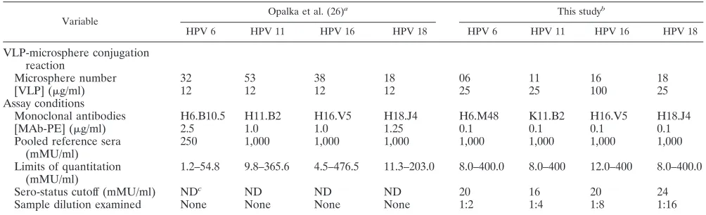
Differences between the proof-of-concept assay described by Opalka et al. (26) and the current validated assay being used to support HPV vaccine clinical trials are shown in Table 1.
Assay validation. Validation of the cLIA for HPV 6, 11, 16, and 18 was performed over 6 days during a 2-week period. Three analysts, using one specific
on August 17, 2020 by guest
http://cvi.asm.org/
Bio-Plex instrument each, generated two assay plates per day either manually or on a “TECAN Genesis Workstation”. Two MAb-PE lots and three separate preparations of VLP-microspheres were examined. From the standard curves generated during the validation, the limits of detection, and the quantifiable ranges of the HPV cLIA were determined for each of the HPV types.
Addition-ally, specification limits for the slope and 50% effective concentration (EC50)
parameters were determined to qualify future assay runs.
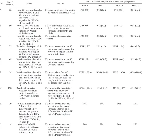
Nine human serum sample panels were tested in the validation of the assay (Table 2). Panels were divided into four groups. Group 1 (panels A, B, and C) contained sera likely to be true negatives. These three panels contained samples from male adolescents, female adolescents, and young adult women (16 to 23 years of age) who reported having two or fewer lifetime sexual partners and who repeatedly tested negative in HPV6, -11, -16, and -18 PCR assays. Group 2 (panels D, E, and F) consisted of sera likely to be true positives. These samples were obtained either from individuals that were enrolled in clinical trials and had received an HPV6, -11, -16, and -18 vaccine or from nonvaccinated subjects with four or more lifetime sexual partners. Panel F was also used to evaluate the dilutability of the assay. Group 3 (panel G) consisted of sera randomly selected from American women who had no history of HPV vaccination. Panel G was used to evaluate the performance of the redefined serostatus cutoff and to provide an estimate of the baseline percent positivity for HPV types 6, 11, 16, and 18 in the primary clinical population. Lastly, group 4 (panels H and I) was used to assess assay robustness and precision between analysts, MAb-PE lots, and VLP-microsphere lots. Panel H (samples with low to medium titers) was tested twice per day per analyst over the 2-week period. Panel I (negative, low, and high spiked ADHS-spiked controls) was tested on each of the 42 validation plates.
Data analysis.The serum antibody titer was obtained by measuring the ability of a test sample to inhibit MAb-PE binding to the VLPs and comparing that value to that of a dilution series of the reference standard. The reference standard dilution series was modeled using the four-parameter logistic function (25), and the test sample concentration was calibrated from the fitted standard curve. The reference sera used for the standard curve were assigned arbitrary antibody concentrations expressed in milli-Merck units per milliliter. Data, in median fluorescence intensity (MFI) units, were processed using either the Bio-Plex Manager 3.0 software (Bio-Rad, Hercules, CA) or using a Microsoft Excel spreadsheet (Redmond, WA).
Since samples were duplicated within a run, intra-assay precision was esti-mated using the variability estimates between the two or three replicates of each
control sample within each run共ˆS2). To determine inter-assay variability,
esti-mates of analyst to analyst variability共ˆA
2
), run within analyst variability关ˆR(A)
2
],
sample by analyst variability共ˆSxA2 ), and sample by run within analyst variability
关ˆSxR(A) 2
] were combined. Variability estimates were obtained on the natural log transformed titers using the MIXED procedure in SAS (SAS Institute Inc., Cary,
NC). An estimate of the total assay precision (% relative standard deviation
[RSD]) was calculated as 100⫻ 关e
冑
ˆS2
⫹ ˆA 2
⫹ ˆR(A) 2
⫹ ˆSxA 2
⫹ ˆSxA 2
⫺1].
RESULTS
Selection of monoclonal antibodies.The MAbs used in the
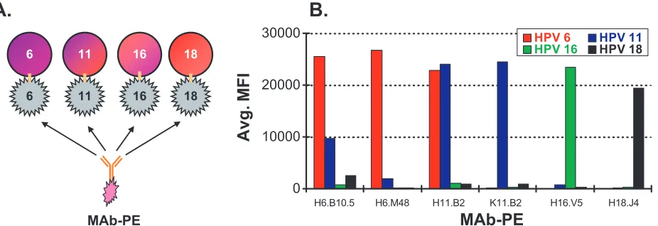
original assay were chosen based on availability, specificity, and assay performance (26). Identifying MAbs that are type-spe-cific and neutralizing to HPV 6 or 11 virions has been difficult, as their respective L1 capsid proteins are greater than 92% identical at the amino acid level and share neutralizing epitopes (9). The HPV 6 and 11 MAbs, H6.B10.5 and H11.B2, have each been shown to bind both HPV 6 and HPV 11 VLPs (9). To improve the analytical specificity by limiting cross-reactivity of the HPV 6 and HPV 11 detection MAbs we examined a previously described MAb for HPV 6 (H6.M48) (8, 11) and a newly developed MAb for HPV 11 (K11.B2) for specificity to its respective VLP. In this experiment, one MAb-PE was added to a well containing the four different VLP-microspheres and allowed to bind to the VLPs (Fig. 1A). The fluorescence signal emitted from the PE-labeled MAb was measured in median fluorescence intensity (MFI). Figure 1B shows the binding specificity of the two new MAbs and the four previously described MAbs in a competitive antigen binding assay. The results show that the H6.M48 and the K11.B2 MAbs have improved specificity over the previously described H6.B10.5 and H11.B2 antibodies. Together, the four MAbs chosen for use in the assay, H6.M48, K11.B2, H16.V5, and H18.J4, all recognize type-specific epitopes on their respective VLPs and show minimal cross-reactivity.
Optimization of the VLP to microsphere conjugation
reac-tion.To adapt the assay previously described by Opalka et al.
(26) for use in large-scale epidemiology and vaccine clinical trials, we first sought to scale up the production of VLP-microspheres from 2.5⫻106to 1.25⫻107microspheres per
TABLE 1. Comparison of assay conditions in the original assay with the validated assay
Variable
Opalka et al. (26)a This studyb
HPV 6 HPV 11 HPV 16 HPV 18 HPV 6 HPV 11 HPV 16 HPV 18
VLP-microsphere conjugation reaction
Microsphere number 32 53 38 18 06 11 16 18
[VLP] (g/ml) 12 12 12 12 25 25 100 25
Assay conditions
Monoclonal antibodies H6.B10.5 H11.B2 H16.V5 H18.J4 H6.M48 K11.B2 H16.V5 H18.J4
[MAb-PE] (g/ml) 2.5 1.0 1.0 1.25 0.1 0.1 0.1 0.1
Pooled reference sera (mMU/ml)
250 1,000 1,000 1,000 1,000 1,000 1,000 1,000
Limits of quantitation (mMU/ml)
1.2–54.8 9.8–365.6 4.5–476.5 11.3–203.0 8.0–400.0 8.0–400 12.0–400 8.0–400.0
Sero-status cutoff (mMU/ml) NDc ND ND ND 20 16 20 24
Sample dilution examined None None None None 1:2 1:4 1:8 1:16
aActivation buffer, PBS (pH 7.4). Microspheres/conjugation, 2.5⫻106microspheres/ml. Conjugation buffer, PBS. Incubation time, 2 h. Wash buffer, PBS plus 0.05%
Tween-20 (pH 7.4). Blocking or storage buffer, histidine buffer (pH 6.2), column A buffer, or PBS⫾1% BSA. Pooled VLP-miscrosphere storage buffer, histidine buffer.
Serostatus cutoff, not determined. Sample dilution examined, no. Detergent, no. Heat inactivation of serum, no. Sample matrix, HPV-negative normal human sera. Incubation time, overnight.
bActivation buffer, 0.1 M NaH
2PO4(pH 6.2). Microspheres/conjugation, 1.25⫻107microspheres/ml. Conjugation buffer, MES. Incubation time, overnight. Wash
buffer, PBS plus 1% Triton X-100 (pH 7.4). Blocking or storage buffer, histidine buffer (pH 6.2)⫹1% BSA. Pooled VLP-miscrosphere storage buffer, PBS⫹1% BSA
⫹0.05% NaN3⫹1% TX100. Detergent, Triton X-100. Heat inactivation of serum, 30⫾2 min at 56⫾2°C. Sample matrix, antibody-depleted human sera. Incubation
time, 15 to 25 h.
cND, not determined.
on August 17, 2020 by guest
http://cvi.asm.org/
reaction. To optimize the scale-up, we compared two buffers: MES, as recommended by Luminex Corporation, and PBS. In general, the MES buffer produced more reproducible results (data not shown). Furthermore, as VLPs have been shown to be more stable at a pH of 6.0 (MES) than at pH 7.4 (PBS), MES was chosen as the preferred coupling buffer.
Optimization of VLP and MAb-PE concentration.We first
sought to determine the MAb-PE and VLP concentrations to maximize sensitivity without sacrificing dynamic range or ro-bustness of the assay. Standard curves were generated by add-ing varyadd-ing concentrations of quadriplex MAb-PE solutions (0.0625 to 10g/ml per MAb type) to microspheres coupled
with 25 to 200g/ml of VLPs. The effect of MAb H16.V5-PE concentration on the standard curves for VLP 16 conjugated at 200 g/ml is shown in Fig. 2A. These results showed that a MAb-PE concentration of 0.1g/ml resulted in reproducible standard curves with good sensitivity and dynamic range.
To determine the optimal concentration of VLP, micro-spheres (1.25⫻107per reaction) were coupled with 500l of
a 25, 50, 100, or 200g/ml solution of HPV VLP 6, 11, 16, and 18, respectively. Each VLP-microsphere preparation was then tested with the same reference sera standard preparation using the optimized MAb-PE concentration of 0.1g/ml. The results for HPV VLP 16 are shown in Fig. 2B. The VLP
concentra-TABLE 2. Serum panels and seropositivity values
Panel No. of
samples Criteria Purpose
No. positive/No. samples with a result and (%) positive
HPV 6 HPV 11 HPV 16 HPV 18
A 56 16 to 23 year old females who reported 1 or 2 lifetime sex partners and were PCR negative for HPV 6, 11, 16, and 18
Primary sample set for setting the clinical serostatus cutoff
0/50 (0.0) 0/48 (0.0) 0/50 (0.0) 0/50 (0.0)
B 96 10 to 12 year old male and female anonymous subjects in Merck clinical studies
To set serostatus cutoff if no differences discovered between adolescents and adults
0/83 (0.0) 0/82 (0.0) 1/85 (1.2) 0/85 (0.0)
C 44 16 to 23 year old female virgins who were PCR negative for HPV 6, 11, 16, and 18
To monitor the serostatus cutoff
0/39 (0.0) 0/38 (0.0) 0/39 (0.0) 0/39 (0.0)
D 72 Females who reported 4 or more lifetime sex partners with higher likelihood of current or past HPV infection
To assess serostatus cutoff and assay performance for subjects of higher risk for natural infection
8/63 (12.7) 1/61 (1.6) 10/63 (15.9) 6/62 (9.7)
E 68 Vaccinated females with low antibody titers as determined in a cRIA for HPV 6, 11, 16, and 18
To assess serostatus cutoff and assay performance for low-titer samples
32/56 (57.1) 42/53 (79.2) 50/55 (90.9) 8/55 (14.5)
F 24 Vaccinated females with antibody titers greater than 100 mMU/ml as determined by a cRIA for HPV 6, 11, 16, and 18
To assess the effect of dilution on antibody titers and to demonstrate the assay’s ability to measure moderate titer samples
20/20 (100.0) 20/20 (100.0) 20/20 (100.0) 20/20 (100.0)
G 192 Randomly selected baseline sera from subjects enrolled in HPV vaccine clinical studies
To validate the serostatus cutoff with expected baseline seroprevalence of ⬍5% for HPV 11 and ⬍15% for HPV 6, 16, and 18
17/168 (10.1) 5/164 (3.0) 22/170 (12.9) 5/169 (3.0)
H 16 Sera from females given 2 doses of a
quadrivalent HPV vaccine that gave a detectable anti-HPV titer as measured in a cRIA for HPV 6, 11, 16, and 18
To assess robustness and precision of the assay between analysts and different lots of MAb-PE and VLP-microspheres
NAa NA NA NA
I 4 Samples of ADHS
spiked with different amounts of AGM reference sera
To assess robustness and precision of the assay between analysts and different lots of MAb-PE and VLP-microspheres
NA NA NA NA
aNA, nonapplicable.
on August 17, 2020 by guest
http://cvi.asm.org/
tions which achieved acceptable sensitivity while maintaining robust assay performance were determined to be 25, 25, 100, and 25g/ml for HPV 6, 11, 16, and 18, respectively. A con-centration of 100 g/ml of HPV16 VLP was chosen as the optimal concentration, because the lower concentrations of VLP resulted in standard curves with lower fluorescence val-ues, reproducibility issval-ues, and small dynamic ranges. Using the final assay conditions of 25, 25, 100, and 25g/ml of VLP 6, 11, 16, and 18, respectively, 0.1g/ml of each MAb-PE, we achieved good sensitivity with a lower limit of quantitation of 8, 8, 12, and 8 mMU/ml for HPV 6, 11, 16, and 18, respectively, and upper limits of quantitation of 400 mMU/ml for all four types.
Sample matrix comparisons. With the concentrations of
VLPs and MAbs determined, we next evaluated which diluent to use as a sample matrix. Because sera from naturally infected individuals typically have very low concentrations of antibodies
to HPV virions (18), the sera must be tested at a high concen-tration. This challenge is compounded by the fact that at high concentrations there are considerable matrix effects caused by interfering substances in serum that vary by individual. These interfering substances can include lipids, cholesterol, proteins, and heterophilic antibodies. Due to the high prevalence of HPV infection in the general serum donor population and inconsistency of serum from different donors, it was difficult to find adequate amounts of HPV negative sera to use as a reli-able, reproducible sample diluent. Therefore, we sought to identify a matrix that most closely resembled HPV negative, normal human serum (NHS), while minimizing matrix effects (Fig. 3). The matrices or diluents evaluated against NHS in-cluded PBS, PBS with 1% BSA, PBS with 1% recombinant human albumin (rHA), normal human serum (NHS), normal goat serum (NGS), defibrinated plasma (DFP), antibody-de-pleted human sera (ADHS), and 3% nonfat milk in PBS. As
FIG. 1. Monoclonal antibody specificity. (A) Experimental design for MAb specificity experiment. A single MAb-PE was incubated with VLP 6, 11, 16, and 18 microspheres. Detection of MAb-PE to any of the four VLP types revealed any cross-reactivity. (B) MAb specificity results. HPV 6, 11, 16, and 18 VLP-microspheres (5,000 VLP-microspheres per type; VLP concentration, 200g/ml) were added in quadriplex to each well. One MAb-PE was added to each well at a concentration of 40g/ml. Detection of fluorescence from the PE-labeled MAbs is shown in median fluorescence intensity (MFI).
FIG. 2. Effect of VLP and monoclonal antibody-PE concentration on assay sensitivity for HPV 16. (A) Effect of different concentrations of H16.V5 MAb-PE (0.0625 to 10.0g/ml) on assay sensitivity and dynamic range. VLP-microspheres used in this experiment were coupled with HPV 16 VLP at a concentration of 200g/ml. (B) Effect of different concentrations of VLP 16 (25 to 200g/ml) on assay sensitivity and dynamic range. The concentration of H16.V5 MAb-PE was 0.1g/ml.
on August 17, 2020 by guest
http://cvi.asm.org/
shown in Fig. 3, the PBS-based matrices produced standard curves that deviated significantly from the NHS standard curves at higher standard concentrations. Across HPV types, the observed matrix effects were generally highest using NGS. Nonfat milk had a similar profile as NHS; however, the dis-parate composition of nonfat milk prevented its use as a sub-stitute for NHS. ADHS and DFP proved to be the most similar to NHS. Because the assay uses sera and not plasma and the fact that ADHS was reproducible from lot to lot (data not shown), we chose to use ADHS as the sample matrix.
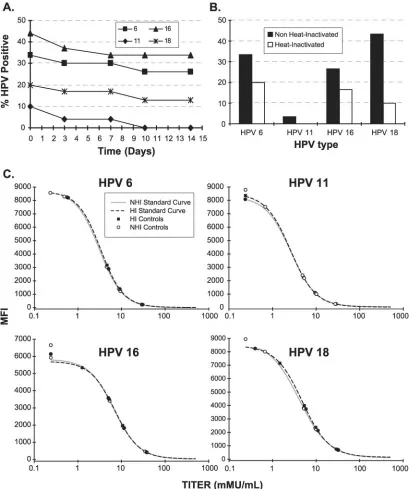
Heat inactivation.A key discovery made during assay
devel-opment was that a heat-labile factor(s) interfered with detec-tion of MAb binding to the VLPs and caused false-positive results. This factor was discovered by the observation that titers decreased and then leveled off on samples that were stored at 4°C for several weeks (Fig. 4A). To eliminate this phenomenon, an experiment was conducted to analyze the effects of heat inactivation on freshly thawed samples. Heat-inactivated samples had antibody titers 48.0%, 5.6%, 28.0%, and 64.3% lower for HPV 6, 11, 16, and 18, respectively, than non-heat-inactivated samples. Using a provisional serostatus
cutoff of 24 mMU/ml for HPV 6, 11, 16, and 18, a pronounced difference in serostatus agreement was observed for all HPV types (Fig. 4B). Heat-inactivating samples that had been stored at 4°C for several weeks had negligible effects on HPV anti-body titers (data not shown). Lastly, heat inactivating the HPV reference serum before spiking it into ADHS had no effect on antibody titers (Fig. 4C) demonstrating that the heat-labile factor was not an antibody and that the HPV antibodies were resistant to heat inactivation. These findings led us to include a heat inactivation step of 56⫾1°C for 30⫾2 min as part of our sample preparation procedure.
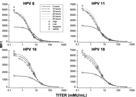
Time determination. Experiments were also performed
comparing incubation times ranging from 1 to 4 h (a same-day assay) and 15 to 25 h (an overnight assay). Thirty-two human serum samples were tested in the HPV cLIA, with an incuba-tion time of 3, 15, 18, 20, 22, or 25 h (Fig. 5). Little difference was seen in both the controls and the sample titers generated from assays incubated between 15 to 25 h. While there was little difference between the test sample titers generated during the 3-h incubation, the maximum MFIs of the standard curves were at least twofold lower than the maximum MFIs of the
FIG. 3. Effect of different sample matrices on assay sensitivity and robustness. Each standard curve was generated using the specified matrix, 100 g/ml VLP-microspheres, and a 0.1-g/ml final MAb-PE concentration. The mixture was incubated overnight (15 to 25 h) and then qualitatively and quantitatively evaluated in comparison to normal human serum (NHS). Abbreviations: bovine serum albumin (BSA), recombi-nant human albumin (rHA), normal human serum (NHS), normal goat serum (NGS), antibody depleted human serum (ADHS), and defibrinated plasma (DFP).
on August 17, 2020 by guest
http://cvi.asm.org/
curves from the 15 to 25 h time points and had a smaller dynamic range. To maintain reproducibility, incubation times were limited to 15 to 25 h after the MAb-PE and VLP-micro-spheres were added to the samples.
Assay validation. Nine human serum sample panels were
tested in the validation of the assay (Table 2). Panels were divided into four groups: group 1 (panels A, B, and C) con-tained sera likely to be true negatives; group 2 (panels D, E,
and F) consisted of sera likely to be true positives; group 3 (panel G) consisted of sera randomly selected from prevacci-nated women from the United States; and group 4 (panels H and I) was used to assess assay robustness and precision be-tween analysts, MAb-PE lots, and VLP-microsphere lots.
From panels H and I, the intra-assay variability (variability within a day across different plates), the interassay variability (variability across different days), and the overall assay
preci-FIG. 4. Effect of heat inactivation of serum samples on HPV antibody titers. (A) Seventy randomly selected retention samples that had never been thawed were thawed at RT and stored at 4°C. The samples were tested in the cLIA on days 0, 3, 7, 10, and 14. (B) Percentage of HPV-positive samples using a serostatus cutoff of 24 mMU/ml for all HPV types. A total of 70 freshly thawed samples were aliquoted into two tubes. One of the tubes was heat inactivated for 30⫾2 min at 56°C, while the other remained at 4°C. Both samples were tested on the same plate in duplicate and read from the same standard curve. (C) Heat inactivation of reference serum has no effect on antibody titers. Reference serum was either heat inactivated or remained at 4°C before spiking into ADHS and tested in the HPV cLIA.
on August 17, 2020 by guest
http://cvi.asm.org/
sion were determined. The overall precision was 21.7%, 20.4%, 23.0%, and 15.9% for HPV 6, 11, 16, and 18, respectively (Table 3). In addition, the use of a TECAN Genesis Worksta-tion resulted in significantly better precision than assays per-formed manually (data not shown). Potential sources of assay
variability were also examined using two different MAb-PE lots, three different VLP-microsphere lots, and three different analysts. No meaningful differences were observed between VLP-microsphere lots or analysts (⬍15%). Different MAb-PE lots had varying impact on the assay (7.3%, 19.4%, 22.8%, and
FIG. 5. Effect of different assay incubation times on assay sensitivity, dynamic range, precision, and robustness. Each curve on the graph represents a four-parameter (4-PL) logistic fit of a 12-point standard curve for HPV 6, 11, 16, and 18. Antibody titers generated from the controls of each standard curve have been plotted on top of each standard curve. VLP-microspheres used in this experiment were coupled with 100g/ml of HPV 16 VLP and 25g/ml of HPV 6, 11, and 18 VLPs. The concentrations of H6.M48-PE, K11.B2-PE, H16.V5-PE, and H18.J4-PE were 0.1 g/ml. All standards were at a final starting concentration of 500 mMU/ml. The mixture of VLP-microspheres, MAb-PE, reference sera, and ADHS was incubated for 3, 15, 18, 20, 22, or 25 h.
TABLE 3. Validation parameter summary table
Assay characteristic HPV type
HPV 6 HPV 11 HPV 16 HPV 18
Extravariability limit (range of 10th root of MFI duplicates) 0.0694 0.0667 0.0611 0.0493
Quantifiable range (mMU/ml) (8, 400) (8, 400) (12, 400) (8, 400)
Upper 99% RMSE limit, 12 point (MFI) 0.3437 0.4049 0.3244 0.2811
Upper 99% RMSE limit, 11 point (MFI) 0.3522 0.4149 0.3324 0.2880
Upper 99% RMSE limit, 10 point (MFI) 0.3627 0.4272 0.3423 0.2966
3Slope limits (MFI/mMU/ml) (1.29, 1.71) (1.1, 1.44) (1.11, 1.68) (1.02, 1.27)
3EC50limits (mMU/ml) (1.1, 4.7) (1.3, 3.3) (3.9, 14.2) (1.5, 6.9)
Serostatus cutoff (mMU/ml) 20 16 20 24
Interassay precision (%RSD) 19.5% 18.0% 21.0% 13.7%
Intraassay precision (%RSD) 8.7% 8.8% 8.3% 7.6%
Total precision (%RSD) 21.7% 20.4% 23.0% 15.9%
3High positive control limits (mMU/ml) (67, 218) (79, 239) (80, 276) (89, 214)
3Low positive control limits (mMU/ml) (20, 62) (23, 70) (22, 76) (27, 65)
3Negative control limits (mMU/ml) (10, 30) (11, 33) (11, 37) (13, 32)
Dilution effect (DE) per twofold increase in titer 12.4% 13.6% 11.8% 4.2%
95% Confidence interval on DE (% bias per twofold dilution) (8%, 17%) (10.8%, 16.4%) (8.8%, 14.8%) (1.7%, 6.8%)
on August 17, 2020 by guest
http://cvi.asm.org/
5.3% for HPV 6, 11, 16, and 18, respectively). From the 42 assay validation runs, the pass/fail specification limits for de-termining assay validity in routine operation were set for the slope, EC50, and root mean square error (RMSE) of the
stan-dard curve parameters and titer ranges for the control samples (Table 3).
Panel F was used to examine the effect of sample dilution on antibody titers. Each sample in panel F was diluted in a twofold dilution series from 1:4 to 1:512. On average, sample titers increased 12.4%, 13.6%, 11.8%, and 4.2% per twofold dilution for HPV 6, 11, 16, and 18, respectively (Table 3).
Because no anti-HPV serum standards exist, we developed panels of sera based on HPV vaccination, likelihood of previ-ous HPV infection, and/or by PCR positivity of cervical swabs to determine the serostatus cutoffs (Table 2). Panels A, B, and C were used to set the serostatus cutoff, and panel G was used to evaluate the performance of the serostatus cutoff for HPV 6, 11, 16, and 18. The serostatus cutoff is the antibody concen-tration that can reliably distinguish a panel of positive samples from a panel of negative samples. The serostatus cutoff was established by determining the lowest titer within a valid as-say’s quantifiable range that provided for an acceptable level of discrimination between the “negative” sera (panels A, B, and C) and the sera likely to contain a varying percentage of true positives (panels D, E, F, and G) (Tables 2 and 3). Using data from runs that passed the acceptance criteria defined in Table 3, the serostatus cutoffs were established at 20, 16, 20, and 24 mMU/ml for HPV 6, 11, 16, and 18, respectively.
DISCUSSION
An immunoassay that measures HPV type-specific antibod-ies simultaneously is preferred to running multiple separate tests. Thus, a competitive immunoassay that simultaneously measures type-specific antibodies to neutralizing epitopes on HPV 6, 11, 16, and 18 was developed. We have modified an HPV Luminex immunoassay to be more sensitive and more robust for testing serum samples from individuals enrolled in epidemiology studies and HPV vaccine clinical trials. The in-crease in sensitivity was necessary to measure the low titers of anti-HPV antibodies in the serum from naturally infected in-dividuals, a requirement for epidemiological and vaccine stud-ies. By analyzing each step of the assay, we have characterized the effects of reagents, timing, and operator variability on the assay. The planned long-term use of the assay required the development of a reference standard to monitor the immune response and to normalize results across runs. In addition, we have developed negative-, low-, medium-, and high-titer qual-ity control panels to monitor the consistency of our results periodically. The assay was formatted for automation so that it could be run on a high-throughput TECAN Genesis Worksta-tion. Following optimization, validation of the assay was per-formed to (i) assess the reproducibility and precision of the assay, (ii) establish a quantifiable range, (iii) assess the effect of dilution, (iv) establish a serostatus cutoff, and (v) evaluate the performance of the serostatus cutoff.
To perform the competitive multiplexed assay, MAbs spe-cific to a single HPV VLP type were needed. To improve the analytical specificity of the assay we switched from the H6.B10.5 MAb to the H6.M48 MAb and from the H11.B2
MAb to the K11.B2 MAb (Fig. 1B). Another advantage of using H6.M48 over H6.B10.5 is that H6.B10.5 was a difficult antibody to purify and to conjugate to PE. Because the HPV cLIA is used to measure the immune response to four sepa-rately manufactured VLPs, it is important that the MAbs rec-ognize type-specific, neutralizing epitopes on each of the VLPs. Ideally, these MAbs would also recognize the immuno-dominant epitopes that are elicited following natural infection, like has been shown for H16.V5 (39).
To reduce the number of steps in the assay and reduce sources of error, we chose to directly conjugate R-phyco-erythrin to the four MAbs. The concentration of MAb and VLP was also optimized. As expected for a competitive immu-noassay, optimization experiments showed that as the MAb concentration (Fig. 2A) or VLP concentration (Fig. 2B) de-creased, the sensitivity of the assay increased. However, if the MAb or VLP concentration was too low, the assay became less reproducible. The final concentrations established were 0.1
g/ml for each of the MAb-PEs and 25, 25, 100, and 25g/ml of VLPs for HPV 6, 11, 16, and 18, respectively. These opti-mized concentrations provided acceptable sensitivity and a broad dynamic range and were robust in routine operation.
Several different sample matrices were evaluated to improve the clinical sensitivity of the assay. Previously, normal human serum was used to limit any differences between the sample and the sample matrix, but identifying large quantities of nor-mal human sera that were negative for HPV 6, 11, 16, and 18 and consistent from donor to donor was extremely difficult. Experiments comparing different sample matrices used to di-lute the samples and reference sera were performed and iden-tified ADHS as the best alternative matrix (Fig. 3). Seven other matrices were also evaluated, including PBS, PBS with 1% BSA, PBS with 1% rHA, NGS, DFP, and 3% nonfat milk in PBS, but none demonstrated the reproducibility and similarity to normal human sera as ADHS. In addition, ADHS was similar in composition to human sera and easy to obtain and was consistent from lot to lot. The PBS-based matrices were not used due to significant deviations from the standard curves generated in NHS. Because of the erratic shapes of the stan-dard curves generated in PBS-based matrices and other matri-ces, such as NGS, ADHS was chosen as the sample diluent to replace NHS.
Several different preanalytical factors can adversely affect results in a clinical assay. These include sample collection and processing variables such as anticoagulants in the collection container, storage and transport temperatures, lipemia, and hemolysis. An important finding during the development of the assay was the observation that a heat-labile factor, present in many individuals’ sera, could interfere with binding of the detection MAb, resulting in higher than expected antibody titers. Heat inactivation of freshly thawed samples significantly decreased the number of positive samples (Fig. 4B) to be more in line with expectations of 5 to 20%, as supported by natural history data (6, 22, 32). The heat-labile factor(s) that caused the false positive results has not yet been identified. Heat inactivation did not impact the titers of samples that had been stored at 4°C for several weeks and did not affect antibodies in the reference standard in comparison to a non-heat-inactivated reference standard (Fig. 4C). These findings led us to include
on August 17, 2020 by guest
http://cvi.asm.org/
a heat inactivation step of 56⫾1°C for 30⫾2 min as part of our sample preparation procedures.
Following assay optimization, a formal validation of the as-say for clinical testing was performed. To examine the asas-say in the target population of females ranging in age from 9 to 45 years and to define the accuracy and precision of the assay, we created nine human serum panels (Table 2). Panels H and I were used to determine the robustness and precision of the assay and also to set specification limits on the controls that are tested in routine operation. Panels A, B, and C contained samples that were most likely to be HPV negative. Panels A and C contained samples from HPV PCR-negative women with two or fewer lifetime sexual partners. Panel B was com-prised of samples from both male and female adolescents ages 10 to 12 and was used to determine whether there were dif-ferences between adults and adolescents. Titer results from these samples were used to determine the background titers of HPV-negative individuals and to set the serostatus cutoffs. The serostatus cutoffs were established by identifying the lowest or next to lowest dilution titer that all of the samples in panels A, B, and C yielded negative results. Using this methodology, the serostatus cutoffs were set at 20, 16, 20, and 24 mMU/ml for HPV6, -11, -16, and -18, respectively. Of note, one sample in panel B yielded a titer of 31 mMU/ml for HPV16 which was greater than the serostatus cutoff of 20 mMU/ml.
Panels D, E, and F included samples from individuals likely to be positive for HPV antibodies because of prior HPV vac-cination or sexual history. These samples were used to assess the assay in the positive region of the standard curve. Panel F (serum from vaccinated individuals) was also used to assess the dilutability of the assay. Results showed that the dilutability of the HPV cLIA ranged from 4.2% to 13.6% per twofold in-crease in dilution for samples diluted from 1:4 to 1:512 (Table 3). Since the upper limit of quantitation for all four types is 400 mMU/ml, dilutions are necessary to calculate titers greater than 400 mMU/ml and have been set at 1:40, 1:400, and 1:4,000. To be conservative, the lowest dilution titer that fell within the quantifiable range was reported.
Panel G consisted of randomly selected baseline sera from subjects enrolled in Merck HPV vaccine clinical trials and were used to assess the newly defined serostatus cutoff. The percent-age of positive samples in panel G was 10.1%, 3.0%, 12.9%, and 3.0% for HPV 6, 11, 16, and 18 which was in line with our expectation of less than 20% for all four HPV types. Previous serologic studies have demonstrated that 20 to 50% of women with HPV DNA do not have detectable type-specific anti-HPV antibodies (6). In our preliminary analysis, about 50% of the women that had a PCR-positive result also had an antibody-positive result.
It is worth noting that the clinical sensitivity and specificity of the validation was performed using predominantly samples from women. Other studies have shown that serology assays may underestimate past or current HPV infection in males (34) and have reported HPV seroprevalence to be generally higher in women than in men (20, 33).
The challenges of developing an HPV serology assay to monitor both natural infection and response to vaccination have also been described by others (18, 38). The most formi-dable challenge for developing a serology assay to measure past or current infection is that not all people seroconvert
following exposure and that no reference panels exist to de-termine the clinical sensitivity and specificity of an assay. Fur-thermore, women with detectable viral DNA in genital epithe-lial cells may not have detectable HPV antibodies, as the antibody response may occur many months after initial infec-tion (5, 35). As the majority of HPV infecinfec-tions of the anogeni-tal tract resolve over 3 to 5 years (30), serologic testing is a better measure of cumulative exposure rather than ongoing infection. Data from ongoing epidemiology studies and clinical trials will provide an opportunity to study the natural history of antibody responses in relationship to HPV DNA detection and clinical outcome, to measure vaccine-induced immune re-sponses over time, and to identify clinically relevant immuno-logic correlates of protection.
In summary, the HPV 6, 11, 16, and 18 cLIA has proven to be a robust, sensitive, and high-throughput assay and is cur-rently being used for epidemiology studies and evaluating the efficacy of prophylactic HPV vaccines.
ACKNOWLEDGMENTS
We thank Natasha Blackshear, Heather Sings, and Janine Bryan for critical review of the manuscript.
REFERENCES
1.American Cancer Society.2004. Cancer facts and figures 4. American Cancer Society, Atlanta, Ga.
2.Atypical Squamous Cells of Undetermined Significance/Low-Grade Squa-mous Intraepithelial Lesions Triage Study (ALTS) Group.2000. Human papillomavirus testing for triage of women with cytologic evidence of low-grade squamous intraepithelial lesions: baseline data from a randomized
trial. J. Natl. Cancer Inst.92:397–402.
3.Bosch, F. X., A. Lorincz, N. Munoz, C. J. Meijer, and K. V. Shah.2002. The causal relation between human papillomavirus and cervical cancer. J. Clin.
Pathol.55:244–265.
4.Bosch, F. X., M. M. Manos, N. Munoz, M. Sherman, A. M. Jansen, J. Peto, M. H. Schiffman, V. Moreno, R. Kurman, and K. V. Shah.1995. Prevalence of human papillomavirus in cervical cancer: a worldwide perspective. Inter-national biological study on cervical cancer (IBSCC) study group. J. Natl.
Cancer Inst.87:796–802.
5.Carter, J. J., L. A. Koutsky, J. P. Hughes, S. K. Lee, J. Kuypers, N. Kiviat, and D. A. Galloway.2000. Comparison of human papillomavirus types 16, 18, and 6 capsid antibody responses following incident infection. J. Infect. Dis.
181:1911–1919.
6.Carter, J. J., M. M. Madeleine, K. Shera, S. M. Schwartz, K. L. Cushing-Haugen, G. C. Wipf, P. Porter, J. R. Daling, J. K. McDougall, and D. A. Galloway.2001. Human papillomavirus 16 and 18 L1 serology compared
across anogenital cancer sites. Cancer Res.61:1934–1940.
7.Chesson, H. W., J. M. Blandford, T. L. Gift, G. Tao, and K. L. Irwin.2004. The estimated direct medical cost of sexually transmitted diseases among
American youth, 2000. Perspect. Sex Reprod. Health36:11–19.
8.Christensen, N. D., J. Dillner, C. Eklund, J. J. Carter, G. C. Wipf, C. A. Reed, N. M. Cladel, and D. A. Galloway.1996. Surface conformational and linear epitopes on HPV-16 and HPV-18 L1 virus-like particles as defined by
mono-clonal antibodies. Virology223:174–184.
9.Christensen, N. D., R. Kirnbauer, J. T. Schiller, S. J. Ghim, R. Schlegel, A. B. Jenson, and J. W. Kreider.1994. Human papillomavirus types 6 and 11 have antigenically distinct strongly immunogenic conformationally
depen-dent neutralizing epitopes. Virology205:329–335.
10.Christensen, N. D., J. W. Kreider, N. M. Cladel, S. D. Patrick, and P. A. Welsh.1990. Monoclonal antibody-mediated neutralization of infectious
hu-man papillomavirus type 11. J. Virol.64:5678–5681.
11.Christensen, N. D., C. A. Reed, N. M. Cladel, K. Hall, and G. S. Leiserowitz.
1996. Monoclonal antibodies to HPV-6 L1 virus-like particles identify con-formational and linear neutralizing epitopes on HPV-11 in addition to
type-specific epitopes on HPV-6. Virology224:477–486.
12.Cook, J. C., J. G. Joyce, H. A. George, L. D. Schultz, W. M. Hurni, K. U. Jansen, R. W. Hepler, C. Ip, R. S. Lowe, P. M. Keller, and E. D. Lehman.
1999. Purification of virus-like particles of recombinant human papillomavi-rus type 11 major capsid protein L1 from Saccharomyces cerevisiae. Protein
Expr. Purif.17:477–484.
13.Gerberding, J. L.2004. Prevention of genital human papillomavirus. Centers for Disease Control and Prevention, Atlanta, Ga.
14.Harper, D. M., E. L. Franco, C. Wheeler, D. G. Ferris, D. Jenkins, A.
on August 17, 2020 by guest
http://cvi.asm.org/
Schuind, T. Zahaf, B. Innis, P. Naud, N. S. De Carvalho, C. M. Roteli-Martins, J. Teixeira, M. M. Blatter, A. P. Korn, W. Quint, and G. Dubin.
2004. Efficacy of a bivalent L1 virus-like particle vaccine in prevention of infection with human papillomavirus types 16 and 18 in young women: a
randomised controlled trial. Lancet364:1757–1765.
15.Hofmann, K. J., J. C. Cook, J. G. Joyce, D. R. Brown, L. D. Schultz, H. A. George, M. Rosolowsky, K. H. Fife, and K. U. Jansen. 1995. Sequence determination of human papillomavirus type 6a and assembly of virus-like
particles in Saccharomyces cerevisiae. Virology209:506–518.
16.Hofmann, K. J., M. P. Neeper, H. Z. Markus, D. R. Brown, M. Muller, and K. U. Jansen.1996. Sequence conservation within the major capsid protein of human papillomavirus (HPV) type 18 and formation of HPV-18 virus-like
particles in Saccharomyces cerevisiae. J. Gen. Virol.77:465–468.
17.Jansen, K. U., and A. R. Shaw.2004. Human papillomavirus vaccines and
prevention of cervical cancer. Annu. Rev. Med.55:319–331.
18.Karem, K. L., A. C. Poon, C. Bierl, R. Nisenbaum, and E. Unger.2002. Optimization of a human papillomavirus-specific enzyme-linked
immunosor-bent assay. Clin. Diagn. Lab. Immunol.9:577–582.
19.Koutsky, L. A., K. A. Ault, C. M. Wheeler, D. R. Brown, E. Barr, F. B. Alvarez, L. M. Chiacchierini, and K. U. Jansen.2002. A controlled trial of a
human papillomavirus type 16 vaccine. N. Engl. J. Med.347:1645–1651.
20.Kreimer, A. R., A. J. Alberg, R. Viscidi, and M. L. Gillison.2004. Gender differences in sexual biomarkers and behaviors associated with human
pap-illomavirus-16, -18, and -33 seroprevalence. Sex Transm. Dis.31:247–256.
21.Kronick, M. N.1986. The use of phycobiliproteins as fluorescent labels in
immunoassay. J. Immunol. Methods92:1–13.
22.Laukkanen, P., P. Koskela, E. Pukkala, J. Dillner, E. Laara, P. Knekt, and M. Lehtinen. 2003. Time trends in incidence and prevalence of human
papillomavirus type 6, 11 and 16 infections in Finland. J. Gen. Virol.84:
2105–2109.
23.Munoz, N., F. X. Bosch, S. de Sanjose, R. Herrero, X. Castellsague, K. V. Shah, P. J. Snijders, and C. J. Meijer.2003. Epidemiologic classification of human papillomavirus types associated with cervical cancer. N. Engl. J. Med.
348:518–527.
24.Neeper, M. P., K. J. Hofmann, and K. U. Jansen.1996. Expression of the major capsid protein of human papillomavirus type 11 in Saccharomyces
cerevisae. Gene180:1–6.
25.O’Connell, M., B. Belanger, and P. Haaland.1992. The four-parameter logistic model for calibration and assay development. Am. Stat. Assoc. Proc.
Biopharm. Section1992:180–185.
26.Opalka, D., C. E. Lachman, S. A. MacMullen, K. U. Jansen, J. F. Smith, N. Chirmule, and M. T. Esser.2003. Simultaneous quantitation of antibodies to neutralizing epitopes on virus-like particles for human papillomavirus types 6, 11, 16, and 18 by a multiplexed Luminex assay. Clin. Diagn. Lab. Immunol.
10:108–115.
27.Parkin, D. M., F. I. Bray, and S. S. Devesa.2001. Cancer burden in the year
2000. The global picture. Eur. J. Cancer37:S4–S66.
28.Peyton, C. L., P. E. Gravitt, W. C. Hunt, R. S. Hundley, M. Zhao, R. J. Apple, and C. M. Wheeler.2001. Determinants of genital human papillomavirus
detection in a US population. J. Infect. Dis.183:1554–1564.
29.Rossi, J. L., L. Gissmann, K. Jansen, and M. Muller.2000. Assembly of human papillomavirus type 16 pseudovirions in Saccharomyces cerevisiae.
Hum. Gene Ther.11:1165–1176.
30.Schlecht, N. F., R. W. Platt, E. Duarte-Franco, M. C. Costa, J. P. Sobrinho, J. C. Prado, A. Ferenczy, T. E. Rohan, L. L. Villa, and E. L. Franco.2003. Human papillomavirus infection and time to progression and regression of
cervical intraepithelial neoplasia. J. Natl. Cancer Inst.95:1336–1343.
31.Staros, J. V., R. W. Wright, and D. M. Swingle.1986. Enhancement by N-hydroxysulfosuccinimide of water-soluble carbodiimide-mediated
cou-pling reactions. Anal. Biochem.156:220–222.
32.Stone, K. M., K. L. Karem, M. R. Sternberg, G. M. McQuillan, A. D. Poon, E. R. Unger, and W. C. Reeves.2002. Seroprevalence of human
papilloma-virus type 16 infection in the United States. J. Infect. Dis.186:1396–1402.
33.Svare, E. I., S. K. Kjaer, B. Nonnenmacher, A. M. Worm, H. Moi, R. B. Christensen, A. J. van den Brule, J. M. Walboomers, C. J. Meijer, N. L. Hubbert, D. R. Lowy, and J. T. Schiller. 1997. Seroreactivity to human papillomavirus type 16 virus-like particles is lower in high-risk men than in
high-risk women. J. Infect. Dis.176:876–883.
34.Thompson, D. L., J. M. Douglas, Jr., M. Foster, M. E. Hagensee, C. Digu-iseppi, A. E. Baron, J. E. Cameron, T. C. Spencer, J. Zenilman, C. K. Malotte, G. Bolan, M. L. Kamb, and T. A. Peterman.2004. Seroepidemiol-ogy of infection with human papillomavirus 16, in men and women attending
sexually transmitted disease clinics in the United States. J. Infect. Dis.190:
1563–1574.
35.Tindle, R. W.2002. Immune evasion in human papillomavirus-associated
cervical cancer. Nat. Rev. Cancer2:59–65.
36.Villa, L. L., R. L. R. Costa, C. A. Petta, R. P. Andrade, K. A. Ault, A. R. Giuliano, C. M. Wheeler, L. A. Koutsky, C. Malm, M. Lehtinen, F. E. Skjeldestad, S.-E. Olsson, M. Steinwall, D. R. Brown, R. J. Kurman, B. M. Ronnett, M. H. Stoler, A. Ferenczy, D. M. Harper, G. M. Tamms, J. Yu, L. Lupinacci, R. Railkar, F. J. Taddeo, K. U. Jansen, M. T. Esser, H. L. Sings, A. J. Saah, and E. Barr.2005. Prophylactic quadrivalent human papilloma-virus (types 6, 11, 16, and 18) L1 papilloma-virus-like particle vaccine in young women: a randomised double-blind placebo-controlled multicentre phase II efficacy
trial. Lancet Oncol.6:271–278.
37.von Krogh, G.2001. Management of anogential warts (condylomata
acumi-nata). Eur. J. Dermatol.11:598–604.
38.Wang, X., M. Sapp, N. D. Christensen, and J. Dillner.2005. Heparin-based ELISA reduces background reactivity in virus-like particle-based
papilloma-virus serology. J. Gen. Virol.86:65–73.
39.Wang, Z., N. Christensen, J. T. Schiller, and J. Dillner.1997. A monoclonal antibody against intact human papillomavirus type 16 capsids blocks the
serological reactivity of most human sera. J. Gen. Virol.78:2209–2215.