4(25): 4289-4309, 2014 SCIENCEDOMAINinternational
www.sciencedomain.org
The Potentiation of Anti-inflammatory Effect
and INOS and COX-2 Gene Expression
Inhibition by Rut in When Complexed
with Cooper
Carla A. H. V. Miyashiro
1, Susana N. Diniz
1, Daniel A. F. de Oliveira
1,
Ivair D. Gonçalves
1, Regina M. S. Pereira
1, Renata G. Silva
1,
Niraldo Paulino
1and Cristina E. Okuyama
1*1Anhanguera University of São Paulo, UNIAN Professional Masters Degree Program in
Pharmacy and Master's Degree and PhD in Biotechnology and Health Innovation Av Maria Candida, 1813, Vila Guilherme Sao Paulo, SP, CEP 02071-013, Brazil.
Authors’ contributions
This work was carried out in collaboration between all authors. Author CEO designed the study, managed the literature searches, performed the statistical analysis, wrote the anti-inflammatory protocol, and revision of the manuscript. Authors CAHVM and IDG performed inflammatory in vivo studies. Author DAFO performed antioxidant assay and author RGS gene expression quantification. Author SND wrote cell culture and gene expression protocols and participated with results discussion and revision of the manuscript. Author RMSP synthesize the rut in complexes and author NP participated with results discussion. All authors read and approved the final manuscript.
Received 19thMarch 2014
Accepted 20thMay 2014
Published 5thJune 2014
ABSTRACT
Aims: The aim of this study was to evaluate both the antioxidant effect and anti-inflammatory activity of a new transition metal coordinated rut in compound, Rutin-Cu2 complex.
Study Design: Flavonoids have proven antioxidant and anti-inflammatory activities. Moreover, recent researches demonstrate that the antioxidant activity of flavonoids is believed to increase when they are coordinated with transition metal ions. Our group has recently synthesized new compounds by the reaction of rut in (a flavonoid) with divalent
metal salts (copper acetate, nickel acetate or iron sulfate), rendering new transitional metal coordinated rut in compounds, named R-Fe1 [(FeC27H32O21S)2], R-Cu2 [C31H32O18Cu.2H20] and R-Ni2 (C31H42O23Ni). In order to investigate the ability of these new compounds in modulating biological activity and to compare if metal coordinated rut in could increase anti-inflammatory activity of rut in alone, we used murine experimental model of peritonitis to measured cell migration andIn vitromodels of antioxidant activity to evaluate radical superoxide scavenging activity and of macrophage cell line culture to quantify nitric oxide production and iNOS and COX-2 gene expression.
Methodology: To characterize physical-chemical the new generated compounds we used elemental analysis, FT-IR and UV/Vis. The antioxidant effect was evaluate by radical superoxide scavenging assay, using NBT methodology. The anti-inflammatory activity of the new compounds were investigated by peritonitis models induced by carrageenan (1%, 4h), bradikynin (10nmol/cavity, 1h), histamine (100µg/cavity, 1h), substance P (20nmol/cavity, 1,5h) and PGE2 (10nmol/cavity, 1h). Total and leucocytes subtypes numbers were evaluated in harvest cells from mice peritoneum after phlogistic agents administration, in controls groups (not treated or treated with dexamethasone or rut in alone) or tested groups (treated with metal coordinated compounds). RAW 264.7 cells were stimulated with LPS on the absence or presence of rut in alone (0.01–90mMr), or Rutin-Cu2 complex (0.01–90mM). The production of NO was measured in culture supernatant after 24h of cell incubation, by Griess assay. And iNOS and COX-2 transcripts were quantified by real time PCR with SYBR GREEN, on cDNAs obtained after 24h of cell incubation, in a step one instrument (Applied bio systems).
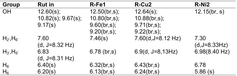
Results:Complex formation was also verified by1H RNM, using DMSO-d
6 as solvent. The proton signals from Hydroxyl groups 5-OH, 7-OH, 3’-OH and 4’-OH shifted to lower and broader frequencies in coordinated complex R-Fe1, R-Cu2 and R-Ni2, compared with signals from free rut in. The results showed that R-Cu2 complex presented a higher superoxide scavenging effect when compared with rut in alone (6.95% and 46.42%, at 10µM; and 51.80% and 71.32%, at 100µM, respectively). The results also showed that R-Cu2 inhibited significantly (P<0.05; ANOVA) cell migration (neutrophils, lymphocytes and monocytes) in peritonitis induced by carrageenan, bradikynin and PGE2, in mice, when compared to controls ones (without treatment or Ru alone treatment). Furthermore, rutin and R-Cu2 significantly (P<0.05, paired t test) inhibited iNOS and COX-2 gene expression in LPS-induced macrophage cells.
Conclusions:Taken together, our results show for the first time that the R-Cu2complex, a metal coordinated rut in compound, produces anti-inflammatory effects in mice, at least in part, by means of increasing the antioxidant activity and inhibition of iNOS and COX-2 gene expression. We suggest that cooper coordinated rut in compound can potentiates some biological properties of this flavonoid and could be more effective for therapeutic treatment of diseases related to oxidative stress.
Keywords: Rut in; rut in-copper complex; antioxidant; anti-inflammatory; gene expression.
1. INTRODUCTION
studied in the attempt to understand which structural effects are more important on the several activities.
Flavonoid anti-inflammatory activity can be related to cellular mechanisms such as arachidonic acid enzyme modulation, and phospholipase A2 (PLA2), cyclooxygenase, lipoxygenase and nitric oxide synthase enzyme inhibition. The inhibition of these enzymes reduces the production of inflammatory mediators such as arachidonic acid, prostaglandins, leukotrienes and nitric oxide [2]. The first PLA2 inhibitor flavonoid found was quercetin, which inhibits PLA2 from human neutrophils [3].
As is well known, there are some metal essentials for all organisms. For example, copper is a well-studied important cofactor of a variety of enzymes that are involved in a variety of biological processes, such as cytochrome C oxidase [4]. Although metals as cooper and nickel in excess could be toxic to life organisms [5,6], but when they were associated with compounds exhibiting antioxidant and anti-inflammatory activities, they could take an interesting part in the struggle against diseases related to damages caused by the increase in free radicals [7]. As demonstrated by Kostyuk et al. [8], iron coordinated flavonoid increase the flavonoid scavenging capacity, indicating that this compound could be beneficial for therapeutic treatment of diseases related to oxidative stress.
The flavonoid naringin has antioxidant and anti-inflammatory activities, and inhibits the proliferation of human mammary tumor cells In vitro [9]. The naringin-copper complex presents increased DPPH radical scavenging activity when compared with naringin alone. In addition, naringin complexed with copper has shown anti-inflammatory activity against carrageen-induced peritonitis in mice, and antitumoral activity in murine melanoma cell line B16F10 and human chronic myelogenous leukemia cell line K562 [7,10].
Rut in is a flavonoid found inDimorphandra mollis, a native plant of Brazil. Rut in (also called vitamin P) is known to exert beneficial effects such as protection against lipid oxidation and free radical scavenging. Rut in is an important potassium modulator [11] and induce vascular regulation meditated by nitric oxide [12,13]. Besides, rut in can also be used in the treatment of anemia, beta-talassemia, cardiovascular diseases and neurodegenerative disorders [14,15], which are diseases associated with the excessive production of reactive oxygen species (ROS) [16].
Our group has recently shown that transition metal complexes bearing flavonoid ligands, such as rut in-Fe (I) and rutin-Cu (II) complexes, can be synthesized. These compounds, as well as naringin and Cu complexes, present higher antioxidant, anti-inflammatory and antitumoral activities when compared with the flavonoid alone [17,18].
Therefore, the objective of this work was to evaluate the antioxidant and anti-inflammatory activities of rut in and rut in derivatives containing transition metals: copper (II)-rut in (R-Cu2), Iron (I)-rut in (R-Fe1), and nickel (II)-rut in (R-Ni2) complexes.
2. MATERIALS AND METHODS
2.1 Antioxidant Activity
was measured. The method was adapted by Nikishimi et al. [19]. The reaction consisted of the test compound (10µM) combined with phenazine methosulfate (PMS-20µM), coenzyme nicotinamide adenine dinucleotide (NADH - 156µM), and nitro blue tetrazolium (NBT-50µM) in phosphate buffer solution (0.1M; pH 7,4) at a final volume of 2.5mL. Among the evaluated composites were copper (II)-Rut in (R-Cu2), Iron (I)-Rut in (R-Fe1), nickel (II)-Rut in (R-Ni2) complexes and Rut in itself, tested at the concentrations of 1, 10 and 100µM. The negative control was a non-NBT run, and the flavonoid itself was used as positive control. The formazan crystals were formed by nitroblue tetrazolium (NBT) reduction in the presence of the superoxide radicals (O2.-), a kind of reactive oxygen species (ROS). The absorbance was measured at 560nm in a spectrophotometer (Femto®). The antioxidant activities of the compounds were demonstrated in superoxide radical scavenging percentage (%).
2.2 Anti-inflammatory Activity–animals
Male Swiss mice (18-35g) from CEDEME, housed at 22±2ºC under a 12h/12h light-dark cycle, were used. Food and water were freely available. The animals were acclimatized to the laboratory for at least 1h before testing and were used once throughout the experiments, which were carried out in accordance with current guidelines for the care of laboratory animals and the ethical guidelines for investigations of experimental plan in conscious animals from Ethical Committee UNIBAN (protocol nº301_08) and Zimmermann [20].
Acute phlogistic-induced inflammatory reaction in the peritoneal cavity of mice. The anti-inflammatory activity was determined by analyzing the total and subtype numbers of leukocyte migration to peritoneal cavity of Swiss mice, after peritonitis induced by phlogistic agents: carrageenan (1%, 4h), bradykinin (BK, 10nmol/cavity, 1h), prostaglandin E2 (PGE2, 10nmol/cavity, 1h), histamine (100µg/cavity, 1h) and substance P (SP, 20nmol/cavity, 1,5h) [10], without (negative controls) or with concomitant administration of dexamethasone (5mg/kg, s.c.), rut in, R-Cu2 and R-Ni2 (16 and 160µmol/kg, p.o.). After the time indicated above for each treatment, the animals were sacrificed, and cells were harvested from the peritoneal cavity and stained with Panotico stain (Laborclin®). The results were expressed as cell number/mL.
2.3 Cell Culture
RAW 264.7 Macrophage: mouse monocytic transformed cell lines mentioned by Raschke et al. [21] were cultivated in RPMI 1640 medium supplemented with sodium bicarbonate 2g/L (“LCG biotecnologia”), streptomycin 100µg/mL, penicillin 100UI/mL and 10% of fetal bovine serum. Cells were maintained at 37ºC and 5% of CO2.
2.3.1 MTT-cell viability assay
2.3.2 Nitric oxide dosage
The effect of the transition metal-Rut in complex on nitric oxide (NO) production can be evaluated in macrophages (RAW 264.7) by Griess assay [23], a protocol for the detection of NO2-in a variety of biological liquid matrices as a measure that reflects NO production. Cells were seeded onto 96-well plates at a density of 1x10⁴cells/well and incubated overnight (37ºC, 5%CO2). Then the media were replaced by fresh media containing the compounds (rutin and R-Cu2) in DMSO or DMSO alone. After 1h, lipopolysaccharide (LPS-Sigma) (1µg/mL) was added and the plate was incubated for another 6, 12 or 24h in 37ºC, 5% CO2. Thereafter, nitrite (NO2) concentration was determined in culture supernatant incubated for 30min with Griess solution. Absorbance was measured at 540nm using a microplate reader (Labtech LT-4000 MS). Results were expressed as NO concentration (µmol/L).
2.3.3 Gene expression quantification-total RNA isolation
For RNA extraction, cells from RAW lineage were incubated with compounds (rutin and R-Cu2) at the concentration of 30mM and stimulated with LPS(1µg/mL) for another 24h at 37ºC, 5%CO2. RNA was isolated from cultured cells using TRIzol reagent (Invitrogen, Carlsbad, CA, USA). The RNA quantity was assessed using nanodrop (NanoDrop Products, Wilmington, DE USA).
2.3.4 Reverse transcription-polymerase chain reaction
Complementary DNA (cDNA) was prepared with Reverse transcription (Fermentas), in a final volume of 50µL, using 500ng total of total RNA, from cultured cells as described above, 400U of reverse transcriptase, 10µL of enzyme buffer, 10µL of 10mM dNTP mixture (all from Invitrogen), 0.2µL of oligo(dT) primer, and 50U of RNase inhibitor (Amersham Biosciences, Uppsala, Sweden). This mixture was incubated at 42ºC, for 60min, and then at 70ºC, for 10min.
2.3.5 Quantitative PCR for iNOS and COX-2 transcript detection
Real-time PCR reactions of cDNAs were performed with SYBR Green I on an ABI PRISM Step one Sequence Detection System (PE Applied Biosystems). Quantification of transcripts iNOS and COX-2 were calculated as delta-delta C(T) method as previously described [24] and normalized by the amount of the beta-actin gene. Results were expressed as 2-(CT) from iNOS and COX-2 expression.
2.4 Rut in Complexes
The rutin complexes were produced in the chemistry laboratory of the Universidade Bandeirante de São Paulo following the patent INPI P. 018110000353 06/01/2001 [17].
2.4.1 Synthesis of complex R-Fe1
2.4.2 Synthesis of complex R-Cu2
A solution of [Cu(CH3COO)2] in methanol was dropped slowly to a solution of rut in in methanol. The filtrated solution was evaporated, and the obtained material purified by chromatography using Sephadex LD 20 and methanol as solvent. A pure green fraction was dried, and pure compound was obtained. Yield: 0.496g, 38%; mp. 242ºC. Experim. data for [C31H32O18Cu.2H20] (Calc.): %C, 46.68 (46.37); %H,4.82 (4.40). IR (cm-1, solid state):(OH) 3419;(C=O) 1655;(C=C) 1599, 1573, 1539, 1534; (C-O-C) 1295;(C-C)807.
2.4.3 Synthesis of complex R-Ni2
By similar procedure, using nickel(II) acetate to metallate rutin. After drying the filtrated solution, previously purified by chromatography using Sephadex LD 20 and methanol as solvent, a pure light brown compound was obtained. Yield: 0.901g, 63%; mp. 285ºC. Experim. data for C31H42O23Ni(Calc.): %C, 44.31(44.00); %H, 5.47(4,26). IR (cm-1, solid state): (OH) 3449; (C=O) 1653; 1505, 1597, 1575, 1558, (C=C) 1456 ; (C-O-C)1296; (C-C)807.
2.5 Statistical Analysis
Data are expressed as means±standard error of mean. Significant differences (*P<0.05) were determined by one-way ANOVA complemented with Bonferroni’s test (for antioxidant activities), Dunnett’s test (for anti-inflammatory effects), and paired t test (for gene expression quantification).
3. RESULTS
3.1 Antioxidant Activity
Superoxide anion radicals are produced by a number of cellular reactions, including several enzyme systems such as lipoxygenase, peroxidase, NADPH oxidase, and xanthine oxidase. The superoxide radical scavenging activities of the compounds are demonstrated (Fig. 1).
The Rut in, R-Cu2, R-Ni2 and R-Fe1 exhibited 51.80%, 71.32%, 53.57% and 52.59% scavenging activity at 100µM, and 6.95%, 46.42%, 21.69%, and 11.01% at 10µM, respectively. The R-Cu2 and R-Ni2 complexes exhibited a statistically significant (P<0.01, ANOVA and Bonferroni post-test) higher scavenging activity when compared with Rut in alone when used at 10µM. The complex R-Fe1 did not show a statistically significant antioxidant effect when compared with Rut in alone. However, R-Cu2 presented the highest scavenging activity (45 and 75%) in more than one concentration (10 and 100µM, respectively).
Fig. 1. Superoxide radical scavenging percentage, in different concentrations (1, 10 and 100µM) of the compounds rutin (Rut- ), R-Cu2 (), R-Ni2 () and R-Fe1 (|||) in 5 minutes. Met-methanol in 30% concentration. DMSO–DMSO in 10% concentration.
**P<0.01, rutin versus compounds.##P<0.01, R-Cu2 versus R-Ni2 and R-Fe1
3.2 Anti-inflammatory Activity-carrageenan-induced Peritonitis
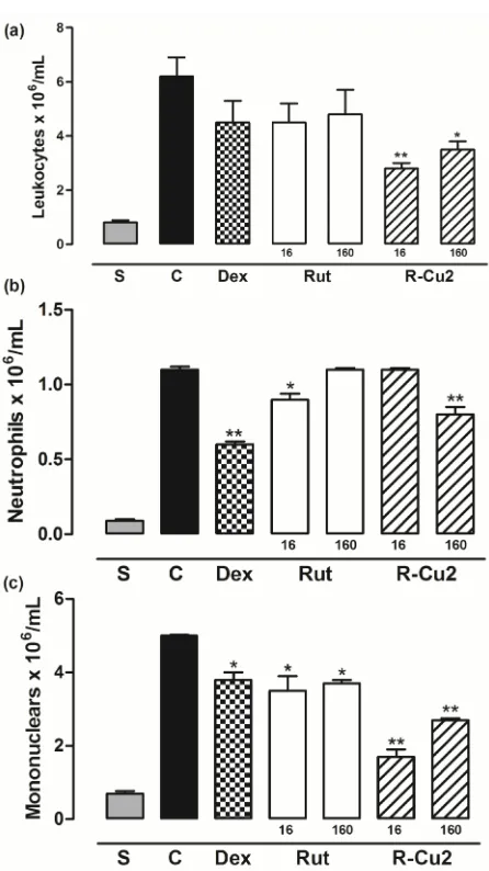
The results of the anti-inflammatory activities analyzed by the migration of leukocytes to the peritoneal cavity of Swiss mice after carrageenan-induced peritonitis are shown in (Fig. 2).
The R-Cu2-treated (16 and 160µmol/kg) group of mice was the only one that inhibited significantly (P<0.05), suggesting a dose-dependent effect in the migration of total leukocyte cells to peritoneal cavity, when compared with control group. The R-Cu2 160μmol/kg group showed significant results that was similar to the positive control dexamethasone group. In addition, the rut in and R-Cu2 effect was compared, and a statistically significant difference (P<0.05) between both compounds was observed. The inhibition (%) of inflammatory response induced by carrageenan in the peritonitis model in mice produced by rut in and R-Cu2 (16 and 160µmol/kg) was 0 and 9.16%, 26.22 and 38.5%, respectively. The R-Ni2 did not change leukocyte migration; hence no further experiment was carried out with this compound.
Fig. 2. Effect of rut in (Rut), R-Cu2 and R-Ni2 (16 and 160µmol/kg, p.o.) on carrageenan-induced peritonitis in mice. Total (a) and differential (b, c) leukocyte counts were evaluated. S=animals treated only with sterile saline, C=animals treated with vehicle plus carrageenan, Dex=animal treated with dexamethasone (5mg/kg, s.c.)
plus carrageenan. Each group (n=5) represents the mean±SEM. Significant differences determined by ANOVA complemented with Dunnett’s test, *P<0.05; **P<0.01, when the control group is compared with the other groups.#P<0.05, when
Rut and R-Cu2 groups are compared
3.3 Anti-inflammatory Activity-histamine-induced Peritonitis
Material 1. Effect of rutin (Rut), R-Cu2 (16 and 160µmol/kg, p.o.) on histamine-induced peritonitis in mice. Total (a) and differential (b, c) leukocyte counts were evaluated.
S=animals treated only with sterile saline, C=animals treated with vehicle plus histamine, Dex = animal treated with dexamethasone (5mg/kg, s.c.) plus histamine. Each group (n=5) represents the mean±SEM. Significant differences determined by ANOVA complemented with Dunnett’s test, *P<0.05; **P<0.01, when the control group
is compared with the other groups
3.4 Anti-inflammatory Activity-bradykinin-induced Peritonitis
Fig. 3. Effect of rutin (Rut), R-Cu2 (16 and 160µmol/kg, p.o.) on bradykinin-induced peritonitis in mice. Total (a) and differential (b, c) leukocyte counts were evaluated. S=animals treated only with sterile saline, C=animals treated with vehicle plus BK, Dex=animals treated with dexamethasone (5mg/kg, s.c.) plus BK. Each group (n=5)
represents the mean±SEM. Significant differences determined by ANOVA complemented with Dunnett’s test, *P<0.05; **P<0.01, when the control group is
compared with the other groups
3.5 Anti-inflammatory Activity-prostaglandin E
2-induced Peritonitis
Fig. 4. Effect of rut in (Rut), R-Cu2 (16 and 160µmol/kg, p.o.) on PGE2-induced peritonitis in mice. Total (a) and differential (b, c) leukocyte counts were evaluated. S=animals treated only with sterile saline, C=animals treated with vehicle plus PGE2, Dex=animals treated with dexamethasone (5mg/kg, s.c.) plus PGE2. Each group (n=5)
represents the mean±SEM. Significant differences determined by ANOVA complemented with Dunnett’s test, *P<0.05; **P<0.01, when the control group is
compared with the other groups
The results of differential leukocyte migration showed that rut in and R-Cu2 significantly inhibited (P<0.05) neutrophils and mononuclear cells after 1 hour of PGE2-induced peritonitis when compared with control group (Fig. 4).
3.6 Anti-inflammatory Activity-substance P-induced Peritonitis
the mononuclear cell migration was reduced by both compounds groups when compared with control group, additional data are given in Online Resource (Material 2).
Material 2. Effect of rut in (Rut), R-Cu2 (16 and 160µmol/kg, p.o.) on substance P-induced peritonitis in mice. Total (a) and differential (b, c) leukocyte counts were evaluated. S = animals treated only with sterile saline, C=animals treated with vehicle
plus substance P, Dex=animals treated with dexamethasone (5mg/kg, s.c.) plus substance P. Each group (n=5) represents the mean±SEM. Significant differences determined by ANOVA complemented with Dunnett’s test, **P<0.01, when the control
group is compared with the other groups
3.7 Nitrite Assay in RAW 264.7 and Cell Viability by Mean MTT
In (Fig. 5B), we have investigated whether the inhibitory effect of the rut in and R-Cu2 on NO production was dose-dependent. The pre-incubation of rut in or R-Cu2 (0.01–10nM) on RAW 264.7 stimulated by LPS reduced dose-dependent NO production, while the 10mM of both, rut in or R-Cu2, almost abolished the NO production in the RAW 264.7 cell line stimulated by LPS. The results also demonstrated that only a small quantity (0.01mM) of R-Cu2 was sufficient to inhibit about 50% of NO production by RAW stimulated cells. The same percentage (50.6%) of NO production inhibition was only seen when cells were treated with higher rut in concentrations (1mM or more) (Fig. 5B).
Fig. 5. Nitrite Assay in RAW 264.7 and cell viability by mean MTT. A) Nitric oxide production from LPS-induced RAW 264.7 cells in 6, 12 and 24 hours. C–control group,
cells with medium; Rut (rut in) and R-Cu2 group in 30mM concentration. B) Increase of the nitric oxide production from LPS-induced RAW 264.7 cells and cell viability in
24 hours. C–control group, cells with medium; Rut (rut in) and R-Cu2 group in 0.01–10mM
It was also demonstrated that cells presented 100% of viability at all concentrations of the treatment compound (Fig. 5b).
3.8 INOS and COX-2 Gene Expression
results showed that LPS induced an increase (delta-delta CT values below one) in COX-2 and iNOS gene expression. However, Rut in and R-Cu2 inhibited (delta-delta CT values above one) COX-2 and, especially, iNOS gene expression in LPS-induced cells (Fig. 6). These results corroborated those presented above, that Rut in and R-Cu2 participated in signaling pathways activated by LPS in macrophages.
Fig. 6. Inducible nitric oxide synthase (iNOS) and cyclooxygenase-2 (COX-2) gene expression levels modulated by LPS, and Rut in (Rut) and Rut in-Cu (R-Cu2)
compounds in RAW 264.7 cells. Results were expressed as 2–(CT)and means that
values below one show an increase in gene expression while values above one demonstrate gene expression inhibition
3.9 Rut in Complexes
The rut in complexes (R-Fe1, R-Cu2 and R-Ni2) were produced following the patent INPI P. 018110000353 06/01/2001 [17]. Complex formation was also verified by 1H RNM, using DMSO-d6 as solvent (Table 1). The structure complexes proposal, in solid state, is given in Online Reource (Materials 3 and 4).
Table 1. NMR data for rut in and complexes R-Fe1, R-Cu2 and R-Ni2
Group Rut in R-Fe1 R-Cu2 R-Ni2
OH 12.60(s);
10.82(s); 9.67(s); 9.17(s)
12.50(br,s); 10.80(br,s); 9.60(br,s); 9.20(br,s);
12.64(s); 10.88(br,s); 9.71(br,s); 9.22(br,s);
12.15(br, s)
H2’,H6’ 7.60
(d, J=8.32 Hz) 7.46(s) 7.60(d,J=8.12 Hz) 7.30(d,J=8.33Hz) H3’,H5’ 6.83
(d, J=8.31 Hz) 6.78 (br,s) 6.9(d, J=8,13Hz) 6.98(8.40 Hz)
H8 6.40(s) 6.32(br,s) 6.43(br,s) 6.78
O
OH OH OH O
OH OH
OH O
O
O O
O
OH OH
OH CH3
O OH
OH OH
CH3
O O O
O HO
OH
OH O
OH
OH OH
O
Fe
Fe H 2+
3+ (H2O)4
(H2O)4
(SO4)
2-x 2
Material 3. Proposed structure for R-Fe1 complex, in solid state
Material 4. Proposed structure for R-Cu2 or R-Ni2 complexes, in solid state. M=Cu2+
4. DISCUSSION
The present study was undertaken to investigate the antioxidant and anti-inflammatory effects of flavonoids and metal-flavonoids complexes, such as Rut in, R-Cu2, R-Ni2, and R-Fe, on superoxide radical scavenging activities, and murine peritonitis models induced by carrageenin, histamine, bradikynin, PGE2, and substance P. This study also investigated the effect of Rut in, R-Cu2, R-Ni2, and R-Fe on the production of nitric oxide in RAW 264.7 macrophage cell lines, linked to the expression of iNOS and COX-2 protein.
Nijveldt et al. [25] showed that a main effect of flavonoids in oxygen-derived scavenging free radicals. The antioxidant activity of rut in was well defined. Noroozi et al. [26] related that rutin has a better antioxidant effect than vitamin C induced by H2O2 in DNA damage in human lymphocyte, and Hanasaki et al. [27] described how rutin has a better antioxidant effect than hydroxyl free radical scavenging mannitol.
Afanas´eva et al. [7] showed that flavonoid-transition metal complexes have better antioxidant and anti-inflammatory activities than flavonoid alone. Kostyuk et al. [8] also related that rut in complexed with iron (II) was an effective superoxide free radical scavenger. The potentialization of the effects that these flavonoid-metal complexes can have can thus be observed.
Most flavonoids are poorly soluble in water and, similar to lipophilic compounds, tends to accumulate in biological membranes, particularly in lipid rafts, where they can interact with different receptors and signal transducers and influence their functioning through modulation of the lipid-phase behavior [28]. The physical-chemical properties of flavonoid–metal complexes demonstrate that flavonoids carboxyl and hydroxyl groups are responsible for metal chelating ability of these compounds and also the flavonoid rutin-complex (R-Cu2) used here. The chelation ability could be facilitated in an alkaline environment (pH 10) due to the deprotonation of hydroxyls [28]. The above-described chelation sites were observed on many flavonoid–metal complexes, such as the complex of quercetin with Cu(II) [29], suggesting that the complexation of flavonoids with metal cations can considerably change their lipophilicity and consequently improve interaction with lipid bilayer [30,31,28]. Studies have showed the capacity of flavonoids to participate in intermolecular and intercellular interactions [32,33]. Although our knowledge of transient metal cations involvement in the process is not completely elucidated, studies that shows the biological significance of these flavonoid-metal ion complexes needs to be explored.
Inflammatory mediators such as NO and pro-inflammatory cytokines are involved in host defense mechanisms: their overproduction contributes to the pathogenesis of several inflammatory diseases including bacterial sepsis, rheumatoid arthritis, chronic inflammation, and hepatitis [35- 37]. During infection and inflammation, high production of NO has shown several modulations of gene expression and in some cases to cause DNA damage [38]. There is plenty of evidence that prostaglandins also play an important role as mediators of the inflammatory response. These mediators are constitutively produced by means of cyclooxygenase 1 (COX) [39]. COX has been found in at least two isoforms: COX-1 and COX-2. COX-1 is expressed constitutively in most tissues and is responsible for the homeostatic production of prostaglandins. COX-2 is induced by several stimuli, including inflammatory stimulus, cytokines and growth factors. After expression COX-2 produces large amounts of pro-inflammatory prostaglandins at the inflammation site, and its uncontrolled activity is thought to play an important role in the pathogenesis of many chronic inflammatory diseases [40].
It has been described that in TNF-α or LPS-stimulated cells, such as RAW 264.7, results in phosphorylation and degradation of the inhibitory protein I-κB, leading to the release of NF-κB from I-NF-κB and increasing its translocation into the nucleus [41]. This is the main mechanism involved in NO, PGE2 production and iNOS, COX-2 expression in RAW 264.7 macrophage cell lines. Our results show that R-Cu2, R-Ni2, and R-Fe are more efficient than rutin in decreasing the inflammatory effects induced by different phlogistic agents In vivoor during the evaluation of NO production in RAW 264.7. These results are in agreement with data from the expression of COX-2 and iNOS, where treatment with R-Cu2 significantly reduced the expression of these enzymes.
Besides, studies demonstrate that macrophage express a voltage-dependent K+(Kv) channels and these ionic channels are involved in immune response and antigen presentation. Using the whole-cell patch clamp technique, Wu et al. [42] analysed the electrophysiological and pharmacological properties of voltage-gated potassium channels in primary rat peritoneal macrophages, showing that peritoneal macrophages express several types of functional voltage-gated K(+) channels. Villalonga et al. [43] shown that Kv1.3 and Kv1.5 are highly expressed in activated macrophages and T-effector memory cells of autoimmune disease patients. In mononuclear phagocytes biophysically and pharmacologically response, such as NFκB activation, NOS and COX2 expression, can be modulate by different Kv1.3/Kv1.5 ratios. In fact, the voltage-dependent potassium channels (Kv) have a crucial function in excitable cells of determining resting membrane potential and controlling action potentials. In addition, they are involved in the activation and proliferation of leukocytes. Blockade of voltage-dependent potassium channels (Kv) by specific antagonists decreases macrophage cytokine production and inhibits proliferation [44] and can be an important target for new drugs to treat inflammation processes, such as asthma and autoimmune disease [45-48]. Bohuslavizki et al. [49] demonstrated thatRuta graveolens extract has ability of blocking potassium channel. Also, other natural compounds like Correolide [50] or 8-prenylnaringenin [51] can blocker Kv 1.3 and modulate the response of these cells during the inflammatory response. Thus, we can suppose that rut in and its complex (R-Cu2) modulate this channel. However further experiments are now underway to confirm this hypothesis.
5. CONCLUSION
means of increasing the antioxidant activity and inhibition of iNOS and COX-2 gene expression. We suggest that cooper coordinated rut in compound can potentiates some biological properties of this flavonoid and could be more effective for therapeutic treatment of diseases related to oxidative stress. New experiments are now in course to identify whether the NFκB pathway is also involved in the anti-inflammatory effect of Rut in and its metal complexes.
CONSENT
Not applicable.
ETHICAL APPROVAL
The animal experiments were carried out in accordance with current guidelines for the care of laboratory animals and the ethical guidelines for investigations of experimental pain in conscious animals from Ethical Committee UNIBAN (protocol nº301_08) and Zimmermann (1983).
ACKNOWLEDGMENT
Cristina E. Okuyama is grateful to Fundação de Amparo à Pesquisa do Estado de São Paulo (FAPESP) and Bandeirante University of São Paulo (UNIBAN) for providing financial support.
COMPETING INTERESTS
Authors have declared that no competing interests exist.
REFERENCES
1. Boots AW, Haenen GR, Bast A. Health effects of quercetin: From antioxidant to nutraceutical. Eur J Pharmacol. 2008;585:325-337.
2. Kim HP, Son KH, Chang HW, Kang SS.Anti-inflammatory plant flavonoids and cellular action mechanisms. J Pharmacol Sci. 2004;96:229-245.
3. Lee TP, Matteliano ML, Middletone E. Effect of quercetin on human polymorphonuclear leukocyte lysosomal enzyme release and phospholipid metabolism. Life Sci. 1982;31:2765–2774.
4. Halliwell B, Gutteridge JMC. Oxygen toxicity, oxygen radicals, transition metal and diseases. Biochem J. 1984;219:1-4.
5. Eckers A, Reimann K, Klotz LO. Nickel and copper ion-induced stress signaling in human hepatoma cells: analysis of phosphoinositide 3'-kinase/Akt signaling. Biometals. 2009;22:307-316.
6. Scott-Fordsmand JJ. Toxicity of nickel to soil organism in Denmark. Rev. Environ. Contam. Toxicol. 1997;148:1.
7. Afanas'eva IB, Ostrakhovitch EA, Mikhal'chik EV, Ibragimova GA, Korkina LG. Enhancement of antioxidant and anti-inflammatory activities of bioflavonoid rut in by complexation with transition metals. Biochem Pharmacol. 2001;61:677-684.
9. Korkina LG, Afanas'ev IB. Antioxidant and chelating properties of flavonoids. Adv Pharmacol. 1997;38:151-163.
10. Pereira R, Madeiros VS, Frode IC. Anti-inflammatory effects of Tacrolimus in a mouse model of pleurisy. Transpl Immunol. 2006;16:105-111.
11. Bohuslavizki Kh, Hansel W, Kneip A, Koppenhofer E, Reimers A. Potassium channel blockers from Ruta-a new approach for the treatment of multiple sclerosis. Gen Physiol Biophys. 1992;11:507-512.
12. Fusi F, Saponara S, Pessina F, Gorelli B, Sgaragli G. Effects of quercetin and rut in on vascular preparations: A comparison between mechanical and electrophysiological phenomena. Eur J Nutr. 2003;42:10-17.
13. Amira S, Rotondo A, Mulè F. Relaxant effects of flavonoids on the mouse isolated stomach: Structure-activity relationships. Eur J Pharmacol. 2008;599:126-130.
14. Williams RJ, Spencer JPE, Rice-Evans C. Flavonoids: Antioxidants or signalling molecules? Free Radical Biol Med. 2004;36:838-849.
15. Grazul M, Budzisz E. Biological activity of metal ions complexes of chromones, coumarins and flavones. Coord Chem Rev. 2009;253:2588-2598.
16. Alcaraz MJ, Hoult JR. Actions of flavonoids and the novel anti-inflammatory flavone, hypolaetin-8-glucoside, on prostaglandin biosynthesis and inactivation. Biochem Pharmacol. 1985;34:2477-2482.
17. Novak EM, Pereira RM, Ikeda NEA, Bydlowski SP, Velosa ASV, Paulino N, Okuyama CE. Method of production of zinc coordination complex rut in, rut in complex zinc, pharmaceutical and/or cosmetic compositions and their uses. INPI patent. n.018110000353, 06/01/2011; 2013.
18. Pereira RM, Andrades NED, Paulino N, Sawaya ACHF, Erbelin MN, Marcucci, MC, Favero GM, Novak EM, Bydlowski SP. Synthesis and characterization of a metal complex containing naringin and Cu, and its antioxidant, antimicrobial, anti-inflammatory and tumor cell cytotoxicity. Molecules. 2007;12:1352-1366.
19. Nikishimi M, Rao NA, Yagi K. The occurrence of superoxide anion in the reaction of reduced phenazine methosulfate and molecular oxygen. Biochem Byoph Res Comm. 1972;46:849-854.
20. Zimmermann M. Ethical guidelines for investigations of experimental pain in conscious animals. Pain. 1983;6:109–110.
21. Raschke W C, Baird S, Ralph P, Nakoinx I. Functional macrophage cell lines transformed by Abelson leukemia virus. Cell. 1978;15:261–267.
22. Mosman T. Rapid colorimetric assay for cellular growth and survival: Application to proliferation and cytotoxicity assays. J Immunol Methods. 1983;65:55-63.
23. Green LC, Wagner DA, Glogowski J, Skipper PL, Wishnok JS, Tannenbaum SR. Analysis of nitrate, nitrite and [15N] nitrate in biological fluids. Anal Biochem. 1984;126:131–138.
24. Livak KJ, Schmittgen TD. Analysis of relative gene expression data using real-time quantitative PCR and the 2(-delta delta C(T)). Method. 2001;25:402-408.
25. Nijveldt RJ, van Nood E, von Hoorn DE, Boelens PG, van Norren K, van Leeuwen PAM. Flavonoids: A review of probable mechanisms of action and potential applications. Am J Clin Nutr. 2001;74:418-425.
26. Noorozi M, Angerson WJ, Lean ME. Effects of flavonoids and vitamin C on oxidative DNA damage in human lymphocytes. Am J Clin Nutr. 1998;67:1210-1218.
27. Hanasaki Y, Ogawa S, Fukui S. The correlation between active oxygens scavenging and antioxidative effects of flavonoids. Free Radic Biol Med. 1994;16:845-850.
29. Torreggani A, Tamba M, Trinchero A, Bonora S. Copper(II)–quercetin complexes in aqueous solutions: Spectroscopic and kinetic properties. J Molec Struct. 2005;759– 766.
30. Ren J, Meng S, Lekka C, Kaxiras E. Complexation of flavonoids with iron: Structure and optical signatures. J Phys Chem. 2008;112:1845–1850.
31. Kim YA, Tarahovsky YS, Yagolnik EA, Kuznetsova SM, Muzafarov EN. Lipophilicity of flavonoid complexes with iron(II) and their interaction with liposomes. Biochem Biophys Res Commun. 2013;431:680–685.
32. Lu J, Gosslau A, Liu AY, Chen KY. PCR differential display-based identification of regulator of G protein signaling 10 as the target gene in human colon cancer cells induced by black tea polyphenol the aflavin monogallate. Eur J Pharmacol. 2008:601:66–72.
33. Lin AH, Leung GP, Leung SW, Vanhoutte PM, Man RY. Genistein enhances relaxation of the spontaneously hypertensive rat aorta by transactivation of epidermal growth factor receptor following binding to membrane estrogen receptors-alpha and activation of a G protein-coupled, endothelial nitric oxide synthase-dependent pathway. Pharmacol Res. 2011;63:181–189.
34. Paulino N, Abreu SRL, Uto Y, Nagasawa H, Hori H, Dirsch V, Vollmar A, Scremin A, Bretz WA. Anti-inflammatory effects of a bioavailable compound, Artepillin C, in Brazilian propolis. Eur J Pharmacol. 2008;587:296-301.
35. Tilg H, Wilmer A, Vogel W, Herold M, Nölchen B, Judmaier G, Huber C. Serum levels of cytokines in chronic liver diseases. Gastroenterology. 1992;103:264-274.
36. Laskin DL, Pendino KJ. Macrophages and inflammatory mediators in tissue injury. Annu Rev Pharmacol Toxicol. 1995;35:655-677.
37. Shapira L, Soskolne WA, Houri Y, Barak V, Halabi A, Stabholz A. Protection against endotoxic shock and lipopolysaccharide-induced local inflammation by tetracycline: correlation with inhibition of cytokine secretion.Infect Immun. 1996;64:825-828. 38. Nguyen T, Brunson D, Crespi CL, Penman BW, Wishnok JS, Tannenbaum SR. DNA
damage and mutation in human cells exposed to nitric oxide in vitro. Proc Natl Acad Sci USA. 1992;89:3030-3034.
39. Smith WL, Marnett LJ and DeWitt DL. Prostaglandin and thromboxane biosynthesis. Pharmacol Ther. 1991;49:153-179.
40. Lee SH, Soyoola E, Chanmugam P, Hart S, Sun W, Zhong H, Liou S, Simmons D, Hwang D. Selective expression of mitogen-inducible cyclooxygenase in macrophages stimulated with lipopolysaccharide. J Biol Chem. 1992;267:25934-25938.
41. Barnes PJ, Karin M. Nuclear factor-kappaB: A pivotal transcription factor in chronic inflammatory diseases. N Engl J Med. 1997;336:1066-1071.
42. Wu BM, Wang XH, Zhao B, Bian EB, Yan H, Cheng H, Lv XW, Xiong ZG, Li J. Electrophysiology properties of voltage-gated potassium channels in rat peritoneal macrophages. Int J Clin Exp Med. 2013;6:166-173.
43. Villalonga N, Escalada A, Vicente R, Sánchez-Tilló E, Celada A, Solsona C, Felipe A. Kv1.3/Kv1.5 heteromeric channels compromisse pharmacological responses in macrophages. Biochem Biophys Res Commun. 2007;352:913-918. 44. Moreno C, Prieto P, Macías Á, Pimentel-Santillana M, de la Cruz A, Través PG, Boscá
L, Valenzuela C. Modulation of voltage-dependent and inward rectifier potassium channels by 15-epi-lipoxin-A4 in activated murine macrophages: Implications in innate immunity. J Immunol. 2013;191:6136-6146.
46. Dong H, Ji Z, Liu M, Wang Y, Bai X, Wang T, Liu Z, Wu Y, Zhang B, Luo Y, Li Z, Dong M. Functional expression of ERG1 potassium channels in rat alveolar macrophages. J Mol Histol. 2013;44:117-124.
47. Leanza L, Zoratti M, Gulbins E, Szabò I. Induction of apoptosis in macrophages via Kv1.3 and Kv1.5 potassium channels. Curr Med Chem. 2012;19:5394-5404.
48. Lam J, Wulff H. The Lymphocyte Potassium Channels Kv1.3 and KCa3.1 as Targets for Immunosuppression. Drug Dev Res. 2011;72:573-584.
49. Bohuslavizki KH, Hänsel W, Kneip A, Koppenhöfer E, Reimers A. Potassium channel blockers from Ruta-a new approach for the treatment of multiple sclerosis. Gen Physiol Biophys. 1992;11:507-512.
50. Koo GC, Blake JT, Shah K, Staruch MJ, Dumont F, Wunderler D, Sanchez M, McManus OB, Sirotina-Meisher A, Fischer P, Boltz RC, Goetz MA, Baker R, Bao J,Kayser F, Rupprecht KM, Parsons WH, Tong XC, Ita IE, Pivnichny J, Vincent S, Cunningham P, Hora D. Jr, Feeney W, Kaczorowski G. Correolide and derivatives are novel immunosuppressants blocking the lymphocyte Kv 1.3 potassium channels. Cell Immunol. 1999;197:99-107.
51. Ostrakhovitch EA, Afanas'ev IB. Oxidative stress in rheumatoid arthritis leukocytes: Suppression by rutin and other antioxidants and chelators, Biochem. Pharmacol. 2001;62:743–746.
© 2014 Miyashiro et al.; This is an Open Access article distributed under the terms of the Creative Commons Attribution License (http://creativecommons.org/licenses/by/3.0), which permits unrestricted use, distribution, and reproduction in any medium, provided the original work is properly cited.
Peer-review history:
The peer review history for this paper can be accessed here: