Article
1
Pseudopterosin Inhibits Proliferation and 3D
2
Invasion in Triple Negative Breast Cancer by
3
Agonizing Glucocorticoid Receptor Alpha
4
Julia Sperlich 1 and Nicole Teusch 1,*
5
1 Bio-Pharmaceutical Chemistry & Molecular Pharmacology, Faculty of Applied Natural Sciences,
6
Technische Hochschule Koeln, Chempark, 51373 Leverkusen, Germany; julia.sperlich@th-koeln.de
7
* Correspondence: nicole.teusch@th-koeln.de; Tel.: +0214-32834-4623
8
9
Abstract: Pseudopterosin, produced by the sea whip of the genus Antillogorgia, possesses a variety
10
of promising biological activities including potent anti-inflammatory effects. However, few studies
11
examined pseudopterosin in the treatment of cancer cells and, to our knowledge, the ability to
12
inhibit triple negative breast cancer (TNBC) proliferation or invasion has not been explored. Thus,
13
we evaluated the as yet unknown mechanism of action of pseudopterosin: Pseudopterosin was
14
able to inhibit proliferation of TNBC. Interestingly, analyzing breast cancer cell proliferation after
15
knocking down glucocorticoid receptor α (GRα) revealed that anti-proliferative effects of
16
pseudopterosin were significantly inhibited when GRα expression was reduced. Furthermore,
17
pseudopterosin inhibited invasion of MDA-MB-231 3D tumor spheroids embedded in an
18
extracellular-like matrix. Remarkably, the knockdown of GRα in 3D tumor spheroids revealed
19
increased ability of cells to invade the surrounding matrix. In a co-culture, encompassing
20
peripheral blood mononuclear cells (PBMC) and MDA-MB-231 cells, production of interleukin 6
21
(IL-6) and interleukin 8 (IL-8) significantly increased compared to monoculture. Notably,
22
pseudopterosin indicated to block cytokine elevation, representing key players in tumor
23
progression, in the co-culture. Thus, our results reveal pseudopterosin treatment as a potential
24
novel approach in TNBC therapy.
25
Keywords: Pseudopterosin; triple negative breast cancer; glucocorticoid receptor alpha;
26
dexamethasone; cell proliferation; 3D invasion; tumor spheroid; co-culture; interleukin 6;
27
interleukin 8
28
29
1. Introduction
30
Breast cancer is still the most common malignancy in woman with one million cases annually
31
worldwide1. Of these, approximately 15% belongs to the triple-negative (ER-/PR-/HER2-) breast
32
cancer (TNBC). TNBC represents the most aggressive breast cancer type, characterized by high
33
proliferation rate, a pronounced potential to metastasize and a shorter survival rate2–4. Furthermore,
34
TNBC lacks effective therapies available for other breast cancer subtypes underlining the significant
35
unmet medical need for identifying novel targets and developing innovative drugs.
36
The tumor microenvironment is increasingly recognized as a major regulator of carcinogenesis.
37
In breast cancer, tumor associated macrophages (TAMs) enhance proliferation and metastasis as
38
well as resistance to chemotherapy by activation of the transcription factor nuclear factor κB
39
(NF-κB), a key factor in regulating inflammatory responses5,6. High expression levels of the NF-κB
40
target genes interleukin 6 (IL-6) or interleukin 8 (IL-8) secreted by macrophages can be correlated
41
with advanced growth of TNBC and poor prognosis7.
42
The pseudopterosins, a family of 31 known related diterpene glycosides, are produced by the
43
sea whip Antillogorgia elisabethae (formerly named Pseudopterosin elisabethae)8. Striking biological
44
activities have been described ranging from anti-inflammation9–11, wound-healing10,11,
45
analgesia-reducing9,12,13 to neuromodulation14. In contrast, to date, little is known regarding
46
anti-tumor effects of pseudopterosin, where only one derivative showed moderate cytotoxic effects
47
on ER+ breast cancer cells and non-small-cell lung cancer cells15.
48
Previously, we have described the potential of pseudopterosin as a novel immune modulator
49
in TNBC acting via NF-κB inhibition and subsequent blockade of cytokine secretion16. Moreover,
50
we identified inhibitory capabilities of pseudopterosin on the NF-κB signaling pathway by
51
agonizing the glucocorticoid receptor α (GRα)16. Accordingly, there is evidence that NF-κB and GRα
52
can physically interact and heterodimerize in breast cancer17. By binding other transcription factors
53
such as NF-κB, GRα can either transactivate or -suppress its target genes18.
54
Although glucocorticoids (GCs) are frequently used to relieve symptoms of cancer treatment
55
related side effects, contradictory effects on breast cancer progression upon GC treatment and with
56
respect to GRα expression have been described19–21. High expression levels of GRα in ER- breast
57
cancer might be associated with drug resistance resulting in an unfavorable clinical outcome22–24. In
58
contrast, a recent analysis demonstrates improved survival independent of the ER status in breast
59
cancer patients receiving GC combined with adjuvant anthracycline-based chemotherapy25. Thus, in
60
the current study we further elucidated the role of GRα in TNBC progression, thereby focusing on
61
pseudopterosin as a novel agent for breast cancer therapy.
62
2. Results
63
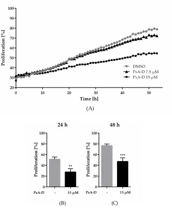
2.1. Pseudopterosin Inhibited Proliferation of Triple Negative Breast Cancer Cells
64
In our previous work we identified the natural product pseudoperosin as a novel inhibitor of
65
NF-κB signaling16, one key pathway in controlling progression of TNBC. As NF-κB is known to
66
regulate various processes in cancer progression such as proliferation, angiogenesis or invasion26–28,
67
the aim of the current study was to further characterize the pharmacological properties of
68
pseudopterosin. First, we investigated a pseudopterosin extract (PsA-D) regarding its effect on
69
breast cancer cell proliferation in MDA-MB-231 cells. To remain within a non-toxic concentration
70
range of PsA-D (IC50 values of cell viability for PsA-D after 24 hours or 48 hours of treatment were
71
31.4 µM and 32.2 µM, respectively; Supplemental Fig. S1A/B), 7.5 and 15 µM of PsA-D were chosen
72
to evaluate anti-proliferative effects (Fig. 1A). As expected, MDA-MB-231 cells treated with DMSO
73
showed a high proliferation rate, represented by a confluency of 78% after 48 hours (Fig. 1A).
74
Notably, a concentration of 15 µM of PsA-D was able to reduce proliferation significantly after 24
75
hours by 1.9 fold and after 48 hours by 1.6 fold compared to DMSO control (Fig. 1B and 1C).
76
Furthermore, preliminary data indicate that pseudopterosin-induced reduction of proliferation is
77
not pERK dependent (Supplemental Fig. S3), which is a key regulator for cell proliferation in
78
80
81
Figure 1. Pseudopterosin inhibited proliferation in triple negative breast cancer cells. (A)
82
Proliferating cells were imaged every hour over a time range of 50 hours with the IncuCyte® ZOOM.
83
Confluency of cells was determined with IncuCyte® software indicated as proliferation in percentage.
84
Cells were treated with either 7.5 µM (triangle) or 15 µM (square) of PsA-D. (B-C) Inhibition of
85
proliferation is shown at selected time points of 24 and 48 hours compared to DMSO control,
86
respectively. The data represent means of three independent experiments. Error bars were calculated
87
using ±SEM. P-values were calculated against DMSO control. Two stars represent a significance of
88
p<0.01 and three stars represent a significance of p<0.001.
89
2.2. Glucocorticoid Receptor Alpha Expression is Essential for Anti-Proliferative Effects of Pseudopterosin
90
In our previous work we hypothesized pseudopterosin to act as an agonist of the glucocorticoid
91
receptor alpha (GRα)16. Subsequently, when downregulating GRα, pseudopterosin failed to inhibit
92
NF-κB target gene expression. Thus, to further explore the role of GRα in the mode-of-action of
93
pseudopterosin, we analyzed the effect of a GRα knockdown on breast cancer cell proliferation.
94
After 72 and 85 hours, treatment with PsA-D inhibited proliferation in non-coding siRNA (nc
95
siRNA) transfected cells by 2 fold, respectively (Fig. 2A and Supplementary Fig. S4). Importantly, in
96
siGRα transfected cells, PsA-D lost its anti-proliferative effect (Fig. 2A). Efficiency of the GRα
97
knockdown using realtime qPCR (up to 88%) is exemplified in Fig. 2B and depicted on the protein
98
level via immunfluorescence analysis in Fig. 2C. In conclusion, our data suggests that GRα
99
expression might be crucial for the anti-proliferative effects of PsA-D.
100
Notably, treatment with the marked GRα ligand dexamethasone showed less potency in
101
nM dexamethasone reduced proliferation by 15% compared to DMSO, respectively (Fig. 2C). After
103
72 hours, PsA-D treatment diminished proliferation by 20%, whereas treatment with 100 nM
104
dexamethasone reduced the proliferation rate by only 9% (Fig. 2D).
105
106
108
Figure 2. Pseudopterosin failed to inhibit breast cancer cell proliferation after knockdown of the
109
glucocorticoid receptor alpha (GRα) and inhibited proliferation of MDA-MB-231 more
110
efficaciously than dexamethasone (Dex). (A) Knockdown of GRα was done with the Lonza
111
Nucleofector 2b device on day one. On day two, the cells were seeded and proliferating cells were
112
imaged with the IncuCyte® ZOOM every hour over a time range of five days. Cell proliferation was
113
determined with IncuCyte® software indicated in percentage. Cells were treated with a concentration
114
of 15 µM of PsA-D. (B) After knockdown of GRα, expression of GRα reduced up to 88.3%, which was
115
confirmed by qPCR analysis at 72 hours. (C) Immunofluorescent analysis of GRα knockdown after
116
72 hours. Scale bars in white show 100 microns in length. (D-E) PsA-D inhibited proliferation after 48
117
and 72 hours more efficaciously than dexamethasone. The data represent means of three
118
independent experiments. Error bars were calculated using ±SEM. Three stars represent a
119
significance of p<0.001 and two stars of p<0.01.
120
2.3. Pseudopterosin Inhibited Invasion into 3D Matrix
121
Breast tumors harbor many devastating characteristics resulting in poor prognosis of patients:
122
high proliferation rate and high histological grade. Furthermore, genetic and epigenetic alterations
123
enable breast cancer cells to migrate and invade the surrounding tissue via a process known as
124
epithelial-to-mesenchymal transition (EMT)30. To explore the effects of pseudopterosin on the
125
invasiveness of MDA-MB-231 cells, we developed a 3D invasion assay, where the cancer cells form a
126
micro-tumor spheroid embedded in extracellular matrix. In the presence of DMSO, the cells
127
immediately started to invade into the 3D matrix by partly disassembling the spheroid core (Fig 3A).
128
In contrast, treatment with PsA-D significantly inhibited the invasion of single cells into the matrix.
129
After 24 hours, the invasive area was reduced significantly by 59%, after 48 hours by 53% and after
130
72 hours by 73% (Fig. 3B-D). Thus, in our experiment we verified the inhibitory properties of
131
pseudopterosin in a 3D assay on TNBC progression, thereby hinting at a better prediction for future
132
134
135
Figure 3. Pseudopterosin inhibited invasion into a 3D matrix. (A) Representative images of
136
invasion of cells into a 3D matrix at 24 hours’ time point. Cells were imaged with IncuCyte® ZOOM
137
over a time range of three days. 3*103 cells per well were seeded into ULA round bottom plates and
138
spheroids were formed for 72 hours. Scale bars in black show 200 microns in length. (B-D) The bar
139
diagrams show three different time points representing six independent experiments. Spheroids
140
P-values were calculated against control (CTRL). Two stars represent a significance of p<0.01 and one
142
star represents a significance of p<0.05.
143
2.4. Down-Regulation of Glucocorticoid Receptor Alpha Expression Increased Invasiveness in TNBC
144
The clinical use of glucocorticoids (GC) is discussed controversially, due to extensive side
145
effects, chemotherapy resistance and survival of cancer cells21,23,31. However, recent literature
146
indicates the beneficial effects of GCs to be strongly dependent on the tumor entity: survival in
147
patients receiving GC combined with anthracycline-based chemotherapy was improved25. In this
148
context, we further investigated the role of GRα in the invasiveness of MDA-MB-231 micro-tumor
149
spheroids (Fig. 4A). The efficiency in GRα knockdown is represented by a reduction of 94% (Fig. 4C).
150
After 72 hours, the spheroids transfected with siGRα showed a significant increase in invasion by
151
27% compared to nc siRNA (Fig. 3B). In conclusion, the knockdown of GRα led to an elevation of
152
invasiveness in MDA-MB-231 cells, suggesting a potential of GRα agonists like pseudopterosin in
153
diminishing TNBC progression.
154
156
Figure 4. Knockdown of the glucocorticoid receptor alpha (GRα) increased invasiveness of triple
157
negative breast cancer. (A) Representative images of tumor cell invasion into a 3D matrix.
158
Knockdown of GRα was performed with the Lonza Nucleofector 2b device on day one. On day
159
three, 3*103 cells per well were seeded into ultra-low-attachment (ULA) round bottom plates.
160
Formation of spheroids was allowed for 72 hours. At t= 0, matrigel was added to the spheroids to
161
start invasion. Scale bars in black show 200 microns in length. (B) The invasion is depicted over a
162
time range of three days and the area of invaded cells into matrigel was calculated with imageJ FIJI at
163
the respective time points. (C) As confirmed by qPCR analysis, GRα expression is reduced up to 94%
164
after 72 hours. The data represent means of three independent experiments. Error bars were
165
calculated using ±SEM. P-values were calculated against nc siRNA control. Two stars represent a
166
significance of p<0.01.
167
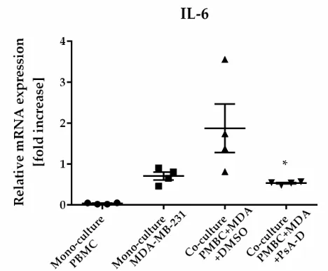
2.5. Pseudopterosin Inhibited Cytokine Release in a Co-Culture of Primary Blood Mononuclear Cells (PBMC)
168
and Triple Negative Breast Cancer Cells
169
The microenvironment plays a critical role in breast cancer carcinogenesis32. Tumor associated
170
macrophages are the drivers of breast cancer cells invasion33,34. A main characteristic of
171
inflammatory breast cancer is the secretion of pro-inflammatory cytokines such as IL-6 or IL-8 by
172
macrophages, regulating angiogenesis and promoting tumor progression35,36. Previously, we
173
verified a blockade of NF-κB-dependent cytokine expression and secretion after pseudopterosin
174
treatment in both, MDA-MB-231 and THP-1 cells 16. In this context, GRα knockdown led to the
175
failure of pseudopterosin to inhibit cytokine expression. Furthermore, as shown previously,
176
stimulation by the TLR4 ligand LPS leads to the production of cytokines and the subsequent
177
secretion into the surrounding “conditioned medium” (CM) 16. Our current data amend a significant
178
reduction of cytokine expression, such as IL-6, IL-8 and TNFα, after PsA-D treatment in peripheral
179
blood mononuclear cells (PBMC) (Supplemental Fig. S5). Medium containing cytokines released by
180
MDA-MB-231 cells, representing the so called “MDA-MB-231 conditioned medium” (M-CM),
181
induced a significant cytokine expression in PBMC. Notably, pseudopterosin treatment was able to
182
block cytokine expression induced by breast cancer cell conditioned media in PBMC. (Supplemental
183
Fig. S5). Thus, to further evaluate the pharmacological effects of pseudopterosin on bidirectional
184
communication, we set up a co-culture encompassing PBMC and MDA-MB-231 cells to analyze the
185
change in IL-6 and IL-8 expression levels. In the co-culture model, PsA-D treatment inhibited IL-6
186
increase of the IL-6 expression level in co-culture increased by 1.9 compared to mono-culture (Fig. 5).
188
As expected, PsA-D treatment reduced IL-6 expression levels by 3.5 fold (Fig. 5). To further explore
189
the agonism of pseudopterosin and GRα in the context of our co-culture model, the focus in future
190
studies will lay in continuing investigations concerning knockdown studies of GRα. Taking
191
together, our data indicate that pseudopterosin has the potential to inhibit the proliferation, the
192
invasiveness and the communication of PBMC and MDA-MB-231 cells in a co-culture model.
193
Thereby, the inhibitory activity of pseudopterosin seems to depend on GRα expression.
194
Table 1. Inhibition of cytokine expression in co-culture of peripheral blood mononuclear cells
195
(PBMC) and MDA-MB-231 cells after pseudopterosin treatment.
196
Mono-culture PBMC
Mono-culture
MDA*
Co-culture PBMC+MDA
+DMSO
Co-culture PBMC+MDA
+PsA-D
P- value1
IL-62 1.09 (± 3.2) 31.7 (± 20.3) 44.6 (± 25.3) 21.2 (± 12.7) 0.02 IL-82 27.1 (± 36.9) 67.9 (± 46.5) 213.9 (± 99.6) 49.5 (± 13.2) 0.22 1 P-values were calculated with ONE-Way ANOVA between ‘co-culture’ and ‘co-culture + PsA-D’.
197
2 The data represent relative mRNA expression values measured with realtime qPCR; * MDA is
198
equivalent for MDA-MB-231 cells.
199
200
Figure 5. Pseudopterosin inhibited cytokine expression in a co-culture of PBMC and
201
MDA-MB-231. Both cell lines were co-cultured at a ratio of 1:1 before treatment with 30 µM PsA-D.
202
Cells were harvested 24 hours after treatment and cytokine expression levels were analyzed with
203
qPCR. Relative mRNA levels were normalized to fold increase. MDA is equivalent for MDA-MB-231
204
cells. Data represent means of four independent experiments. Standard deviation was calculated
205
using ±SEM. P-values were calculated between ‘co-culture’ and ‘co-culture + PsA-D’ using Dunnett’s
206
multiple comparisons test.
207
3. Discussion
208
For pseudopterosin effective biological activities in various therapeutic areas including
209
anti-inflammatory effects are described9–11. This study aimed to explore the inhibitory capabilities of
210
pseudopterosin on distinct features of triple negative breast cancer (TNBC), namely the ability to
211
invade surrounding tissue and the contribution to rapid tumor progression. For TNBC, a disease
212
with a high unmet medical need and a low survival rate, we demonstrated previously a novel
213
Furthermore, suggested by the translocation of GRα, we revealed a role of GRα activation upon
215
pseudopterosin treatment. In the current study, GRα again indicated to play a role in mediating
216
pseudopterosin induced inhibition of breast cancer cell proliferation.
217
Among others, NF-κB is an important regulator in the development of the mammary glands37.
218
However, chronic inflammation in general and inflammation in the tumor microenvironment in
219
particular, caused by NF-κB up-regulation over a long time range, increases aggressiveness,
220
invasivenes38,39 and correlates with poor prognosis in breast cancer patients40. As our data suggest
221
pseudopterosin to inhibit constitutive NF-κB activity in TNBC cells16, we further examined effects of
222
pseudopterosin on blocking invasion. Adipocytes in breast tumors are described to secrete high
223
amounts of collagen leading to increased tumor growth41. Despite of using equivalently high
224
collagen concentrations, which is known to reduce drug sensitivity42, pseudopterosin displayed
225
strong anti-invasive properties. Moreover, in a GRα knockdown, invasiveness in breast cancer
226
tumor spheroids increased.
227
Gene expression analysis of breast tumors revealed a down-regulation of genes involved in cell
228
differentiation, whereas genes promoting tumorigenesis were up-regulated43. However, mutations
229
alone cannot explain the high malignancy and the complexity of the tumor. The tumor
230
microenvironment is the most important factor of why immune cells undergo a reprogramming
231
step, thereby promoting tumor progression. The discovery that normal mammary epithelial cells
232
cooperate with innate immune cells for invasive processes, led to the discovery that macrophages
233
are the drivers of intravasation from invasive breast tumors by establishing the tumor
234
microenvironment33,44. Thereby, extracellular matrix (ECM), stromal cells such as endothelial and
235
immune cells, fibroblasts and adipocytes are the main components of the microenvironment45.
236
Additionally, tumor associated macrophages (TAMs) play a critical role in the tumor
237
microenvironment by secreting second messengers such as IL-8 or IL-6 via NF-κB activation, thus
238
promoting the tumor microenvironment and regulating angiogenesis which in turn correlates with
239
poor outcome and malignant features in breast cancer35,36,46,47. Paradoxically, cytotoxic chemotherapy
240
further initiates TAM recruitment into invasive carcinoma48, where co-culture with breast cancer
241
cells results in high IL-6 levels leading to activation of cancer stem cells49. We confirmed elevated
242
IL-6 and IL-8 expression levels as a result of co-cultivating PBMC and MDA-MB-231 cells, where
243
pseudopterosin was able to significantly block cytokine expression and henceforth the
244
communication of both cell types.
245
In the clinics, glucocorticoids are used to reduce allergic reactions or nausea during
246
chemotherapy due to up-regulation of anti-inflammatory signals 50–52. On tumor cells, the synthetic
247
GRα ligand dexamethasone (Dex) has been described to reduce cell proliferation by decreasing ERK
248
phosphorylation in ER+ breast cancer cells, possibly via the mechanism of transactivation51. ERK is a
249
key regulator of proliferation and remodels the chromatin structure29. To our knowledge,
250
anti-proliferative effects of Dex where as yet not observed in MDA-MB-231 cells. In contrast, Dex
251
was described to increase tumor growth and act pro-proliferative53. However, in our study, we not
252
only observed anti-proliferative effects after Dex treatment, but also witnessed improved
253
anti-proliferative effects of pseudopterosin treatment compared to Dex. Interestingly, preliminary
254
data indicate that the mechanism of action of pseudopterosin seems to be distinct from Dex, as the
255
phosphorylation status of ERK did not change in the presence of pseudopterosin.
256
To date, GRα signaling can be divided into two distinct pathways: the so-called
257
“transactivation”, reflecting target gene expression, and the “transrepression”, representing the
258
downregulation of parallel signaling pathways such as NF-κB activation. Prominent metabolic side
259
effects of glucocorticoid treatment might be ascribed to transactivation of GRα54. In contrast, positive
260
effects of glucocorticoids include reduced migration and a reduction in proteins associated with
261
chemotherapy resistance in TNBC cells, which might be explained by transrepression of GRα55–57.
262
The mechanism of the transrepressive process of GRα can have different origins: GRα can
263
heterodimerize and bind directly to the p65/p50 dimer58 or GRα recruits histone deacetylases to the
264
promotors of inflammatory genes59. GRα transrepression is thereby defined as a direct interaction
265
independent of IκB, p50 or p65 regulation of expression54. Thus, up-regulation of IκBα expression60
267
or repression of IL-8 by transcriptional inhibition of NF-κB are correlated with transactivation of
268
GRα54. After GRα knockdown, we observed increased invasiveness in tumor spheroids and a lack of
269
pseudopterosin to inhibit proliferation or invasion. Thus, we suggest the expression of GRα to be
270
beneficial in maintaining a less invasive phenotype in TNBC and propose pseudopterosin to address
271
the mechanism of transrepression by agonizing GRα.
272
In conclusion, we demonstrated inhibitory effects of pseudopterosin on pronounced
273
characteristics of TNBC including tumor cell proliferation and invasion. Our results imply
274
pseudopterosin as a potential therapeutic basis suitable for targeting TNBC. Future studies will
275
focus on investigating the molecular function including transrepressive effects of GRα in mediating
276
pseudopterosin-dependent pharmacological actions.
277
4. Materials and Methods
278
4.1. Cell Culture and Reagents
279
The origin of the extract of pseudopterosin A to D isolated from A. elisabethae (subsequently
280
named PsA-D) was kindly provided by Dr. Russell Kerr (University of Prince Edward Island,
281
Marine Natural Products Lab, Canada) as described in our previous work16. U0126 inhibitor was
282
purchased from Selleckchem (Houston, U.S.). MDA-MB-231 breast cancer cells were obtained from
283
European Collection of Authenticated Cell Cultures (ECACC, Salisbury, UK) and grown in
284
humidified atmosphere containing 5% CO2 in RPMI medium. Medium was supplemented with 15 %
285
FCS, 100 units ml-1 penicillin and 100 µg ml-1 units streptomycin. PBMCs were purchased from
286
STEMCELL Technologies (Vancouver, Canada) and cultured in the presence of 5% CO2 in RPMI
287
along with 10% FCS, penicillin and streptomycin. Staurosporin was purchased from Sigma-Aldrich
288
(St. Louis, USA) and medium and antibiotics from Life Technologies (Gibco, Carlsbad, U.S.).
289
4.2. Realtime Cell Proliferation
290
MDA-MB-231 breast cancer cells were seeded at a density of 1*105 cells per ml in 96-well image
291
lock plates (Sartorius, Goettingen, Germany) and images were taken every hour for a time frame of
292
five days with the IncuCyte® Zoom from Sartorius (Goettingen, Germany). Confluency of cells was
293
determined using the software of IncuCyte® Zoom (Version 2016B).
294
4.3. Knockdown Studies
295
Glucocorticoid receptor alpha (GRα) siRNA (siGRα) sc-35505 was purchased from Santa Cruz
296
Biotechnology (Dallas, U.S.). Silencer® Select Negative Control No. 2 siRNA (nc siRNA) was
297
obtained from Life Technologies (Carlsbad, U.S.). 1*106 cells were transfected with 300 nM siRNA
298
using the Nucleofector 2b device (Lonza, Basel, Switzerland) using the X-013 protocol for
299
transfection of MDA-MB-231 cells. After different time points, cells were harvested and expression
300
upon knockdown of interest was analyzed using quantitative realtime PCR, respectively.
301
4.4. Quantitative Realtime PCR
302
To determine cytokine or GRα expression levels after co-culture or knockdown, the following
303
primers were used (purchased from Eurofins, Ebersberg, Germany): IL-6 forward
304
(GGCACTGGCAGAAAACAACC), IL-6 reverse (GCAAGTCTCCTCATTGAATCC) IL-8 forward:
305
(ACTGAGAGTGATTGAGAGTGGAC), IL-8 reverse: (AACCCTCTGCACCCAGTTTTC), GAPDH
306
forward: (TGCACCACCAACTGCTTAGC), GAPDH reverse: (GGCATGGACTGTGGTCATGAG),
307
GR forward: (AAAAGAGCAGTGGAAGGACAGCAC) GR reverse:
308
(GGTAGGGGTGAGTTGTGGTAACG). Total RNA was isolated with “RNase Mini kit” from
309
QIAGEN (Hilden, Germany) according to the manufacturer’s instructions and reverse transcriptase
310
PCR was performed using “Reverse Transcription Kit” from Promega (Darmstadt, Germany).
311
protocol: Pre-incubation at 95° for 900 seconds, amplification was performed over 45 cycles (95° for
313
15 seconds, 55° for 25 seconds and 72° for 10 seconds). No-template controls served as negative
314
controls. CT values were calculated according to the 2-ΔΔCT method61. Sample values were normalized
315
to the house-keeping gene GAPDH (glyceraldehyde 3-phosphate dehydrogenase).
316
4.5. 3D Invasion Assay
317
To study MDA-MB-231 invasion into an extracellular matrix such as matrigel (Corning, New
318
York, U.S.), spheroids of MDA-MB-231 were generated for 72 hours starting with 3*103 cells and
319
0.25% matrigel in an ultra-low-attachment (ULA) plate (Corning, New York, U.S.). Invasion was
320
initiated by addition of matrigel in a ratio of 1:1 volume to the spheroids. Images were taken with the
321
IncuCyte® Zoom (Sartorius, Goettingen, Germany) to create a time lapse movie or the Axio Vert.A1
322
microscope (Zeiss, Oberkochen, Germany) every 24 hours for a time frame of three days. Image
323
analysis was done with imageJ makro “Analyze Spheroid Cell Invasion in 3D matrix” by Volker
324
Baecker62 (FIJI distribution63).
325
4.6. Co-culture Studies
326
Co-culture of PBMC and MDA-MB-231 cells: PBMC were freshly thawed for each experiment.
327
1*106 cells of MDA-MB-231 were seeded on day one and incubated with PsA-D for 20 minutes on
328
day two. Treatment was followed by addition of PBMC cells to the MDA-MB-231 cells at a ratio of
329
1:1. Finally, cells were harvested at day three and analyzed for cytokine expression by realtime PCR.
330
4.7. Preparation of PsA-D Mixture
331
A. elisabethae was collected from South Bimini Island, as described I our previous work16: the
332
extract was dried and extracted in EtOAc/MeOH (1:1) for 48 hours and subjected to silica gel
333
chromatography eluting with hexanes and EtOAc to afford a mixture of PsA-D. The ratio was
334
determined to be 85:5:5:5 (PsA:B:C:D) by LC-MS analysis.
335
4.8. Immunofluorescent staining
336
After treatment according to 4.3, cells were fixed with −10 °C cold methanol for 5 minutes and
337
treated with 0.1% Triton™ X-100 for 15 minutes. Antibodies were purchased from Santa Cruz
338
Biotechnology (Dallas, Texas, USA): primary antibody (sc-8992 GR (H-300)) incubated 1:50 for 24
339
hours overnight at 4°C and secondary antibody (sc-2012 IgG-FITC (fluorescein isothiocyanate)) was
340
incubated 1:100 for 2.5 hours at room temperature For staining the cell nuclei
341
4′,6-Diamidin-2-phenylindol (DAPI, Sigma Aldrich, St. Louis, USA) was incubated for 5 min at room
342
temperature at a concentration of 3 µM. . Cells were washed three times with PBS following each
343
incubation step.
344
4.9. Statistical Analysis
345
All data shown represent at least three independent experiments. Error bars show ±SEM of all
346
the means of triplicate values. Figures and statistical analysis were generated with Graphpad Prism
347
v. 6.07 (Graphpad Software, San Diego, USA) using one-way-ANOVA and the underlying Dunnett's
348
multiple comparisons test. P<0.05 was chosen to define statistically significant differences.
349
Supplementary Material: Figure S1: Cell Viability of MDA-MB-231 cells after pseudopterosin
350
treatment. Figure S2: Cell viability assessment of PBMC cells after pseudopterosin treatment. Figure
351
S3: Pseudopterosin did not change ERK phosphorylation status in MDA-MB-231 cells. Figure S4:
352
Pseudopterosin failed to inhibit breast cancer cell proliferation after knockdown of the
353
glucocorticoid receptor alpha (GRα) after 72 hours. Figure S5: Pseudopterosin inhibited bidirectional
354
communication between triple negative breast cancer (TNBC) and peripheral blood mononuclear
355
Acknowledgments: The research project is financed by a grant to Nicole Teusch provided by the
357
Ministry of Culture and Science of the federal state of North Rhine-Westphalia, Germany.
358
Furthermore, Ph.D. training of Julia Sperlich was financed by the graduate program in
359
Pharmacology and Experimental Therapeutics at the University of Cologne which is financially and
360
scientifically supported by Bayer. We would like to thank Lars Frangenberg for excellent technical
361
assistance. Prof. Dr. Russell Kerr, from the University of Prince Edward Island, Canada, has kindly
362
provided the PsA-D extract originating from A. elisabethae collected in the Bahama Islands.
363
Author Contributions: Nicole Teusch and Julia Sperlich developed the scientific concept and
364
designed the experiments. Julia Sperlich performed the experiments and analyzed the data. Nicole
365
Teusch and Julia Sperlich wrote the manuscript.
366
Conflicts of Interest: The authors declare no conflict of interest.
367
References
368
(1) Stewart, B. W., Wild, C. P.; IARC; World Cancer Report 2014. World Heal. Organ. 2014, 1–631 DOI:
369
9283204298.
370
(2) Pal, S. K.; Childs, B. H.; Pegram, M. Triple negative breast cancer: Unmet medical needs. Breast Cancer
371
Res. Treat. 2011, 125 (3), 627–636 DOI: 10.1007/s10549-010-1293-1.
372
(3) Biswas, T.; Efird, J. T.; Prasad, S.; James, S. E.; Walker, P. R.; Zagar, T. M. Inflammatory TNBC Breast
373
Cancer: Demography and Clinical Outcome in a Large Cohort of Patients with TNBC. Clin. Breast Cancer
374
2016, 16 (3), 212–216 DOI: 10.1016/j.clbc.2016.02.004.
375
(4) Matsumoto, H.; Koo, S.; Dent, R.; Tan, P. H.; Iqbal, J. Role of inflammatory infiltrates in triple
376
negative breast cancer: Table 1. J. Clin. Pathol. 2015, 68 (7), 506–510 DOI: 10.1136/jclinpath-2015-202944.
377
(5) Pollard, J. W. Macrophages define the invasive microenvironment in breast cancer. J. Leukoc. Biol.
378
2008, 84 (3), 623–630 DOI: 10.1189/jlb.1107762.
379
(6) Rolny, C.; Mazzone, M.; Tugues, S.; Laoui, D.; Johansson, I.; Coulon, C.; Squadrito, M. L.; Segura, I.;
380
Li, X.; Knevels, E.; et al. HRG inhibits tumor growth and metastasis by inducing macrophage polarization
381
and vessel normalization through downregulation of PlGF. Cancer Cell 2011, 19 (1), 31–44 DOI:
382
10.1016/j.ccr.2010.11.009.
383
(7) Hartman, Z. C.; Poage, G. M.; Den Hollander, P.; Tsimelzon, A.; Hill, J.; Panupinthu, N.; Zhang, Y.;
384
Mazumdar, A.; Hilsenbeck, S. G.; Mills, G. B.; et al. Growth of triple-negative breast cancer cells relies upon
385
coordinate autocrine expression of the proinflammatory cytokines IL-6 and IL-8. Cancer Res. 2013, 73 (11),
386
3470–3480 DOI: 10.1158/0008-5472.CAN-12-4524-T.
387
(8) Berrué, F.; McCulloch, M. W. B.; Kerr, R. G. Marine diterpene glycosides. Bioorganic Med. Chem. 2011,
388
19 (22), 6702–6719 DOI: 10.1016/j.bmc.2011.06.083.
389
(9) Mayer, a M.; Jacobson, P. B.; Fenical, W.; Jacobs, R. S.; Glaser, K. B. Pharmacological
390
characterization of the pseudopterosins: novel anti-inflammatory natural products isolated from the
391
Caribbean soft coral, Pseudopterogorgia elisabethae. Elsevier Sci. 1998, 62 (26), PL401-L407 DOI:
392
10.1016/S0024-3205(98)00229-X.
393
(10) Ata, A.; Kerr, R. G.; Moya, C. E.; Jacobs, R. S. Identification of anti-inflammatory diterpenes from the
394
marine gorgonian Pseudopterogorgia elisabethae. Tetrahedron 2003, 59 (23), 4215–4222 DOI:
395
10.1016/S0040-4020(03)00515-5.
396
(11) Correa, H.; Valenzuela, A. L.; Ospina, L. F.; Duque, C. Anti-inflammatory effects of the gorgonian
397
Pseudopterogorgia elisabethae collected at the Islands of Providencia and San Andrés (SW Caribbean). J.
398
Inflamm. (Lond). 2009, 6, 5 DOI: 10.1186/1476-9255-6-5.
399
(12) Look, S. a.; Fenical, W.; Matsumoto, G. K.; Clardy, J. The pseudopterosins: A new class of
400
antiinflammatory and analgesic diterpene pentosides from the marine sea whip Pseudopterogorgia
401
elisabethae (Octocorallia). J. Org. Chem. 1986, 51, 5140–5145 DOI: 10.1021/jo00376a016.
402
(13) Look, S. A.; Fenical, W.; Jacobs, R. S.; Clardy, J.; Jacobst, R. S.; Clardyt, J. O. N. The pseudopterosins :
403
Anti-inflammatory and analgesic natural products from the sea whip Pseudopterogorgia elisabethae. Proc.
404
Natl. Acad. Sci 1986, 83 (17), 6238–6240 DOI: 10.1073/pnas.83.17.6238.
405
(14) Caplan, S. L.; Zheng, B.; Dawson-Scully, K.; White, C. A.; West, L. M. Pseudopterosin a: Protection of
406
synaptic function and potential as a neuromodulatory agent. Mar. Drugs 2016, 14 (3), 1–14 DOI:
407
(15) Rodríguez, I. I.; Shi, Y. P.; García, O. J.; Rodríguez, A. D.; Mayer, A. M. S.; Sánchez, J. a.;
409
Ortega-Barria, E.; González, J. New pseudopterosin and seco-pseudopterosin diterpene glycosides from
410
two Colombian isolates of Pseudopterogorgia elisabethae and their diverse biological activities. J. Nat. Prod.
411
2004, 67 (10), 1672–1680 DOI: 10.1021/np049802o.
412
(16) Sperlich, J.; Kerr, R.; Teusch, N. The Marine Natural Product Pseudopterosin Blocks Cytokine Release
413
of Triple-Negative Breast Cancer and Monocytic Leukemia Cells by Inhibiting NF-κB Signaling. Mar. Drugs
414
2017, 15 (262), 1–16 DOI: 10.3390/md15090262.
415
(17) McKay, L. I.; Cidlowski, J. A. Molecular Control of Immune / Inflammatory Responses : Interactions
416
Between Nuclear Factor-κB and Steroid Receptor-Signaling Pathways. Endocr. Rev. 1999, 20 (4), 435–459
417
DOI: 10.1210/edrv.20.4.0375.
418
(18) Moutsatsou, P.; Papavassiliou, A. G. The glucocorticoid receptor signalling in breast cancer. J. Cell.
419
Mol. Med. 2008, 12 (1), 145–163 DOI: 10.1111/j.1582-4934.2007.00177.x.
420
(19) Conzen, S. D. Minireview: nuclear receptors and breast cancer. Mol. Endocrinol. 2008, 22 (10), 2215–
421
2228 DOI: 10.1210/me.2007-0421.
422
(20) Hall, R. E.; Lee, C. S.; Alexander, I. E.; Shine, J.; Clarke, C. L.; Sutherland, R. L. Steroid hormone
423
receptor gene expression in human breast cancer cells: inverse relationship between oestrogen and
424
glucocorticoid receptor messenger RNA levels. Int. J. Cancer 1990, 46 (6), 1081–1087 DOI:
425
10.1002/ijc.2910460622.
426
(21) Pan, D.; Kocherginsky, M.; Conzen, S. D. Activation of the glucocorticoid receptor is associated with
427
poor prognosis in estrogen receptor-negative breast cancer. Cancer Res. 2011, 71 (20), 6360–6370 DOI:
428
10.1158/0008-5472.CAN-11-0362.
429
(22) Chen, Z.; Lan, X.; Wu, D.; Sunkel, B.; Ye, Z.; Huang, J.; Liu, Z.; Clinton, S. K.; Jin, V. X.; Wang, Q.
430
Ligand-dependent genomic function of glucocorticoid receptor in triple-negative breast cancer. Nat.
431
Commun. 2015, 6, 8323 DOI: 10.1038/ncomms9323.
432
(23) West, D. C.; Pan, D.; Tonsing-Carter, E. Y.; Hernandez, K. M.; Pierce, C. F.; Styke, S. C.; Bowie, K. R.;
433
Garcia, T. I.; Kocherginsky, M.; Conzen, S. D. GR and ER Coactivation Alters the Expression of
434
Differentiation Genes and Associates with Improved ER+ Breast Cancer Outcome. Mol. Cancer Res. 2016, 14
435
(8), 707–719 DOI: 10.1158/1541-7786.MCR-15-0433.
436
(24) Skor, M.; Wonder, E.; Kocherginsky, M.; Goyal, A.; Hall, B.; Cai, Y.; Conzen, S. D. Glucocorticoid
437
receptor antagonism as a novel therapy for triple-negative breast cancer. Clin. Cancer Res. 2013, 25 (8), 713–
438
724 DOI:10.1158/1078-0432.CCR-12-3826 .
439
(25) Lin, C. H.; Chuang, P. Y.; You, S. L.; Chiang, C. J.; Huang, C. S.; Wang, M. Y.; Chao, M.; Lu, Y. S.;
440
Cheng, A. L.; Tang, C. H. Effect of glucocorticoid use on survival in patients with stage I–III breast cancer.
441
Breast Cancer Res. Treat. 2018, No. 0123456789, 1–10 DOI: 10.1007/s10549-018-4787-x.
442
(26) Cai, C.; Yao, Z. Activation of NF-κB in Human Breast cancer and its Role in Cell Proliferation and
443
Progresssion. Chinese J. Clin. Oncol. 2006, 3 (1), 5–10 DOI: 10.1007/s11805-006-0063-7.
444
(27) Badr, C.; Niers, J. M.; Tjon-Kon-Fat, L.-A.; Noske, D. P.; Wurdinger, T.; Tannous, B. Real-time
445
monitoring of NF-kappaB activity in cultured cells and in animal models. Mol Imaging 2009, 8 (5), 278–290
446
DOI: 10.1080/13880200601028339.
447
(28) Smith, S. M.; Lyu, Y. L.; Cai, L. NF-κB affects proliferation and invasiveness of breast cancer cells by
448
regulating CD44 expression. PLoS One 2014, 9 (9) DOI: 10.1371/journal.pone.0106966.
449
(29) Chambard, J. C.; Lefloch, R.; Pouysségur, J.; Lenormand, P. ERK implication in cell cycle regulation.
450
Biochim. Biophys. Acta - Mol. Cell Res. 2007, 1773 (8), 1299–1310 DOI: 10.1016/j.bbamcr.2006.11.010.
451
(30) Neophytou, C.; Boutsikos, P.; Papageorgis, P. Molecular Mechanisms and Emerging Therapeutic
452
Targets of Triple-Negative Breast Cancer Metastasis. Front. Oncol. 2018, 8 (February) DOI:
453
10.3389/fonc.2018.00031.
454
(31) Mondal, S. K.; Jinka, S.; Pal, K.; Nelli, S.; Dutta, S. K.; Wang, E.; Ahmad, A.; AlKharfy, K. M.;
455
Mukhopadhyay, D.; Banerjee, R. Glucocorticoid Receptor-Targeted Liposomal Codelivery of Lipophilic
456
Drug and Anti-Hsp90 Gene: Strategy to Induce Drug-Sensitivity, EMT-Reversal, and Reduced Malignancy
457
in Aggressive Tumors. Mol. Pharm. 2016, 13 (7), 2507–2523 DOI: 10.1021/acs.molpharmaceut.6b00230.
458
(32) Hanahan, D.; Weinberg, R. a. Hallmarks of cancer: The next generation. Cell 2011, 144 (5), 646–674
459
DOI: 10.1016/j.cell.2011.02.013.
460
(33) Harney, A. S.; Arwert, E. N.; Entenberg, D.; Wang, Y.; Guo, P.; Qian, B.-Z.; Oktay, M. H.; Pollard, J.
461
cell intravasation stimulated by Tie2Hi macrophage-derived VEGFA. Cancer Discov. 2015, 5 (9), 932–943
463
DOI: 10.1158/2159-8290.CD-15-0012.
464
(34) Grugan, K. D.; McCabe, F. L.; Kinder, M.; Greenplate, A. R.; Harman, B. C.; Ekert, J. E.; van Rooijen,
465
N.; Anderson, G. M.; Nemeth, J. a; Strohl, W. R.; et al. Tumor-associated macrophages promote invasion
466
while retaining Fc-dependent anti-tumor function. J. Immunol. 2012, 189 (11), 5457–5466 DOI:
467
10.4049/jimmunol.1201889.
468
(35) Mohamed, M. M.; El-Ghonaimya, E. A.; Nouhb, M. A.; Schneiderc, R. J.; Sloaned, B. F.; El-Shinawie,
469
M. Cytokines secreted by macrophages isolated from tumor microenvironment of inflammatory breast
470
cancer patients possess chemotactic properties. Int J Biochem Cell Biol. 2014, 46 (1), 138–147 DOI:
471
10.1016/j.biocel.2013.11.015 .
472
(36) Sanguinetti, A.; Santini, D.; Bonafè, M.; Taffurelli, M.; Avenia, N. Interleukin-6 and pro inflammatory
473
status in the breast tumor microenvironment. World J. Surg. Oncol. 2015, 13 (1), 4–9 DOI:
474
10.1186/s12957-015-0529-2.
475
(37) Cao, Y.; Karin, M. NF-kappaB in mammary gland development and breast cancer. J. Mammary Gland
476
Biol. Neoplasia 2003, 8 (2), 215–223 DOI: 10.1002/jcp.24285.
477
(38) Porta, C.; Larghi, P.; Rimoldi, M.; Grazia Totaro, M.; Allavena, P.; Mantovani, A.; Sica, A. Cellular
478
and molecular pathways linking inflammation and cancer. Immunobiology 2009, 214 (9–10), 761–777 DOI:
479
10.1016/j.imbio.2009.06.014.
480
(39) E. Goldberg, J.; L. Schwertfeger, K. Proinflammatory Cytokines in Breast Cancer: Mechanisms of
481
Action and Potential Targets for Therapeutics. Curr. Drug Targets 2010, 11 (9), 1133–1146 DOI:
482
10.2174/138945010792006799.
483
(40) DeNardo, D. G.; Coussens, L. M. Inflammation and breast cancer. Balancing immune response:
484
Crosstalk between adaptive and innate immune cells during breast cancer progression. Breast Cancer Res.
485
2007, 9 (4), 1–10 DOI: 10.1186/bcr1746.
486
(41) Iyengar, P.; Espina, V.; Williams, T. W.; Lin, Y.; Berry, D.; Jelicks, L. A.; Lee, H.; Temple, K.; Graves,
487
R.; Pollard, J.; et al. Adipocyte-derived collagen VI affects early mammary tumor progression in vivo,
488
demonstrating a critical interaction in the tumor/stroma microenvironment. J. Clin. Invest. 2005, 115 (5),
489
1163–1176 DOI: 10.1172/JCI23424.
490
(42) Armstrong, T.; Packham, G.; Murphy, L. B.; Bateman, A. C.; Conti, J. A.; Fine, D. R.; Johnson, C. D.;
491
Benyon, R. C.; Iredale, J. P. Type I Collagen Promotes the Malignant Phenotype of Pancreatic Ductal
492
Adenocarcinoma. 2004, 10, 7427–7437DOI: 10.1158/1078-0432.CCR-03-0825 .
493
(43) Allinen, M.; Cai, L.; Brennan, C.; Lahti-Domenici, J.; Huang, H.; Porter, D.; Hu, M.; Chin, L.;
494
Richardson, A.; Schnitt, S.; et al. Molecular characterization of the tumor microenvironment in breast
495
cancer. Cancer Cell 2004, 6 (1), 17–32 DOI: 10.1016/j.ccr.2004.06.010.
496
(44) Bonde, A. K.; Tischler, V.; Kumar, S.; Soltermann, A.; Schwendener, R. A. Intratumoral macrophages
497
contribute to epithelial-mesenchymal transition in solid tumors. BMC Cancer 2012, 12 (1), 35 DOI:
498
10.1186/1471-2407-12-35.
499
(45) Place, A. E.; Jin Huh, S.; Polyak, K. The microenvironment in breast cancer progression: Biology and
500
implications for treatment. Breast Cancer Res. 2011, 13 (6) DOI: 10.1186/bcr2912.
501
(46) Agrawal, A. K.; Pielka, E.; Lipinski, A.; Jelen, M.; Kielan, W.; Agrawal, S. Clinical validation of
502
nuclear factor kappa B expression in invasive breast cancer. Tumor Biol. 2018, 40 (1), 1–10 DOI:
503
10.1177/1010428317750929.
504
(47) Dolcet, X.; Llobet, D.; Pallares, J.; Matias-Guiu, X. NF-kB in development and progression of human
505
cancer. Virchows Arch. 2005, 446 (5), 475–482 DOI: 10.1007/s00428-005-1264-9.
506
(48) DeNardo, D. G.; Brennan, D. J.; Rexhepaj, E.; Ruffell, B.; Shiao, S. L.; Madden, S. F.; Gallagher, W. M.;
507
Wadhwani, N.; Keil, S. D.; Junaid, S. A.; et al. Leukocyte complexity predicts breast cancer survival and
508
functionally regulates response to chemotherapy. Cancer Discov. 2011, 1 (1), 54–67 DOI:
509
10.1158/2159-8274.CD-10-0028.
510
(49) Zhou, N.; Zhang, Y.; Zhang, X.; Lei, Z.; Hu, R.; Li, H.; Mao, Y.; Wang, X.; Irwin, D. M.; Niu, G.; et al.
511
Exposure of tumor-associated macrophages to apoptotic MCF-7 cells promotes breast cancer growth and
512
metastasis. Int. J. Mol. Sci. 2015, 16 (6), 11966–11982 DOI: 10.3390/ijms160611966.
513
(50) Keith, B. D. Systematic review of the clinical effect of glucocorticoids on nonhematologic malignancy.
514
BMC Cancer 2008, 8 (1), 84 DOI: 10.1186/1471-2407-8-84.
515
the MCF-7 breast cancer cell line. Mol. Med. Rep. 2015, No. 10, 4051–4054 DOI: 10.3892/mmr.2015.3920.
517
(52) King, E. M.; Chivers, J. E.; Rider, C. F.; Minnich, A.; Giembycz, M. A.; Newton, R. Glucocorticoid
518
Repression of Inflammatory Gene Expression Shows Differential Responsiveness by Transactivation- and
519
Transrepression-Dependent Mechanisms. PLoS One 2013, 8 (1) DOI: 10.1371/journal.pone.0053936.
520
(53) Gündisch, S.; Boeckeler, E.; Behrends, U.; Amtmann, E.; Ehrhardt, H.; Jeremias, I. Glucocorticoids
521
augment survival and proliferation of tumor cells. Anticancer Res. 2012, 32 (10), 4251–4262.
522
(54) Newton, R.; Holden, N. S. Separating Transrepression and Transactivation: A Distressing Divorce for
523
the Glucocorticoid Receptor? Mol. Pharmacol. 2007, 72 (4), 799–809 DOI: 10.1124/mol.107.038794.
524
(55) Ferrand, N.; Stragier, E.; Redeuilh, G.; Sabbah, M. Glucocorticoids induce CCN5/WISP-2 expression
525
and attenuate invasion in oestrogen receptor-negative human breast cancer cells. Biochem. J. 2012, 447 (1),
526
71–79 DOI: 10.1042/BJ20120311.
527
(56) Honorat, M.; Mesnier, A.; Pietro, A. Di; Lin, V.; Cohen, P.; Dumontet, C.; Payen, L. Dexamethasone
528
down-regulates ABCG2 expression levels in breast cancer cells. Biochem. Biophys. Res. Commun. 2008, 375
529
(3), 308–314 DOI: 10.1016/j.bbrc.2008.07.149.
530
(57) Fietz, E. R.; Keenan, C. R.; López-Campos, G.; Tu, Y.; Johnstone, C. N.; Harris, T.; Stewart, A. G.
531
Glucocorticoid resistance of migration and gene expression in a daughter MDA-MB-231 breast tumour cell
532
line selected for high metastatic potential. Sci. Rep. 2017, 7 (October 2016), 1–13 DOI: 10.1038/srep43774.
533
(58) Altonsy, M. O.; Sasse, S. K.; Phang, T. L.; Gerber, A. N. Context-dependent cooperation between
534
nuclear factor κB (NF-κB) and the glucocorticoid receptor at a TNFAIP3 intronic enhancer: A mechanism to
535
maintain negative feedback control of inflammation. J. Biol. Chem. 2014, 289 (12), 8231–8239 DOI:
536
10.1074/jbc.M113.545178.
537
(59) Ito, K.; Barnes, P. J.; Adcock, I. M. Glucocorticoid receptor recruitment of histone deacetylase 2
538
inhibits interleukin-1beta-induced histone H4 acetylation on lysines 8 and 12. Mol. Cell. Biol. 2000, 20 (18),
539
6891–6903 DOI: 10.1128/MCB.20.18.6891-6903.2000.
540
(60) Auphan, N.; DiDonato, J.; Rosette, C.; Helmberg, A.; Karin, M. Immunosuppression by
541
Glucocorticoids : Inhibition of NF-κB Activity Through Induction of IκB Synthesis. Science (80-. ). 1995, 270
542
(1), 286–290DOI: 10.1126/science.270.5234.286 .
543
(61) Livak, K. J.; Schmittgen, T. D. Analysis of relative gene expression data using real-time quantitative
544
PCR and. Methods 2001, 25, 402–408 DOI: 10.1006/meth.2001.1262.
545
(62) Baecker, V. ImageJ Macro Tool Sets for Biological Image Analysis. In ImageJ User and Developer
546
Conference; 2012; pp 1–6.
547
(63) Schindelin, J.; Arganda-Carreras, I.; Frise, E.; Kaynig, V.; Longair, M.; Pietzsch, T.; Preibisch, S.;
548
Rueden, C.; Saalfeld, S.; Schmid, B.; et al. Fiji: An open-source platform for biological-image analysis. Nat.
549
Methods 2012, 9, 676 DOI:10.1038/nmeth.2019.


![catena Poly[[[diaquacopper(II)] bis[μ 1,1′ (butane 1,4 diyl)diimidazole κ2N3:N3′]] dinitrate]](data:image/gif;base64,R0lGODlhAQABAIAAAP///wAAACH5BAEAAAAALAAAAAABAAEAAAICRAEAOw==)