Dissertation
Neural circuits mediating aversive olfactory
conditioning in
Drosophila
Zur Erlangung des akademischen Grades Doktor der Naturwissenschaften
(Dr. rer. nat.)
Fakultät für Biologie
Ludwigs-Maximilians-Universität München
Dana Shani Galili
2
Die vorliegende Arbeit wurde im Max-Planck-Institut für Neurobiologie in der Arbeitsgruppe
von Dr. Hiromu Tanimoto, in der Zeit vom 1. Marz 2009 bis 31. Juli 2014 ausgeführt.
Erstgutachter: Prof. Dr. Mark Huebener
Zweitgutachter: Prof. Dr. Andreas Herz
3 In memory of my grandfatherDavid Holzer, 1928-2014.
לש ורכזל יבס
,רצלוה דוד 1928-2014
4 Contents
Contents ... 4
1 List of publications and declaration of self-contributions ... 6
2 Table of figures ... 7
3 List of abbreviations ... 8
4 Zusammenfassung ... 9
5 Summary ... 11
6 Introduction ... 13
6.1 Neural pathways of odor and reinforcement ... 13
6.1.1 Olfactory pathway ... 13
6.1.2 Electric shock sensation ... 15
6.1.3 Temperature sensation ... 16
6.2 Learning and memory in Drosophila ... 18
6.2.1 Classical conditioning of fruit-flies ... 18
6.2.2 Reinforcement pathway: the role of dopamine ... 20
6.2.3 The role of mushroom bodies as coincidence detectors ... 21
6.2.4 Importance of timing for associative learning: Trace, delay and relief learning ... 23
6.3 Advantages of fly neuroscience: the genetic tool box ... 26
6.4 Aims of the thesis: exploring the CS and US representations in the nervous system ... 28
7 Results ... 29
7.1 First paper: Galili DS, Lüdke A, Galizia CG, Szyszka P, Tanimoto H. (2011) Olfactory trace conditioning in Drosophila. J Neurosci. 31(20):7240-8. ... 29
7.2 Second paper: Galili DS, Dylla KV, Lüdke A, Friedrich AB, Yamagata N, Wong JY, Ho CH, Szyszka P, Tanimoto H. (2014) Converging circuits mediate temperature and shock aversive olfactory conditioning in Drosophila. Curr Biol. 24(15):1712-22. ... 29
8 Discussion ... 30
8.1 Olfactory trace conditioning in Drosophila ... 30
8.1.1 Trace and delay memories are based on the same odor perception ... 30
8.1.2 Odor trace may be less salient than the odor itself ... 30
8.1.3 Identity and location of the odor trace ... 31
8.1.4 Former experience improves trace conditioning ... 33
5
8.2.1 Temperature and shock sensation are separate ... 35
8.2.2 Detection of increased temperature during reinforcement learning occurs via a specific sensory pathway ... 36
8.2.3 Dopamine integrates aversive reinforcement of both temperature and shock (Figure 9A) 37 8.2.4 Overlapping subsets of dopaminergic neurons for temperature and shock reinforcement 38 8.3 General conclusions and outlook ... 40
9 References ... 41
10 Acknowledgements ... 50
11 Curriculum Vitae ... 51
6
1 List of publications and declaration of self-contributions
Galili DS, Lüdke A, Galizia CG, Szyszka P, Tanimoto H. (2011) Olfactory trace conditioning in Drosophila. J Neurosci. 31(20):7240-8.
D.S.G., A.L., C.G.G., P.S., and H.T. designed the research;
D.S.G. and A.L. collected the data: D.S.G. performed the experiments in figures 1, 2, 3A, 3B, 3C, 5, 7; ~80% of the data. A.L. performed the experiments in figures 3D, 3E, 4; ~20% of the data.
D.S.G., A.L., C.G.G., P.S., and H.T. analyzed the data: D.S.G. together with H.T. analyzed the data for figures 1, 2, 3A, 3B, 3C, 5, 7. ~80% of the data. A.L., C.G.G. and P.S. analyzed ~20% of the data (Figures 3D, 3E, 4).
D.S.G., A.L., P.S., and H.T. wrote the paper.
Self-contribution: I collected and analyzed ~80% of the data.
Galili DS, Dylla KV, Lüdke A, Friedrich AB, Yamagata N, Wong JY, Ho CH, Szyszka P, Tanimoto H. (2014) Converging circuits mediate temperature and shock aversive olfactory conditioning in Drosophila. Curr Biol. 24(15):1712-22.
D.S.G. and H.T. designed the research;
D.S.G., K.V.D., J.H.W., C.H.H., N.Y. and A.B.F. collected the data: K.V.D. performed the experiments described in figure 4 and Figure S3 (~15% of the data). J.H.W., C.H.H., N.Y. collected the data for Figure 6 E, F; 7C; S6C (~5% of data). A.B.F. contributed confocal images for Figure 6 D and Figure S5 (~5% of data). D.S.G. collected the data for Figures 1, 2, 3, 5, 6 A, B, C, 7, S1, S2, S4, S6 A, B (~75% of data).
Self-contribution: I collected the data for Figures 1, 2, 3, 5, 6 A, B, C, 7, S1, S2, S4, S6 A, B (~75% of data). D.S.G. and H.T. analyzed the data, with the exception of Figure 4 and Figure S3 which were analyzed by K.V.D., P.S. and A.L. (~15% of the data).
7 2 Table of figures
Figure 1: Overview of the olfactory system of Drosophila ... 14
Figure 2: Temperature sensation in flies ... 18
Figure 3: Classical olfactory conditioning in Drosophila ... 19
Figure 4: Dopaminergic neurons innervate the mushroom bodies (MBs) and play a role in learning and memory ... 21
Figure 5: Schematic representation of the molecular mechanisms involved in aversive olfactory conditioning ... 23
Figure 6: Behavioral design and memory performance in delay and trace conditioning ... 25
Figure 7: Simplified schematic of the olfactory conditioning circuit in flies ... 28
Figure 8: Possible locations of the odor trace ... 33
8 3 List of abbreviations
CS Conditioned Stimulus
US Unconditioned Stimulus
ORN Olfactory Receptor Neuron
MB Mushroom Body
KC Kenyon Cells
TRP Transient Receptor Potential
cAMP cyclic Adenosine Monophosphate
AC Anterior Cell
UAS Upstream Activating Sequence
9 4 Zusammenfassung
Die Assoziation eines sensorischen Umweltreizes mit einem Verstärkungsreiz, auch Belohnungs- oder Bestrafungslernen genannt, verschafft Tieren einen Überlebensvorteil. Während dieses assoziativen Lernens wird der sensorische Reiz (konditionierter Reiz; CS) zeit- und raum- nahe zu dem verstärkenden Reiz (nicht konditionierter Reiz; US) präsentiert, sowohl auf neuronaler als auch auf Verhaltensebene. Auf diese Weise wird der CS mit einem positiven oder negativen Vorhersagewert aufgeladen, der das zukünftige Antwortverhalten des Tieres gegenüber dem CS negativ oder positiv beeinflusst.
In meiner Doktorarbeit verwendete ich die olfaktorische Konditionierung, das Bestrafungslernen, am Modellorganismus Drosophila um herauszufinden, wo und wie diese Reize im Nervensystem abgebildet werden um eine Assoziation zu ermöglichen.
10
und des physiologischen Duftgeneralisierungsprofils im Antennallobus deuten darauf hin, dass die zum assoziativen Lernen benutzte Duftspur in einer den ORN nachfolgenden Gehirnstruktur codiert ist.
Der zweite Teil meiner Doktorarbeit beschäftigt sich mit der Repräsentation von bestrafenden Reizen (US) und deren zu Grunde liegenden neuronalen Signalwegen. Dafür habe ich eine Duft-Temperatur-Konditionierungseinrichtung etabliert, welche vergleichbar ist zur Duft-Elektroschock-Konditionierung. Somit konnte ich auch die den beiden Gedächtnissen zu Grunde liegenden neuronalen Signalwege besser vergleichen. Es war mir möglich einen spezifischen sensorischen Signalweg zu beschreiben, der für erhöhte Temperatur als Bestrafung benötigt wird: die sogenannten AC-Neuronen, die als Wärmesensoren dienen durch Exprimieren des Rezeptors dTrpA1. Trotz unterschiedlicher sensorischer Signalwege für Duft-Temperatur- und Duft-Elektroschock-Konditionierung, konvergieren beide in einem zentralen Signalweg: den Dopamin-Neuronen, welche generell die Verstärkerinformation weiterleiten. Obwohl eine gemeinsame Population von Dopamin-Neuronen Informationen von Temperatur- und Elektroschockbestrafung übermittelt, ist die Population, die für Temperaturbestrafung zuständig ist, kleiner, und wahrscheinlich in der Population von Dopamin-Neuronen, die Elektroschockbestrafung weiterleiten, mit inbegriffen. Daraus ziehe ich folgenden Schluss: Dopamin-Neurone integrieren unterschiedliche schädliche Reize in einen generellen Bestrafungs-Signalweg.
11 5 Summary
For all animals it is highly advantageous to associate an environmental sensory stimulus with a reinforcing experience. During associative learning, the neural representation of the sensory stimulus (conditioned stimulus; CS) converges in time and location with that of the reinforcer (unconditioned stimulus; US). The CS is then affiliated with a predictive value, altering the animal’s response towards it in following exposures. In my PhD thesis I made use of olfactory aversive conditioning in Drosophila to ask where these two different stimuli are represented and how they are processed in the nervous system to allow association.
In the first part of my thesis, I investigated the presentation of the odor stimulus (CS) and its underlying neuronal pathway. CS-US association is possible even when the US is presented after the physical sensory stimulus is gone ('trace conditioning'). I compared such association of temporally non-overlapping stimuli to learning of non-overlapping stimuli ('delay conditioning'). I found that flies associate an odor trace with electric shock reinforcement even when they were separated with a 15 s gap. Memories after trace and delay conditioning have striking similarities: both reached the same asymptotic learning level, although at different rates, and both memories have similar decay kinetics and highly correlated generalization profiles across odors. Altogether, these results point at a common odor percept which is probably kept in the nervous system throughout and following odor presentation. In search of the physiological correlate of the odor trace, we used in vivo calcium imaging to characterize the odor-evoked activity of the olfactory receptor neurons (ORNs) in the antennal lobe (in collaboration with Alja Luedke, Konstanz University). After the offset of odor presentation, ORNs showed odor-specific response patterns that lasted for a few seconds and were fundamentally different from the response patterns during odor stimulation. Weak correlation between the behavioral odor generalization profile in trace conditioning and the physiological odor similarity profiles in the antennal lobe suggest that the odor trace used for associative learning may be encoded downstream of the ORNs.
12
pathway: the dopamine neurons, generally signaling reinforcement in the fly brain. Although a common population of dopamine neurons mediates both reinforcement types, the population mediating temperature reinforcement is smaller, and probably included within the population of dopamine neurons mediating shock reinforcement. I conclude that dopamine neurons integrate different noxious signals into a general aversive reinforcement pathway.
13 6 Introduction
6.1 Neural pathways of odor and reinforcement
The Drosophila melanogaster brain has been studied for decades, and therefore much is known about specific neuronal pathways encoding sensory stimuli. In my project, I focus on the association of olfactory information with aversive reinforcement, applying either electric shock or increased temperature. The pathway underlying olfactory sensation has been intensively studied and is well described; however pathways underlying aversive stimuli sensation, especially for electric shock, are still poorly described although electric shock is widely used for aversive conditioning experiments.
6.1.1 Olfactory pathway
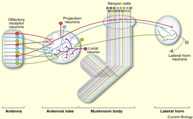
In Drosophila, odors are detected by olfactory receptor neurons (ORNs), which are located in sensilla on the antenna and maxillary palps. In general, all the ORNs of one receptor type project onto one glomerulus of the antennal lobe (Hallem et al., 2004), where they synapse with projection neurons as well as with local interneurons (Figure 1). Projection neurons further transmit odor information to the lateral horn and the mushroom body (MB). Within the MB calyx, projection neurons synapse onto Kenyon cells (KCs), the major intrinsic neurons. The lateral horns are considered centers for innate olfactory behaviors, whereas the MBs are regarded as centers for learned olfactory behaviors (Tanaka et al., 2004) , as will be discussed later (chapter 6.2.3). The output regions of MBs and lateral horns are not yet well characterized, but include premotor areas (Ito et al., 1998), and eventually control odor avoidance and attraction behaviors. The olfactory pathway is overviewed in figure 1.
14
it is transmitted from ORNs to projection neurons (Wilson and Laurent, 2005, Root et al., 2008, Tanaka et al., 2009, Olsen et al., 2010, Das et al., 2011). The population of local neurons in the antennal lobe is highly diverse and includes neurons innervating single, multi or all glomeruli (Chou et al., 2010). The majority of local neurons are inhibitory although some excitatory local neurons are also important for olfactory modification (Olsen et al., 2007, Shang et al., 2007, Huang et al., 2010). Lateral connections between the glomeruli might add to odor modification (Bhandawat et al., 2007, Masse et al., 2009). In the MBs, the olfactory information is further processed by a 10 to 1 convergence of input from projection neurons to KCs (Turner et al., 2008, Caron et al., 2013). This convergence and a high firing threshold leads to sparse firing of KCs for each given odor (Turner et al., 2008). Lateral connections between KCs, and between KCs and modulatory neurons innervating the MBs may further modify olfactory representations and behaviors. Some of these modulatory neurons are important for olfactory memory formation and consolidation, such as the GABAergic anterior paired lateral neuron (Liu and Davis, 2009), or the dorsal paired medial neuron (Keene et al., 2004, Yu et al., 2005). To conclude, representation of most odors starts as a unique combination of many active ORNs, and becomes sparse as it reaches the KCs. This sparseness separates representations of different odors from each other and serves a basis for memory formation. Representations of different odors do indeed become less correlated as they progress through the olfactory system(Turner et al., 2008).
15
Olfactory receptor neurons in the antennae and maxillary palps send axons to specific glomeruli in the antennal lobe. All olfactory receptor neurons expressing the same odorant receptor (same color) converge at a single glomerulus. There they form synaptic contacts with projection neurons and local neurons. Projection neurons send axons either directly to the lateral horn (green projection neuron) or via the calyx of the mushroom bodies (red and blue projection neurons), where they form synapses with Kenyon cells. Figure is taken from (Masse et al., 2009).
6.1.2 Electric shock sensation
Electric shock is a potent noxious stimulus which is easy to control, hence application of electric shock is the most common way of inducing aversion in animals and humans (Handwerker and Kobal, 1993). Nociceptors are the sensory receptors that respond to different kinds of noxious stimuli which can potentially damage tissues. In the hairy skin of mammals there are a few types of nociceptors. Some are polymodal receptors, responding to thermal, mechanical and irritant chemical stimuli. Other types of nociceptors are only sensitive to one modality and even to a specific intensity of that modality by having distinct activation thresholds. Sensory receptors are located in peripheral nerve endings and send sensory information through many types of fibers. These fibers include thickly myelinated A-beta-fibers carrying motor response and/or low threshold mechanical information, as well as thinly myelinated A-delta-fibers or C-fibers carrying information from thermo-receptors and nociceptors. Transcutaneous electrical stimulation of peripheral nerves excites almost all of these fibers, rendering the response non-specific for any modality (Baumgartner et al., 2012). Electric shock can excite the full spectrum of peripheral nerve fibers (depending on the stimulation method). Additionally, the sensory nerve endings are probably not recruited when an electrical stimulus is given, and the signal bypasses a transduction mechanism (Handwerker and Kobal, 1993). Being a potent and unnatural stimulus, electric shock might recruit general sensory pathways rather than dedicated ones.
16
Nociception studies in Drosophila larvae led to the discovery of polymodal nociceptors in their body walls, the class IV multiple-dendritic neurons (Hwang et al., 2007). These neurons detect both noxious heat and noxious mechanical stimuli and are responsible for avoidance of larvae from increased temperature, mechanical pain, dry surfaces and bright light (Hwang et al., 2007, Xiang et al., 2010, Im and Galko, 2012, Johnson and Carder, 2012). These cells express Painless, Piezo, Pickpocket and dTrpA1, all proteins that function in perception of noxious mechano- and/or thermal stimuli (Zhong et al., 2010, Neely et al., 2011, Hwang et al., 2012, Kim et al., 2012, Zhong et al., 2012). The role of some proteins in aversive sensation is also multimodal: Painless is involved in thermal, mechanical and chemical nociceptive perception (Zhong et al., 2010), and dTrpA1 in the sensation of mild heat, noxious heat and chemical avoidance (Rosenzweig et al., 2005, Kang et al., 2010, Neely et al., 2011, Kang et al., 2012).While larvae share a single class of neurons for sensing different modalities of aversive stimuli, no such neurons have been identified in adult flies. For example, the expression pattern of painless in adults is distributed in the central and peripheral nervous system (Xu et al., 2006). Other transmitters and proteins are involved in mechanical nociception: histamine (Buchner et al., 1993, Melzig et al., 1996), Pickpocket (Zhong et al., 2010) and Piezo (Kim et al., 2012) but their cellular distribution is not fully characterized. It is not known whether electric shock stimulation is sensed through a specific sensory pathway, or if it leads to unspecific activation such as it is the case in mammals (Handwerker and Kobal, 1993, Baumgartner et al., 2012). Electric shock may be sensed by any of the identified pain receptors, or by novel ones.
6.1.3 Temperature sensation
17
and closing transitions of a channel are sufficiently different. Accordingly, thermal sensitivity is possible when the energy needed for channel opening is much greater than that needed for closing, such as in the case of thermo-sensitive TRP channels (Voets et al., 2004). Each of these channels opens at a distinct temperature, allowing for graded activation of variable receptors with temperature elevation. While dTrpA1 and GR28B are receptors for mild heat (higher than 27°C), pyrexia and painless are only activated by temperatures higher than 38°C or 40°C, respectively (reviewed by Sokabe and Tominaga, 2009). Other proteins and channel-subunits are involved in thermal sensation although they are probably not direct temperature receptors, such as Straightjacket (Neely et al., 2010), histamine receptors (Hong et al., 2006), the cAMP pathway in the mushroom bodies (Hong et al., 2008, Kang et al., 2011), amnesiac (Aldrich et al., 2010) and others. The main characterized thermo-sensory membrane proteins are summarized in Figure 2A.
18 Figure 2: Temperature sensation in flies
A. Threshold activation temperature of thermo-sensory membrane proteins. Flies narrowly distribute around their preferred temperature, 24°C. dTrpA1 is activated above 27°C and contributes to avoidance of warmer temperatures. Pyrexia is activated above 38°C and prevents paralysis during high temperature stress. Painless is activated above 40°C, and is essential for avoiding hazardous temperatures. These channels belong to the TrpA subfamily and possess temperature sensitivity. GR28B is a thermal receptor belonging to the gustatory receptor family, with a similar activation threshold to dTrpA1. Straightjacket is a sub-unit of a calcium channel needed for sensation of high temperatures, with a similar threshold to Painless. B. Schematic of two pathways for elevated temperature reception in the fly brain. Red: the internal AC neurons which express dTrpA1 (arrows mark cell bodies). Black: the antennal hot cells which express GR28B (arrowheads mark cell bodies). VP2 and VP3 glomeruli of the antennal lobes (black innervation sites) are innervated both by hot cells and by AC neurons. AC neurons mostly terminate in the posterior protocerebrum (red innervation sites). Blue: antenna; purple: antennal lobes; Green: MBs. Adapted from Galili et al., 2014, figure 3A there, with changes.
6.2 Learning and memory in Drosophila
It is important for animals to rapidly detect meaningful stimuli in their surroundings, and use these to predict the value of co-occurring stimuli. Indeed, fruit-flies can learn to associate overlapping stimuli, remember and use this knowledge in following exposures. Since the pioneering work of Seymour Benzer beginning in the 1960’s, learning and memory in Drosophila melanogaster has been a research topic in laboratories world-wide. Benzer and his colleagues were first to use behavioral genetics in flies to dissect the factors important for behaviors such as vision, locomotion, sexual function, circadian rhythm, learning and memory.
6.2.1 Classical conditioning of fruit-flies
19
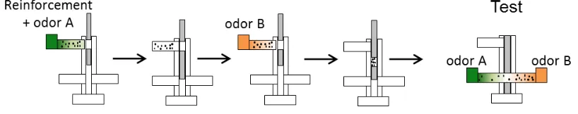
unconditioned stimulus (US), which led to a measurable behavioral response, salivation. After the CS-US coupling, the animal started responding to the CS with a conditioned response (Pavlov, 1911). Classical conditioning has been studied extensively in many model organisms since, including humans. A rather neutral CS is coupled to a meaningful US. The US can either be aversive and elicit an avoidance response or appetitive and elicit attraction. Fruit-flies can easily be trained and tested with classical conditioning (Quinn et al., 1974, Tully and Quinn, 1985). The most commonly used CSs for flies are olfactory or visual cues. Here, I will focus on aversive olfactory classical conditioning. Multiple stimuli can serve as aversive USs for Drosophila flies: mechanical vibration (Folkers, 1982, Eschbach et al., 2011, van Swinderen, 2011), courtship refusal (Siegel and Hall, 1979, Gailey et al., 1984), bitter substances and irritant chemicals (Gerber and Hendel, 2006, Schnaitmann et al., 2010, El-Keredy et al., 2012), the initial effect of ethanol (Kaun et al., 2011), humidity (Le Bourg, 2005), elevated temperature (Wolf and Heisenberg, 1991, Wustmann and Heisenberg, 1997, Ofstad et al., 2011), and electric shock (Quinn et al., 1974) (for more information see reviews: (Davis, 1996, Pitman et al., 2009)). The most commonly used aversive classical conditioning paradigm in flies is coupling an odor (CS) to electric shock (US), followed by a test for conditioned odor avoidance (Figure 3; Quinn et al., 1974, Tully and Quinn, 1985). This coupling leads to strong and stable odor avoidance, which is easy to quantify based on the distribution of flies between two tubes, one containing the CS and the other a control odor (or odorless solution). Depending on the training design, the associative memory can last as long as a few days (Tully et al., 1994). Much of the current knowledge about the neural circuit of olfactory conditioning in flies emerged from using electric shock olfactory conditioning (reviewed in: Davis, 2005), which I also utilized during my PhD project.
Figure 3: Classical olfactory conditioning in Drosophila
20
6.2.2 Reinforcement pathway: the role of dopamine
Although not much is known about electric shock sensation in flies (see chapter 6.1.2 “electric shock sensation”), it is widely used as a potent aversive reinforcer in olfactory conditioning. There are multiple lines of evidence that electric shock reinforcement information is eventually conveyed to dopaminergic neurons (Schwaerzel et al., 2003, Riemensperger et al., 2005, Kim et al., 2007, Claridge-Chang et al., 2009, Aso et al., 2010, Waddell, 2013). During training, these dopaminergic neurons are thought to signal reinforcement information to the axons of KCs, and modulate the strength of synaptic connections between KCs and their output partners (Figure 4). Dopamine type 1 receptors on KC axons play a role in this plasticity, resulting in alteration of the conditioned odor avoidance behavior (Kim et al., 2007). Recently, specific subsets of dopaminergic neurons were identified to play a role in olfactory conditioning with electric shock (Claridge-Chang et al., 2009, Aso et al., 2010, Aso et al., 2012).
21
Figure 4: Dopaminergic neurons innervate the mushroom bodies (MBs) and play a role in learning and memory
A. Three clusters of dopaminergic neurons directly innervate the MBs: PAM, PPL1, PPL2ab (nomenclature according to Nassel and Elekes, 1992). B. Four types of dopaminergic neurons were found important for memory formation in olfactory shock conditioning (Aso et al., 2010, Aso et al., 2012). From the PPL1 cluster the neurons MB-V1, MB-MV1 (both depicted in green), MB-MP1 (depicted in blue). Additionally, from the PAM cluster the neuron MB-M3 (depicted in orange) (Figure modified from Aso et al., 2012).
However, there is only sparse evidence for the role of dopamine in signaling aversive reinforcers other than electric shock (Unoki et al., 2005, Honjo and Furukubo-Tokunaga, 2009). In some cases dopamine was not found necessary for conditioning of flies with variable aversive reinforcers (Sitaraman et al., 2008, Yarali and Gerber, 2010). In addition, serotonin is involved in reinforcement signaling in
Drosophila and other insects (Sitaraman et al., 2008, Wright et al., 2010, Sitaraman et al., 2012). Particularly, thermal reinforcement in Drosophila place learning requires serotonin, but not dopamine (Sitaraman et al., 2008). Therefore, it is unknown whether the role of dopaminergic neurons in shock learning is restricted to signaling electric shock, or if they are part of a general aversive reinforcement pathway. To test this question, it is important to compare aversive conditioning with different reinforcers under otherwise similar experimental conditions.
6.2.3 The role of mushroom bodies as coincidence detectors
22
dopaminergic neurons which innervate KCs axons in different regions of the lobes. When both olfactory and reinforcement events reach the KCs simultaneously (or within a short range, as will be discussed), output from these activated KCs onto MB-output neurons is thought to be strengthened. The KCs project to a variety of target regions including premotor areas (Ito et al., 1998). Strengthened output is thought to mediate conditioned odor avoidance during the test period, when odor is encountered without reinforcement (reviewed in: Gerber et al., 2004). Indeed, blocking MB-output specifically during test period impaired memory performance, indicating that MB-output is needed for behavior execution (Dubnau et al., 2001, McGuire et al., 2001).
Behavioral screens of flies were used to discover genes involved in learning and memory. By inducing random mutations into the fly genome, many learning mutants were isolated in the past decades, among them dunce and rutabaga which are a part of the cAMP signaling pathway (Dudai et al., 1976, Duerr and Quinn, 1982, Dudai et al., 1983, Shotwell, 1983). It has been suggested that the type I adenylate cyclase, encoded in flies by the rutabaga gene, acts as a molecular coincidence detector between odor and reinforcement information, since rutabaga mutants are impaired in olfactory learning (Davis et al., 1995, Zars et al., 2000, Gerber et al., 2004). Activation of this enzyme requires both calcium influx and active G-protein; therefore it is capable of integrating information from two independent sources such as neuronal depolarization following odor activation (calcium influx), and dopamine receptor activation following reinforcement (G-protein activation), (Tomchik and Davis, 2009), as summarized in Figure 5. Restoring rutabaga expression specifically to the MBs of rutabaga
mutant flies was enough to restore the learning ability (Zars et al., 2000). Activation of adenylate cyclase leads to the conversion of ATP to cAMP. cAMP, a major signal transducer of the cell, then activates protein kinase A, which in turn initiates a phosphorylation cascade leading to the induction of genes involved in learning (Davis et al., 1995, Connolly et al., 1996, Davis, 1996). Along with rutabaga, other genes in the cAMP signaling pathway have proven important for intact olfactory learning: amnesiac
(encoding a product similar to adenylate cyclase activating peptides), dunce (cAMP phosphodiesterase),
23
Figure 5: Schematic representation of the molecular mechanisms involved in aversive olfactory conditioning
CS information reaches the Kenyon cell via projection neurons releasing acetylcholine (Ach), leading to opening of voltage-gated calcium channels and increased intracellular calcium. Calcium binds to calmodulin and activates the
rutabaga protein adenylate cyclase (AC). The shock US induces dopamine (DA) release, which binds to the alpha
subunit of the G protein coupled receptor (GPCR),and activates AC. During coincident occurrence of CS and US, the two types of input act synergistically on AC, triggering cAMP signaling, and leading to the strengthening of the output from this Kenyon cell (Busto et al., 2010).
6.2.4 Importance of timing for associative learning: Trace, delay and relief learning
For efficient associative learning to take place, an overlap between CS and US presentation is beneficial. However, animals can also associate two temporally separated stimuli. This ability is important, since often in nature the cause and outcome of stimuli are not contiguous. In many behaviors critical for survival such as searching for a mate or food, or escaping predators, there can be a temporal gap between the sensory stimulus and the reward or punishment it predicts. In 1927, Ivan Pavlov had already noted that dogs were able to learn the CS-US association in classical conditioning, even after training with a whistle-sound CS and a food reward US, separated by several minutes (Pavlov, 1927). The conditioned salivary response was delayed in proportion to the duration of the CS-US interval, suggesting that the dogs learned the existence of the gap and anticipated the US.
24
aversive US, and show attraction towards it. This type of learning is termed relief learning (Tanimoto et al., 2004, Yarali and Gerber, 2010). Thus, the relative timing of CS and US presentation is a critical parameter of learning. Trace conditioning was studied in many organisms, using different paradigms (Rescorla, 1988, Figure 1 there). In general, aversive learning is highest when the CS and the US overlap, such as in delay conditioning, and it is lower for trace conditioning. The shape of the CS-US interval function in olfactory learning of flies (Figure 6B) is strikingly similar to those in mammals, across different conditioning paradigms with different stimuli (Rescorla, 1988, Figure 1 there), suggesting that stimuli traces are kept for a variety of modalities and are commonly used for conditioning by many species.
Are the memories formed during delay and trace conditioning governed by different neuronal pathways? In mammals, delay and trace eyeblink conditioning engage similar brain circuits, but in trace conditioning there is additionally a requirement of the hippocampus (Solomon et al., 1986, Woodruff-Pak and Disterhoft, 2008) and in humans also a state of awareness is required (Clark and Squire, 1998, Clark et al., 2002, Christian and Thompson, 2003). Therefore, trace memory was proposed to be qualitatively and anatomically distinct from delay memory, and serve as a model of declarative memory (Clark et al., 2002, Woodruff-Pak and Disterhoft, 2008). However, other views have been proposed (LaBar and Disterhoft, 1998). The demands on neural resources increase with task complexity for both trace and delay conditioning (Knuttinen et al., 2001, Carter et al., 2003). Thus the differential requirement of the hippocampus for trace conditioning might be a result of task complexity and not of the discontinuity between stimulus and reinforcement (Carrillo et al., 2000, Beylin et al., 2001, Walker and Steinmetz, 2008, Kehoe et al., 2009). In addition, a few examples of trace conditioning in humans without awareness have been observed (Bekinschtein et al., 2009, Arzi et al., 2012). Therefore, the anatomical and mechanistic distinction between the associative memory in delay and trace neuronal pathways in mammals is still an open question.
25
stimulus termination? The engram of the olfactory trace and its temporal properties are unknown. In order to tackle these open questions it is important to first establish a robust experimental design where trace and delay conditioning can be compared.
Figure 6: Behavioral design and memory performance in delay and trace conditioning
A. Behavioral design of delay (upper) and trace (lower) conditioning. While in delay conditioning CS and US presentations overlap, in trace conditioning a there is a stimulus-free gap between them. The time between CS-onset and US-CS-onset is termed the CS-US interval. B. Memory performance after aversive conditioning in Drosophila as a function of CS-US interval. Blue bar indicates the 15 s odor CS, its end is indicated by a dashed line. US is 4 electric shocks of 90 V within 16 s. Delay conditioning occurs when the CS-US interval≤15 s, and trace conditioning when the CS-US interval>15 s. Figure is modified from (Dylla et al., 2013). Data for B is taken from (Tanimoto et al., 2004).
26
6.3 Advantages of fly neuroscience: the genetic tool box
Drosophila melanogaster has long been a popular model organism for biological and developmental studies. In recent decades, the fruit fly has also become an important model organism in behavioral neuroscience for several reasons. Drosophila are small, inexpensive, easy to cultivate, have a short generation time and perform highly stereotyped behaviors. Flies have a numerically reduced nervous system compared to mammals (~105 vs. ~1011 cells in a Drosophila and a human brain, respectively). In addition to the anatomical simplicity, studies in Drosophila are supported by a wealth of genetic techniques (Venken and Bellen, 2005). With only four pairs of chromosomes and knowledge of the complete sequence of the genome, genetic analysis of behaviour is facilitated. Among the most common genetic techniques used in Drosophila is the generation of mutant flies. Hundreds of mutants that affect behavioral functions have been identified in genetic screens. Usually, mutagenic treatments such as the feeding of specific chemicals or UV-radiation allow an unbiased generation of mutations (Ashburner, 1989). Another method to induce mutations is by randomly inserting transposable genetic elements (P-elements) into the fly genome (O'Kane and Gehring, 1987). Simple behaviors can be tested across many mutants, to identify specific genes required for these behaviors. Using a genetic screen, the gene period was among the first to be discovered (Konopka and Benzer, 1971). This gene plays a role in circadian rhythm of flies, and demonstrated that mutations in a single gene can alter circadian behavior, a first step towards a molecular analysis of circadian rhythms in several species. Generation and use of mutant flies tremendously advanced our understanding of the genetic basis of behaviour, yielding principles applicable to several other species. Since the fly genome is sequenced, targeted mutations are also possible by inserting P-elements into known locations in the genome, thus disrupting genes of interest. Different types of insertion and deletion mutations were generated using specifically designed P-elements (Hummel and Klambt, 2008).
27
two components are brought together with a simple genetic cross. In the progeny of the cross, the effector transgene will be expressed specifically in a defined set of cells that have the tissue specific promoter activated and express the GAL4 protein (Brand and Perrimon, 1993). Common effector transgenes used in Drosophila neuroscience research under UAS control include blockers of neuronal activity such as the potassium channel Kir2.1 (Baines et al., 2001) or the light-chain of tetanus toxin (Sweeney et al., 1995); neuronal activators such as the temperature-activated channel dTrpA1 (Hamada et al., 2008); and marker proteins such as GFP linked to mCD8 to allow plasma membrane labelling (Lee and Luo, 1999). For knocking-down specific genes in a restricted cellular population, one can exploit the RNA interference method, and express a double stranded RNA sequence complementary to the sequence of a gene of interest, under UAS control (Enerly et al., 2003). Expression of any of these transgenes in a specific cellular population by using the GAL4-UAS transcription system enables labelling, regulating, blocking or activating specific subsets of neurons, and testing the effect of these manipulations on behaviour.
Many tools were developed to complement and control the GAL4/UAS expression system, enabling specific spatial and temporal manipulations. By splitting the GAL4 construct into two parts, the DNA-binding domain and the activation domain, it is possible to use an intersection of different lines and thus reduce the number of labelled cells, in some cases to the level of single cells (Luan et al., 2006). Additionally, the yeast protein GAL80, a repressor of GAL4, binds the transactivation domain of GAL4 and prevents GAL4 activated transcription. The use of GAL80 allows for refinement of the expression pattern (Lee and Luo, 1999). A further modification of GAL80 allows for exact temporal control of the repression: the temperature sensitive version of GAL80 only represses GAL4 at certain temperatures (McGuire et al., 2003), providing an “on-off” temporal switch for GAL4 expression.
28
6.4 Aims of the thesis: exploring the CS and US representations in the nervous system
What happens to CS representation after CS is gone? What is the US representation in different modalities?
My PhD project focuses on two open questions in the field of Drosophila olfactory learning (figure 7). The first question concerns the temporal requirement for the CS-US association. What do flies learn when a gap is inserted between the odor and the reinforcement? In order to answer this question, I established a robust paradigm for trace conditioning of flies using odor and electric shock which were temporally separated. Flies can learn the association even when a gap of 15 seconds is separating the end of odor presentation from the beginning of electric shock (Galili et al., 2011). I study the commonalities and differences between trace conditioning and delay conditioning (when odor and shock overlap), and look for the identity and location of the olfactory trace. The second part of my project concerns the neuronal representation of reinforcement. Almost all of the current knowledge on aversive reinforcement pathway was derived using electric shock. However, we do not know the sensory correlates of shock sensation. Electric shock is eventually signaled through dopamine neurons, but the generality of this representation was never tested. I compared electric shock reinforcement with a more ecologically relevant reinforcement: increased temperature. I established olfactory temperature conditioning, using the same experimental set-up and design as olfactory shock conditioning, and used
genetic manipulations to compare the sensory and central neurons needed for forming these memories.
Figure 7: Simplified schematic of the olfactory conditioning circuit in flies
29 7 Results
7.1 First paper: Galili DS, Lüdke A, Galizia CG, Szyszka P, Tanimoto H. (2011) Olfactory trace conditioning in Drosophila. J Neurosci. 31(20):7240-8.
http://www.jneurosci.org/content/31/20/7240.long DOI: 10.1523/JNEUROSCI.6667-10.2011.
7.2 Second paper: Galili DS, Dylla KV, Lüdke A, Friedrich AB, Yamagata N, Wong JY, Ho CH, Szyszka P, Tanimoto H. (2014) Converging circuits mediate temperature and shock aversive olfactory conditioning in Drosophila. Curr Biol. 24(15):1712-22.
30 8 Discussion
8.1 Olfactory trace conditioning in Drosophila
I showed that flies can associate an odor CS with a shock US even when a gap is separating them, in olfactory trace conditioning. Olfactory stimuli are often hard to switch-off as odor molecules may linger in the set-up in the form of residual odor, preventing a clean stimulus offset. I controlled for precise odor presentation, by testing whether flies are able to form an association between reinforcement and the residual odor, and then chose an odor which did not lead to learning of the residual. Thus the odor trace I studied originated from the nervous system of the fly itself and not the apparatus (Galili et al., 2011, Fig. 2).
8.1.1 Trace and delay memories are based on the same odor perception
By comparing the behavioral parameters of trace and delay memories, I discovered that they share striking commonalities. My data suggest that both memory forms engage similar odor percepts, although odor saliency may decline between odor presentation and its trace. This means that the odor percept probably does not undergo qualitative changes after offset. This conclusion is based on three findings: (1) Trace and delay conditioning have similar generalization profiles (Galili et al., 2011, Fig. 5). When one odor is presented during training and a new odor is presented during test, flies generalize their conditioned odor avoidance response depending on the degree of perceived similarity between the new odor and the trained one. With four different odors, the generalization index after training with trace or with delay conditioning was identical (correlation coefficient of r2=0.99), suggesting the odor itself and its trace share a common (or extremely similar) percept. (2) Trace and delay memories reached the same asymptote in memory acquisition after sufficient trials (Galili et al., 2011 Fig. 6A). See discussion in the next chapter 9.1.2. (3) Trace and delay memories have similar decay patterns. When starting from similar levels of initial memory, both memories decay within the same duration and their decay curves are not significantly different (Galili et al., 2011, Fig. 6B). Together, these results suggest that a similar perception of odor quality may be engaged in both trace- and delay memories.
8.1.2 Odor trace may be less salient than the odor itself
31
This difference might be explained by a reduced CS salience in trace conditioning. One of the classical models of learning acquisition (Rescorla and Wagner, 1972) suggests that learning improvement during acquisition depends on three factors: the asymptotic level of memory acquisition, the strength of the reinforcement and the saliency of the stimulus. In the trace conditioning paradigm I established, the asymptotic levels are identical between trace and delay acquisition, and the same shock stimulus was used as reinforcement; hence the difference in the learning rates can be attributed to different saliencies of the sensory stimuli. According to Rescorla and Wagner (1972), CS saliency specifically changes the rate, but not the asymptote of learning acquisition. During trace conditioning, the salience of the odor stimulus probably decays after CS presentation until the US is applied. So the odor trace is probably fading with time [this fits also to the lower memory scores after longer intervals, as seen in the CS-US interval function, Figure 6B, and (Galili et al., 2011; Figure 3B there)]. In insect olfactory learning, changing odor saliency was indeed reported to effect memory formation (Pelz et al., 1997, Masek and Heisenberg, 2008, Yarali et al., 2009). In these studies, higher odor intensity supported stronger association, implying that the saliency of the odor can determine its effectiveness for memory formation. Together, my data suggest that the odor trace retains stimulus specificity but might be less salient than that odor during its presentation.
8.1.3 Identity and location of the odor trace
32
other than persistent calcium signals. For example, long-lasting biochemical modifications or “tags” (Perisse and Waddell, 2011, Dylla et al., 2013) can signal an odor trace. ‘Eligibility traces’ are transient synaptic changes which can theoretically last for several seconds, for example the activation of an enzyme with slow kinetics, important for synaptic plasticity. In the model suggested by Izhikevich (2007), after stimulus presentation, the slow kinetics of synaptic plasticity are sensitive to changes in the extracellular dopamine concentration signaling reinforcement, during a critical period of a few seconds. The synaptic ‘eligibility trace’ creates a time window in which a global diffusive reinforcement signal in the form of extracellular dopamine can selectively influence the right synapses at the right time (Izhikevich, 2007). Alternatively, subtle network activity, such as changes in the correlation of glomerular spontaneous activity following odor presentation (Galan et al., 2006) might also be an underlying mechanism for maintaining a sensory trace for a short while. In a study by Galan et al. (2006), a single odor presentation changed the relative timing of spontaneous firing across glomeruli, recapitulating the odor-induced activation, and this modification lasted for a few minutes. Both these scenarios suggest a trace representation which is reminiscent of the stimulus itself, a prediction which fits my results of trace and delay memories sharing a common odor percept (see chapter 9.1.1). Either a synaptic ‘eligibility trace’ or a modification of the network activity could represent a sensory trace in any of the neuronal relays along the olfactory pathway, without being reflected in our calcium imaging. Alternatively, the trace can be maintained as an increased firing in other neurons along the olfactory system, besides ORNs or projection neurons. Calcium imaging may be used to test for activity related to trace maintenance in these regions. Reasonable candidates are the local neurons in the antennal lobe, or the Kenyon cells of the MBs (Figure 8). Modulatory neurons of the olfactory system like the anterior paired lateral neuron (Liu and Davis, 2009)or the dorsal paired medial neuron (Waddell et al., 2000) may also play a role in keeping the olfactory trace.
33
the main GABAergic innervation to the MB lobes through the GABA-A receptors, resistance to dieldrin (Rdl; Liu and Davis, 2009)). Rdl levels in the MBs were negatively correlated to olfactory memory performance (Liu et al., 2007). In preliminary experiments I knocked-down the GABA-A receptors Rdl in the MBs and trained flies for delay or trace conditioning. Decreased levels of Rdl receptors in the MBs improved trace conditioning, compared to delay conditioning (data not shown). These data hint that inhibition of the MBs may contribute to the fading of the olfactory trace, hence trace duration may be prolonged when inhibition of the MBs is blocked. Future experiments are needed to dissect the contribution of the MBs and MB-inhibition to olfactory traces and trace conditioning (Figure 8).
Figure 8: Possible locations of the odor trace
During odor presentation, olfactory sensory neurons (ORNs) send olfactory information to projection neurons in the antennal lobe, which further transmit the information to the mushroom bodies (MBs; upper panel; see olfactory pathway chapter 6.1.1). After the odor presentation is over, the odor trace is maintained in the nervous system. Our collaborator from Konstanz University, Alja Luedke, showed that the odor trace is not kept in calcium signals of ORNs or projection neurons (Galili et al., 2011; and personal communication). Possibly, the odor trace is located in the firing patterns or in biochemical alterations (eligibility traces) of the Kenyon cells of the MBs (Perisse and Waddell, 2011, Shuai et al., 2011, Dylla et al., 2013). Figure taken from Perisse and Waddell (2011).
8.1.4 Former experience improves trace conditioning
Woodruff-34
Pak and Disterhoft, 2008), and here is shown for the first time in insects (Galili et al., 2011, Szyszka et al., 2011). Successful trace conditioning with a short time interval between CS and US extends the interval that can be bridged in subsequent trials, enabling learning with gaps that were too long for naïve flies. Thus it appears that flies benefit from being familiar with the gap between the stimuli. There are two possible mechanisms to explain how the experience gained from trace conditioning training can help bridge a longer gap in subsequent trials. The first mechanism is altering the CS representation pathway. Behavioral experience could enhance the CS saliency, so that greater importance will be assigned to the fading CS trace by the fly during later trials. Enhanced saliency would result in a stronger association (Rescorla and Wagner, 1972). At the level of neuronal activity, associative learning may change the dynamics of odor-evoked activity and strengthen or extend the odor trace until the US arrival. Indeed, calcium imaging in odor-activated dopamine neurons revealed that olfactory conditioning prolonged the response to the trained but not to the control odor (Riemensperger et al., 2005). An alternative possible mechanism is a temporal shift of the onset of activity in reinforcement neurons, after the initial training. In this case the activation of US-representing neurons would start already during CS presentation. This is the case in monkeys, which can be trained to anticipate the arrival of a future US in trace conditioning, and show earlier activation of reinforcement neurons following training ('US anticipation', reviewed by Schultz, 2006). Insects can also learn to anticipate a US after associative conditioning (Gil et al., 2007, Gil et al., 2008). It was shown that honeybees’ experience with increasing magnitudes of reinforcement during associative learning changed their subsequent behavior towards the CS; the animals spent longer time inspecting CS that predicted a bigger reward, indicating they learned to expect the US (Gil et al., 2007). Since flies can learn trace conditioning with a single training trial (Galili et al., 2011), US anticipation can be excluded as an explanation for 1-trial trace learning. Nonetheless, after several trials, US anticipation may develop. Whether experience with trace conditioning alters the CS pathway by prolonging the trace, or the US pathway by earlier activation of US-representation is still an open question. These proposed mechanisms may also act together to improve consecutive trials.
8.2 Comparing the neural circuits of aversive reinforcers: temperature and shock
35
commonalities and differences between the neural representations of these aversive reinforcements: temperature and shock. I found that the sensory correlates of temperature and shock perception are different: elevated temperature is sensed through Anterior Cell (AC) neurons expressing the thermo-sensitive cation channel dTrpA1, while shock sensation does not require these neurons. Although reception engages different pathways, eventually both shock and temperature reinforcement converge onto dopaminergic neurons, generally signaling aversive reinforcement. Within the small population of dopaminergic neurons necessary for both temperature and shock conditioning, a smaller subgroup of neurons seems to represent temperature than those representing shock.
8.2.1 Temperature and shock sensation are separate
36
8.2.2 Detection of increased temperature during reinforcement learning occurs via a specific sensory pathway
37
Although I showed the separation of sensory pathways to specific circuits regarding reinforcement signaling, other functions may be shared between these sensory pathways. For example, glomeruli VP2 and VP3 of the antennal lobe are suggested to be commonly innervated by both AC neurons and hot cells (Gallio et al., 2011, Tanaka et al., 2012). It was also shown that a few Kenyon cells get thermal information from glomeruli VP2 and VP3 through projection neurons (Caron et al., 2013, Figure 3 there). It is an open question which behavioral functions this circuit does serve, but the role of the MB in thermal behaviors may depend on these inputs (Hong et al., 2008). To conclude, mild temperature increase activates separate sensory pathways, and can lead to different behavioral outputs, some are segregated between these inputs and some may be shared.
8.2.3 Dopamine integrates aversive reinforcement of both temperature and shock (Figure 9A)
I showed that dopaminergic neurons are required for both shock and temperature reinforcement signaling. Dopamine neurons are not needed for innate perception, but for assigning the appropriate aversive value of the reinforcement to the olfactory stimulus (Galili et al., 2014; Figure 5). Evidence from my anatomical characterization suggest that AC neurons contact dopamine neurons, possibly transmitting temperature reinforcement information during conditioning (Galili et al., 2014; Figure 6). My results join a line of research on the role of dopamine in reinforcement signaling, both in vertebrates and invertebrates. Many studies in recent years support a role for the dopamine system in both negative and positive reinforcement signaling. In insects, aversive learning of electric shock is conveyed by dopaminergic neurons (Schwaerzel et al., 2003, Riemensperger et al., 2005, Kim et al., 2007, Vergoz et al., 2007, Claridge-Chang et al., 2009, Aso et al., 2010, Waddell, 2013). Aversive conditioning using quinine taste in fly larvae (Honjo and Furukubo-Tokunaga, 2009) or saline solution in water-deprived crickets (Unoki et al., 2005) also depend on dopaminergic neurons. Although reward signaling in flies was initially attributed to octopamine (Schwaerzel et al., 2003, Schroll et al., 2006), recent papers show that reward learning depends additionally on dopaminergic neurons (Kim et al., 2007, Burke et al., 2012, Liu et al., 2012a, Perry and Barron, 2013) that are probably innervated by octopaminergic neurons signaling the appetitive value of sweet taste.
38
The convergence of different inputs to dopaminergic neurons may be conserved among different animal species beyond insects. Similar to the organization in the fly, midbrain dopaminergic neurons in mammals have been shown to respond to different punishing events in rodents and primates (Brischoux et al., 2009, Bromberg-Martin et al., 2010, Lammel et al., 2011, Wang and Tsien, 2011, Zweifel et al., 2011). Although dopamine in mammals was affiliated with reward signaling, it is emerging that dopamine plays a role in signaling aversive conditioned stimuli as well (Matsumoto and Hikosaka, 2009, Schultz, 2010, Lammel et al., 2011, Zweifel et al., 2011). Thus, dopamine may be a signal encoding general motivational values for decision making regardless of stimuli and valence.
Temperature and shock are sensed by different sensory pathways, converging to the same dopamine system. A similar circuit configuration was described for sugar reinforcement in flies. Sugar includes two separate reinforcing qualities- sweet taste and nutritional value. These qualities are sensed by separate pathways and converge onto the dopaminergic system, which signals reinforcement (Burke et al., 2012). The sweet taste is probably signaled by octopamine and conveyed to dopaminergic PAM neurons, while nutritional value is sensed independently of octopamine, and eventually signaled by dopamine too (Burke et al., 2012). Similarly, in the case of temperature and shock, sensory pathways differ and reinforcement signaling converges to the dopaminergic system. This convergence may be a typical property of reinforcement systems. While the stimulus identity is initially separated, a general system attaches a value to an environmentally relevant reinforcer. A simplified model is suggested in Figure 9B.
8.2.4 Overlapping subsets of dopaminergic neurons for temperature and shock reinforcement
39
subsets of neurons within the TH-D’-GAL4 may separately be responsible for shock- and for temperature olfactory memories. In order to find out whether there are specific dopaminergic neurons which signal temperature reinforcement, I screened a library of driver lines labeling variable sub-populations, small clusters or single dopaminergic neurons (data not shown). However, I did not find drivers which specifically impaired temperature but not shock memories. Thus, the signal for temperature reinforcement may be included within the population of dopaminergic neurons signaling shock reinforcement. It is possible, that the artificial electric shock stimulus recruits more than one specific pathway (Claridge-Chang et al., 2009, Aso et al., 2010, Aso et al., 2012), while the ecologically relevant thermal stimulus recruits a more specific subset of dopaminergic neurons within those, to signal reinforcement.
Figure 9: Summary and model for sensory and reinforcement signaling
40 8.3 General conclusions and outlook
Brains have the unique ability to change and adapt according to environmental experience. Animals must learn to attach values to meaningful sensory stimuli they are exposed to, in order to approach stimuli which predict positive outcome, and avoid those which predict negative outcome. Hence the study of associative learning is an important and fundamental field in neuroscience. Associative learning involves detection and encoding of two stimuli: the CS and the US. My results provide important insights into the neuronal pathways transmitting both these stimuli, until they converge and modulate behavioral output. In my PhD project I studied two phenomena in the field of olfactory conditioning in
41 9 References
Aldrich BT, Kasuya J, Faron M, Ishimoto H, Kitamoto T (2010) The amnesiac gene is involved in the regulation of thermal nociception in Drosophila melanogaster. J Neurogenet 24:33-41.
Alekseyenko OV, Chan YB, Li R, Kravitz EA (2013) Single dopaminergic neurons that modulate aggression in Drosophila. Proc Natl Acad Sci U S A 110:6151-6156.
Arzi A, Shedlesky L, Ben-Shaul M, Nasser K, Oksenberg A, Hairston IS, Sobel N (2012) Humans can learn new information during sleep. Nat Neurosci 15:1460-1465.
Ashburner M (1989) Drosophila: A Laboratory Handbook. New York: Cold Spring Harbor Laboratory. Aso Y, Herb A, Ogueta M, Siwanowicz I, Templier T, Friedrich AB, Ito K, Scholz H, Tanimoto H (2012)
Three dopamine pathways induce aversive odor memories with different stability. PLoS genetics 8:e1002768.
Aso Y, Siwanowicz I, Bracker L, Ito K, Kitamoto T, Tanimoto H (2010) Specific dopaminergic neurons for the formation of labile aversive memory. Curr Biol 20:1445-1451.
Baines RA, Uhler JP, Thompson A, Sweeney ST, Bate M (2001) Altered electrical properties in Drosophila neurons developing without synaptic transmission. J Neurosci 21:1523-1531.
Baumgartner U, Greffrath W, Treede RD (2012) Contact heat and cold, mechanical, electrical and chemical stimuli to elicit small fiber-evoked potentials: merits and limitations for basic science and clinical use. Neurophysiologie clinique = Clinical neurophysiology 42:267-280.
Bekinschtein TA, Shalom DE, Forcato C, Herrera M, Coleman MR, Manes FF, Sigman M (2009) Classical conditioning in the vegetative and minimally conscious state. Nat Neurosci 12:1343-1349. Beylin AV, Gandhi CC, Wood GE, Talk AC, Matzel LD, Shors TJ (2001) The role of the hippocampus in
trace conditioning: temporal discontinuity or task difficulty? Neurobiol Learn Mem 76:447-461. Bhandawat V, Olsen SR, Gouwens NW, Schlief ML, Wilson RI (2007) Sensory processing in the Drosophila
antennal lobe increases reliability and separability of ensemble odor representations. Nat Neurosci 10:1474-1482.
Brand AH, Perrimon N (1993) Targeted gene expression as a means of altering cell fates and generating dominant phenotypes. Development 118:401-415.
Brischoux F, Chakraborty S, Brierley DI, Ungless MA (2009) Phasic excitation of dopamine neurons in ventral VTA by noxious stimuli. Proc Natl Acad Sci U S A 106:4894-4899.
Bromberg-Martin ES, Matsumoto M, Hikosaka O (2010) Dopamine in motivational control: rewarding, aversive, and alerting. Neuron 68:815-834.
Buchner E, Buchner S, Burg MG, Hofbauer A, Pak WL, Pollack I (1993) Histamine is a major mechanosensory neurotransmitter candidate in Drosophila melanogaster. Cell and tissue research 273:119-125.
Burke CJ, Huetteroth W, Owald D, Perisse E, Krashes MJ, Das G, Gohl D, Silies M, Certel S, Waddell S (2012) Layered reward signalling through octopamine and dopamine in Drosophila. Nature 492:433-437.
Busto GU, Cervantes-Sandoval I, Davis RL (2010) Olfactory learning in Drosophila. Physiology (Bethesda) 25:338-346.
Caron SJ, Ruta V, Abbott LF, Axel R (2013) Random convergence of olfactory inputs in the Drosophila mushroom body. Nature 497:113-117.
Carrillo MC, Gabrieli JDE, Disterhoft JF (2000) Selective effects of division of attention on discrimination conditioning. Psychobiology 28:293-302.
42
Chou YH, Spletter ML, Yaksi E, Leong JC, Wilson RI, Luo L (2010) Diversity and wiring variability of olfactory local interneurons in the Drosophila antennal lobe. Nat Neurosci 13:439-449.
Christian KM, Thompson RF (2003) Neural substrates of eyeblink conditioning: acquisition and retention. Learn Mem 10:427-455.
Claridge-Chang A, Roorda RD, Vrontou E, Sjulson L, Li H, Hirsh J, Miesenbock G (2009) Writing memories with light-addressable reinforcement circuitry. Cell 139:405-415.
Clark RE, Manns JR, Squire LR (2002) Classical conditioning, awareness, and brain systems. Trends Cogn Sci 6:524-531.
Clark RE, Squire LR (1998) Classical conditioning and brain systems: the role of awareness. Science 280:77-81.
Connolly JB, Roberts IJ, Armstrong JD, Kaiser K, Forte M, Tully T, O'Kane CJ (1996) Associative learning disrupted by impaired Gs signaling in Drosophila mushroom bodies. Science 274:2104-2107. Das S, Sadanandappa MK, Dervan A, Larkin A, Lee JA, Sudhakaran IP, Priya R, Heidari R, Holohan EE,
Pimentel A, Gandhi A, Ito K, Sanyal S, Wang JW, Rodrigues V, Ramaswami M (2011) Plasticity of local GABAergic interneurons drives olfactory habituation. Proc Natl Acad Sci U S A 108:E646-654.
Davis RL (1996) Physiology and biochemistry of Drosophila learning mutants. Physiological reviews 76:299-317.
Davis RL (2005) Olfactory memory formation in Drosophila: from molecular to systems neuroscience. Annu Rev Neurosci 28:275-302.
Davis RL, Cherry J, Dauwalder B, Han PL, Skoulakis E (1995) The cyclic AMP system and Drosophila learning. Molecular and cellular biochemistry 149-150:271-278.
de Belle JS, Heisenberg M (1994) Associative odor learning in Drosophila abolished by chemical ablation of mushroom bodies. Science 263:692-695.
Dubnau J, Grady L, Kitamoto T, Tully T (2001) Disruption of neurotransmission in Drosophila mushroom body blocks retrieval but not acquisition of memory. Nature 411:476-480.
Dudai Y, Jan YN, Byers D, Quinn WG, Benzer S (1976) dunce, a mutant of Drosophila deficient in learning. Proc Natl Acad Sci U S A 73:1684-1688.
Dudai Y, Uzzan A, Zvi S (1983) Abnormal activity of adenylate cyclase in the Drosophila memory mutant rutabaga. Neuroscience letters 42:207-212.
Duerr JS, Quinn WG (1982) Three Drosophila mutations that block associative learning also affect habituation and sensitization. Proc Natl Acad Sci U S A 79:3646-3650.
Duffy JB (2002) GAL4 system in Drosophila: a fly geneticist's Swiss army knife. Genesis 34:1-15.
Dylla KV, Galili DS, Szyszka P, Ludke A (2013) Trace conditioning in insects-keep the trace! Frontiers in physiology 4:67.
El-Keredy A, Schleyer M, Konig C, Ekim A, Gerber B (2012) Behavioural analyses of quinine processing in choice, feeding and learning of larval Drosophila. PLoS One 7:e40525.
Enerly E, Larsson J, Lambertsson A (2003) Silencing the Drosophila ribosomal protein L14 gene using targeted RNA interference causes distinct somatic anomalies. Gene 320:41-48.
Eschbach C, Cano C, Haberkern H, Schraut K, Guan C, Triphan T, Gerber B (2011) Associative learning between odorants and mechanosensory punishment in larval Drosophila. J Exp Biol 214:3897-3905.
Folkers E (1982) Visual learning and memory of Drosophila melanogaster wild type CS and the mutants dunce1, amnesiac, turnip and rutabaga. Journal of Insect Physiology 28:535–539.
Gailey DA, Jackson FR, Siegel RW (1984) Conditioning Mutations in DROSOPHILA MELANOGASTER Affect an Experience-Dependent Behavioral Modification in Courting Males. Genetics 106:613-623. Galan RF, Weidert M, Menzel R, Herz AV, Galizia CG (2006) Sensory memory for odors is encoded in
43
Galili DS, Dylla KV, Lüdke A, Friedrich AB, Yamagata N, Wong JH, Ho CH, Szyszka P, Tanimoto H (2014) Converging circuits mediate temperature and shock aversive olfactory conditioning in Drosophila. Curr Biol 24.
Galili DS, Ludke A, Galizia CG, Szyszka P, Tanimoto H (2011) Olfactory trace conditioning in Drosophila. J Neurosci 31:7240-7248.
Galizia CG, Szyszka P (2008) Olfactory coding in the insect brain: molecular receptive ranges, spatial and temporal coding. Entomologia Experimentalis et Applicata 128:81-92.
Gallio M, Ofstad TA, Macpherson LJ, Wang JW, Zuker CS (2011) The coding of temperature in the Drosophila brain. Cell 144:614-624.
Gerber B, Hendel T (2006) Outcome expectations drive learned behaviour in larval Drosophila. Proc Biol Sci 273:2965-2968.
Gerber B, Tanimoto H, Heisenberg M (2004) An engram found? Evaluating the evidence from fruit flies. Curr Opin Neurobiol 14:737-744.
Gil M, De Marco RJ, Menzel R (2007) Learning reward expectations in honeybees. Learn Mem 14:491-496.
Gil M, Menzel R, De Marco RJ (2008) Does an insect's unconditioned response to sucrose reveal expectations of reward? PLoS One 3:e2810.
Hallem EA, Ho MG, Carlson JR (2004) The molecular basis of odor coding in the Drosophila antenna. Cell 117:965-979.
Hamada FN, Rosenzweig M, Kang K, Pulver SR, Ghezzi A, Jegla TJ, Garrity PA (2008) An internal thermal sensor controlling temperature preference in Drosophila. Nature 454:217-220.
Handwerker HO, Kobal G (1993) Psychophysiology of experimentally induced pain. Physiological reviews 73:639-671.
Heisenberg M, Borst A, Wagner S, Byers D (1985) Drosophila mushroom body mutants are deficient in olfactory learning. J Neurogenet 2:1-30.
Hong ST, Bang S, Hyun S, Kang J, Jeong K, Paik D, Chung J, Kim J (2008) cAMP signalling in mushroom bodies modulates temperature preference behaviour in Drosophila. Nature 454:771-775.
Hong ST, Bang S, Paik D, Kang J, Hwang S, Jeon K, Chun B, Hyun S, Lee Y, Kim J (2006) Histamine and its receptors modulate temperature-preference behaviors in Drosophila. J Neurosci 26:7245-7256. Honjo K, Furukubo-Tokunaga K (2009) Distinctive neuronal networks and biochemical pathways for
appetitive and aversive memory in Drosophila larvae. J Neurosci 29:852-862.
Huang J, Zhang W, Qiao W, Hu A, Wang Z (2010) Functional connectivity and selective odor responses of excitatory local interneurons in Drosophila antennal lobe. Neuron 67:1021-1033.
Hummel T, Klambt C (2008) P-element mutagenesis. Methods Mol Biol 420:97-117.
Hwang RY, Stearns NA, Tracey WD (2012) The ankyrin repeat domain of the TRPA protein painless is important for thermal nociception but not mechanical nociception. PLoS One 7:e30090.
Hwang RY, Zhong L, Xu Y, Johnson T, Zhang F, Deisseroth K, Tracey WD (2007) Nociceptive neurons protect Drosophila larvae from parasitoid wasps. Curr Biol 17:2105-2116.
Im SH, Galko MJ (2012) Pokes, sunburn, and hot sauce: Drosophila as an emerging model for the biology of nociception. Developmental dynamics : an official publication of the American Association of Anatomists 241:16-26.
Isono K, Morita H (2010) Molecular and cellular designs of insect taste receptor system. Frontiers in cellular neuroscience 4:20.
Ito I, Ong RCY, Raman B, Stopfer M (2008) Sparse odor representation and olfactory learning. Nature neuroscience 11:1177-1184.
44
Izhikevich EM (2007) Solving the distal reward problem through linkage of STDP and dopamine signaling. Cereb Cortex 17:2443-2452.
Johnson WA, Carder JW (2012) Drosophila nociceptors mediate larval aversion to dry surface environments utilizing both the painless TRP channel and the DEG/ENaC subunit, PPK1. PLoS One 7:e32878.
Jones WD, Cayirlioglu P, Kadow IG, Vosshall LB (2007) Two chemosensory receptors together mediate carbon dioxide detection in Drosophila. Nature 445:86-90.
Kang J, Kim J, Choi KW (2011) Novel cytochrome P450, cyp6a17, is required for temperature preference behavior in Drosophila. PLoS One 6:e29800.
Kang K, Panzano VC, Chang EC, Ni L, Dainis AM, Jenkins AM, Regna K, Muskavitch MA, Garrity PA (2012) Modulation of TRPA1 thermal sensitivity enables sensory discrimination in Drosophila. Nature 481:76-80.
Kang K, Pulver SR, Panzano VC, Chang EC, Griffith LC, Theobald DL, Garrity PA (2010) Analysis of Drosophila TRPA1 reveals an ancient origin for human chemical nociception. Nature 464:597-600.
Kaun KR, Azanchi R, Maung Z, Hirsh J, Heberlein U (2011) A Drosophila model for alcohol reward. Nat Neurosci 14:612-619.
Keene AC, Stratmann M, Keller A, Perrat PN, Vosshall LB, Waddell S (2004) Diverse odor-conditioned memories require uniquely timed dorsal paired medial neuron output. Neuron 44:521-533. Kehoe EJ, Ludvig EA, Sutton RS (2009) Magnitude and timing of conditioned responses in delay and trace
classical conditioning of the nictitating membrane response of the rabbit (Oryctolagus cuniculus). Behav Neurosci 123:1095-1101.
Keleman K, Vrontou E, Kruttner S, Yu JY, Kurtovic-Kozaric A, Dickson BJ (2012) Dopamine neurons modulate pheromone responses in Drosophila courtship learning. Nature 489:145-149.
Kim SE, Coste B, Chadha A, Cook B, Patapoutian A (2012) The role of Drosophila Piezo in mechanical nociception. Nature 483:209-212.
Kim YC, Lee HG, Han KA (2007) D1 dopamine receptor dDA1 is required in the mushroom body neurons for aversive and appetitive learning in Drosophila. J Neurosci 27:7640-7647.
Knuttinen MG, Power JM, Preston AR, Disterhoft JF (2001) Awareness in classical differential eyeblink conditioning in young and aging humans. Behavioral neuroscience 115:747-757.
Kong EC, Woo K, Li H, Lebestky T, Mayer N, Sniffen MR, Heberlein U, Bainton RJ, Hirsh J, Wolf FW (2010) A pair of dopamine neurons target the D1-like dopamine receptor DopR in the central complex to promote ethanol-stimulated locomotion in Drosophila. PLoS One 5:e9954.
Konopka RJ, Benzer S (1971) Clock mutants of Drosophila melanogaster. Proc Natl Acad Sci U S A 68:2112-2116.
LaBar KS, Disterhoft JF (1998) Conditioning, awareness, and the hippocampus. Hippocampus 8:620-626. Lammel S, Ion DI, Roeper J, Malenka RC (2011) Projection-specific modulation of dopamine neuron
synapses by aversive and rewarding stimuli. Neuron 70:855-862.
Le Bourg E (2005) Humidity as an aversive stimulus in learning in Drosophila melanogaster. Learning & behavior 33:265-276.
Lee T, Luo L (1999) Mosaic analysis with a repressible cell marker for studies of gene function in neuronal morphogenesis. Neuron 22:451-461.
Lee Y, Lee J, Bang S, Hyun S, Kang J, Hong ST, Bae E, Kaang BK, Kim J (2005) Pyrexia is a new thermal transient receptor potential channel endowing tolerance to high temperatures in Drosophila melanogaster. Nature genetics 37:305-310.