Rearrangement of Upstream Regulatory Elements Leads to Ectopic
Expression
of the Drosophila mulleri
Adh-2
Gene
Dean Falb, Janice Fischer' and Tom Maniatis
Department of Biochemistry and Molecular Biology, Haruard University, Cambridge, Massachusetts 02138 Manuscript received June 3, 1992
Accepted for publication September 3, 1992
ABSTRACT
The Adh-2 gene of Drosophila mulleri is expressed in the larval fat body and the adult fat body and hindgut, and a 1500-bp element located 2-3 kb upstream of the Adh-2 promoter is necessary for maximal levels of transcription. Previous work demonstrated that deletion of sequences between this upstream element and the Adh-2 promoter results in Adh-2 gene expression in a novel larval tissue,
the middle midgut. In this study we show that the upstream element possesses all of the characteristics of a transcriptional enhancer: its activity is independent of orientation, it acts on a heterologous promoter, and it functions at various positions both 5' and 3' to the Adh-2 gene. Full enhancer function can be localized to a 750-bp element, although other regions possess some redundant activity. The ectopic expression pattern is dependent on the proximity of at least two sequence elements. Thus, tissue-specific transcription can involve complex proximity-dependent interactions among combinations of regulatory elements.
T
HE Drosophila alcohol dehydrogenase (Adh)genes are expressed in the fat body of larvae and adults and in a variety of other tissues (URSPRUNG, SOFER and BURROUGHS 1970; DICKINSON 1980a;
GOLDBERG, POSAKONY and MANIATIS 1983; BATTER-
HAM et al. 1983; FISCHER and MANIATIS 1986). T h e
Adh locus of Drosophila mulleri consists of two closely linked Adh genes and a pseudogene (FISCHER and
MANIATIS 1985; Figure 1). Adh-1 is expressed mainly in larvae, Adh-2 in late larvae and in adults, and pseudogene transcripts are detected in adults
(FISCHER and MANIATIS 1985, 1986).
T h e D. mulleri Adh-1 and Adh-2 genes are regulated in D. melanogaster P element transformants almost exactly as they normally are in D. mulleri, and each gene is regulated independently when introduced into the
D.
melanogaster genome on separate DNA frag- ments (FISCHER and MANIATIS 1986). A detailed analysis of the Adh-1 regulatory elements has been carried out, and promoter-enhancer interactions in- volved in the tissue-specific expression of the gene in larvae have been characterized (FISCHER and MANIA-Adh-2 is normally expressed in the larval and adult fat body, and in the adult hindgut (BATTERHAM et al.
1983; FISCHER and MANIATIS 1986). However, in
P
element transformants containing an Adh-2 gene with 2.3 k b of 5'- and 0.6 kb of 3'-flanking sequences Adh-2 is expressed in the appropriate stage- and tissue- specific manner, but at 10-20-fold reduced levels TIS 1988).
Cambridge Center, Cambridge, Massachusetts 02 142. Genetics 134: 1071-1079 (December, 1992)
'
Present address: Whitehead Institute for Biomedical Research, Nine(FISCHER and MANIATIS 1986). Thus, a 1.5-kb regu- latory element located 2.3 kb upstream of the Adh-2
transcription start site is required for normal levels of
Adh-2 gene expression (FISCHER and MANIATIS 1986). Surprisingly, high levels of expression in a novel tissue, the larval middle midgut, were observed when the Adh-2 regulatory sequences upstream of the pseu- dogene were placed 1.2 kb upstream of the Adh-2
transcription start site, 1.1 kb closer than normal to the Adh-2 promoter (FISCHER and MANIATIS 1986). Here we report an investigation of the basis for this novel expression and an analysis of the cis-sequences required for maximal levels of expression in larvae and adults. We find that the region upstream of the pseudogene contains a tissue-specific enhancer. More-
over, we find that the cis-acting sequence require- ments for the novel expression of Adh-2 are complex. T h e high level of Adh-2 expression observed in the larval middle midgut is a consequence of proximity- dependent interactions among at least two regulatory elements: one region of the enhancer and additional DNA sequences between -1.2 and -0.8 kb upstream
of the Adh-2 transcription start site. Thus, Adh-2 tran- scription can be activated in a novel tissue through the juxtaposition of existing regulatory elements.
MATERIALS AND METHODS
Construction of P element-transformant lines: Test con- structs were microinjected into ryJo6 or A d l # " c n ; f l (FCR) embryos (SPRADLING and RUBIN 1982; RUBIN and SPRA-
DLING 1982) and transformant lines were established in an FCR background exactly as described previously (FISCHER
1072 D. Falb, J. Fischer and T. Maniatis
produces low levels of unspliced mRNA but no ADH protein (BENYAJATI et al. 1982). All P element constructs were injected into FCR embryos except for 123adh2, 234adh2, 12adh2,32adh2,34adh2 and enh6, which were injected into y J o 6 embryos. The FCR strain was abandoned as a microin- jection host because of serious problems associated with its viability, fertility and transformation efficiency in our hands.
P element transformed lines were maintained over balancer chromosomes (FISCHER and MANIATIS 1988) or as homozy- gotes for the introduced genes. Drosophila strains were grown on standard cornmeal food at 25 O
.
Plasmid constructions: All test constructs were ligated into the HpaI site of the P element transformation vector Carnegie 20 (RUBIN and SPRADLINC 1983) or into the XbaI
site of C20X, a derivative of Carnegie 20 containing an XbaI
linker in the HpaI site (provided by Paul Macdonald). The
h s l , hs2, 1234adh2, enhl, enh5, enh6, 12adh2, adh2 and
4321adh2 constructs were in the same transcriptional ori- entation in Carnegie 20 as the ry gene, and 123adh2, 234adh2, enh2, enh3 and enh4 were in the opposite orienta- tion. The orientations of the remaining constructs were not determined. The DNA fragments within the different test gene constructs are described in the legends to Figures 2, 3 and 4. Standard procedures were employed for all enzymatic reactions and cloning manipulations (see MANIATIS, FRITSCH and SAMBROOK 1982). The constructions of pSP6-atub and pSP6-Ad2 were previously described (FISCHER and MANIA-
TIS 1985, 1986).
Ql234adh2 is a 2.5-kb BamHI fragment from pSP6qBam cloned into the BamHI site of pucAdh2. pucAdh2 is the 2.8- kb NruI-EcoRI fragment of Adh-2 blunted into the SmaI site of pucl2. pSP6QBam is the 2.5-kb Sad-NruI Adh-2 frag- ment blunted and ligated into the BamHI site of pSP64 with BamHI linkers. 4321Qadh2 is the 5.5-kb Sad-EcoRI Adh-2
genomic fragment.
4321adh2 is a 1.5-kb Sad-XmnI fragment blunted into the blunt BamHI site of pucAdh2. 1234adh2 was con- structed in the same way, except that the blunt 1.5-kb Sad-
XmnI fragment is ligated into pucAdh2 in the opposite orientation.
Histochemistry: ADH histochemical staining (UR- SPRUNG, SOFER and BURROUGHS 1970) was performed ex- actly as previously described (FISCHER and MANIATIS 1988). At least 10 individuals from each transformant line were tested. The larval and adult tissues were stained for approx- imately 30 min. Histochemical staining allows a qualitative determination of the tissue distribution of ADH expression and is not used as a quantitative measure of ADH expression levels.
RNA analysis: Total nucleic acid preparations, RNAse protection experiments (ZINN, DIMAIO and MANIATIS
1983), and transcription of ’*P-labeled SP6 RNA probes (MELTON et al. 1984) were performed exactly as previously described (FISCHER and MANIATIS 1985, 1986, 1988). Prior to transcription, pSP6-atub was linearized with HinfI, re- sulting in a 100-nucleotide (nt) probe, and pSP6-Ad2 was
linearized with HpaII, resulting in an 270-nt probe (Figure 1). Visible a-tubulin protected bands vary with hybridization and digestion conditions and with autoradiograph exposure time. Expression levels can vary between different transfor- mant lines of the same DNA construct due to chromosomal position effects, and between individuals of the same trans- formant line due to slight differences in age and in the mRNA preparations. To control for these differences, RNAse protection assays were carried out at least three times with different mRNA preparations. Densitometry val- ues were determined by the use of an LKB Bromma Ultros- can X L laser densitometer from autoradiographs exposed
A
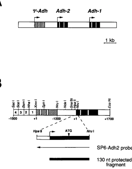
Y-Adh Adh-2 Adh- 1
r
r
r
I
1 kb c_
4 1 3 1 2 1 1
4 SP6-Adh2 probe
-
130 nt protected fragmentFIGURE 1 .-Diagram of the D. mulleri Adh locus. (A) Shown is a 9.3-kb region containing the D. mulleri Adh locus. The black regions indicate exons, the open areas within exons introns, and the white regions intergenic sequences. The striped regions indicate exons of the pseudogene. Arrows indicate transcription start sites and the direction of transcription. In D. melanogaster P element transform- ants, Adh-1 is expressed primarily in the larval stages, and Adh-2
primarily in third instar larvae and adults (FISCHER and MANIATIS
1986). In addition, there is a pseudogene ( q - A d h ) upstream of Adh- 2 which is transcribed in adults, but not translated. (B) Restriction map of the Adh-2 gene and flanking sequences. Shown is a diagram of the 5.5-kb Sac I-EcoR I fragment of the D. mulleri Adh locus, containing the Adh pseudogene and Adh-2, used to transform the
4 3 2 1 q a d h 2 lines (Figure 2). Striped boxes represent the exons of the pseudogene, black boxes are Adh-2 exons, open boxes within exons are introns, and white regions are intergenic sequences. The numbers in the white boxes at the left end indicate the four subregions into which the Adh-2 enhancer has been divided (Figure
4). The direction of transcription is from left to right. Restriction endonuclease recognition sites used to construct the various test genes are indicated and referred to elsewhere. Note that the figure is not drawn to scale. The extent ( N r d - H p a I I ) of the RNA probe (SP6-Ad2) complementary to Adh-2 is indicated by the leftward pointing arrow. The 130-nt fragment of the first exon of Adh-2
protected by the SP6-Adh-2 probe is indicated by the black bar.
within the linear range of the film. Adh message levels were normalized to a-tubulin levels in the same samples and in the same lanes.
RESULTS
Previous studies demonstrated that the 5.5-kb Sad-
EcoRI fragment containing Adh-2 with 3.7 kb of 5”flanking sequences (including the pseudogene)
a n d 0.6 kb of 3”flanking sequences (Figure 2;
Ectopic Drosophila Adh Expression 1073
4321adh2 (2)
7
+
r- adh24321Y (5) 413121 1 IY-Adh I
FIGURE 2.-Adh-2 gene constructs. The expression patterns of
4321tadh2, adhaand 4321adh2 were reported previously (FISCHER
and MANIATIS 1986). (Here, 4321qadh2 and 4321adh2 are named
t a d h 2 and 5 ‘ q a d h 2 , respectively.) The numbers of independent transformant lines bearing each construct are indicated in the parentheses. The boxes containing the numbers represent the 1.5- kb Sad-XmnI fragment containing the Adh-2 enhancer, normally located upstream of the pseudogene. The order of the numbers in the boxes indicates the orientation of the enhancer fragment, with
4 3 2 N a d h 2 representing the genomic configuration. The arrows indicate the start sites of transcription and the boxes labeled q - A d h
and Adh-2 represent the respective transcription units. The
4 3 2 1 t a d h 2 construct is a 5.5-kb Sad-EcoRI fragment of the D. mulleri Adh locus (Figure 1) containing the pseudogene and 1.5 kb of 5”flanking sequences. Adh-2 was constructed with 1.2 kb of 5‘- and 0.6 kb of 3”flanking sequences. The adh2 structure is a 2.9-kb
NruI-EcoRI fragment containing the Adh-2 gene with 1.2 kb of 5’- and 0.6 kb of 3”flanking sequences. The four remaining constructs all contain the 2.9-kb NruI-EcoRI of Adh-2 and the additional DNA sequences indicated upstream or downstream of this Adh-2 gene fragment. Message levels were quantitated with densitometry, with detectable message levels indicated by a
+,
and very low or unde- tectable levels by a -.+
levels are 16-20-fold greater than-
levels (data not shown).expression in the larval and adult fat body, and in the adult hindgut (FISCHER and MANIATIS 1986). Analysis of two additional Adh-2 gene constructs showed that sequences upstream of the pseudogene are required for normal levels of Adh-2 transcription (FISCHER and
MANIATIS 1986). First, transformants containing an
Adh-2 gene truncated at
-
1200 from the transcription start site (adh2, Figure 2) showed an approximately 20-fold lower level of Adh-2 transcripts than the4 3 2 I q a d h 2 transformants. Second, when 1.5 kb of sequences (referred to as the Adh-2 upstream regula- tory region) normally located upstream of the pseu- dogene were added 5’ to the adh2 construct
(4321adh2, Figure 2), normal levels of Adh-2 expres- sion were restored.
A tissue-specific enhancer is upstream of the pseu- dogene: We performed several experiments to deter- mine if the Adh-2 upstream regulatory region has the properties of a tissue-specific enhancer. First, we con- structed genes similar to 4321adh2 and 4 3 2 1 q a d h 2 ,
except that the region upstream of the pseudogene was placed in reverse orientation (1234adh2 and
:
1:
9 1 2 3 4 a d h 2 , Figure 2) or 3’ to the Adh-2 gene
(adh24?21*, Figure 2).
T h e levels of Adh-2 and tubulin control messages in RNA prepared from larval and adult transformants were determined by quantitative RNAse protection. As summarized in Figure 2, the expression levels of the Adh-2 genes in 1234adh2 and a d h 2 4 3 2 1 q trans- formants are similar to those in 4321adh2. Thus, the upstream regulatory region of Adh-2 can function as an enhancer in either orientation 5’ to the Adh-2
promoter, and also downstream of the Adh-2 gene.
To determine if the Adh-2 upstream regulatory region could activate transcription from a heterolo- gous promoter, we placed the 2.5-kb SacI-NruI frag- ment containing the pseudogene and the upstream regulatory region ( h s l , Figure 3) or just the 1.5-kb upstream regulatory region (hs2, Figure 3) 5’ to an
hsp70/AdhF hybrid gene (DUDLER and TRAVERS 1984). T h e hybrid gene contains the hsp70 promoter and 5”untranslated sequences (-68 to +198) joined to the
D.
melanogaster AdhF gene (at +9 from the proximal transcription start site) and thus lacks its own promoter and regulatory sequences (FISCHER and MANIATIS 1988). T h e hsp70 sequences provide a “neu- tral” promoter for our experiments, as the hsp70 gene is not normally expressed in a stage- or tissue-specific manner (MASON, HALL and GAUSZ 1984). Consistent with these observations, the hsp70/AdhF gene used in these experiments is not expressed in P element trans-r-+lg8 +g
Spacer (0.6 kb) h s p n
1
Larvae
1
Adults
1
FB MMGl FB HG
I
hsl (3) 1 4 1 3 1 2 l 1 I Y
hs2 (9) 1 4 1 3 1 2 I 1 A/;-
1
1
1
1 1
1
FIGURE 3.-hs constructs. The hsp70/AdhF hybrid gene is a 2.5- kb XhoI-XbaI fragment of the plasmid R68 (DUDLER and TRAVERS
1984), containing 0.6 kb of Xenopus 5 s “spacer” DNA, the hsp70
promoter and 5”untranslated region from -68 to +198 from the transcription start site, and AdhF gene sequences from +9 of the proximal promoter transcription start site to the XbaI site 0.7 kb downstream of the Adh-2 polyadenylation site. In h s l , the 2.6-kb
SacI-NruI of 91234adh2 (Figure 2), and in hs2, the 1.5-kb SacI-
XmnI fragment in 1234adh2 (Figure 2), are ligated via EamHI
linkers upstream of the hsp70/AdhF gene. The numbers in paren- theses indicate the number of independent transformed lines of each construct analyzed. Expression of the hsp70/AdhF gene in larval and adult tissues was assayed by histochemical staining. De-
1074 D. Falb, J. Fischer and T. Maniatis
formants without heat shock (DUDLER and TRAVERS 1984; FISCHER and MANIATIS 1988).
In hsl and hs2 transformants, ADH activity was detected by histochemical staining in the larval and adult fat body and in the adult hindgut (Figures 3, 6, and data not shown). These results, taken together with those above, show that there is a tissue-specific enhancer within the 1.5-kb SacI-Xmnl fragment up- stream of the pseudogene.
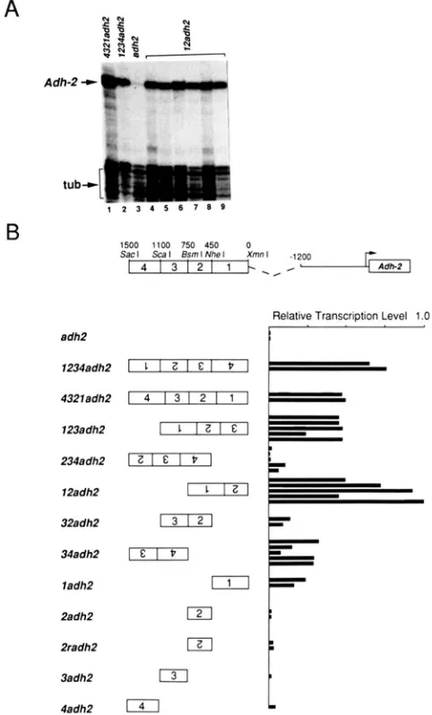
Localization of the enhancer: We attempted to
localize the Adh-2 enhancer within the 1.5-kb SacI-
XmnI fragment by placing various fragments of this region 1.2 kb upstream of the Adh-2 transcription start site (4321 constructs, Figure 4), and assaying the level of Adh-2 transcripts relative to tubulin in adult transformants by quantitative RNAse protection.
We found that the 750-bp BsmI-XmnI fragment in
12adh2 (Figure 4) is the smallest region tested that retains the ability to confer wild type levels of tran- scription on the Adh-2 promoter. However, the DNA fragments in the 34adh2, 32adh2, and 1adh2 trans- formants also display some enhancer activity. Notably, the 34adh2 construct does not contain any enhancer DNA sequences in common with the 12adh2 con- struct. Thus, while the 750-bp BsmI-XmnI fragment in 12adh2 can restore normal levels of Adh-2 expres- sion, the 750-bp SacI-BsmI fragment upstream also contains sequences sufficient to elevate the Adh-2 tran- scription level.
Deletion of the pseudogene results in high levels of transcription in a novel tissue: Preliminary exper- iments demonstrated that the 1.5 kb upstream region confers wild-type levels of expression on the
-
1200Adh-2 construct (FISCHER and MANIATIS 1986). Sur- prisingly, ADH staining activity was observed not only in the fat body but also in a novel tissue, the middle midgut of 4321adh2 transformant larvae.
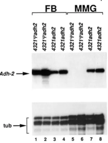
To determine if the ADH histochemical staining activity observed in the middle midgut represents a significant change in the level of transcription in this tissue, we analyzed R N A prepared from isolated fat body and middle midgut tissues of 43219udh2 and
4321adh2 transformants by quantitative RNAse pro- tection. As shown in Figure 5, while Adh-2 transcripts are detected only in the fat body of 43219adh2 larvae, similar levels of Adh-2 transcripts are detected in the fat body and middle midguts of 4321adh2 transform- ants. Thus, deletion of the pseudogene, which juxta- poses the Adh-2 upstream regulatory region with se- quences 1.2 kb upstream of the Adh-2 transcription start site results in high levels of ectopic Adh-2 tran- scription in the middle midgut.
To determine if the novel expression seen in the
4321adh2 transformants is dependent on the orien- tation of the enhancer, w e investigated the ADH histochemical staining patterns and Adh-2 transcript
Adh-2 +
B
Relative Transcription Level 1 .O
I
12adh2
32adh2
13121
ladh2
2adh2
121
2radh2 IZI
3adh2
131
4adh2
141
L
FIGURE 4.-Adh-2 mRNA transcripts in adults transformed with 4321 genes. (A) Adh-2 transcripts in adult flies of various 4321 transformants were analyzed by quantitative RNAse mapping using the SP6-AdhP and SP6-atub probes (see MATERIALS A N D METHODS).
Relevant bands are marked to the left of the autoradiograms. (R)
The 4321 genes were constructed by ligating various portions of the 1.5 kb SacI-Xmnl fragment located upstream of the pseudogene
to the -1200 adh2 construct. The boxes containing the numbers indicate the regions which the I .5-kb element is divided into, and the numbers over the boxes indicate the approximate length of each segment. The normalized levels ofAdh-2 mRNA in total adults were determined by RNAse protection analysis, as described in
MATERIALS A N D METHODS. The normalized level of Adh-2 message
in lane 6 (part A) was arbitrarily designated a 1 *’ in order to calculate
the relative transcription level (RTL) in each of the other lines. Each bar in the histogram represents the R T L calculated for an independent transformant line. The RTLs mainly reflect the levels of Adh-2 transcripts in the fat body, as it is the largest of the Adh- 2-expressing tissues.
distributions in 91234adh2 and 1234adh2 larvae. ADH activity staining (Figure 6C and data not shown) and Adh-2 transcripts (see Figure 8) were detected in both the fat body and middle midgut of 91234adh2
FIGURE 5.-Adh-2 transcripts in the larval fat body and and middle midguts o f 43219udh2 and 432Zndh2 transformants. Adh-2
transcripts in early third instar larval fat .body (FB) and middle midgut (MMG) of two independent lines each of the 43219adh2
and 4321adh2 transformants were detected by RNAse protection assays. Total nucleic acid preparations from dissected fat body and middle midguts were hybridized separately with the RNA probes SP6-Adh2 (Figure 1) and SP6-atub (MATERIALS AND METHODS) which protect 130- and 90-nt segments o f the Adh-2 and a,-tubulin genes, respectively. The tissues from which the total nucleic acid was prepared were from the same larvae (approximately 10). The tubulin probe served as an internal control to show that there are similar amounts of mRNA in all o f the preparations, as equal amounts of each RNA preparation were assayed with each of the two probes. Consistent with the histochemical staining results (FISCHER and MANIATIS 1986), Adh-2 transcripts were detected in the middle midguts of 4321adh2, but not 4321qadh2 transformed larvae.
These results demonstrate that the novel expression cannot be due to the fortuitous creation of a small sequence element that induces middle midgut expres- sion by the deletion of the pseudogene sequences, as the construction of the q 1 2 3 4 a d h 2 and 1234adh2
genes resulted in a different junction sequence than in the 4321adh2 gene (Figure
2).
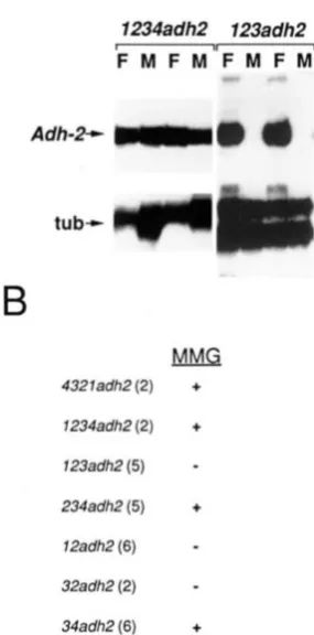
Enhancer region 4 is necessary for middle midgut expression: In the course of localizing the enhancer, we found that although 23adh2 and 12adh2 trans- formants express high levels of ADH in the larval fat body (Figures 4 and 7 and data not shown), no ADH histochemical staining was observed in the middle midgut (Figure
7).
RNAse protection analysis of iso- lated fat body and middle midguts of 1234adh2 and123adh2 transformant larvae show that expression of
Adh-2 in the middle midgut is dependent on the presence of region 4 (Figure
7).
Middle midgut expression was also observed with 234adh2 and34adh2. As summarized in Figure
7,
the presence of region 4 in conjunction with regions 3 and 2, or region3 alone results in middle midgut expression. T h e level of expression with region 4 alone was too low to assay by histochemical staining, so we do not know whether region 4 alone is sufficient for middle midgut expres- sion. Thus, it appears that various combinations of regions 1, 2 and 3 result in fat body expression, while the addition of region 4 results in both fat body and middle midgut expression.
Adh-2 expression in the larval middle midgut
depends on the position of the enhancer upstream
of the Adh-2 promoter: Next we addressed the ques- tion of why middle midgut expression of Adh-2 is not observed when the enhancer is present in its genomic context upstream of the pseudogene. We designed several experiments to distinguish between the follow- ing two alternative explanations. First, the failure of the wild type Adh-2 gene to be expressed in the larval middle midgut could be due to a blocking effect of the pseudogene sequences. Alternatively, larval mid- dle midgut expression may depend solely on the dis- tance between the enhancer and the Adh-2 promoter and upstream sequences.
To determine if the pseudogene sequences prevent the enhancer from activating transcription in the lar- val middle midgut, we constructed Adh-2 genes in which the 1.1 kb of pseudogene sequences were re- placed by either a 1 .O-kb fragment of esc gene coding region ( e n h l ; Figure 8) or a 1.0-kb fragment of the CY- tubulin coding region (enh2; Figure 8). ADH activity (data not shown) and Adh-2 transcripts (Figure 8) were detected in the larval fat body, but not in the middle midguts of enhl and enh2 transformant larvae. Thus, the failure to see larval middle midgut expression in larvae transformed with the 4 3 2 N a d h 2 (genomic) construct, in which the pseudogene sequences were present, was not because the pseudogene sequences blocked the action of the enhancer in this tissue. This conclusion is also supported by the observation that in a d h 2 4 3 2 l q transformants, which contain an Adh-2
gene with the enhancer 3’ to the gene (Figure 2), ADH activity in larvae is observed only in the fat body (data not shown).
T h e above observations suggest that the novel expression may be a consequence of placing the en- hancer at
-
1.2 kb from the Adh-2 transcription start site. To test the stringency of this position require- ment, we constructed enh3, enh4 and enh5, in which the 1.5-kb SacI-XmnI fragment containing the en- hancer was located at -1 800, -800 and -200 bp, respectively, from the Adh-2 transcription start site (Figure 8). ADH activity (data not shown) and Adh-2transcripts were detected in the larval fat body, but not in the middle midguts of the enh3, enhl and enh5
1076 D. Falb, J. Fischer and T. Maniatis
FIGURE 6.-ADH activity in larval tis- sues. Third instar larval tissues were dis- sected, fixed, and histochemically stained for ADH activity (MATERIALS AND METH-
ODS). The drawing in the upper left corner is oriented similarly to the dissections in the photographs. The labeled tissues are the gastric caecae (gc), anterior midgut (amg), middle midgut (mmg). hindgut (hg), Malpi- ghian tubules (mt) and fat body (%). Each
of these cell types has a different embryo- logic origin, with the possible exception of
the Malpighian tubules and hindgut, which may arise from the same cells (POULSEN
1950; CAMPOS-ORTEGA and HARTENSTEIN 1985). Most of the fat body, a large struc- ture which runs the entire length of the larva, was removed to reveal the other tis- sues. The AdF6cn;
fl
un6) strain pro- duces no ADH protein and does not stain (A). The transformant lines in (B) and (C) are indicated in the lower left corner ofeach photograph. The arrows in each pho- tograph point to the fat body, and the stained middle midgut (mmg) is indicated in (C).
positioned at a fairly precise location relative to the
Adh-2 promoter.
Sequences between -1.2 and -0.8 kb of the Adh-2
transcription start site are required for expression in the larval middle midgut: We next determined whether the juxtaposition of the enhancer with spe- cific sequences at
-
1.2 kb from the Adh-2 transcrip tion start site is necessary for larval middle midgut expression, or if this effect is simply due to the spacing between the enhancer and sequences near the Adh-2transcription start site. We constructed enh6 (Figure
8), in which the 1.5-kb enhancer fragment was placed at
-
1.2 kb upstream of the Adh-2 transcription start site, with the sequences between -1.2 and -0.8 kbreplaced by a 0.4-kb fragment of spacer DNA. Adh-2
transcripts were detected in the fat body, but not in the middle midgut of enh6 transformant larvae. Thus, the enhancer must be positioned at a certain distance from regulatory elements in the -1.2- and -0.8-kb region upstream of the Adh-2 transcription start site to activate transcription in the larval middle midgut.
DISCUSSION
We have shown that the Adh-2 gene of
D.
mulleri,normally expressed in the larval and adult fat body and in the adult hindgut, is regulated by a tissue- specific enhancer that can function in association with the hsp70 promoter in these tissues. We have also localized the activity of the Adh-2 enhancer to a 750- bp element which activates wild type levels of tran- scription from the Adh-2 promoter. Furthermore, this enhancer can function in either orientation upstream
of the Adh-2 gene, and also when placed 3' to the
Adh-2 coding region.
In addition to the activities mentioned above, the
Adh-2 enhancer can also activate transcription in the larval middle midgut. This activity requires three conditions to be satisfied. T h e Adh-2 enhancer must contain region 4, the enhancer must be positioned at -1 200, and the sequences between -1200 and -800 must be present. This conclusion is based on the following observations. First, replacing the pseudo- gene with DNA segments of similar lengths does not result in ectopic expression of Adh-2, indicating that sequences within the pseudogene are not responsible for repressing expression in the middle midgut. Sec- ond, when the enhancer is placed at different distances from the Adh-2 transcription start site or when se- quences between -800 and -1200 are replaced by heterologous sequences, Adh-2 expression is seen only in the fat body of third instar larvae. Finally, when region 4 of the enhancer is deleted, expression in the middle midgut is abolished, while fat body expression is unchanged. Interestingly, expression in the middle midgut is observed regardless of the orientation of the enhancer at
-
1200, even though this changes the distance between individual enhancer elements and the Adh-2 promoter. Thus, the enhancer can be con- sidered a regulatory unit, and the relative position of elements within this unit with respect to the Adh-2promoter does not appear to be important, as long as the unit is located at -1 200.
FIGURE 7.-Expression levels in the middle midgut. (A) Adh-2
transcripts in early third instar larval fat body (F) and middle midgut
(M) of several 1234ndh2 and 123adh2 transformant lines were detected by RNAse protection (MATERIALS AND METHODS). Total
nucleic acid prepared from dissected fat body and middle midguts was hybridized simultaneously with the SP6-Adh2 and SP6-atub RNA probes, which protect 130- and 90-nt RNA fragments of the
Adh-2 and a-tubulin genes, respectively. The tissues from which the total nucleic acid was prepared were from the same larvae (approx- imately IO). The autoradiograms were deliberately overexposed to show that there is no Adh-2 mRNA detectable in the middle midguts of several of the transformant lines. Relevant bands are marked to the left of the autoradiogram. (B) ADH histochemical staining in
4 3 2 1 gene transformant larvae. Third instar larval tissues were
dissected, fixed, and histochemically stained for ADH activity. Detectable staining is indicated by a
+
and undetectable staining by a-.
The numbers in parentheses following the construct names indicate the number of independent transformant lines analyzed. Note that this is a qualitative measurement and does not indicate the quantitative differences in Adh expression levels shown in Figure 4.enhancer (regions 1, 2 and 3) and an additional ele- ment (region 4). Other examples of this phenomenon are the expression of the D. mulleri Adh-I gene
(FISCHER and MANIATIS 1988) and the D. melanogaster
Adh gene (CORBIN and MANIATIS 1989; FALB 1991). Second, the tissue specific activity of an enhancer can be profoundly affected by its proximity to other reg- ulatory elements, and its distance from the promoter. Novel cell-type specificities have previously been reported in cases where a tissue-specific enhancer is linked to a heterologous promoter (SWANSON et al. 1985; SHERMOEN et al. 1987). In contrast, our studies
demonstrate that expression in novel tissues can be generated by rearranging the existing regulatory ele- ments of a single gene.
One interpretation of these results is that transcrip- tional specificity is determined by a combinatorial mechanism (SWANSON et al. 1985). Support for this idea comes from experiments on the dopa decarbox- ylase (Ddc) gene. Ddc expression in neurons requires the combination of one cis-acting element used also in glial cells and another element used only in neurons
(BEALL and HIRSH 1987). Similarly, the combination of the Adh-l larval fat body specific enhancer (box B)
and a particular promoter element (box A) activates transcription in three larval tissues in addition to the fat body (FISCHER and MANIATIS 1988).
There is much species variation in the tissue speci- ficity of expression of Adh genes, even among closely related species (DICKINSON 1980a; BATTERHAM et al. 1983; RABINOW and DICKINSON 1986; FISCHER and
MANIATIS 1986). All Drosophila species examined so
far express Adh in the fat body, but expression in a range of other cell types varies, and some species express Adh only in the fat body (DICKINSON 1980a). Evidence from genetic crosses (DICKINSON and CAR- SON 1979; DICKINSON 1980b) and P element transfor- mation experiments (FISCHER and MANIATIS 1986;
BRENNAN and DICKINSON 1988) suggests that the var- iations in tissue specificity are due to differences in the Adh cis-acting regulatory sequences, rather than to the presence of different trans-acting Adh transcrip- tion factors in each species. In fact, based on the results presented here, these variations could be due, at least in part, to rearrangement of existing regula- tory elements during evolution, rather than to alter- ations in the binding sites of transcription factors.
These observations, taken together with the finding that Adh-I is regulated by larval fat body specific enhancers (FISCHER and MANIATIS 1986), suggested that the enhancers which regulate Adh are under evolutionary pressure to accommodate the levels and type of transcription factors present in the fat body
(FISCHER and MANIATIS 1986). Thus, it was hypothe- sized that Adh genes in general are regulated by fat body specific enhancers (FISCHER and MANIATIS
1986). Indeed, the proximal and distal promoters of the D. melanogaster Adh gene are regulated by fat body specific enhancers (CORBIN and MANIATIS 1989;
FISCHER and MANIATIS 1988; FALB 199 1). T h e addi- tional tissue specificity (adult hindgut) of the Adh-2
1078 D. Falb, J. Fischer and T. Maniatis
FIGURE 8.-enh constructs. (A) The enhl-enh6 constructs all contain the 2.9- kb Nrul-EcoRI fragment in Adh-2, and the additional DNA sequences indicated u p stream or downstream of this gene frag- ment. In enhl and enh2 the 1.5-kb en- hancer fragment is separated by, respec- tively, a 1.0-kb BamHl fragment of the coding of the Drosophila esc gene (pro- vided by GARY STRUHL), or a 1 .O-kb ScaI-
BamHl fragment of pDmtctl (KALFAYAN
and WENSINK 1982). containing coding sequences of the Drosophila al-tubulin gene. In the enh3-enh5 constructs, the Adh-2 enhancer fragment is upstream of a 3.5-kb Sphl-EcoRl fragment, a 2.5-kb Ndel-EcoRI fragment, or a 1.9-kb SnaBI- EcoRl fragment, respectively, containing Adh-2 and different extents of 5”flanking sequences as indicated. In enh6, the Adh-2 enhancer fragment is separated from the Adh-P gene fragment in enh4 by a 0.4-kb fragment of the cad gene coding sequence (provided by PAUL MACDONALD). (B) Adh- 2 transcripts in early third instar larval fat body (F) and middle midgut (M) of the indicated transformant lines were de- tected by RNAse protection as described in Figure 7.
broadened in conjunction with Adh promoter ele- ments.
We thank T. ABEL and A. MICHELSON for helpful discussions, advice, and critical reading of the manuscript. This work was supported by a grant from the National Institutes of Health to T.M.
LITERATURE CITED
BAITERHAM, P., J. A. LOVETT, W. STARMER and D. T. SULLIVAN, 1983 Differential regulation of duplicate alcohol dehydro- genase genes in Drosophila mojauensis. Dev. Biol. 9 6 346-354. BEALL, C. J., and J. HIRSH, 1987 Regulation of the Drosophila
dopa decarboxylase gene in neuronal and glial cells. Genes Dev. 1: 5 10-520.
BENYAJATI, C., A. R. PLACE, N. WANG, E. PENTZ and W. SOFER, 1982 Deletions at intervening splice sites in the alcohol de- hydrogenase gene of Drosophila. Nucleic Acids Res. 10: 7261- 7272.
BRENNAN, M. D., and W. J. DICKINSON, 1988 Complex develop- mental regulation of the Drosophila aflinidisjuncta alcohol de- hydrogenase gene in Drosophila melanogaster. Dev. Biol. 125:
CAMPOS-ORTEGA. J. A., and V. HARTENSTEIN, 1985 The Embryonic Development of Drosophila melanogaster. Springer-Verlag, Ber- lin.
CORBIN, V., and T. MANIATIS, 1989 The role of specific enhancer- promoter interactions in the Drosophila Adh promoter switch. Genes Dev. 3: 2 191 -2200.
64-74.
DICKINSON, W. J., 1980a Evolution ofpatterns of gene expression in Hawaiian picture-winged Drosophila. J. Mol. Evol. 1 6 73- 94.
DICKINSON, W. J.. 1980b Complex cis-acting regulatory genes demonstrated in Drosophila hybrids. Dev. Genet. 1: 229-240. DICKINSON, W. J., and H. L. CARSON, 1979 Regulation of the
tissue specificity of an enzyme by a cis-acting genetic element: evidence from interspecific Drosophila hybrids. Proc. Natl. Acad. Sci. USA 85: 4559-4562.
DUDLER, R., and A. A. TRAVERS, 1984 Upstream elements nec- essary for optimal function of the hsp70 promoter in trans- formed flies. Cell 3 8 391-398.
FALB, D., 199 1 Mechanisms of tissue-specific Adh gene expression in Drosophila. Ph.D. Thesis, Harvard University.
FISCHER, J. A., and T. MANIATIS, 1985 Structure and transcrip tion of the Drosophila mulleri alcohol dehydrogenase genes. Nucleic Acids Res. 13: 6899-6917.
FISCHER, J. A., and T. MANIATIS, 1986 Regulatory elements in- volved in Drosophila Adh gene expression are conserved in divergent species and separate elements mediate expression in different tissues. EMBO J. 5: 1275-1289.
FISCHER, J. A., and T. MANIATIS, 1988 Drosophila Adh: a pro- moter element expands the tissue specificity of an enhancer. Cell 53: 451-461.
KALFAYAN, L., and P. C. WENSINK, 1982 Developmental regula- tion of Drosophila a-tubulin genes. Cell 2 9 91-98.
MANIATIS, T., E. F. FRITXH and J. SAMBROOK, 1982 Molecular Cloning: A Laboratoy Manual. Cold Spring Harbor Laboratory,
Cold Spring Harbor, N.Y.
MASON, P. J., L. M. C. HALL and J. GAUSZ, 1984 The expression of heat-shock genes during normal development in Drosophila melanogaster. Mol. Gen. Genet. 1 9 4 73-78.
MELTON, D. A., P. A. KRIEG, M. R. REBACLIATI, T. MANIATIS, K. ZINN and M. R. GREEN, 1984 Efficient in vivo synthesis of biologically active RNA and RNA hybridization probes from plasmids containing a bacteriophage SP6 promoter. Nucleic Acids Res. 12: 7035-7056.
POULSEN, D. F., 1950 pp. 168-274 in Biology of Drosophila, edited by M. DEMEREC. Hafner Publishing, New York.
RABINOW, L., and W. J. DICKINSON, 1986 Complex cis-acting regulators and locus structure of Drosophila tissue-specific Adh variants. Genetics 112: 523-537.
RUBIN, G. M., and A. C. SPRADLING, 1982 Genetic transformation of Drosophila with transposable element vectors. Science 218:
348-353.
RUBIN, G . M., and A. C. SPRADLING, 1983 Vectors for P-element- mediated gene transfer in Drosophila. Nucleic Acids Res. 11:
SHERMOEN, A. W., J. JONGENS, S. W. BARNETT, K. FLYNN and S .
K. BECKENDORF, 1987 Developmental regulation by an en- hancer from the sgs-4 gene of Drosophila. EMBO J. 6: 207- 214.
SPRADLING, A. C., and G . M. RUBIN, 1982 Transposition of cloned P elements into Drosophila germ line chromosomes. Science
SWANSON, L. W., D. M. SIMMONS, J. ARRIZA, R. E. HAMMER, R. BRINSTER, M. G . ROSENFELD and R. M. EVANS, 1985 Novel developmental specificity in the nervous system of transgenic animals expressing growth hormone fusion genes. Nature 317:
363-366.
URSPRUNG, H., W. H. SOFER and N. BURROUGHS, 1970 Ontogeny and tissue distribution of alcohol dehydrogenase in Drosophila melanogaster. Wilhelm Roux’s Arch. 164: 201-208.
ZINN, K., D. DIMAIO and T. MANIATIS, 1983 Identification of two distinct regulatory regions adjacent to the human &inter- feron gene. Cell 34: 865-879.
6341-6351.
218: 341-347.