Fine Structure Recombinational Analysis
of
Cloned Genes
Using
Yeast Transformation
Sam Kunes,
Hong
Ma, Karen Overbye,’ Maurice
S.
Fox and David Botstein
Department of Biology, Massachusetts Institute of Technology, Cambridge, Massachusetts 021 39 Manuscript received July 1, 1986
Revised copy accepted September 15, 1986
ABSTRACT
We describe a general method for analyzing the genetic fine structure of plasmid-borne genes in yeast. Previously we had reported that a linearized plasmid is efficiently rescued by recombination with a homologous restriction fragment when these are co-introduced by DNA-mediated transfor- mation of yeast. Here, we show that a mutation can be localized to a small DNA interval when members of a deletion series of wild-type restriction fragments are used in the rescue of a linearized mutant plasmid. The resolution of this method is to at least 30 base pairs and is limited by the loss of a wild-type marker with proximity to a free DNA end. As a means for establishing the nonidentity of two mutations, we determined the resolution of two-point crosses with a mutant linearized plasmid and a mutant homologous restriction fragment. Recombination between mutations separated by as little as 100 base pairs was detected. Moreover, the results indicate that exchange within a marked interval results primarily from one of two single crossovers that repair the linearized plasmid. These approaches to mapping the genetic fine structure of plasmids should join existing methods in a robust approach to the mutational analysis of gene structure in yeast.
ECOMBINANT DNA technology currently ap-
R
plied to the yeast, Saccharomyces cerevisiae, in- cludes convenient methods that facilitate a mutational analysis of gene structure and function (reviewed inBOTSTEIN and DAVIS 1982). To isolate new mutations,
a cloned gene residing on a plasmid can be mutagen- ized and introduced via transformation into an appro- priate yeast host to screen for altered gene function. T h e plasmid of a transformant displaying a mutant phenotype can be returned to a n Escherichia coli host, permitting isolation of the mutant gene for direct analysis and further manipulation.
This basic methodology lacks a simple, sensitive method for mapping to a high resolution a mutation o n a plasmid in yeast. Even with the use of in vitro mutagenesis, new mutations need to be localized un- ambiguously to the gene of interest, t o be distin- guished from silent or modifying lesions, and to be localized to DNA segments small enough to be con- venient for sequence analysis. Furthermore, localizing genetic regions of substantial importance to gene
function is of immediate interest in a mutational anal- ysis.
To devise a n approach to the high resolution map- ping of plasmids, we utilized the known property of free DNA ends as efficient substrates for homologous recombination in yeast (ORR-WEAVER, SZOSTAK and
ROTHSTEIN 198 1 ; reviewed in ORR-WEAVER a n d
SZOSTAK 1985). We have reported (KUNES, BOTSTEIN
Present address: Biotechnica International Inc., 85 Bolton Street, Cam- bridge, Massachusetts 02 140.
Genetics 115: 73-81 (January, 1987)
and Fox 1984, 1985) that a plasmid DNA broken in
a sequence absent from the yeast genome is efficiently rescued by recombination with a homologous restric- tion fragment included during yeast transformation. Here, we describe some features of recombination with these substrates a n d utilize this reaction in two approaches to plasmid fine structure analysis, two- point heteroallelic crosses and deletion mapping.
MATERIALS AND METHODS
Strains and media: Yeast strains DBY1226 ( M A T a h i d - 5 1 9 met8-1 leu2-3,112 ura3-3) and DBY1227 ( M A T a h i d - 5 1 9 met8-1 leu2-3,112 u r a 3 - 1 8 ) were constructed in this laboratory by standard methods (SHERMAN, FINK and LAW- RENCE 1979). Yeast strain DBY2052 ( M A T a 1ys2-801 1eu.2- 3, I I 2 ura3-52 hxk1::LEUZ hxk2-202) is a derivative of DBY 13 15 constructed by disruption of H X K l with the cloned LEU2 gene (RATZKIN and CARBON 1977; H. MA and D. BOTSTEIN, unpublished data) and deletion of the H X K P
locus. The hxk2 deletion allele, hxk2-202, is a derivative of the cloned H X K 2 gene (see below) constructed i n vitro by removing the H X K Z coding region from between the flank- ing sites Sac1 and XbaI (see pRB313 in Figure 5 for the positions of these sites). The genomic H X K Z locus was replaced with this deletion allele by recombination (see BOTSTEIN and DAVIS 1982 for details), yielding strain DBY2052. As a result of the presence of the hxkl and hxk2 mutations, this strain cannot grow on fructose as a sole carbon source, but can grow on glucose due to the presence of glucokinase.
(MILLER 1972) containing, when appropriate, 100 pg am- picillin/ml (Sigma).
Plasmids: Plasmids pRB28, pRB29 and pRB30 (Figure 1) were constructed by FALCO et al. (1 982). Plasmid pSK265 (Figure 3) is a derivative of pRB30 constructed by removing the BamHI, BglII fragment of 5' URA3 flanking sequence, and incorporating a Sad-linker (Collaborative Research, Waltham, Massachusetts) in the recircularized product. Plas- mid pRB45 and its derivative plasmids containing URA3'lacZ gene fusions (pRB70, pRB72, pRB73, pRB74, pRB75, pRB76, pRB79, pRB80, pRB81 and pRB82) were
constructed by ROSE and BOTSTEIN (1 983). Plasmid pRB3 13 (Figure 2 ) is a derivative of the yeast vector YCp50 (C. MANN and R. W. DAVIS, personal communication) contain- ing a 3.8-kb yeast EcoRI fragment bearing the HXKB gene, isolated by STACHELEK et al. (1 986). This plasmid comple- ments the chromosomal deletion allele of HXK2 described above, restoring the ability of strain DBY2052 to utilize fructose as a sole carbon source. Mutant derivatives of pRB3 13 lackingHXK2 complementing activity were isolated after transformation of strain DBY2052 with plasmid DN,4 mutagenized by amplification in an E. coli mutD host (ERLICH and Cox 1980). T h e resulting mutations were mapped to small DNA intervals within the cloned HXK2 gene (see Figure 2 ) by the deletion method described in the text. A derivative of pRB3 13 with a known hxk2 mutation, pRB430 (hxk2-430), was constructed in vitro by deleting 4 base pairs (bp) at the HXK2 KpnI site (nucleotide 266-269, see Figure
2 ) . T h e 3.8 kb HXK2 EcoRI fragment was also subcloned in the vector YIp5 (BOTSTEIN et al. 1979) to construct pRB309. Deletion series of HXK2 were generated by Ba131 partial digestion of pRB309 linearized at either the Sac1 site or XbaI site flanking HXK2, with incorporation of a BglII-linker (Collaborative Research) in the recirculation of the linear molecules. T h e resulting deletions were shown to extend into the HXKB coding sequence to various points as deter- mined by restriction analysis, ending at the site of the BglII- linker inserted during the construction.
Recombinant DNA methods were performed essentially as described by MANIATIS, FRITSCH and SAMBROOK ( 1 982). DNA preparation and restriction enzyme cleavage: Plas- mid DNA was isolated from E. coli by a modification (RAM- BACH and HOGNESS 1977) of the method of CLEWELL and HELINSKI (1969). Plasmid DNA was further purified by banding once in a CsCl/ethidium bromide density gradient (RADLOFF, BAUER and VINOGRAD, 1967). Supercoiled plas- mid DNA was stored in T E buffer (10 mM Tris, pH 8.0, 1
mM Na2EDTA) at 4" o r -20". Yeast DNA was isolated by the method of HOLM et al. (1986) from cultures grown in liquid SD medium with selection for plasmid maintenance. Chicken erythrocyte DNA was obtained from Calbiochem- Behring (La Jolla, California). After dissolution in TE, it
was sonicated to an average molecular weight of 5 X IO',
phenol/chloroform ( l : l , v/v, equilibrated to pH 8.0) ex- tracted three times, precipitated in ethanol three times and stored in T E buffer at 4'.
Restriction enzymes were used according to the recom- mendations of the manufacturer (New England Biolabs, Beverly, Massachusetts).
Yeast transformation: Yeast transformation by the spher- oplasting method was performed essentially as described by HINNEN, HICKS and FINK (1978) with the exception that STC buffer (1 M sorbitol, 10 mM Tris, pH 7.5, 10 mM CaClp) was substituted for 1 M sorbitol in the third wash after
treatment with glusulase (Dupont Pharmaceuticals, Wil- mington, Delaware). T h e final suspension contained spher- oplasts at a concentration of 1 X 10⁹/ml, of which typically
10% were colony-forming units in regeneration agar with
coniplete supplements. In an individual transformation, 100 pl of spheroplast suspension was added to 10 pl of T E buffer containing plasmid DNA and 10 pg sonicated chicken eryth- rocyte (carrier) DNA, prepared as described above. Plating in regeneration agar on SD selective medium was followed by incubation at 30" for 4-5 days. Selection for Leu+ transformants was made in the presence of a growth limiting quantity (1 pg/ml) of leucine. Under these conditions, with a plasmid containing both the URA3 and LEU2 genes (pRB28), a similar yield was obtained with selection for either Ura+ o r Leu' transformants.
Yeast transformation by the alkali cation method of ITO
et al. (1 983) was performed as modified by Kuo and CAMP- BELL (1983) using 0.1 M lithium acetate. Each transforma- tion included 50 pg carrier DNA.
RESULTS
Two-point heteroallelic crosses in a plasmid-
borne URA3 gene: Previously we reported (KUNES,
BOTSTEIN and Fox 1985) that a high frequency of transformants results after co-transformation of yeast with a plasmid linearized in a region lacking homology with the yeast genome and a homologous restriction fragment that spans the plasmid's two-chain break. More than 95% of these transformants were shown to have restored the linearized plasmid to its parental circular form via recombination with the homologous restriction fragment.
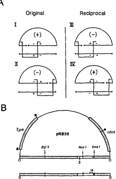
We investigated the possible resolution of a two- point cross where a mutant restriction fragment is used in the rescue of a mutant linearized plasmid. In principle, the frequency of wild-type recombinants that result in such a cross could depend on the order of the markers with respect to the free DNA ends of the substrates. As shown in Figure 1, there must be at least two crossovers, one on each side of the break of the linearized plasmid, in order to form a viable circular product. If these were indeed single crossover events, a wild-type recombinant plasmid could be expected only with the mutant alleles proximal to the free DNA ends that flank the marked interval (see Figure 1). Thus, in the original cross depicted in Figure 1, wild-type recombinant plasmids would result with the cleavage site of the linearized plasmid on the left side of the marked interval, and double mutant plasmid recombinants would result with the cleavage on the right side. In the reciprocal cross, however, cleavage of the linearized plasmid on the right side of the marked interval, but not on the left, would result in the formation of wild-type recombinants. With the plasmid cleaved within the marked interval (not shown), wild-type recombinants would be observed in both the original and reciprocal crosses.
These expectations were tested in a study of recom- bination between two homologous plasmids distin- guished by the mutant allele of their resident yeast
ura3 gene. Plasmids pRB29 and pRB3O (Figure 1) are autonomous yeast plasmids containing a cloned yeast
Yeast Plasmid Fine Structure 75
A
Original Reciprocal
I I , I
- I + 1
+ ( - I
I '
R
U
FIGURE 1 .-Formation of a URA3 recombinant plasmid by two single crossovers between a linearized plasmid and homologous restriction fragment. A, As indicated by the brackets, at least two crossovers, one on each side of the break in the linearized plasmid, are required to yield a circular product. In the original cross, with the plasmid linearized on the left of the marked interval (I), wild- type recombinants are formed by a single crossover that falls within the marked interval. With the plasmid linearized on the right of the marked interval (11), a double mutant circular product is formed by a single crossover within the marked interval; no wild-type recombinants are expected. In the reciprocal cross, a wild-type circular product is formed with the plasmid linearized on the right side of the marked interval (IV), but not with cleavage on the left side (111). The crosses which yield wild-type recombinants are those with the two mutant alleles proximal to the free DNA ends that flank the marked interval. B, Plasmids pRB28 (U+3), pRB29
(ura3-18), and pRB3O ( u r d - 3 ) were constructed by FALCO, ROSE and BOTSTEIN (1983). These plasmids are derivatives of pBR322 (BOLIVAR et al. 1977) containing the cloned yeast LEU2 gene (RATZKIN and CARBON 1977) and a yeast 2 pm circle segment that renders them autonomous in yeast (BEGGS 1978; BROACH and HICKS
1980). The mutation u r d 3 (am) has been located at nucleotide 245 (ROSE, GRISAFI and BOTSTEIN, 1983) in the URA3 coding sequence (open box between arrowheads). The mutation ura3-18
(am) has been mapped (FALCO, ROSE and BOTSTEIN 1983; LOSSON and LACROUTE 1979) to the region indicated by a bracket between nucleotides 880 and 1030. The plasmids have a unique BglII cleavage site about 2.0 kb to the left of the u r d - 3 , u r d - 1 8 interval, a unique NcoI site within the interval (nucleotide 432 of the (IRA3
sequence), and a unlque SmaI slte to the right of the interval (nucleotide 1106 of the U R A 3 sequence). Shown below the uru3-3 plasmid is the 5.5-kb ura3-18 BamHI fragment that spans the two- chain break of the linearized plasmids. Restrictlon sites are denoted as follows: 0. BamHI; B, EcoRI.
3 (am) gene insert or a ura3-18 (am) gene insert, respectively. Each plasmid has a unique site for cleav- age by BglIl, SmaI, and NcoI located, respectively, on
the left, on the right, or inside, of the ura3-3, ura3- 18 marked interval (Figure 1). T h e 5.5-kb BamHI fragment generated by BamHI, EcoRI cleavage of these plasmids spans the two-chain break of the line- arized plasmids.
T h e original cross was performed by transforming the ura-3-3 leu2 strain DBY 1226 with the ura3-3
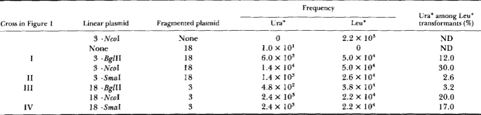
plasmid in linearized form and the ura3-18 plasmid in fragmented (BamHI, EcoRI-cleaved) form. Separate addition of the ura3-3 plasmid linearized within the marked interval, or the fragmented ura3-18 plasmid resulted in few, or no, Ura+ transformants (Table 1). Co-transformation with these two DNAs resulted in a high frequency of transformants on either Ura+ or Leu+ selection. Of the Leu+ transformants, 30% dis- played the Ura+ phenotype. All of 1 8 Ura+ transform- ants tested were found to be Leu+; both phenotypes were unstable, and co-segregated. These 18 trans- formants were further shown to harbor a plasmid of the parental structure (data not shown). On co-trans- formation with the u r d - 3 plasmid linearized on the left of the marked interval and the fragmented ura3- 18 plasmid, 12% of the resulting Leu+ transformants were found to be Ura+. However, with the ura3-3
plasmid linearized on the right side of the marked interval, a significantly smaller fraction, 2.6%, were found to be Ura+. Were URA3 recombinants to arise solely via single exchange events, no URA3 recombi- nants would be expected in this latter cross. T h e results are thus consistent with the formation of URA3
recombinants primarily, but not exclusively, by a sin- gle crossover required for the repair of the linearized plasmid.
T h e reciprocal cross was performed by transform- ing the ura3-18 leu2 strain DBY1227 with the ura3- 18 plasmid in linearized form and the ura3-3 plasmid in fragmented form. On addition of the fragmented
ura3-3 plasmid with the ura3-I8 plasmid linearized within the marked interval, the frequency of URA3
recombinants differed only modestly from that in the original cross (Table 1). However, relative to the outcome in the original cross, the URA3 recombinant frequency was decreased about fourfold with the
ura3-18 plasmid linearized on the left of the marked interval and increased more than sixfold with the
ura3-18 plasmid linearized on the right of the marked interval. This outcome, in agreement with the expec- tations outlined above, clearly demonstrates that the resolution of a two-point cross depends on the order of the markers with respect to the free DNA ends of the substrates.
Two-marker recombination in a plasmid-borne
TABLE 1
Two-marker recombination in a plasmid-borne URA3 gene: frequency of URA3 recombinants
Frequency
Ura+ among Leu+ Cross in Figure 1 Linear plasmid Fragmented plasmid Ura+ Leu+ transformants (%)
3 .NcoI
None
I 3 .BgllI
3 - N C O 1
11 3 .SmaI
111 18 .BglII
18 .NcoI
IV 18 .SmaI
None 18 18 18 18 3 3 3
0 1.0 x 10’ 6.0 x io3 1.4 x io4
1.4 x 10³
2.4 x 10³ 4.8 X 10’ 2.4 X 10’
2.2 x io3
5.0 x i o 4
5.0 x io4 2.6 x io4 3.8 x io4
2.2 x i o 4 2.2 x io4
0
ND ND 12.0 30.0 2.6 3.2 20.0 17.0
~~~
The plasmid crosses illustrated in Figure 1 (I, 11, 111, or IV) are indicated in the table. Plasmids pRB3O (ura3-3) and pRB29 (uru3-18), described in Figure 1, are denoted as “3,” and 18,” respectively. The ura3-3 leu2-3,112 strain DBY 1226 was the host for crosses involving linearized pRB30 (ura3-3), and the uru3-18 leu2-3,112 strain DBY 1227 was the host for crosses involving linearized pRB29 (ura3-18). Hence, the formation of URA3 recombinants via plasmid integration was prevented. Transformation was carried out by the spheroplasting method with DNA mixtures containing 10 pg sonicated carrier DNA, and when indicated, 100 ng of a linearized (BglII-, NcoI- or SmaI-digested) plasmid DNA and 4 pg of a fragmented (BamHI-, EcoRI-digested) plasmid DNA. The frequency of Ura’ transformants and Leu’ transformants was determined by direct selection. For each cross, approximately 500 Leu+ transformants were scored for the Ura+ phenotype.
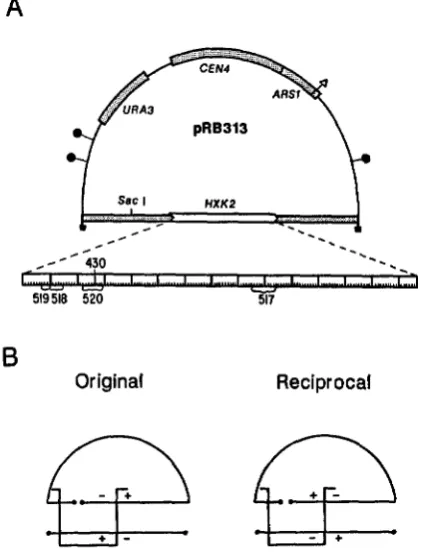
mosomal URA? locus contributed to the outcome of those crosses, a similar analysis was carried out with the cloned gene for yeast hexokinase 11, HXK2, in a strain where the chromosomal locus had been deleted. Two-marker crosses were performed by co-trans- forming the hxk2 deletion strain DBY2052 with one mutant plasmid linearized on the left of the marked interval and a heteroallelic mutant plasmid in frag- mented form (see Figure
2).
In each case where the cross was performed with both dispositions of mark- ers, a four- to 20-fold higher frequency of HXK2recombinants occurred with the mutant alleles proxi- mal to the free DNA ends that flank the marked interval (Table 2), consistent with the result of the preceding analysis of two-marker recombination in the URA? gene. Furthermore, with this disposition of markers, the frequency of HXK2 recombinants in- creased with the size of the marked interval, from 0.9 to 20.4%, a feature that is clearly expected. T h e yield of HXK2 recombinants in the reciprocal crosses, with one exception, showed no dependence on the size of the marked interval, yielding approximately 1 % HXK2 recombinants in each case. T h e one exception was in the case of the smallest interval, the cross of
hxk2-518 with hxk2-519, which resulted in 0.2% HXK2
recombinants. T h e result suggests that, if multiple exchange accounts for the low frequency of HXK2
recombinants detected in the reciprocal crosses, mul- tiple events are insensitive to the size of the marked interval, except at very short distances.
Marker loss due to the proximity of a two-chain
break: We describe below a second approach to plas-
mid fine structure analysis that is based on an assay for marker rescue. If, as indicated above, single cross- overs that repair a linearized plasmid often occur at sites distant from the molecule’s original free DNA ends, a genetic marker near a DNA end would often
be lost. This would diminish the marker rescue fre- quency. T h e expectation was tested by examining the effect of the cleavage position of the linearized plas- mid on the rescue of a marker residing on a homolo- gous restriction fragment. We would expect that with the cleavage site of the linearized plasmid close to the resident marker, an increased proportion of the trans- formants would display the restriction fragment’s al- lele. T h e um?-18 plasmid (Figure l ) was linearized with either BgZII, NcoI, or SmuI, as described above, and introduced along with the fragmented URA3 plas- mid into the uru3-18 leu2 strain DBY1227. As shown in Table 3, the fraction of Leu+ transformants dis- playing the Ura+ phenotype was highest (96%) with the uru?-I8 plasmid cleaved at the site nearest to the mutation, and lowest (58%) when linearized at the site furthest from uru3-18. In the reciprocal experiment, with the URA3 plasmid in linearized form and the
uru3-18 plasmid fragmented, the inverse result is ob- tained, with the greatest yield of Ura+ transformants occurring with the URA3 plasmid linearized at the site furthest from the wild-type marker. As a control, a co-transformation was performed with a linearized plasmid and homologous restriction fragment that were both wild-type URA3. T h e observation that 99%
of the Leu+ transformants displayed the Ura+ pheno- type indicates that the chromosomal marker, uru?-18,
is rarely contributed to the repaired plasmid. These observations are extended in the experiments which follow.
Deletion mapping of cloned genes by a marker
rescue assay: One approach to mapping a mutation
Yeast Plasmid Fine Structure 77
TABLE 2
Two-marker recombination in a plasmid-borne HXK2 gene: frequency of HXK2 recombinants
A
ORA3
pRB313
Original Reciprocal
I I
+ J - - 1 +
FIGURE 2.-Two-point heteroallelic crosses in a plasmid-borne HXK2 gene. A, Plasmid pRB3 13 is a derivative of the yeast CEN4
A R S l vector YCp50 containing the HXK2 gene on a 3.8-kb yeast EcoRI DNA fragment isolated by STACHELEK et al. (1986). The expanded scale of the HXKZ coding region (1470 base pairs) shows the approximate map positions of mutations in derivatives of pRB313 lacking HXK2 complementing activity in yeast. The mu- tants were localized by the deletion mapping method described in the text (see Figure 5) to the regions indicated by brackets, with the exception of hxk2-430, which is a 4-bp deletion (nucleotides 266-269) introduced at the HXKZ KpnI site. Restriction sites are denoted as follows: M, EcoR1; 0, BglI; A, BglII. B, Two-marker crosses with the hxk2 derivatives of pRB3 13 were performed with a mutant plasmid linearized to the left of the marked interval by
SacI cleavage and a heteroallelic hxk2 plasmid fragmented by diges- tion with EcoRI, BglI, and BglII. The hxk2 EcoRl fragment gener- ated by the latter cleavage spans the break of the Sad-linearized plasmid. The original cross, with the mutant alleles proximal to the free DNA ends that flank the marked interval, and the reciprocal cross, are shown.
s
approach would be to select for a second functional gene on the linearized mutant plasmid, and screen the resulting transformants for rescue of the wild-type
allele. A mutation could be unambiguously localized between the deletion endpoints of any two restriction fragments provided that wild-type information lo- cated very close to the molecular end of a restriction fragment is included in the plasmid product often enough to be detected.
This approach was tested by mapping a mutation,
ura3-3, that has been localized by DNA sequence
analysis (ROSE, GRISAFI and BOTSTEIN 1983). A nested deletion set of the wild-type
URA3
gene was con- structed by ROSE and BOTSTEIN (1 983) in the form of a group of plasmids bearing URA3‘lacZ gene fusions. These plasmids differ in the amount of 3’ URA3Hxk+ among
plasmid plasmid transformants
Linearized Fragmented Ura+
Cross in Figure 2 marker marker (%)
Original 519 518 0.9
Reciprocal 518 519 0.2
Original 518 520 4.2
Original 518 430 5.6
Reciprocal 430 518 1
.o
Original 430 517 12.2
Reciprocal 517 430 1.1
Reciprocal 517 518 1
.o
Original 518 517 20.4
Two-marker crosses were carried out by transforming the ura3 hxk2-202 strain DBY2052 by the alkali cation method with DNA mixtures containing 50 fig of sonicated carrier DNA, 100 ng of a Sad-linearized hxk2 mutant plasmid and 4 fig of a second mutant plasmid fragmented by cleavage with EcoRI, BglI, and BglII. T h e recombination substrates generated by these cleavages and the disposition of markers in each cross, original or reciprocal, are illustrated in Figure 2. For each cross, Ura+ transformants occurred at a high frequency on SD medium, of which approximately 1000 were screened for the Hxk’ phenotype.
coding sequence removed at the site of joining to
’ l a d , and can be cleaved at this junction to generate a fragment that contains a fraction of the
URA3
gene, and that extends to a constant endpoint (Figure 3).T h e
URA3
deletion restriction fragments are homol- ogous to the autonomous 247-03-3, LEU2 plasmid pSK265 (Figure 3), and span the two-chain break resulting from cleavage of pSK265 at its unique SacIsite.
Transformation of the ura3-3 leu2 strain, DBY 1226, by separate addition of Sad-linearized pSK265 or the
URA3
deletion fragments produced few, if any, Ura+ transformants (not shown). T h e combination of linearized pSK265 with any of theURA3 deletion fragments resulted in a high frequency
of Leu+ transformants. With the presence of those
TABLE 3
Effect of linearized plasmid cleavage site position on rescue of a restriction fragment markef
Fragmented plasmid Ura+ among Leu’
Linear plasmid alleleb allele transformants
+
-NcoI+
18 .NcoI
+
18 . S ~ U I
+
+
.BglI 18+
.NcoI 18+
.SmaI 1818 -BglII
+
99 58 80 96 76 37 21
a See the legend to Table 1 for experimental details.
3
a.
b.
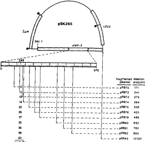
FIGURE 3.-A general method of deletion mapping for plasmid- borne genes. Plasmid pSK265, bearing a u r d - 3 gene insert (open box between arrowheads), is linearized at a Sac1 site spanned by the homologous deletion restriction fragments, a and b. These frag- ments are members of a deletion set derived from the wild-type
URA? gene constructed by ROSE and BOTSTEIN (1983) in the form of a group of plasmids bearing URA3’lacZ gene fusions. T h e ura3- 3 mutation is mapped by co-transforming yeast with the linearized mutant plasmid and each of the deletion restriction fragments, with selection for Leu+ transformants. Transformants bearing the Ura+ phenotype would result with fragment b , but not with fragment a ,
thus localizing the mutation between the endpoints of the two deletions. Restriction sites are denoted as in Figure 1.
URA3 deletion fragments expected to include the wild-type allele, more than 12% of the Leu+ trans- formants displayed the Ura+ phenotype (Figure 4). T h e fraction of transformants that were Ura+ in- creased with the distance of the wild-type allele from the end of the URA? deletion fragments, from 12 to
63%, a feature consistent with marker loss, as de-
scribed above. With the combination of linearized pSK265 and those URA? deletion fragments not ex- pected to include the wild-type allele, fewer than 0.1 %
of the resulting Leu+ transformants were found to be Ura+. Thus, the ura? mutation is placed by the end- points of pRB72 and pRB73 URA? sequence in an interval of 34 bp, consistent with the previously de- termined location of the ura3-? mutation.
Bidirectional deletion mapping of uncharacter-
ized hxh2 mutations: T h e deletion method was fur-
ther demonstrated by mapping uncharacterized mu- tations in the hxk2 gene. Two nested deletion sets of
HXK2, one from each end of the gene, were con- structed as described in MATERIALS AND METHODS.
T h e mapping experiment was performed by adding the hxk2 plasmid linearized at a site 5‘ or 3’ of the
hxk2 gene along with the HXK2 3’ deletion fragments or the HXK2 5’ deletion fragments, respectively (Fig- ure 5 ) . With these cleavages, each member of the deletion sets produced a HXK2 fragment that spanned the break in the linearized mutant plasmid. Figure 5 indicates the result of replica-plating the resulting Ura+ transformants of DBY2052 (ura3 hxk2-202) to a
EUZ
_ _ _ - - - -
,Ira(lmen!sd delellon Plasmid Bndpomt
--
- - - _ -
_ _ - _ _ _ _
PflB7O 171_ _ _ _ _ _ - _ _
_ _ _
--pR072 241- - _ - _ - _ _ _
-
_ _ _ _
pRB73 275- -
- - - _ - _ _ _ _ _ _ _
pR074 284- -
- - - - - _ - - _ _ PRB79 488- -
-
- - - -- -
- - - OR081 780 38 - - --
- - --pRB62 88363
_ _ _
-
- PRB45 121001__
Ure‘ 1x1
FIGURE 4.-Deletion mapping of a plasmid-borne ura3-3 muta- tion. T h e expanded scale of the ura3 gene (open box between arrowheads) shows the 1 170-bp sequenced region and the position of the ura3-3 amber mutation (nucleotide 245; ROSE, GRISAFI and BOTSTEIN 1983). A 3’ deletion series of the URA3 gene was con- structed by ROSE and BOTSTEIN (1983) in the form of a group o f
plasmids bearing URA3’lacZ gene fusions (see Figure 3). B a m H I ,
EcoRI cleavage of these plasmids generates a deletion restriction fragment that spans the break in SacI-linearized pSK265. T h e vertical lines appending the expanded URA3 scale and the adjoining horizontal dashed lines indicate the extent of each deletion. T h e nucleotide position of each deletion endpoint {ROSE and BOTSTEIN 1983) is given. Yeast strain DBY1226 (ura3-3 leu2-3,112) was transformed with a DNA mixture containing 100 ng of Sacl- linearized pSK265,4 pg of a fragmented lJRA3‘lacZ fusion plasmid, and 10 r g sonicated carrier DNA. Each transformation gave rise to about 1 X lo4 Leu+ transformants. T h e fraction of about 400 Leu+ transformants that were Ura+ is given in the figure column beneath the known position of ura3-3.
medium on which HXK2 function is required for growth. The resulting deletion map, constructed with deletions from both ends of the gene, is self-consistent, and accurately predicts the location of hxk2-430, the
hxk2 mutation constructed in vitro by deletion at the
HXK2 KpnI site. Secondly, the frequency of marker- rescue markedly decreases as the deletion endpoints approach the respective sites of the mutations from either direction, as was observed in mapping the ura?- ? mutation above. T h e frequency of marker rescue is sufficient, though, to unambiguously localize the mu- tations to the smallest intervals attempted, those of approximately 100 bp.
DISCUSSION
An analysis of genetic recombination between a linearized plasmid and homologous restriction frag-
ment: We examined the effect of the position of the
Yeast Plasmid Fine Structure 79
S ddetlon
-
-501- -
-48846
-
39-
wild-typeHxk+(%I Hxk+(%)
FIGURE 5.-Bidirectional deletion mapping of mutations in a plasmid-borne hxk2 gene. Hxk- derivatives of pRB313 (Figure 2) and HXKZ 3’ deletions and 5’ deletions were isolated as described in MATERIALS AND METHODS. Digestion of the HXKZ deletion deriv-
atives with BglII and EcoRI generates 3’ deletion fragments that span the pRB313 Sac1 site, or 5’ deletion fragments that span the pRB3 13 Xbal site. The vertical lines appending the expanded scale of the HXKZ coding region indicate the deletion endpoints of these fragments, as determined by restriction analysis (to an accuracy of about 25 bp in either direction). The dashed horizontal lines indicate the extent of the HXKZ material deleted. The ura3 strain DBY2052, in which the HXK2 locus is deleted, was transformed by the alkali cation method with a DNA mixture containing 100 ng of an appropriately linearized hxk2 mutant plasmid and 2 p g of a fragmented HXK2 deletion plasmid. Approximately 1000 Ura+ transformants were assayed for the Hxk+ phenotype (growth with fructose as sole carbon source). The Hxk+ fraction (in percentage of total transformants) recovered with each deletion fragment is given in a column beneath the apparent map position of the respec- tive mutation.
ment. T h e outcome of this analysis is consistent with the formal notion that a pair of single crossovers that repair the linearized plasmid are primarily responsible for generating recombinants in the marked interval.
As shown in Figure 1, with the two mutant alleles proximal to the free DNA ends that flank the marked interval, a single crossover that falls within the marked interval yields a wild-type recombinant plasmid. With the wild-type alleles proximal to these ends, a double mutant plasmid would result. This nonreciprocality was observed in the analysis of two-marker recombi- nation in the plasmid-borne genes
URA?
andHXKB,
with wild-type recombinants detected in the latter crosses at a reduced frequency. We therefore con- clude that multiple exchange is uncommon during recombinational repair.
T h e above analysis implies that a crossover would
100 200 3W 400 500 600 700 EO1 (+I marker distance to DNA end (basepairs)
FIGURE 6.-The dependence of marker-rescue fresuencv on
.
,
marker distance to a free DNA end. The fraction of Leu+ trans- formants displaying the Ura+ phenotype, determined in mapping the ura3-3 mutation in pSK265 (Figure 4), is plotted as a function of the distance of the wild-type marker from the variable (BamHI)end of the deletion restriction fragments. The Ura+ fraction result- ing with the longest wild-type fragment (63% Ura’ with fragmented pRB45) is not shown in the figure.
in some outcomes separate a marker from the viable circular product, resulting in marker loss. This was observed as the site of cleavage of the linearized plasmid was varied with respect to the site of the resident marker. With cleavage of the linearized plas- mid near the site of the resident marker, an increased proportion of the resulting transformants bore the allele of the homologous restriction fragment. T h e effect of distance to a free DNA end on marker rescue was also observed on deletion mapping mutations in the plasmid-borne genes, URA? and
HXKB.
In map- ping the ura3-? mutation, the frequency of rescue of the wild-type marker declined rapidly as the end of the URA? deletion fragment approached the site of the wild-type marker. A plot of this dependence (Fig- ure 6 ) indicates that the half-maximal rescue fre- quency, 30%, occurred with the wild-type allele about300 bp from the varied end.
These observations are reminiscent of polarized marker segregation of bacteriophage T 4 terminal het- erozygotes (DOERMANN and BOEHNER 1963; DOER-
MANN and PARMA 1967; MOSIG et al. 197 1). Recom- bination between the terminally redundant phage
DNA ends, which is required for phage growth
(STREISINGER, EDGAR and DENHARDT 1964), results
in marker loss, with markers proximal to the ends of the infecting phage lost with the greatest frequency.
Loss of genetic information from free DNA ends
loci studied, the frequency of meiotic gene conversion events decays for markers distributed across the locus, an observation termed polarity (see FOGEL, MORTI- MER and LUSNAK 1981 for a review). SZOSTAK et al.
( 1 983) have proposed that polarity originates with the
creation of a two-chain break at a specific site. They suggest that double strand exonucleolytic degradation away from the site of this break erases two strands of parental information, which are replaced by copy from an intact homologue during repair. Yeast mating type switching is associated with the introduction of a two-chain break at the MAT locus (STRATHERN et al.
1982). It was suggested that exonucleolytic degrada- tion of the resulting DNA ends removes the resident allele, leaving free DNA ends that initiate copy of an allele at either silent locus, HML or HMR. A similar model has been proposed for the transposition of an intron in the yeast mitochondrial 21s rRNA gene (ZINN and BUTOW 1985).
A consonant explanation for polarized marker loss
in recombination between a linearized plasmid and a homologous restriction fragment is that exonucleo- lytic degradation acts on the ends of these molecules after transformation. T h e resulting new DNA ends would be substrates for the invasion of a homologous duplex, initiating repair that completes the recombi- nation event. Alternatively, a recombination enzyme may enter a free DNA end and act at some distance from the end to initiate or resolve exchange, as has been proposed to account for the Chi, cos interaction in RecBC-mediated bacteriophage lambda recombi- nation (SMITH et al. 198 I ; KOBAYASHI et al. 1982).
The use of plasmid fine structure mapping in a
mutational analysis of gene structure: We have
shown that the yield of wild-type recombinants in two- point heteroallelic crosses depends on the map orien- tation of the two-markers with respect to the free
DNA ends of the substrates. Since this orientation would not be known with uncharacterized mutants, two crosses would need to be performed, as wild-type recombinants could result in either the original or reciprocal cross. Though the maximum resolution of this approach is not clear, we have shown that muta- tions may be resolved when separated by at least 100 bp (for example, in the cross between hxk2-518 and
hxk2-519, Table 2). With mutations that recombine, the analysis reveals the marker's order on the physical map.
In a second approach to plasmid fine structure analysis, mutations in plasmid-borne ura3 and hxk2
genes were localized to small DNA intervals, of less than 50 bp in some cases, by using members of a deletion set of wild-type restriction fragments in the rescue of a linearized mutant plasmid. With selection for a second, functional gene on the linearized mutant plasmid, several thousand transformants are readily
screened by replica-plating to an indicator medium, enabling the detection of marker-rescue frequencies of at least 0.1 %. When a direct selection for marker rescue is possible, even lower frequencies may be detected.
While the maximum resolution of the deletion map- ping approach is not clear, we have used a replica- plating assay to unambiguously map mutations within a 22-bp interval (our unpublished results). On the other hand, as shown in Figure 6, the marker rescue frequency declines rapidly as the wild-type informa- tion comes to within about 40 bp from the restriction fragment end. It thus remains possible that a mutation may be located several base pairs outside of the DNA
interval indicated by a deletion mapping experiment. A second consideration is the effect the mutation itself may have on the marker rescue frequency. We have shown that point mutations and very short deletions may be mapped with high resolution. Substantially larger deletion mutations are found to pose no prob- lem with the use of this method (C. KAISER, personal communication).
This fine structure mapping methodology for cloned genes should join existing recombinant DNA technology in a robust approach to the analysis of gene structure in yeast. A gene clone, in general easily isolated from yeast, can be used to construct a null or conditional defect of the chromosomal locus that could be complemented by the wild-type clone resid- ing on a plasmid. Screening a "agenized population of plasmids for failure of complementation permits the isolation of a large number of mutations that are putatively in the cloned gene. T h e deletion mapping method described here would allow these mutations to be localized unambiguously to the gene of interest, to be distinguished from silent or modifying lesions, and to be localized to DNA segments small enough to be conveniently sequenced. Furthermore, it would immediately localize genetic regions of substantial im- portance to gene function, and facilitate the identifi- cation of regions of diverse function.
This work was supported by grants from the National Institutes of Health to M.S.F. (AI05388) and to D.B. (GM21253 and GM18973). S.K. was supported by training grant GM07287 from the National Institutes of Health.
LITERATURE CITED
BEGGS, J. D., 1978 Transformation of yeast by a replicating hybrid plasmid. Nature 275: 104-109.
BOLIVAR, F., R. L. RODRIQUEZ, P. J. GREENE, M. C. BETLACH, H. L. HEYNEKER, H. W . BOYER, J. H . CROSA and S. FALKOW, 1977 Construction and characterization of new cloning ve- hicles, 11. A multipurpose cloning system. Gene 2: 95-1 13.
Principles and practice of
recombinant DNA research with yeast. pp. 607-636. In: The Molecular Biology of the Yeast Saccharomyces: Metabolism and Gene
Expression. Edited by J. STRATHERN, E. JONES and J. BROACH. Cold Spring Harbor Laboratory Press, Cold Spring Harbor, New York.
BOTSTEIN, D., S. C. FALCO, S. E. STEWART, M. BRENNAN, S. SCHERER, D. T . STINCHCOMB, K. STRUHL and R. W. DAVIS, 1979 Sterile host yeasts (SHY): a eukaryotic system of biolog- ical containment for recombinant DNA experiments. Gene 8:
Replication and recombina- tion functions associated with the yeast plasmid 2 pm circle. Cell 21: 501-508.
Supercoiled circular DNA protein complex in E. coli: purification and induced conversion to an open circular DNA form. Proc. Natl. Acad. Sci. USA 62: 1159-1 166.
An experimental anal- ysis of bacteriophage T 4 heterozygotes, I. Mottled plaques from crosses involving six rII loci. Virology 21: 551-567.
Recombination in bacteriophage T4. J. Cell. Comp. Physiol. 70 (Suppl. 1): 147-
164.
ERLICH, H. A. and E. C. COX, 1980 Interaction of an Escherichia coli mutator gene with a deoxyribonucleotide effector. Mol. Gen. Genet. 178: 703-708.
FALCO, S. C., M. ROSE and D. BOTSTEIN, 1983 Homologous recombination between episomal plasmids and chromosomes in yeast. Genetics 1 0 5 843-856.
FALCO, S. C., Y. LI, J. R. BROACH and D. BOTSTEIN, 1982 Genetic consequences of chromosomally integrated 2 pm plasmid in yeast. Cell 2 9 573-584.
Mechanisms of meiotic gene conversion, or “wanderings on a foreign strand.” pp. 289-339. In The Molecular Biology of the Yeast Saccharomyces: Li$e Cycle and Inheritance. Edited by J. STRATHERN, E. JONES
and J. BROACH. Cold Spring Harbor Laboratory Press, Cold Spring Harbor, New York.
HINNEN, A., J. B. HICKS and G. R. FINK, 1978 Transformation of yeast. Proc. Natl. Acad. Sci. USA 75: 1929-1933.
HOLM, C., D. W. MEEKS-WAGNER, W. L. FANGMAN and D. BOT- STEIN, 1986 A rapid, efficient method for isolating DNA from yeast. Gene 42: 169-173.
Trans- formation of intact yeast cells treated with alkali cations. J. Bacteriol. 153: 163-168.
KOBAYASHI, l., H. MURIALDO, J. M. CRASEMANN, M. M. STAHL and F. W. STAHL, 1982 Orientation of cohesive end site cos de- termines the active orientation of Chi sequence in stimulating recA, recBC-mediated recombination in phage X lytic infec- tions. Proc. Natl. Acad. Sci. USA 7 9 5981-5985.
KUNES, S., D. BOTSTEIN and M. S. FOX. Formation of inverted dimer plasmids after transformation of yeast with linearized plasmid DNA. Cold Spring Harbor Symp. Quant. Biol. 4 9
Transformation of yeast with linearized plasmid DNA: formation of inverted dimers and recombinant plasmid products. J. Mol. Biol. 184:
375-387.
Kuo, C. and J. L. CAMPBELL, 1983 Cloning of Saccharomyces cermisiae DNA replication genes: isolation of the CDC8 gene 17-24.
BROACH, J. R. and J. B. HICKS, 1980
CLEWELL, D. B. and D. R. HELINSKI, 1969
DOERMANN, A. H. and L. BOEHNER, 1963
DOERMANN, A. H. and D. H. PARMA, 1967
FOGEL, S., R. K. MORTIMER and K. LUSNAK, 1981
ITO, H., Y. FUKUDA, K. MURATA and A. KIMWA, 1983
617-628.
KUNES, S., D. BOTSTEIN and M. S. FOX, 1985
and two genes that compensate for the cdc8-1 mutation. Mol. Cell. Biol. 3: 1730-1737.
LOSSON, R., and F. LACROUTE, 1979 Interference of nonsense mutations with messenger RNA stability. Proc. Natl. Acad. Sci. USA 76: 5134-5137.
Molecular Cloning: A Laboratory Manual. Cold Spring Harbor Laboratory Press, Cold Spring Harbor, New York.
MILLER, J. H., 1972 Experiments in Molecular Genetics. Cold Spring Harbor Laboratory Press, Cold Spring Harbor, New York. MOSIG, G., R. ERHING, W. SCHLIEWEN and S. BOCK, 1971 T h e
patterns of recombination and segregation in terminal regions of T 4 DNA molecules. Mol. Gen. Genet. 113: 51-91. ORR-WEAVER, T. L. and J. W. SZOSTAK, 1985 Fungal recombi-
nation. Microbiol. Rev. 4 9 33-58.
ORR-WEAVER, T. L., J. W. SZOSTAK and R. J. ROTHSTEIN, 1981 Yeast transformation: a model system for the study of recombination. Proc. Natl. Acad. Sci. USA 78: 6354-6358.
A dye-buoyant- density method for the detection and isolation of closed circular duplex DNA: the closed circular DNA in HeLa cells. Proc. Natl. Acad. Sci. USA 57: 1514-1521.
Translation of Drosophila melanogaster sequences in Escherichia coli. Proc. Natl. Acad. Sci.
RATZKIN, B. and J. CARBON, 1977 Functional expression of cloned yeast DNA in E. coli. Proc. Natl. Acad. Sci. USA 7 4 487-49 1. ROSE, M. and D. BOTSTEIN, 1983 Structure and function of the yeast URA3 gene. Differentially regulated expression of hybrid beta-galactosidase from overlapping coding sequences in yeast. J. Mol. Biol. 1 7 0 883-904.
ROSE, M., P. GRISAFI and D. BOTSTEIN, 1983 Structure and func- tion of the yeast URA3 gene: expression in Escherichia coli. Gene
SHERMAN, F., G. R. FINK and C. W. LAWRENCE, 1974 Methods in Yeast Genetics. Cold Spring Harbor Laboratory Press, Cold Spring Harbor, New York.
SMITH, G. R., D. W. SCHULTZ, A. TAYLOR and K. TRIMAN, 1981 Chi sites, RecBC enzyme, and generalized recombina- tion. Stadler Genet. Symp. 13: 25-29.
STACHELEK, C., J. STACHELEK, J. SWAN, D. BOTSTEIN and W. KONIGSBERG, 1986 Identification, cloning, and sequence de- termination of genes specifying hexokinase A and B from yeast. Nucleic Acids Res. 14: 945-963.
STRATHERN, J. N., A. J. S. KLAR, J. B. HICKS, J. A. ABRAHAM, J. M. IVY, K. A. NASMYTH and C. MCGILL, 1982 Homothalic switching of yeast mating type cassettes is initiated by a double- stranded cut in the MAT locus. Cell 31: 183-192.
STREISINGER, G., R. S. EDGAR and G. H. DENHARDT, 1964 Chromosome structure in phage T4, I. Circularity of the linkage map. Proc. Natl. Acad. Sci. USA 51: 775-779. SZOSTAK, J. W., T. L. ORR-WEAVER, R. J. ROTHSTEIN and F. W.
STAHL, 1983 T h e double-strand-break repair model for re- combination. Cell 33: 25-35.
ZINN, A. R. and R. A. BUTOW, 1985 Nonreciprocal exchange between alleles of the yeast mitochondrial 21s rRNA gene: kinetics and the involvement of a double-strand break. Cell 4 0
MANIATIS, T., E. F. FRITSCH and J. SAMBROOK, 1982
RADLOFF, R., W. BAUER and J. VINOGRAD, 1967
RAMBACH, A. and D. S. HOGNESS, 1977
USA 7 4 5041-5045.
2 9 113-124.
887-895.