Electronic Thesis and Dissertation Repository
8-14-2012 12:00 AM
On sensorimotor function and the relationship between
On sensorimotor function and the relationship between
proprioception and motor learning
proprioception and motor learning
Jeremy D. Wong
The University of Western Ontario
Supervisor Paul Gribble
The University of Western Ontario Graduate Program in Neuroscience
A thesis submitted in partial fulfillment of the requirements for the degree in Doctor of Philosophy
© Jeremy D. Wong 2012
Follow this and additional works at: https://ir.lib.uwo.ca/etd
Part of the Systems Neuroscience Commons
Recommended Citation Recommended Citation
Wong, Jeremy D., "On sensorimotor function and the relationship between proprioception and motor learning" (2012). Electronic Thesis and Dissertation Repository. 713.
https://ir.lib.uwo.ca/etd/713
This Dissertation/Thesis is brought to you for free and open access by Scholarship@Western. It has been accepted for inclusion in Electronic Thesis and Dissertation Repository by an authorized administrator of
(Spine title: Proprioception and motor learning)
(Thesis format: Integrated-Article)
by
Jeremy Wong
Graduate Program in Neuroscience
A thesis submitted in partial fulfillment of the requirements for the degree of Doctor
of Philosophy
School of Graduate and Postdoctoral Studies
Western University
London, Ontario, Canada
c
CERTIFICATE OF EXAMINATION
Supervisor
______________________________ Dr. Paul Gribble
Supervisory Committee
______________________________ Dr. Melvyn Goodale
______________________________ Dr. David Sherry
Examiners
______________________________ Dr. Elaine Chapman
______________________________ Dr. Jody Culham
______________________________ Dr. Matthew Heath
______________________________ Dr. Stephen Lomber
The thesis by
Jeremy D Wong
entitled:
On sensorimotor function and the relationship between proprioception
and motor learning
is accepted in partial fulfillment of the requirements for the degree of
Doctor of Philosophy
Date__________________________ _______________________________
Chair of the Thesis Examination Board
Research continues to explore the mechanisms that mediate successful motor control.
Behaviourally-relevant modulation of muscle commands is dependent on sensory
sig-nals. Proprioception – the sense of body position – is one signal likely to be crucial
for motor learning. The present thesis explores the relationship between human
pro-prioception and motor learning.
First we investigated changes to sensory function during the adaptation of arm
movements to novel forces. Subjects adapted movements in the presence of directional
loads over the course of learning. Psychophysical estimates of perceived hand position
showed that motor learning resulted in sensed hand position becoming biased in the direction of the experienced load. This biasing of perception occurred for four different
perturbation directions and remained even after washout movements. Therefore,
motor learning can result in systematic changes to proprioceptive function.
In a second experiment we investigated proprioceptive changes after subjects
learned highly accurate movements to targets. Subjects demonstrated improved
acu-ity of the hand’s position following this type of motor learning. Interestingly, improved
acuity did not generalize to the entire workspace but was instead restricted to local
positions within the region of the workspace where motor learning occurred. These
results provide evidence that altered sensory function from motor learning may also
include sensory acuity improvements.
Subsequently the duration of acuity improvements was assessed. Improved acuity
of hand position was observed immediately after motor learning and 24h later, but
was not reliably different from baseline at 1h or 4h. Persistent sensory change may
thus be similar to retention of motor learning and may involve a sleep-dependent
prove motor learning. Subjects had to match the position and speed of desired
tra-jectories. At regular intervals during motor motor learning, subjects were presented
with the desired trajectory either only visually, or with both vision and passive
pro-prioceptive movement through the desired trajectory using a robot. Subjects who
received proprioceptive guidance indeed performed better in matching both
veloc-ity and position of desired movements, suggesting a role for passive proprioceptive
training in improving motor learning.
motor learning, proprioception, arm movements, psychophysics, force-field, robotic
manipulandum, human
A version of Chapter 2 has been published: Ostry DJ, Darainy M, Mattar AA, Wong
J, Gribble PL (2010). Somatosensory plasticity and motor learning. J Neurosci,
30(15): 5384-93. A subset of the experiments were performed in Montreal Quebec,
where data collection and analysis were performed by AAM and MD. Writing of the
manuscript was performed in collaboration with (principally) DJO and AAM, and
also with the remaining authors. This author collaborated in the experimental design
of motor learning tasks, sensory tests, and control experiments, and was responsible
for all data and analysis collected for Experiment 2.
A version of Chapter 3 has been published: Wong JD, Wilson ET, Gribble PL
(2011). Spatially selective enhancement of proprioceptive acuity following motor
learning. J Neurophysiol, 105(5):2512-21. ETW assisted with experimental design,
data collection and manuscript revisions. PLG assisted in experimental design, data
collection and manuscript writing.
S Climans assisted with data collection and analysis of chapter 4.
DA Kistemaker assisted with the experimental design and manuscript revision of
chapter 5. PLG assisted in experimental design and manuscript revision.
I would like to thank Paul Gribble for his role as my supervisor throughout my
graduate years. He has been a training signal for scholarship, experimental design,
critical thinking, programming, and writing, and I continue to work on these skills
with him in mind as an exemplar. He has also been incredibly generous and patient,
giving me a truly enjoyable studentship.
Nicole Malfait helped me very much in the first two graduate years with her
brilliant ideas and foresight. I strive to design such clever experimental manipulations,
and to be as thoughtful a labmate.
Nicholas Cothros was a model for me of how to sharply consider and present new
ideas clearly, and was an extremely fun and kind labmate for much of my graduate
years. I am quite certain I will never be able to explain things as well and certainly
not with so many excellent accents.
I have benefitted extraordinarily from Dinant Kistemaker’s willingness to teach,
particularly physics biomechanics and Matlab, but also things about which I do not
know anything like evolution and driving. From Dinant I am also learning to have
scientific confidence, one of the hardest lessons to learn. Above all, he has been a
generous and fun friend. I hope to intertwingle my interests as successfully as he has
done.
Alvin Chin and Heather McGregor are the newest Gribble-lab members and it
has been a pleasure to work with them and be impressed at their ability to learn
new things so fast. They had no choice in being the first junior graduate students I
occasionally tried to help, and hopefully I learned a bit about how to be a contributing
lab member.
genetically) both a broad curiosity that drives a desire to understand (and do/fix)
things that most people leave to those directly trained (occasionally there are real
limits), and a specific interest in human psychology via fascinating discussions about
(anonymous) patients. They have been generous to their children to a degree that is
rather unmatched to my knowledge. My brothers have always been a source of ideas
and support. I envy how quick-thinking and thoughtful they are, and growing up
with them continues to influence me.
Finally, Elizabeth has provided me with so much. She truly has given me the
sharpest and clearest scientific critiques, the most needed support, the best jokes and
the most beautiful smile for a time period almost precisely equalling the duration of
my studies in motor control, and I really hope that she continues to spend most of
her time with me climbing, cooking and singing and so much smiling.
1 Introduction 1
1.1 Overview . . . 2
1.2 Literature Review . . . 6
1.2.1 Motor adaptation in the upper limb . . . 6
1.2.2 Proprioception . . . 13
1.2.3 Perceptual changes resulting from motor learning . . . 23
1.2.4 Physiological bases for proprioceptive changes from motor learn-ing . . . 27
1.3 Summary . . . 32
2 Somatosensory plasticity and motor learning 47 2.1 Introduction . . . 48
2.2 Materials and Methods . . . 49
2.3 Results . . . 59
2.4 Discussion . . . 76
3 Proprioceptive acuity and motor learning 87 3.1 Introduction . . . 88
3.2.2 Apparatus . . . 90
3.2.3 Experimental Protocol . . . 90
3.2.4 Movement task . . . 92
3.2.5 Proprioceptive Measurement . . . 92
3.3 Results . . . 94
3.3.1 Movement Accuracy . . . 96
3.3.2 Proprioceptive Acuity . . . 98
3.3.3 Spatial Specificity . . . 101
3.3.4 Control Tests . . . 103
3.4 Discussion . . . 109
3.5 Acknowledgments . . . 116
4 Retention of proprioceptive acuity 125 4.1 introduction . . . 126
4.2 Methods . . . 127
4.2.1 Subjects . . . 127
4.2.2 Apparatus . . . 127
4.2.3 Experimental Protocol . . . 128
4.2.4 Movement task . . . 129
4.2.5 Proprioceptive measurement . . . 129
4.3 Results . . . 132
4.3.1 Motor Learning . . . 132
4.3.2 Proprioceptive Acuity . . . 132
4.5 Acknowledgments . . . 138
5 Proprioceptive training 142 5.1 Abstract . . . 143
5.2 Introduction . . . 144
5.3 Materials and Methods . . . 145
5.4 Results . . . 150
5.5 Discussion . . . 161
5.6 Acknowledgments . . . 168
6 General Discussion 174 6.1 Sensorimotor control . . . 177
6.2 Interpreting proprioceptive bias . . . 179
6.3 What is the anatomical basis for motor learning and proprioceptive change? . . . 182
6.4 Proprioception of desired trajectories improves motor learning: testing optimal control . . . 184
6.5 Further studies: proprioceptive bias . . . 187
6.6 Further studies: testing a peripheral model for sensory prediction . . 189
7 Ethics approval 198
8 Vita 206
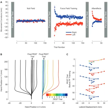
2.1 Force-field learning and the perception of limb position. . . 51
2.2 Following motor learning, the perceptual boundary shifts in a direction opposite to the applied force. . . 63
2.3 The presence of a perceptual shift depends on motor learning. . . 69
2.4 After motor learning, movements follow trajectories that are aligned with shifted perceptual boundaries. . . 72
2.5 During perceptual tests, subjects only apply lateral force immediately after force-field learning. . . 74
2.6 Force-field learning and perceptual testing with lateral movements. . 77
3.1 Experimental apparatus. . . 91
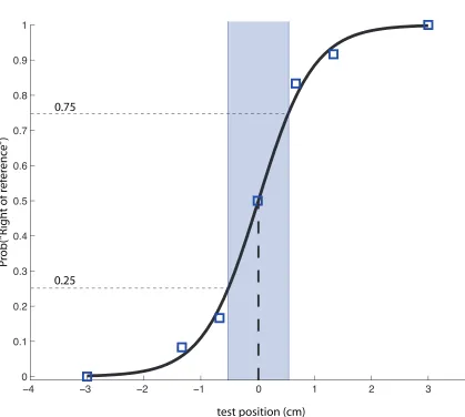
3.2 Example psychometric function. . . 95
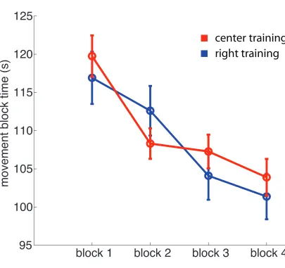
3.3 Motor learning: movement time. . . 97
3.4 Movement accuracy measurements. . . 99
3.5 Motor learning: movement accuracy. . . 100
3.6 Proprioceptive acuity. . . 102
3.7 Tuning of proprioception as a function of learning location. . . 104
3.8 Passive controls. . . 107
4.1 Experimental apparatus. . . 128
4.2 Example psychometric function. . . 131
4.3 Total time to target interception. . . 133
4.4 Changes to proprioceptive acuity. . . 135
5.1 Experimental apparatus. . . 147
5.2 Motor learning. . . 152
5.3 Mean tangential velocity. . . 154
5.4 Positional error. . . 155
5.5 Cross correlation index: REV and ACT. . . 158
5.6 Positional error: REV and ACT. . . 160
5.7 Cursive writing experiment. . . 162
5.8 Cross correlation index: cursive writing task. . . 163
6.1 Interhemispheric transfer . . . 181
Introduction
Investigations into the neural control of movement frequently feature human
adap-tation to novel movement contexts. These adaptive changes are often characterized
in terms of kinematic, electromyographic and dynamic measures, such as movement
kinematics, muscle activation, and delivered force and torque.
1.1
Overview
The studies presented in this thesis explore the relationship between motor
learn-ing and proprioceptive function, uslearn-ing the paradigm of human reachlearn-ing movements
made with the upper limb. This relationship has been measured by assessing
move-ment kinematics, and psychophysical estimates of human proprioceptive threshold.
Both motor learning tasks (sometimes in the presence of external, perturbing forces)
and psychophysical tests were undertaken using a graspable robotic manipulandum
(inMotion Technologies).
The studies described in Chapter Two assess the degree to which
propriocep-tion systematically varies during the adaptapropriocep-tion to novel external loads. During the
learning of novel movements it is often observed that subjects make specific and
sys-tematic changes to motor commands to adapt to novel perturbations. It is not known
if these motor changes occur as a result, or in the presence of, similar changes to our
sensory system. Indeed it may be that sensory changes are an integral part to the
motor adaptation process.
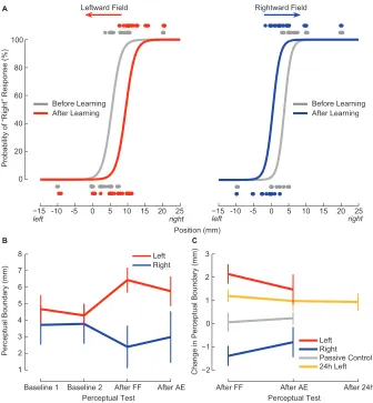
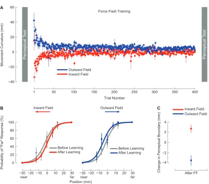
We performed a series of empirical studies that examined if the sense of hand
po-sition was systematically varied during the production of reaching movements made
perturbing loads and reduced the kinematic deviations made along the force-field’s
direction. We performed psychophysical estimates of proprioception before and after
learning, and found that subjects’ perceived location of their hand was systematically
altered in a consistent fashion across subjects experiencing the same force-field. After
learning, subjects were more likely to perceive the position of their hand as being
lo-cated in the direction of the experienced external load. We explored the generality of
the effect on altered perception by measuring both static and dynamic
psychophys-ical estimates, and found that subjects demonstrated perceptual changes in both
instances, data which support the notion for a generalized shift in perceptual
esti-mates. We also tested if this effect was related to motor learning itself by estimating
sensory function in subjects who did not perform active movement but were instead
passively moved by the robot through pre-recorded movements made by other
sub-jects. We found that these subjects did not demonstrate any sensory changes, which
supports the notion that sensory changes are related to motor learning.
InChapter Three, we further explore the idea that motor learning might change
sensory function, specifically in the context of motor tasks requiring high precision.
Many tasks require the motor system to generate precise motor commands to bring
the hand accurately from one location to another. Little is known about how this
change in motor ability relates to perceived hand position, leaving open the question
of perceptual function following training in these tasks.
Again using the robotic manipulandum, we provided subjects with the task of
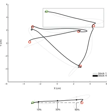
accurately positioning their hand within targets arrayed randomly in a small patch
(10 × 10 cm square) of the arm’s workspace, for approximately 10 minutes. Sensory
Perceptual acuity – the slope of the psychophysical threshold, and a measure of
how consistently stimuli were perceived by a subject – was improved after motor
learning, relative to baseline. This increase in perceptual acuity was shown also
to depend on the location of motor learning, as subjects who performed accurate
reaching movements in a location 25 cm away from the perceptual testing location
did not demonstrate the same improvements to sensory function. Again, we tested if
this perceptual effect depended on active movement by measuring sensory changes for
subjects who passively experienced pre-recorded movements made by other subjects.
Proprioceptive acuity improvement was not observed for these subjects, a finding that
supports the idea that active movement is required to mediate change in perceptual
function.
Lastly we attempted to test whether learning itself was the cause of sensory
per-ceptual change. To do this, we provided new subjects with a motor task that required
active movement, but which reduced the possibility of learning of accurate movements.
To do this, targets were presented open-loop to subjects, and no visual information
was provided about subjects’ hand position. We observed no reliable increases in
movement accuracy for these subjects, and also found no changes to sensory
func-tion. Taken together, changes to the perceptual acuity of hand function depend on
spatially-specific, active, learning of novel accurate movement.
The experiment inChapter Fourexplores the extent to which the improvements
to sensory sensitivity observed in Chapter Three persist following motor learning. It
has previously been shown that learned motor commands may persist for long periods
of time post-learning. In Chapter Two, we observed 24 hour persistence of sensory
about the persistence of the perceptual acuity change observed in Chapter Three.
We measured proprioceptive sensitivity at baseline and then again at 0, 1, 4
and 24 hours following the motor task requiring accurate movement described in
Chapter Three. Perceptual acuity was again shown to be significantly improved
relative to baseline immediately following motor learning. Subsequent tests revealed
that acuity was again improved 24 h following learning, relative to baseline measures.
No-movement control subjects did not show the same sensory improvements at any
time after motor learning. Taken together, these results support the notion that
proprioceptive changes as a result of motor learning have some persistence greater
than immediately following movement itself, and perhaps suggest that motor learning
and sensory processes have similar retention characteristics.
In Chapter Fivewe invert the relationship between proprioceptive function and
motor learning, and ask whether proprioceptive training might improve motor
learn-ing. It remains an open question if proprioceptive information about desired
move-ment can be used by the motor system to generate more accurate movemove-ments.
To address this question, we provided subjects with a motor learning task
com-posed of two kinds of trials – demonstration trials where subjects were shown the
desired movement, and active movements where subjects were asked to perform the
desired arm movement themselves. Demonstration trials were interleaved throughout
learning, and either featured only visual information (control subjects) or both visual
and proprioceptive information (experimental subjects) about the desired trajectory
to determine the effect of proprioceptive information on motor learning. Subjects
performed arm movements over three days. Subjects were provided with feedback
this movement over time. Notably, experimental subjects receiving proprioceptive
information on demonstration trials showed improved ability to reproduce desired
movement. This improvement was measured in terms of matching desired movement
velocity, where subjects showed significantly greater performance, and also in terms
of positional error, where subjects were better able to reduce positional error.
In-terestingly, experimental subjects demonstrated greater benefit from training than
an additional group of subjects who received an active form of proprioceptive
train-ing: during demonstration trials these subjects actively attempted to move along the
desired trajectory. These results support the idea that proprioceptive information
provided to a passive arm can assist in the learning of novel movements.
1.2
Literature Review
1.2.1
Motor adaptation in the upper limb
Studies in motor learning have investigated the extent to and means by which the
motor system adapts to changes in the dynamical behaviour of the human body. Tool
use is an oft-cited paradigm in which human motor control is exceptional. A
hand-held object, with its own unique dynamic properties, changes the relationship between
muscle force and limb movement. Despite this fact, we are capable of effortlessly
interfacing with myriad objects with considerable skill, and seemingly without effort.
How this adaptive behaviour is achieved is not understood. A broad research aim in
motor control is to understand how the brain adapts muscle commands to compensate
for perturbing forces both from the environment (of which gravitational and frictional
that result from interaction torques between connected limb segments. The principal
goal of motor control might be defined as the investigation of adaptations made by
the human motor system to compensate for these perturbations and thereby obtain
successful movement.
One well-studied avenue of motor adaptation has been the application of
exter-nally generated forces to the human arm. These forces are typically applied via
a handle grasped in the subject’s hand (or in some cases directly to the shoulder
and elbow joints) and are commonly termed force-fields. An early study of motor control observed adaptations to reaching movements, the characteristics of which are
illustrative of and relevant to current motor control studies. In Shadmehr and
Mussa-Ivaldi (1994), subjects generated reaching movements to targets in the presence of
a velocity-dependent force-field. Movements first made in the force-field were shown
to be curved, as these external forces pushed the limb away from a regular,
approxi-mately straight trajectory. Throughout the training phase, subjects learned to adapt
muscle forces to counteract the robot forces, and therefore regained the straight
reach-ing movements made in baseline trials (also called null trials to denote zero applied force). The curvature during special after-effect trials – trials presented infrequently
at randomized intervals, in which the force-field was removed entirely – illuminated
the specific nature of adaptation, because these movements were mirror-reflected
im-ages of early, perturbed movement. Therefore, subjects had learned to apply forces
to precisely counteract external loads. Finally, the concept of generalization was
also addressed in this paper (although limited in scope), by investigating how other
movements were affected by the force-field training. Deviations in hand-path were
motor adaptations resulted in changed motor commands to a subset of movements,
rather than to a single specific reach. These concepts of magnitude and generaliza-tion of motor adaptation continue to be investigated, and both are addressed in some capacity in the experiments contained in succeeding chapters.
Neural correlates of arm movements
It is known that the region of cortex identified as primary motor cortex (MI) is involved in reaching movements, based on the results of lesion, electrical
stimula-tion, and single cell recording experiments (Porter and Lemon, 1993). Thus some
neurally-implemented calculation that occurs in this area is important for reaching
movement, but the nature of this calculation continues to be poorly understood. The
prior hypothesis might be simply that MI neural activation directly results in
mus-cle activation, and therefore musmus-cle force (given that neurons in this area project
directly to motor neurons via the corticospinal tract), and this indeed was an early
finding of monkey neurophysiological preparations (Evarts, 1968). In a subsequent
influential study, Georgopoulos et al. (1982) attempted to determine if neural firing
is related to movement direction. By measuring mean neural firing activity during
the reaction time (RT, from cue presentation to non-zero hand velocity), movement
time (MT, non-zero velocity) and total time (RT+MT), the authors were able to
determine that neural activity in 75% of all cells was directionally tuned to
move-ment direction in either or both of RT and MT, and that the activity of many cells
showed well-behaved cosine tuning functions featuring peak activation for a specific
from the direction of maximal activity). Thus a reasonable argument was made for
the idea that MI activity is also related to movement direction.
Recent studies about cortical activity during reaching movements have more fully
explored two aspects of this early work. First, Georgopoulos et al. (1982) does not
address whether motor neurons in factencode direction itself or some other covariate; despite references made to the earlier Evarts study, no attempt was made to determine
the degree to which correlations with direction would appear if MI activity simply
coded for the open-loop activation of muscles. Other studies have gone on to show
significant correlations between MI activity and movement velocity, arm position,
acceleration, movement preparation, target position, distance to target, muscle
coac-tivation, serial order, visual target position, and visual target position (see Todorov
2000; Mussa-Ivaldi 1988 for a review of these correlated parameters), presumably
be-cause kinematic parameters are related by physical laws. Indeed, it has been shown
that an artificial neural network of MI implemented such that neurons send
mus-cle activation commands to a simulated two-joint arm will feature activation in the
simulated neurons that correlates with these movement parameters (Todorov, 2000).
Moreover, changes in the activity of MI neurons have also been observed when joint
configuration (but not direction of hand movement) is altered for reaching movements
(Scott et al., 1997). Errors in preferred-direction-based representations of MI activity
have even been shown to be correlated with the rate of work done at a joint (Scott
et al., 2001), evidence for MI activity related to muscle activation itself.
Second, the conclusions in Georgopoulos et al. (1982) were not based on
time-varying correlations of movement direction with neural firing but rather mean firing
to which this coarse analysis holds for the precise temporal variation of MI activation
during movement. In fact, collapsing data across time obscures important aspects
of MI activation which cast doubt on such simplification. Most basically, neural
activation is significant before movement (even accounting for transmission delays
from neural conduction and the activation dynamics of muscle). The accepted theory
for this activation is that this activity is merely sub-threshold activation of preferred-direction-tuned neurons (Bastian et al., 1998; Riehle and Requin, 1989; Cisek, 2006).
This hypothesis is testable: if MI neurons encode movement direction, neural activity
both before and during movement should be well-correlated. However the preferred
direction of a given MI neuron can change substantially between preparatory and
movement epochs (see Churchland et al. 2010 for review) such that, on a population
level, only weak correlations exist for neural activity between epochs.
At least four posterior hypotheses might be made about the relationship between
pmovement neural activity and movement activity: neural activation is not
re-lated to the same parameter at each epoch; neural activation is rere-lated to the same
parameter but the (directional) preference changes between epochs; neural activity
before movement is epiphenomenal; and finally it might be that they are related in a
non-obvious manner at the level of single neuron activation. This issue continues to
be unsolved. Recent proposals have employed powerful mathematical techniques to
represent neural activity at different epochs as linear dynamical models of neural
ac-tivity over time (Churchland et al., 2010). In such models, acac-tivity at a given neuron
for (discretized) time-points is linearly related to firing of other neurons in the
popu-lation. These models have been proposed as mechanistic models of neural activity –
such an approach, these models can match two important aspects of empirical data:
population activity at each epoch is well captured, while activity of a given neuron at
each epoch (preparatory vs movement times) is only weakly correlated. The degree
to which this approach succeeds in fact due to the power of linear dynamical models
remains to be quantitatively assessed (though argued against in the above paper).
Among regions of the brain considered as a whole, activity in MI is most
com-monly associated with the execution of movement. A number of other brain regions
have been implicated in the computation of various aspects of movement. Proposed
roles for these contributing brain regions are briefly outlined here. Ipsilateral
cerebel-lum may be involved in generating precise commands to control grasping and tool-use
movements (Imamizu et al., 2000). Pre-motor dorsal area neurons respond in a similar
fashion to primary motor cortical neurons (and when activation is represented with
preferred-direction vectors, demonstrate similar encoding), while pre-motor ventral
neurons may encode information about target position and reach directions in visual
space, motor space, or both (for review see Hoshi and Tanji, 2007). Supplementary
motor area neurons have been associated with self-generated (as opposed to externally
triggered) movement, the coding of movement sequences, and abstract cognitive
con-trol of movement (Nachev et al., 2008, for review). Posterior parietal cortical neurons
may be specifically involved in coordinating looking and reaching movements (Dean
et al., 2012). Regions of the cerebellum have been proposed as sites of plastic changes
for the learning of internal models of novel dynamics (Diedrichsen et al., 2005)
includ-ing tool use (Imamizu et al., 2000), and a role in motor learninclud-ing of motor sequences
Motor learning: observational learning
A powerful mechanism by which the motor system links motor control with action
observation has been identified in visual observational learning. When we observe the
actions of others, there is measurable activation of the same neural circuitry involved
in executing the action ourselves (Gallese et al., 1996). Thus, so-called mirror neurons
show similar activation when performing an action, or when observing it. In a recent
study the authors proposed that such observations are capable of informing low-level
information about the dynamical properties of a task. In this study by Mattar and
Gribble (2005) the authors had subjects watch videos of other subjects performing
reaching movements with a robotic manipulandum as they learned to adapt to a a
force-field that depended on movement velocity (termedviscous) and exerted a force perpendicular to object motion (termed curl). Subjects (both movie watchers and controls) were then required to perform reaching movements themselves in the
force-field. Observers of the learning videos demonstrated a performance improvement over
the first eight movements, at the beginning of the motor learning phase, compared to
control subjects who had not undergone observational learning, and also to controls
who had watched videos of subjects only making movements in a null field (and who
therefore acquired no novel motor commands). Interestingly, subjects who observed
the opposite force-field showed poorer initial performance. Additional groups of
sub-jects who were actively engaged in a motor task during video observation did not
show modifications to their own performance as strongly, a fact that argues for this
effect requiring engagement of the motor system itself. Taken together these results
support the idea that visual observation of a motor learning task being learned can
1.2.2
Proprioception
Signals underlying proprioception
Historically the sense of body position has been considered to be the composite of
several distinct anatomical sensors. Four primary classes of receptors - joint receptors,
skin afferents, Golgi tendon organs, and muscle spindles - are all proposed to provide
information about the body. The functional roles of each primary receptor has been
debated.
It is currently held that muscle spindles serve a primary role for position sense of
the body. Muscle spindles are situated in parallel with extrafusal (force-generating)
muscle fibres, and have been characterized as sending afferent signals about the
kine-matic properties of muscle - position, velocity, and acceleration, in some nonlinear
relationship. Signals from these receptors are known to reach somatosensory cortex
(Oscarsson and Rosen, 1963), respond during muscle lengthening across a wide range
of joint angles (Prochazka and Gorassini, 1998b; Dimitriou and Edin, 2008a,b), and
can be entrained during sinusoidal stretching (and vibration) of the tendon (Brown
et al., 1967). Tendon vibration, when applied externally, has been shown to
pro-vide an illusory sense of motion when performed on humans. (Goodwin et al., 1972;
McCloskey et al., 1983).
Golgi tendon organs may also play a role in proprioception. These sensory organs
are placed in series at the junction of muscle and tendons. Their afferent behaviour
has been differentiated from muscle spindle afferents in a critical way during the
above vibration preparations: Golgi tendon organs have been observed to be very
muscle fibres (Brown et al., 1967). Partly as a result, Golgi tendon organs have been
traditionally proposed to signal muscle force (Appenteng and Prochazka, 1984).
It has however been observed that in the real musculoskeletal system, the existence
of spring-like tendons means that tendon-length itself must be known (or calculated)
for the motor system to in turn know the current state of the body. Given the fact
that tendons are passive structures and exhibit nonlinear spring-like behaviour, force
at the muscle-tendon junction is proportional to the product of tendon stiffness and
(a power law relation of) tendon length. Therefore an alternative interpretation of
tendon signals considers Golgi tendon organs as in fact signalling tendon length. This
role has been proposed explicitly in a recent modelling study (Kistemaker et al., 2012).
Joint receptors were originally thought to fulfill the role now ascribed to muscle
spindles, that is to provide necessary information about joint angle change across
the entire range of a joint. However, subsequent studies identified that in fact joint
receptors signal identically for opposite ends of the joint range, and also feature
low/absent mid-range responses (Burgess et al., 1982). In addition, both temporary
and permanent disruption of receptor function via anaesthesis (Clark et al., 1979) or
surgically-mediated destruction (Grigg et al., 1973) have been shown to leave
move-ment perception and generation relatively unimpaired. Thus joint receptors have
been relegated to subsidiary roles, perhaps as limit detectors signalling extreme joint
angles (Ferrell et al., 1987).
Behavioural experiments of proprioception
Investigating human proprioception has typically been performed by measuring both
experimental methods have been employed.
Perhaps the most direct attempt to assess proprioception as a source of
sen-sory information in arm movements was performed by van Beers et al. (1998). The
authors began with the following assumption: in bimanual visually-guided reaching
movements, there are 3 principle sources of information necessary for movement
plan-ning: 1) proprioception of the left arm, 2) proprioception of the right arm, and 3)
visual information. While it might be argued that proprioception in a stationary arm
is qualitatively different than proprioception of a moving arm, the authors argued
that by allowing very slow movements (and even corrective movements), errors from
movement itself were minimal, and thus assumed that variability was the same for
a stationary arm used as a target, or a moving arm used as an indicator (the right
arm was used as both target and indicator, while the left arm was always only used
for indication). Therefore, the authors attempted to quantify the bias and variance
of each of the three information sources. Estimates were made by fitting a model of
pointing accuracy during reaching to several pointing locations. Subjects performed
three sets of pointing movements: 1) to visual targets with an unseen left hand, 2)
to visual targets with the unseen right hand, and 3) to the right hand with the left
hand, without vision. In each case one of the sources of information was therefore
removed, and regression coefficients were computed for each information source. The
authors demonstrated that proprioception was more accurate in radial (shoulder)
di-rections rather than azimuthal didi-rections. Proprioception was also shown to be higher
for targets closer to the body (in particular near the ipsilateral shoulder). Both of
these patterns can be understood by the geometry of the arm - greater total angular
has been observed again in several recent studies (Wilson et al., 2010; Fuentes and
Bastian, 2010).
A number of studies have targeted inter-hemispheric differences in proprioception
(Goble et al., 2006; Goble and Brown, 2008a; Sainburg, 2002). Some evidence exists
that proprioception of the non-dominant left arm benefits from superior position sense
to that of the right arm. Differences in proprioception between limbs were reported
in Goble et al. (2006) in a task in which subjects were required to return their limb
to a remembered joint angle. Limb matching was better for the non-preferred left
arm. A subsequent study by the same lab replicated this finding and also observed
better performance for remembered visual matching with the preferred right arm
(Goble and Brown, 2008a), which supports a recent hypothesis proposing that the
non-preferred arm has a specific role for stabilizing an object based on proprioceptive
feedback, while the preferred arm uses visual guidance for motor control (Sainburg,
2002; Goble and Brown, 2008a).
Other studies have investigated proprioception in the context of sensorimotor
in-tegration in visuomotor tasks. The use of proprioceptive signals might be particularly
important in movements where proprioceptive signals are superior to visual signals.
This has been observed to indeed be true for movement directions where relatively
low visual information exists, such as radial movements away and toward the body
(van Beers et al., 2002, 1998).
Misalignment of proprioception and vision has been shown to occur in a number of
instances (Brown et al., 2003; Wolpert and Kawato, 1998; Wann and Ibrahim, 1992).
Brown et al. (2003) showed that when subjects make reaching movements between
hand drift gradually over time from their original positions, while other movement
parameters – movement direction and amplitude – remained constant. Brown et al.
(2003) note that this is paradoxical because of two seemingly inconsistent facts: first,
there is evidence that the motor system has apoor estimate of the arm’s position since the hand is no longer successfully reaching the desired stationary targets (apparently
unbeknownst to subjects); second however, there is evidence that the motor system
has in fact a verygood estimate of the hand’s position, since very accurate information about the arm’s position is required to make movements that maintain the same
length and direction of the original goal movement. Drift between proprioceptive
arm position and visual target has also been shown by Wann and Ibrahim (1992)
who had subjects make unseen reaches every 15 seconds to visual targets. Drift
has not always been observed: Desmurget et al. (2000) did not find any decrease
in accuracy in a hand-position-matching task over 20 seconds. It remains unclear
what constitutes the perceptual estimate of drift. Desmurget et al. (2000) suggest
one potential culprit. The authors note that both sensory and efference copy signals
about recent movement – as distinct from perception of static position – are typically
included in the experiments for which drift has been reported, and in fact it is the
differential use of these signals over time that may be the source of observed drift.
Isolating proprioception
A general difficulty in the interpretation of proprioceptive studies is the separation of
proprioception from other methodological components. Seldom have these methods
clearly disentangled proprioception itself from coordinate transformations implicit in
re-sponses that require a motor or active movement aspect, rere-sponses that require require
inter-hemispheric transfer of information for contralateral limb-matching, memory of
remembered positions, and/or a combination of the above. Therefore, any
measure-ment of proprioceptive threshold may be confounded with other sources of variation:
sensory noise in the visual and/or motor systems themselves, or higher level variations
from transformation of different sensory sources to a common coordinate system.
These methodological variations are of particular importance for the studies
in-cluded in this thesis because we are interested in measuring proprioception in the
presence of motor learning. We carefully chose particular aspects of our sensory
test-ing paradigm to avoid confoundtest-ing estimates of proprioception with other aspects
potentially affected by motor learning. Three major methodological variations in
recent behavioural studies of proprioception serve as useful categorizations of the
re-maining literature, and identify components of sensory testing to be avoided in the
psychophysical estimation procedure we employ in Chapters Two, Three, and Four.
Active motor responses
A number of methods have required that subjects indicate sensed position by
per-forming an active movement (van Beers et al., 1998; Wann and Ibrahim, 1992; Chieffi
et al., 2004; Goble et al., 2006; Goble and Brown, 2008a). Active movements here
have included pointing to an unseen hand, or matching a target limb position or joint
angle deviation. Naturally, any difference between the planned movement endpoint
and the actual movement endpoint (resulting from an error in the computed
mo-tor command, or noise in efferent neural signals to muscles) will contribute to the
One aspect of active movement that can affect proprioceptive estimates is
move-ment velocity of active responses (Adamovich et al., 1998; Chieffi et al., 2004). For
example, in the former study, the authors observed that when subjects attempted
to replicate an imposed passive movement of the limb, overshooting of the passive
movement was observed when replication movements were performed with greater
movement speed.
Muscle activation has also been shown to influence proprioceptive accuracy. In
studies of proprioception of previously visited arm positions, accuracy to those
loca-tions held with active muscle contraction was greater than localoca-tions made via passive
movements (Adamovich et al., 1998).
It has also been suggested that proprioception might share some aspect of the
(anatomically-derived) what/where dichotomy in the visual system, a fact that would
support the notion that fundamental differences exist between proprioception for
ac-tive movement and proprioception for perception (Volpe et al., 1979; Paillard et al.,
1983; Rossetti et al., 1995; Dijkerman and Haan, 2007). Using specific
neuroanatom-ical case studies, a dissociation has been observed separating two different kinds of
responses to tactile stimuli: conscious reporting of, and directed movement to
dif-ferent positional stimuli. In two case studies analogous of blindsight (Paillard et al.,
1983; Rossetti et al., 1995), experimenters observed in each case a patient able to
move accurately to a tactile stimulus, despite a complete lack of conscious awareness
(i.e. as indicated by verbal reporting) of the proprioceptive testing site. Lesions
were identified to exist in the left posterior cortical area and ventral posterior
nu-cleus of the thalamus, respectively. This contrasts with other case studies reporting
successfully detect (Head and Holmes, 1911; Rapp et al., 2002), which when taken
together with the previous study suggest that detection and location are
doubly-dissociable processes. Recently, however, the double-dissociation hypothesis for the
sense of touch was tested directly in a vibrotactile experiment (Harris et al., 2004). In
this experiment, experimenters fit responses of normal subjects to different signal
de-tection models to determine whether psychophysical responses could be explained by
independent parallel processes, or serial processes. Only the serial model successfully
described subject responses, leading the authors to conclude that somatosensation
for action and perception are not mutually independent processes but rather
localiza-tion is subsequent to deteclocaliza-tion. The authors speculate that somatosensory blindsight
analogs may well be confounded by different response characteristics in yes-no as
opposed to forced-choice psychophysical paradigms (for an exception to measures of
true blindsight with controlled psychophysical estimates, see Azzopardi and Cowey
1997).
Vision in proprioception
Visual presentation of stimuli has been used in a number of proprioception
experi-ments, either as a means of presenting targets, or as a response method itself
(Desmur-get et al., 2000; Sittig et al., 1985; Smeets et al., 2006; Vindras et al., 1998). Responses
made to or with some visual reference involve coordinate transformations into a
com-mon space (van Beers et al., 1998), meaning that 1) determining proprioception
dis-tinct from vision is not straightforward, and 2) errors in coordinate transformation
implicit in any perceptual judgment might again be attributed to sensory variation.
bet-ter remembered than blinded (and passive) positions (Goble and Brown, 2008b) and
also the reverse (Darling and Miller, 1993). In Darling and Miller (1993), matching
movements to targets presented either visually (visible before movement onset) or
kinaesthetically (blind guidance of the hand to a tactile object), subjects were better
at moving toward kinaesthetic targets.
Inter-hemispheric transfer
Another method used for proprioceptive assessment is via joint matching either
be-tween limbs, or with the same limb (to remembered positions). Joint matching is
more accurate when made to ipsilateral remembered positions than to contralateral
positions either remembered or simultaneously presented (Adamo and Martin, 2009;
Goble et al., 2006), thereby supporting the simple hypothesis that there is a cost of
integrating two sensory signals that is greater than comparing a remembered signal
to a current signal from the same sensor.
Psychophysics and proprioceptive sensitivity
The psychophysical estimation of sensory threshold is the measurement of subjective
responses at various levels of stimulus intensity. The mapping between perception
and stimulus intensity provides estimations of both perceptual acuity – the range of
stimuli for which changes in perception occur – and also perceptual threshold: the
stimulus intensity at which subjects detect with 50% probability. This is the method
we employ for the estimation of proprioception in subsequent chapters.
By choosing to perform psychophysical estimates we diverge somewhat from the
lit-erature cited above. The most important reason for this choice has been because
the use of two-alternative, forced-choice psychophysical tests is a good way of
mea-suring proprioception while avoiding movement and visual components that would
confound hypothetically observed changes in proprioceptive function following motor
learning. It is known that motor learning certainly results in altered movements, and
motor learning also systematically enhances vision (Brown et al., 2007), and thus
such components must be avoided in estimates of sensory function.
Measuring the sense of human proprioception is slightly different from other
psy-chophysical estimation procedures. At a basic level, estimates of human psychophysics
require the delivery of stimuli across an input range; psychophysics of human vision
or audition involve presenting sensory stimuli of sound or light inputs of varying
am-plitude or wavelength via precision display and acoustic equipment. Estimation of
proprioceptive threshold faces a unique challenge by the obvious fact that the human
apparatus must be physically moved across any range. Where other perceptual
judg-ments require the stimuli at particular wavelengths/amplitudes to be systematically
(Laming and Laming, 1992) or algorithmically (Taylor and Creelman, 1967) varied,
in proprioceptive measurements the transitions between each stimuli must also be constrained. A robotic apparatus for stimulus presentation is useful for such tasks.
Additionally, the dimension along which stimuli might be varied is also open for
consideration: in which coordinate system should the stimuli be varied? Cartesian
distance (perhaps relative to some initial location), joint angle, or muscle length,
are all potential domains (likewise with their time derivatives). The benefit of a
given coordinate system might depend on how proprioception is of interest. Joint
the body; Cartesian perturbations are useful for considering perceptual differences
that might be use-dependent, or for interpreting the functional relevance of such
perceptual differences. Ultimately, proprioceptive signals are believed to be mediated
at a muscle level, the length of which however cannot be directly known without
knowledge of or assumptions about additional states of the system, including muscle
activation and tendon length (see Dimitriou and Edin 2010).
1.2.3
Perceptual changes resulting from motor learning
Several recent investigations have focused on the degree to which movements modify
our perceptual abilities, particularly in the context of motor learning. Does
learn-ing new motor commands to generate novel movement result in changes in human
perception?
In the visual system, developmental findings suggest that this effect of movement
on perception may be a feature of normal development. It has been shown in infant
studies that accurate reaching movements to objects are made early at 5 months of
age (von Hofsten and Spelke, 1985). Prediction of visual motion happens slightly
later, at 7 months (Kim and Spelke, 1992). Visual motion prediction was assessed by
measuring the duration infants spent gazing at objects accelerating at appropriate or
inappropriate rates, the authors inferred the degree to which infants were habituated
to different visual accelerations. Seven month olds, but not five month olds,
demon-strated a habituated response to objects accelerating appropriately under gravity, and
spent longer time observing visual motion. Therefore, long-term visual adaptation
occurs a few months later than the time during which which infants learn
visual adaptations thus allows for the possibility that reaching movements play a role
in the natural development of visual perception.
Other investigations have been made into altered visual sensitivity directly from
motor learning itself. After subjects learned a novel gait, subjects demonstrated
in-creased visual sensitivity to the visual perception of such gait patterns (Casile and
Giese, 2006). Subjects were required to learn novel phase differences between hands
(180 degrees normally separate the contralateral limbs) of either 225 or 270 degrees.
These tasks were learned blind-folded, and only with the assistance of verbal
instruc-tions and haptic guidance from experimenters. Following this motor learning period
subjects were given a visual discrimination task in which two point-light displays
were shown of human walking. The experimenters specifically altered the degree of
phase difference between the two displays, and subjects were required to respond
in a forced-choice paradigm whether the two point-light displays of visual motion
depicted identical gait patterns. It was observed that subjects who had learned the
motor task showed lower detection thresholds for gait phase differences – and, further,
subjects who were better able to learn the novel movement patterns were also better
at detecting visual differences. Importantly, because subjects were blindfolded
dur-ing their own learndur-ing of movement, the increased sensitivity to visual gait differences
after learning was not confounded by subjects simply training their visual system on
processing (their own) novel gait patterns. This study demonstrates an effect of
mo-tor learning on visual processing in a restricted sense: altered visual perception was
restricted to vision of that movement itself.
One recent study investigated motor learning effects on visual processing, and in
oc-curred more generally (Brown et al., 2007), and not specifically restricted to vision of
learned movements as in the previous study. Subjects performed reaching movements
with a robotic manipulandum and the robot was programmed such that the force at
the hand was constant and unidirectional. One group of subjects received a
force-field pushing their hand leftward, another group experienced a rightward force-force-field,
and a final group of subjects experienced a null force-field (0 output force). First,
subjects learned to make straight reaches to targets while in the presence of the
force-field. Next, with the force-field still turned on, subjects were required to reach out
and intercept visual targets that moved left-to-right across the display, with constant
acceleration. Therefore, the visual motion of these targets was carefully chosen to
be either congruent, or incongruent, to the experienced force-field. The authors
hy-pothesized that motor learning of a force-field systematically alters visual prediction:
subjects who have learned reaches in a rightward force-field will also predict visual
objects to accelerate rightward, as though the objects are moving under the same
force-field.
Indeed, subjects whose force-field experience was congruent to visual object
mo-tion were significantly better at successfully intercepting the visual targets. This
benefit was observed to be independent of interception behaviour in two ways. First,
subject groups showed no reliable differences in interception kinematics. Second, a
control experiment showed that changes to visual perception were not tied to reaching
movements alone. In a task that required subjects to make button-press responses
to indicate the position of moving objects, the timing (early, accurate, or late) of
these button presses depended on the direction of the force-field experienced during
systematically altering how the brain perceives visual motion.
Increased visual sensitivity can also develop near the functional end of learned
tools (Brown et al., 2011). After learning to move a novel pointer-like tool
ballis-tically to targets, subjects performed a visual detection experiment in which they
were asked to respond as soon as they saw a cursor appear near the tip of the tool.
Only subjects who performed these active, ballistic movements showed an increase
in visual sensitivity, as indicated by reduced response times. No visual sensitivity
improvements were observed for control subjects who made no movements with the
tool, nor for subjects who received passive guidance of their arm through reaching
trajectories.
Proprioceptive changes resulting from motor learning
While motor effects on the visual system have been studied more frequently, recently
some studies have investigated the effects of motor learning on the proprioceptive
sys-tem. One such study investigated perceptual changes following the motor adaptation
to a visuomotor rotation (Cressman and Henriques, 2009). In this study, subjects
performed a set of point-to-point reaching movements while the visual representation
of hand position (the arm itself was not visible) was gradually altered such that the
represented position appeared rotated from actual hand position. Before and after
this learning phase, subjects performed a psychophysical test session during which
they reported the perceived position of their hand as being left or right of a displayed
visual cursor. After learning a visual rotation field, subjects demonstrated that the
perceived position of their hand was directly related to the rotation magnitude
between perceived hand position and visual cursor, the observed change represents
a change in the calibration between vision and proprioception: neither vision nor
proprioception are uniquely implicated in this change.
Evidence for long-term changes to proprioceptive function as a result of motor
learning might be observed by assessing proprioception in different locations of the
arm’s workspace, thereby probing for differences dependent upon arm use. Relatively
few studies have been reported that assess how proprioception may vary across the
workspace of the limb (Fuentes and Bastian, 2010; van Beers et al., 1998; Wilson
et al., 2010), and no perceptual differences (beyond those admitting of a simple
ge-ometric explanation) have been observed. In contrast, we know a great deal about
other sensory systems that are characterized by differential anatomic sensor density,
resulting in greater visual (Wald, 1945), acoustic (Davis and Kranz, 1964), and haptic
sensitivity (Bolanowski et al., 1988; Verrillo, 1963; Weinstein, 1968) across a subset
of the input domain.
1.2.4
Physiological bases for proprioceptive changes from
mo-tor learning
Central plasticity of sensory areas
Neuroplasticity is central to the development of human motor function and, likewise,
to skill acquisition in the mature motor system. Work to date on motor learning has
focused almost exclusively on plasticity in motor systems: on how motor systems
ac-quire new abilities, how learning occurs during motor development, and how learning
function affect the somatosensory system is largely unknown. Numerous studies have
shown central activity of sensory neurons during movement, presumably since activity
in somatosensory cortex neurons varies systematically changes with body position.
In area SI 3b of monkey somatosensory cortex, approximately 50% of neurons with
tactile receptive fields of skin have been reported to modulate their activity during
reaching movement (Prud’homme and Kalaska, 1994; Cohen et al., 1994). These
neurons fired most strongly to tactile manipulations but still showed activity during
reaches without direct tactile stimulation. The probability of task-related activity
was related to particular response properties of given neurons, in particular
sensitiv-ity to direct stretching of the skin, implying that movement-induced skin stretch is
the mechanical event resulting in cortical signals.
Neurons in primary somatosensory cortex (SI) vary systematically with
move-ment. This conforms with a priori expectations about cortical areas that appear to
wire directly to peripheral sensors (Oscarsson and Rosen, 1963). Interestingly
how-ever, these neurons did not seem to reflect only the kinematics of the arm, since
neural activity was not only affected by current limb kinematics; instead, these
neu-rons showed strong hysteresis effects. This was observed in a task where monkeys
made reaching movements to a common central target. Neuronal activity seemed to
reflect previously-experienced movement. It could be that somatosensory cortex
in-deed reflects sensory history; inin-deed the authors note that hysteresis effects observed
in a particular neuron were often larger when previous movements included the
neu-ron’s preferred direction. A different earlier study found that activity for many of
the neurons in somatosensory areas (1, 2, and 5) changed firing while the arm was
be-fore changes in electromyographs (Soso and Fetz, 1980; Nelson, 1987; Lebedev et al., 1994).
Finally, it is also well known that sensory representations can be heavily
modu-lated by training. Several studies have investigated adaptations to vibrotactile sensory
learning (Recanzone et al., 1992), precision grasping (Xerri et al., 1999) and even
sim-ple tactile stimuli from maintained contact between finger and object surface (Jenkins
et al., 1990). Each of these studies observed rather similar changes to tactile
represen-tations in area 3b of somatosensory cortex. The most recent paper Xerri et al. (1999)
required monkeys to make precision grasps to apprehend small banana-flavoured food
pellets placed in wells, over a period of 3 months. The monkeys demonstrated change
in prehensile behaviour over the course of learning to feed on the pellets, often
switch-ing number of graspswitch-ing fswitch-ingers (typically reducswitch-ing number of involved fswitch-ingers) and
ultimately resulting in a greater proportion of successful grasps. In 3b, representation
of a given glabrous skin area nearly doubled (on average; relative to untrained
finger-tips or contralateral fingertip areas); receptive fields for individual neurons in the area
simultaneously decreased to less than half original size. These findings demonstrate
the nature and extent of cortical plasticity that may occur during the acquisition of
a motor skill.
Segmental basis for changes to proprioceptive function during movement
Sensory sensitivity is modulated dynamically via descending signals from the brain,
and this may provide one mechanism by which the motor system mediates functional
sensory change during movement. Efferent innervation of sense organs occurs in many
in-nervation of semicircular canals (Purcell and Perachio, 1997; Warr, 1975) and retinal
cells (Honrubia and Elliott, 1970) modulate the signals about head orientation and
the visual field. Muscle spindles, widely regarded to be the primary source of
in-formation about the position of the limbs, also receive modulating efferent signals.
It has been proposed that such signals may account for the sensory consequences of
self-generated action (Bays et al., 2005; Blakemore et al., 1998; Wolpert and
Flana-gan, 2001) and augment the functional dynamic range of the sensor (Scott and Loeb,
1994; Windhorst, 2007).
Models of spindle afferent signals designed to infer what property is encoded by
spindles during movement itself have had only partial success. In one study of
chronic-recordings of normal stepping in cats (Prochazka and Hulliger, 1998), the key variable
achieving good prediction of afferent signals was muscle velocity, with power law
functions being the best fit (spindle firing rate being approximately proportional to
the square root of muscle velocity). These data were collected from hamstring muscles
- a muscle that is not significantly recruited during the stepping phase. When the
experiment was repeated with a heavily-recruited muscle - triceps surae - it was shown
that this relationship between muscle length and encoded signal does not hold well
(Prochazka and Gorassini, 1998a). In humans, spindle afferent signals recorded during
finger tapping and grasping studies have found muscle velocity and acceleration may
be encoded by spindles (Dimitriou and Edin, 2008a,b).
Spindles are significantly innervated by descending signals that functionally
mod-ify the behaviour of the sensor. Gamma fusimotor drive is putatively responsible for
changing the mechanical properties of the muscle spindle and thereby altering
component of learning novel movement may in fact be learning appropriate fusimotor
signals to control our sensory apparatus (Dimitriou and Edin, 2010).
Gamma fusimotor efferents have been classified into two types: static and dynamic
(for a review, see Hulliger 1984). Descending activation from these differing fibre types
has been differentiated based on the ability to either increase or decrease sensitivity
during the ramp phase of ramp and hold movements.
In humans there has been some evidence that afferent signals may be modulated
by efferent commands. In Hospod et al. (2007), experimenters tested the hypothesis
that spindle signals might change systematically when subjects were required to
at-tend to that part of their body. Interestingly researchers observed a variety of changes
to spindle afferents, including increased discharge variability, changes in spontaneous
(baseline) activity, and decreases in depth of signal modulation. The authors
spec-ulated that since muscle EMG was zero (and subjects were told to remain passive
during these imposed movements), that fusimotor drive is responsible for modulation
of afferent signals.
A separate study has found further evidence that spindle sensitivity can be
modi-fied based on movement context (Davis et al., 2011). In this study it was hypothesized
that a stressful movement context would affect spindle reflex amplitudes. Subjects
were asked to stand at the edge of a platform either during a low stress condition
(less than 1 metre from the ground), or high stress condition (suspended 3 m from
the floor). During quiet standing at these differing heights, subjects received
reflex-inducing achilles tendon taps, while both reflex amplitude and cortical evoked
poten-tials were recorded. Reflex amplitude was shown to increase during the high stress
This suggests that either spindle afferents themselves, or spinal reflex gains, are
mod-ulated during the maintenance of stance under stressful conditions. A recent
subse-quent study by the same research group replicated the experiment but in addition
to the tendon-tap reflex measures, H-reflex magnitude was also recorded (Horslen
et al., 2011). H-reflexes are elicited by surface-electrical stimulation of afferent
neu-rons, causing a monosynaptic motor reflex. By essentially sending artificial sensory
input to the spinal cord and measuring the resulting reflex magnitude, changes to
spinal reflex gains themselves may be inferred. No change in H-reflex magnitude was
observed, which suggests that modulation of the afferent sensory signal occurs at the
level of the spindle itself.
The literature cited above shows that anatomic substrates of perception support
differential sensitivity, efferent neural signals provide top-down modulation of
sensi-tivity, and behavioural context may play a role in this descending modulation.
1.3
Summary
In this chapter we have reviewed issues related to the motor system’s capacity for
motor adaptation, and provided an outline for the current characterizations of human
proprioception. We also highlighted recent research investigating the how sensory
function might be altered with motor learning, and explored current knowledge about
the central and segmental bases for sensory plasticity.
Proprioception and its relationship to motor learning has not been completely
studied. While other work has explored sensory integration of vision and
motor learning, the effect of motor learning on proprioception itself remains an open
question. Indeed it may be that the nature of changes in the perceived position of
the hand after motor learning depend on the nature of the type of motor adaptation
itself. Nothing is known about changes to the perceived position of the hand in the
presence of external load, or even following the simple task of increasing movement
accuracy to targets in the absence of an external load.
Similarly, it is not known if proprioceptive information can assist motor learning.
While the ability of the motor system to adapt movements has been shown to benefit
from visual information, it is not known if proprioceptive information about a desired