Kok Pin Ng, MRCP
Tharick A. Pascoal, MD
Sulantha
Mathotaarachchi, MSc
Chang-Oh Chung, MD
Andréa L. Benedet, MSc
Monica Shin, MSc
Min Su Kang, BSc
Xiaofeng Li, MD
Maowen Ba, MD
Nagaendran Kandiah,
FRCP
Pedro Rosa-Neto, MD,
PhD
Serge Gauthier, MD
For the Alzheimer
’
s
Disease Neuroimaging
Initiative
Correspondence to Dr. Gauthier: serge.gauthier@mcgill.ca
Supplemental data at Neurology.org
Neuropsychiatric symptoms predict
hypometabolism in preclinical Alzheimer
disease
ABSTRACT
Objective:
To identify regional brain metabolic dysfunctions associated with neuropsychiatric
symptoms (NPS) in preclinical Alzheimer disease (AD).
Methods:
We stratified 115 cognitively normal individuals into preclinical AD (both amyloid and
tau pathologies present), asymptomatic at risk for AD (either amyloid or tau pathology present),
or healthy controls (no amyloid or tau pathology present) using [
18F]florbetapir PET and CSF
phosphorylated tau biomarkers. Regression and voxel-based regression models evaluated the
relationships between baseline NPS measured by the Neuropsychiatric Inventory (NPI) and
base-line and 2-year change in metabolism measured by [
18F]fluorodeoxyglucose (FDG) PET.
Results:
Individuals with preclinical AD with higher NPI scores had higher [
18F]FDG uptake in the
posterior cingulate cortex (PCC), ventromedial prefrontal cortex, and right anterior insula at
base-line. High NPI scores predicted subsequent hypometabolism in the PCC over 2 years only in
individuals with preclinical AD. Sleep/nighttime behavior disorders and irritability and lability were
the components of the NPI that drove this metabolic dysfunction.
Conclusions:
The magnitude of NPS in preclinical cases, driven by sleep behavior and irritability
domains, is linked to transitory metabolic dysfunctions within limbic networks vulnerable to the
AD process and predicts subsequent PCC hypometabolism. These findings support an emerging
conceptual framework in which NPS constitute an early clinical manifestation of AD
pathophysiology.
Neurology®2017;88:1814–1821GLOSSARY
AD5Alzheimer disease;ADNI5Alzheimer’s Disease Neuroimaging Initiative;ADNI-mem5Alzheimer’s Disease Neuro-imaging Initiative memory composite score;AI5anterior insula;AR-AD5asymptomatic at risk for Alzheimer disease;CDR5 Clinical Dementia Rating;[18F]FDG5[18F]fluorodeoxyglucose;MCI5mild cognitive impairment;NPI5Neuropsychiatric
Inventory;NPS5neuropsychiatric symptoms;PCC5posterior cingulate cortex;p-tau5phosphorylated tau;SN5salience network;SUVR5standardized uptake value ratio;vmPFC5ventromedial prefrontal cortex.
Neuropsychiatric symptoms (NPS) represent a common feature of mild cognitive impairment
(MCI) and dementia phases of Alzheimer disease (AD).
1In these patients, NPS are associated
with a poorer outcome in cognition and functional state.
2,3Although there is an emerging
conceptual framework supporting NPS as noncognitive symptoms of preclinical AD,
4the role
of NPS as early indicators of AD pathophysiologic progression remains unclear. Studies
con-ducted in patients with AD indicate that NPS are associated with metabolic dysfunction in brain
networks subserving mood and cognition.
5Therefore, further studies focusing on the
associa-tion between NPS and AD pathophysiologic abnormalities are of paramount importance given
From the Translational Neuroimaging Laboratory (K.P.N., T.A.P., S.M., C.-O.C., A.L.B., M.S., M.S.K., P.R.-N.) and Alzheimer’s Disease Research Unit (K.P.N., X.L., M.B., P.R.-N., S.G.), McGill University Research Centre for Studies in Aging, Montreal, Quebec, Canada; Department of Neurology (K.P.N., N.K.), National Neuroscience Institute, Singapore; Montreal Neurological Institute (P.R.-N.); Department of Neurology and Neurosurgery (P.R.-N.), McGill University, Montreal, Quebec, Canada; Department of Neurology (X.L.), The Second Affiliated Hospital of Chongqing Medical University, Chongqing; and Department of Neurology (M.B.), Yantai Yuhuangding Hospital Affiliated to Qingdao Medical University, Shandong, PR China.
Data used in preparation of this article were obtained from the Alzheimer’s Disease Neuroimaging Initiative (ADNI) database. The ADNI investigators contributed to the design and implementation of ADNI and/or provided data. The ADNI list is available at Neurology.org. Go to Neurology.org for full disclosures. Funding information and disclosures deemed relevant by the authors, if any, are provided at the end of the article. The Article Processing Charge was funded by the authors.
a growing body of evidences proposing subtle
NPS as manifestations of emergent disease
progression.
6[
18F]fluorodeoxyglucose ([
18F]FDG) PET is
a technique that is sensitive in detecting changes
in resting metabolism associated with the
neu-ropsychiatric conditions
7and metabolic declines
commonly observed in neurodegenerative
con-ditions.
8In AD, hypometabolism has been
con-sidered a part of disease pathophysiology and
interpreted as synaptic abnormality or neuronal
injury. Thus, modeling the magnitude of
symp-toms as a function of changes in brain
metabo-lism constitutes a valuable strategy to investigate
the neural correlates of NPS in preclinical AD.
Here, in a longitudinal observation of
cog-nitively normal individuals stratified by
hall-mark AD biohall-markers to identify individuals
with preclinical AD with the highest risk of
progression to clinical AD,
9we test the
hypothesis that NPS are associated with
met-abolic abnormalities in limbic regions and
predict pathophysiologic progression in
indi-viduals with preclinical AD.
METHODS Data used in the preparation of this article were obtained from the Alzheimer’s Disease Neuroimaging Initiative (ADNI) database (adni.loni.usc.edu). The ADNI was launched in 2003 as a public-private partnership led by principal investi-gator Michael W. Weiner, MD. The primary goal of ADNI has been to test whether serial MRI, PET, other biological markers, and clinical and neuropsychological assessment can be combined to measure the progression of MCI and early AD.
In this study, we selected cognitively normal individuals with baseline Neuropsychiatric Inventory (NPI) and Clinical Dementia Rating (CDR) scores, CSF phosphorylated tau (p-tau181p) measurements, [18F]florbetapir PET imaging, both
baseline and 2-year follow-up [18F]FDG PET imaging, and ADNI memory composite score (ADNI-mem). We defined cognitively normal individuals as those with a Mini-Mental State Examination score of$24, CDR score of 0, and absence of any neuropsychiatric diseases such as depression, MCI, and dementia. The participants were then stratified on the basis of the recent definition of preclinical AD9into 3 groups: healthy controls, no amyloid or tau pathology present; asymptomatic at risk for AD (AR-AD), either amyloid or tau pathology ent; or preclinical AD, both amyloid and tau pathologies pres-ent. With this definition,9the risk of progression to clinical AD is particularly high in preclinical AD. In AR-AD, which may represent cognitively normal individuals who do not follow the temporal sequence in the amyloid cascade hypothesis,10 the risk of clinical evolution still needs to be determined. The inclusion/exclusion criteria adopted by ADNI can be found at www.adni-info.org (accessed October 2016).
Standard protocol approvals, registrations, and patient consents.The ADNI study was approved by the Institutional Review boards of all of the participating institutions. Informed written consent was obtained from all participants at each site.
Neuropsychological assessments. The neuropsychological assessments were performed by certified raters using standard-ized ADNI protocols. The CDR, NPI, and ADNI-mem da-tasets used in this study were obtained from the ADNI files CDR.csv, NPI.csv, and UWNPSYCHSUM_01_28_15-5.csv, respectively. In this study, the NPI was used to measure NPS of the participants. The NPI is an instrument that assesses behavioral disturbances occurring in patients with dementia.11 Both the severity and frequency of each symptom are mea-sured, and this information is obtained from a caregiver familiar with the individual’s behavior. ADNI-mem is a vali-dated composite memory score derived from data from the ADNI neuropsychological battery.12 Briefly, a modern psy-chometric approach was used to analyze the Rey Auditory Verbal Learning Test, AD Assessment Scale–cognition, Mini-Mental State Examination, and Logical Memory tests to obtain a composite memory score. In the ADNI-mem test, lower scores reflect poorer performance. Details of the ADNI pro-tocols for the neuropsychological assessments and the methods for developing the ADNI-mem can be found at www.adni-info.org (accessed October 2016).
CSF analysis.CSF p-tau181pwas measured with the Luminex
multiplex platform (Luminex, Austin, TX) and Innogenetics INNO-BIA AlzBio3 (Innogenetics, Ghent, Belgium) immu-noassay reagents. The CSF biomarker datasets used in this study were obtained from the ADNI file UPENNBIOMK5-8. csv. An ADNI published cutoff of CSF p-tau181p.23 pg/mL
was used to define the presence of tau pathology.13Details of the ADNI methods for the acquisition and measurement of CSF can be found at www.adni-info.org (accessed October 2016).
MRI and PET methods.MRI and PET standard acquisition protocols were described in the ADNI website at http://adni. loni.usc.edu/methods/ (accessed October 2016). T1-weighted MRI images corrected for field distortions were processed with the CIVET image processing pipeline,14and the PET images were processed with an established image processing pipeline.15 In summary, the preprocessed images from the ADNI database were spatially normalized to the Montreal Neurological Insti-tute 152 standardized space with the use of transformations obtained for PET native to MRI native space and the MRI native to the Montreal Neurological Institute 152 space. Subsequently, the [18F]florbetapir PET standardized uptake value ratio (SUVR) and the [18F]FDG PET SUVR maps were generated with the cerebellum gray matter and the pons, respectively, as reference regions. [18F]florbetapir PET images were then normalized for the white matter SUVR. The global brain glucose uptake and the global amyloid deposition were defined as the average SUVR calculated from several brain regions characteristic to AD, including the precuneus, pre-frontal, orbitopre-frontal, parietal, temporal, anterior, and poste-rior cingulate cortices in the [18F]FDG and [18F]florbetapir PET images, respectively. Because we do not have access to the pathology data, we used the best operational point of the receiver operating characteristic curve contrasting controls and ADNI patients with AD (n590)16and calculated [18 F]flor-betapir PET SUVR.1.15 as the threshold for positive amy-loid pathology. With this threshold, fewer than one-third of the controls were amyloid positive, which is consistent with the literature.17
of baseline demographics and cognitive scores were summarized and compared between the 3 groups using analysis of variance for continuous measurements andx2tests for categorical measure-ments. Linear regression models evaluated the associations of NPI scores with baseline global [18F]FDG SUVR and globalD[18F] FDG SUVR over 2 years in each group. Percentage change in [18F]FDG SUVR was defined as {(D[18F]FDG5[18F]FDG base-line2[18F]FDG follow-up)/[18F]FDG baseline}. An interaction term was added to the regression models to evaluate the interac-tions of NPI and the biomarker groups onD[18F]FDG SUVR:
D½18FFDG SUVR5b
01b1ðbaseline NPIÞ
1b2ðbiomarker groupsÞ
1b3ðbaseline NPI3biomarker groupsÞ
1covariates1e
Linear regression models also evaluated the associations of individual subcomponents of the NPI withD[18F]FDG. In a sec-ondary analysis, we used linear regression models to evaluate the associations between NPI scores and ADNI-mem at baseline and DADNI-mem over 2 years in each group as follows: [D ADNI-mem 5 (ADNI-mem baseline 2 ADNI-mem follow-up)/ ADNI-mem baseline].
Voxel-based regression models were used to test the associa-tions of NPI scores with baseline [18F]FDG SUVR andD[18F] FDG SUVR over 2 years in each group:
Baseline ½18FFDG SUVR5b
01b1ðbaseline NPIÞ
1covariates1e
D½18FFDG SUVR5b
01b1ðbaseline NPIÞ1covariates1e
We extracted andzscored the mean SUVR at baseline and at follow-up from the regions where significant associations between NPI scores and D[18F]FDG were observed. We defined the presence of hypometabolism if the mean [18F]FDG SUVR was significantly lower (95% confidence interval) than the biomarker-negative individuals, under the assumption that these individuals had a normal metabolic trajectory.
Bonferroni correction was used to correct the aforementioned linear regression analysis for multiple-comparison tests. The sta-tistical parametric maps presented in this study were false discov-ery rate corrected for multiple comparisons with a threshold of
p,0.001. All statistical models presented here were corrected for age, sex, education, andAPOEe4 status.
RESULTS
Baseline demographics,
APOE
status, and
biomarker characteristics of the 3 groups are
summa-rized in the table.
We found that individuals with preclinical AD
with higher NPI scores had higher [
18F]FDG uptake
in the posterior cingulate cortex (PCC), ventromedial
prefrontal cortex (vmPFC), and right anterior insula
(AI) at baseline (figure 1). However, this finding was
not observed in the AR-AD or healthy control group
at baseline. NPI scores did not have an effect on
baseline global [
18F]FDG uptake in any of the groups.
We further found that high NPI scores predicted
global [
18F]FDG uptake decline over 2 years in the
preclinical AD group (
b
5
0.52, SE
5
0.17,
p
5
0.01) but not in the AR-AD or healthy control
group. A significant interaction between NPI and
the groups confirmed that this effect was higher in
those with preclinical AD than the other individuals
(
b
5
0.44, SE
5
0.19,
p
5
0.02). Subcomponent
analysis indicated that only the combined sleep/
nighttime behavior disorders and irritability/lability
components of NPI predicted global [
18F]FDG
uptake decline in the preclinical AD group (
b
5
0.51, SE
5
0.17,
p
5
0.008). NPI scores did not
predict ADNI-mem scores in any of the groups at
baseline or over 2 years.
The voxel-based analysis indicated that high NPI
scores predicted regional [
18F]FDG uptake decline over
2 years in the PCC and vmPFC of individuals with
preclinical AD (figure 2). The mean [
18F]FDG uptake
at the 2-year follow-up declined below normality in the
PCC (
t
5
2.89,
p
5
0.006) (figure 3B) but remained
within a normal range, compared with
biomarker-negative individuals, in the vmPFC (figure 3A).
Table Baseline demographics and sample characteristics
Healthy controls (n522) AR-AD (n560) Preclinical AD (n533) pValue
Age, mean (SD), y 75.16 (7.17) 74.08 (6.54) 76.86 (6.24) 0.15
Male, n (%) 14 (63.6) 34 (56.7) 13 (39.4) 0.15
Education, mean (SD), y 17.77 (2.70) 16.38 (2.73) 16.15 (2.33) 0.06
MMSE score, mean (SD) 29.05 (1.49) 29.17 (1.12) 28.91 (0.94) 0.58
APOEe4 carriers, n (%) 1 (4.5) 16 (26.7) 12 (36.4) 0.02
Follow-up, mean (SD), y 2.01 (0.07) 1.99 (0.17) 2.02 (0.16) 0.55
CSF p-tau181p, mean (SD), pg/mL 19.38 (2.63)a,b 36.28 (15.08)b,c 46.21 (16.67)a,c ,0.001
[18F]florbetapir, mean (SD) SUVR 1.06 (0.03)b 1.08 (0.08)b 1.30 (0.09)a,c
,0.001
Abbreviations: AD5Alzheimer disease; AR-AD5asymptomatic at risk for Alzheimer disease; MMSE5Mini-Mental State Examination; p-tau181p5phosphorylated tau; SUVR5standardized uptake value ratio.
pvalues were assessed with analyses of variance for each variable except sex andAPOEe4, for which ax2test was
performed.
DISCUSSION
Our study showed that the magnitude
of NPS is linked to a transient metabolic dysfunction
in limbic networks that are vulnerable to early AD
pathophysiology in individuals with preclinical AD.
While individuals with preclinical AD with higher
NPI scores had higher [
18F]FDG uptake in the
PCC, vmPFC, and right AI at baseline, high NPI
scores predicted subsequent hypometabolism in the
PCC. These metabolic dysfunctions were driven by
the sleep/nighttime behavior disorders and irritability
components of the NPI.
The finding of worse NPS with higher [
18F]FDG
uptake in the PCC, vmPFC, and right AI in
individ-uals with preclinical AD at baseline suggests a link
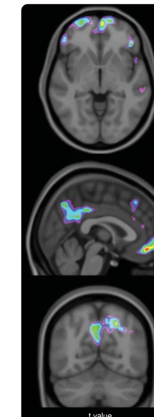
Figure 1 Individuals with preclinical AD with worse NPS have higher [18F]FDG
uptake in PCC, vmPFC, AI at baseline
Statistical parametric map overlaid on a structural MRI scan shows regions in the PCC, vmPFC, and right AI where higher [18F]FDG uptake was found in individuals with preclinical AD
with both amyloid and tau pathologies and higher NPI scores at baseline. The analysis was corrected for age, sex, educa-tion, andAPOEe4 status and multiple comparisons corrected with a false discovery rate corrected atp,0.001. AD5
Alzheimer disease; AI5anterior insula; [18F]FDG5[18F]
fluo-rodeoxyglucose; NPI5Neuropsychiatric Inventory; NPS5
neuropsychiatric symptoms; PCC5posterior cingulate cor-tex; vmPFC5ventromedial prefrontal cortex.
Figure 2 NPI predicts 2-year [18F]FDG uptake
decline in PCC and vmPFC of individuals with preclinical AD
Statistical parametric map overlaid on a structural MRI scan showed regions in the PCC and vmPFC where 2-year [18F]FDG uptake decline occurred as a function of baseline
NPI in individuals with preclinical AD with both amyloid and tau pathologies. The analysis was corrected for age, sex, education, andAPOEe4 status and multiple comparisons corrected with a false discovery rate corrected atp ,
0.001. AD5Alzheimer disease; [18F]FDG5[18F]
fluoro-deoxyglucose; NPI5Neuropsychiatric Inventory; PCC5
between specific brain regions involving early AD
pathophysiology and behavioral control. Our results
are in line with recent evidence from both functional
and metabolic imaging studies. For example, a
posi-tive correlation has been shown between increased
functional connectivity in the anterior cingulate and
right insula regions of the salience network (SN) and
irritability, agitation, disinhibition, aberrant motor
behavior, and euphoria symptoms in patients with
AD,
19while NPS changes have been proposed to
reflect aberrant increases in SN functional
connectiv-ity.
20Most recently, NPS are found to be associated
with increased glucose metabolism in the left insula,
anterior cingulate gyrus, and superior frontal gyrus of
patients with early-onset AD.
5Together, these results
support a metabolic dysfunction in limbic networks
affected by early AD pathophysiology, leading to NPS
manifestations.
The AI, which is a key node of the SN, plays a key
role in generating appropriate behavioral responses by
integrating affective, homeostatic, and higher-order
cognitive processes.
21Hyperactivity in the AI may
lead to enhanced salience detection, which is associated
with NPS such as irritability or anxiety,
19,22while
hypo-activity in the AI is associated with cognitive slowing
and attentional deficits.
23The vmPFC regulates
behav-ioral responses particularly in the context of changing
reinforcement contingencies, emotional regulation, and
decision-making tasks.
24,25Higher NPI scores and
high-er [
18F]FDG uptake in the right AI and vmPFC at
baseline may explain the enhanced neuropsychological
presentations in these cognitively normal individuals
who have both amyloid and tau pathologies.
Our findings of higher [
18F]FDG uptake in the
PCC in individuals with preclinical AD with higher
NPI scores at baseline may represent a change in
tissue metabolism in response to amyloid-related
neu-rotoxicity to stabilize neuropsychiatric manifestations
in the early stages of the disease.
26The PCC, a central
part of the default mode network, is a highly
con-nected and metabolically active region that plays a key
role in supporting cognition.
27While
hypometabo-lism in the PCC has been consistently observed in
early AD pathophysiology,
28our findings suggest an
early and transient neuronal compensation as a
man-ifestation of cognitive reserve to preserve function
that has been shown previously in cognitively normal
individuals and those with MCI in association with
amyloid deposition.
26,29As amyloid pathology
pro-gressively accumulates in the brain, metabolic decline
occurs in response to the demands of the neuronal
injury.
26In line with this concept, we showed that
individuals with preclinical AD with worse NPS had
subsequent hypometabolism in the PCC at the 2-year
follow-up.
In addition, sleep/nighttime behavior disorders
and irritability/lability were the NPI components that
drove the metabolic dysfunctions in individuals with
preclinical AD. Both symptoms have been shown to
be associated with early AD pathophysiology. For
example, in the National Alzheimer
’
s Coordinating
Center data, noncognitive symptoms of early AD
presented in 3 phases, and both irritability and
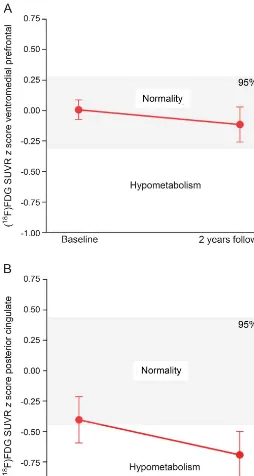
Figure 3 NPI predicts hypometabolism in PCC but not in vmPFC in individuals with preclinical AD
The dots represent thez-scored mean [18F]FDG SUVR in (A) vmPFC and (B) PCC in individuals
with preclinical AD at baseline and the 2-year follow-up. The 2-year follow-up mean [18F]FDG
uptake in PCC was lower (95% CI) than the mean of biomarker-negative individuals at follow-up, which suggests the presence of hypometabolism. However, the 2-year follow-up mean [18F]FDG uptake in vmPFC remained within normal range in relation to
biomarker-negative individuals. AD5Alzheimer disease; CI5confidence interval; [18F]FDG5[18F]
nighttime behavior changes were found to be some of
the first symptoms to occur.
4In a longitudinal study
of the Massachusetts Alzheimer
’
s Disease Research
Center Cohort in which participants were cognitively
normal, had subjective cognitive concerns, or were
diagnosed with MCI, worse affective factor score
pre-dicted time to progression to a worse diagnosis in all 3
groups, driven by depression, agitation, and
irritabil-ity.
30In the Mayo Clinic Study of Aging, baseline
irritability in cognitively normal adults increased the
risk of developing MCI over a median of 5 years
(hazard ratio 1.84, 95% confidence interval 1.31
–
2.58).
31Irritability and lability are NPS linked to
abnormal emotional processing, associated with the
vmPFC and PCC.
25,32In fact, reduced volume of the
PCC gray matter has been shown to contribute to
emotional instability.
33Sleep disturbances are
com-mon symptoms of AD and may be early indicators of
amyloid pathology and dementia.
34Sleep deprivation
can exacerbate amyloid pathology and mediate
neu-ronal injury, while circadian dysfunction may also
lead to cerebral oxidative stress and synaptic
dam-age.
34Worse sleep quality is also shown to be
associ-ated with amyloid deposition in the preclinical AD
stage.
35Patients with obstructive sleep apnea and
day-time somnolence have decreased brain metabolism in
the precuneus, middle and posterior cingulate gyrus,
and parieto-occipital and prefrontal cortices,
36while
healthy adults with dream enactment behavior have
significantly lower glucose metabolism in brain
re-gions, including the PCC.
37Hence, the finding of
sleep/nighttime behavior disorders and irritability/
lability symptoms predicting hypometabolism in the
PCC supports these NPS as early manifestations of
AD pathophysiology.
Although baseline NPI score predicted [
18F]FDG
uptake decline, it did not predict memory decline
using the ADNI-mem. Given that our study
popu-lation is composed of a group of highly educated
individuals, high cognitive reserve and education
might play a role in stabilizing the cognitive function
in the early stages of the disease.
9,38,39In addition,
NPS may precede cognitive manifestations in early
AD pathophysiology. In the Alzheimer
’
s Disease
Cooperative Study Prevention Instrument Project,
the presence of baseline behavioral symptoms
pre-dicts conversion to a CDR score
$
0.5 over 4 years
in cognitively healthy older individuals,
40while in
the National Alzheimer
’
s Coordinating Center Data,
a significantly earlier presence of NPS is found in
cognitively normal people who progressed in CDR
score compared to those who did not progress.
4This
further supports NPS as early manifestations of AD
pathophysiology.
There are limitations to our study. Although NPI
is the most widely used instrument to measure NPS,
it was initially designed for dementia studies, not for
preclinical AD studies.
11Hence, the sensitivity of
NPI in diagnosing NPS in individuals with preclinical
AD is not known. In addition, given that the NPI is
based on responses from an informed caregiver, the
NPI scores may not accurately reflect the NPS of
study participants. The ADNI database is made up
of highly educated individuals who volunteered to
participate in the study that focused on AD research.
This may introduce selection bias in that the study
population is a self-selected group of individuals who
may have concerns about their cognition. The
self-selected character of our study population and the
relatively small sample size of our study limit the
generalizability of our findings to a broader
commu-nity. Therefore, our findings will need to be
con-firmed in a larger population-based cohort.
Our findings support an emerging conceptual
framework that NPS, driven by sleep behavior and
irritability domains, are early manifestations of AD
pathophysiology. Therefore, early NPS may further
contribute to the characterization of the preclinical
AD stage.
AUTHOR CONTRIBUTIONS
Kok Pin Ng: study concept, design, analysis and interpretation of data, compose figures and manuscript draft. Tharick A. Pascoal: study design, analysis and interpretation of data, compose figures and manuscript draft. Sulantha Mathotaarachchi, Chang-Oh Chung, Andréa L. Benedet, Monica Shin, Min Su Kang: image data processing and manuscript draft. Xiaofeng Li, Maowen Ba: manuscript draft. Nagaendran Kandiah: study concept, design and manuscript draft. Pedro Rosa-Neto, Serge Gauthier: study concept, design, study supervision, and critical review of manu-script for intellectual content.
STUDY FUNDING
Southern California. ADNI data are disseminated by the Laboratory for Neuro Imaging at the University of Southern California.
DISCLOSURE
K. Ng, T. Pascoal, S. Mathotaarachchi, C. Chung, A. Benedet, M. Shin, M. Kang, X. Li, M. Ba, N. Kandiah, and P. Rosa-Neto report no disclo-sures relevant to the manuscript. S. Gauthier received honoraria for serv-ing on the scientific advisory boards of Alzheon, Axovant, Lilly, Lundbeck, Novartis, Schwabe, and TauRx and on the Data Safety Mon-itoring Board of a study sponsored by Eisai and studies run by the Alz-heimer’s Disease Cooperative Study and by the Alzheimer’s Therapeutic Research Institute. Research was funded by CIHR and NIH. Go to Neurology.org for full disclosures.
Received October 14, 2016. Accepted in final form February 17, 2017.
REFERENCES
1. Lyketsos CG, Carrillo MC, Ryan JM, et al. Neuropsychi-atric symptoms in Alzheimer’s disease. Alzheimers Dement 2012;7:532–539.
2. Fischer CE, Ismail Z, Schweizer TA. Delusions increase functional impairment in Alzheimer’s disease. Dement Geriatr Cogn Disord 2012;33:393–399.
3. Teng E, Lu PH, Cummings JL. Neuropsychiatric symp-toms are associated with progression from mild cognitive impairment to Alzheimer’s disease. Dement Geriatr Cogn Disord 2007;24:253–259.
4. Masters MC, Morris JC, Roe CM.“Noncognitive” symp-toms of early Alzheimer disease : a longitudinal analysis. Neurology 2015;84:617–622.
5. Ballarini T, Iaccarino L, Magnani G, et al. Neuropsychi-atric subsyndromes and brain metabolic network dysfunc-tions in early onset Alzheimer’s disease. Hum Brain Mapp 2016;37:4234–4247.
6. Ismail Z, Smith EE, Geda Y, et al. Neuropsychiatric symp-toms as early manifestations of emergent dementia: pro-visional diagnostic criteria for mild behavioral impairment. Alzheimers Dement 2016;12:195–202.
7. Ruider H, Krell-Roesch J, Stokin G, et al. FDG-PET of the brain and neuropsychiatric symptoms in normal cog-nitive aging: the Mayo Clinic Study of Aging. J Alzheimers Dis 2016;53:1609–1616.
8. Mosconi L, Berti V, Glodzik L, Pupi A, De Santi S, De Leon MJ. Pre-clinical detection of Alzheimer’s disease using FDG-PET, with or without amyloid imaging. J Alzheimers Dis 2010;20:843–854.
9. Dubois B, Hampel H, Feldman HH, et al. Preclinical Alzheimer’s disease: definition, natural history, and diagnostic criteria. Alzheimers Dement 2016;12:292–
323.
10. Edmonds EC, Delano-Wood L, Galasko DR, Salmon DP, Bondi MW; Alzheimer’s Disease Neuroimaging Ini-tiative. Subtle cognitive decline and biomarker staging in preclinical Alzheimer’s disease. J Alzheimers Dis 2015; 47:231–242.
11. Cummings JL, Mega M, Gray K, Rosenberg-Thompson S, Carusi DA, Gornbein J. The Neuropsychiatric Inven-tory: comprehensive assessment of psychopathology in dementia. Neurology 1994;44:2308–2314.
12. Crane PK, Carle A, Gibbons LE, et al. Development and assessment of a composite score for memory in the Alz-heimer’s Disease Neuroimaging Initiative (ADNI). Brain Imaging Behav 2012;6:502–516.
13. Shaw LM, Vanderstichele H, Knapik-Czajka M, et al. Cerebrospinal fluid biomarker signature in Alzheimer’s
Disease Neuroimaging Initiative subjects. Ann Neurol 2009;65:403–413.
14. Zijdenbos AP, Forghani R, Evans AC. Automatic“ pipe-line”analysis of 3-D MRI data for clinical trials: applica-tion to multiple sclerosis. IEEE Trans Med Imaging 2002; 21:1280–1291.
15. Pascoal TA, Mathotaarachchi S, Mohades S, et al. Amyloid-b and hyperphosphorylated tau synergy drives metabolic decline in preclinical Alzheimer’s disease. Mol Psychiatry 2017;22:306–311.
16. Fawcett T. An introduction to ROC analysis. Pattern Rec-ognit Lett 2006;27:861–874.
17. Jack CR, Lowe VJ, Weigand SD, et al. Serial PIB and MRI in normal, mild cognitive impairment and Alz-heimer’s disease: implications for sequence of patholog-ical events in Alzheimer’s disease. Brain 2009;132: 1355–1365.
18. R Development Core Team. R: a language and environ-ment for statistical computing. R Found Stat Comput 2015;1:409.
19. Balthazar MLF, Pereira FRS, Lopes TM, et al. Neuropsy-chiatric symptoms in Alzheimer’s disease are related to functional connectivity alterations in the salience network. Hum Brain Mapp 2014;35:1237–1246.
20. Zhou J, Seeley WW. Network dysfunction in Alzheimer’s disease and frontotemporal dementia: implications for psy-chiatry. Biol Psychiatry 2014;75:565–573.
21. Menon V, Uddin LQ. Saliency, switching, attention and control: a network model of insula function. Brain Struct Funct 2010;214:655–667.
22. Stein MB, Simmons AN, Feinstein JS, Paulus MP. Increased amygdala and insula activation during emotion processing in anxiety-prone subjects. Am J Psychiatry 2007;164:318–327.
23. Blanc F, Colloby SJ, Cretin B, et al. Grey matter atro-phy in prodromal stage of dementia with Lewy bodies and Alzheimer’s disease. Alzheimers Res Ther 2016;8: 31.
24. Zald DH, Andreotti C. Neuropsychological assessment of the orbital and ventromedial prefrontal cortex. Neuropsy-chologia 2010;48:3377–3391.
25. Etkin A, Büchel C, Gross JJ. The neural bases of emotion regulation. Nat Rev Neurosci 2015;16:693–700. 26. Ashraf A, Fan Z, Brooks DJ, Edison P. Cortical
hyperme-tabolism in MCI subjects: a compensatory mechanism? Eur J Nucl Med Mol Imaging 2014;42:447–458. 27. Leech R, Sharp DJ. The role of the posterior cingulate
cortex in cognition and disease. Brain 2014;137:12–32. 28. Johnson Ka, Jones K, Holman BL, et al. Preclinical pre-diction of Alzheimer’s disease using SPECT. Neurology 1998;50:1563–1571.
29. Ossenkoppele R, Madison C, Oh H, Wirth M, Van Berckel BNM, Jagust WJ. Is verbal episodic memory in elderly with amyloid deposits preserved through altered neuronal function? Cereb Cortex 2014;24: 2210–2218.
30. Donovan NJ, Amariglio RE, Zoller AS, et al. Subjective cognitive concerns and neuropsychiatric predictors of pro-gression to the early clinical stages of Alzheimer disease. Am J Geriatr Psychiatry 2014;22:1642–1651.
32. Phillips ML, Drevets WC, Rauch SL, Lane R. Neurobi-ology of emotion perception II: implications for major psychiatric disorders. Biol Psychiatry 2003;54:515–528. 33. Hazlett EA, New AS, Newmark R, et al. Reduced anterior
and posterior cingulate gray matter in borderline person-ality disorder. Biol Psychiatry 2005;58:614–623. 34. Musiek ES, Xiong DD, Holtzman DM. Sleep, circadian
rhythms, and the pathogenesis of Alzheimer disease. Exp Mol Med 2015;47:e148.
35. Ju YES, McLeland JS, Toedebusch CD, et al. Sleep quality and preclinical Alzheimer disease. JAMA Neurol 2013;70: 587–593.
36. Yaouhi K, Bertran F, Clochon P, et al. A combined neuro-psychological and brain imaging study of obstructive sleep apnea. J Sleep Res 2009;18:36–48.
37. Caselli RJ, Chen K, Bandy D, et al. A preliminary fluo-rodeoxyglucose positron emission tomography study in healthy adults reporting dream-enactment behavior. Sleep 2006;29:927–933.
38. Roe CM, Xiong C, Miller JP, Morris JC. Education and Alzheimer disease without dementia: support for the cognitive reserve hypothesis. Neurology 2007;68:223– 228.
39. Stern Y. Cognitive reserve and Alzheimer disease. Alz-heimer Dis Assoc Disord 2006;20:S69–S74.
40. Banks SJ, Raman R, He F, et al. The Alzheimer’s Disease Cooperative Study Prevention Instrument Project: longi-tudinal outcome of behavioral measures as predictors of cognitive decline. Dement Geriatr Cogn Dis Extra 2014;4: 509–516.