International Journal of Women’s Health 2018:10 251–256
International Journal of Women’s Health
Dove
press
submit your manuscript | www.dovepress.com 251
O r I g I n a l r e s e a r c H
open access to scientific and medical research
Open access Full Text article
Intrapartum ultrasound: viewpoint of midwives
and parturient women and reproducibility
adrielle Van adrichem1
ellen Faes1
Kristof Kinget2
Yves Jacquemyn1
1antwerp University Hospital UZa,
antwerp University Ua – asTarc, edegem, Belgium; 2KlIna Hospital,
Brasschaat, Belgium
Introduction: Vaginal examination (VE) is known to be subjective in interpretation and is considered uncomfortable by many women. Intrapartum ultrasound aims to be more objective and less invasive. The purpose of this study was to evaluate the acceptability of introducing intrapartum ultrasound to both midwives and parturients. Furthermore, we wanted to evalu-ate the reproducibility of different measurements when introduced de novo among operators without prior ultrasound experience.
Subjects and methods: This study determined and interobserver variability of intra-partum ultrasound in nulliparous women in labor. Ultrasound examinations were performed independently by a midwife and a gynecologist. The symphysis–head distance (SHD) and the angle of progression (AOP) were measured by translabial ultrasound. Structured questionnaires were given to midwives and parturients. Intraclass correlation coefficient (ICC) and limits of agreement (LA) were calculated to evaluate variability.
Results: A total of 33 patients were included; of whom, 28 filled in the questionnaire. A total of 19 midwives working on a delivery ward were asked to respond to the questionnaire, and 13 returned the forms. Midwives clearly continued to prefer VE over ultrasound, the majority evaluated translabial ultrasound as easy to use, but some declared to be unable to use it. The majority of patients, 71%, preferred ultrasound over VE. Reproducibility of intrapartum trans-labial ultrasound was good; ICC for interobserver variability was 0.603 (p=0.001) for SHD, and ICC for intraobserver variability was 0.844 (p0.001) and 0.914 (p0.001) for SHD and AOP, respectively.
Conclusion: Patients prefer ultrasound over VE; midwives tend to stick to trusted VE. Repro-ducibility of intrapartum ultrasound in non-experienced operators is good.
Keywords: intrapartum, ultrasound, labor
Introduction
For centuries, progression of labor has been evaluated by vaginal examination (VE), describing dilation, effacement, consistency and position of the cervix and descent of the fetal head. Interobserver variability of VE is large, and it is a traditional but unreliable
method.1–4 Furthermore, the vaginal examination can be experienced as intimidating and
uncomfortable by a woman in labor. In recent years, intrapartum ultrasound has become increasingly popular. During labor, ultrasound can be performed by transabdominal, trans-labial and transperineal way. Measurements for dilation, effacement, position and descent
of the fetal head have been developed.2,3,5,6 Ultrasound during labor has been proven to be
able to predict labor outcomes in primiparous women with prolonged first stage of labor.7
Moreover, intrapartum ultrasound evaluation is superior to VE in terms of identifying the
correct fetal head position.4 The symphysis–head distance (SHD) and the angle of
progres-sion (AOP) have been the most studied measurements by transperineal ultrasound.7–10
correspondence: Yves Jacquemyn Department of Obstetrics and gynaecology, antwerp University Hospital UZa, Wilrijkstraat 10, 2650 edegem, Belgium Tel +32 3 821 5945 Fax +32 3 825 5883
email yves.jacquemyn@uza.be
Journal name: International Journal of Women’s Health Article Designation: Original Research
Year: 2018 Volume: 10
Running head verso: Van Adrichem et al
Running head recto: Intrapartum ultrasound: viewpoint DOI: 155865
International Journal of Women's Health downloaded from https://www.dovepress.com/ by 118.70.13.36 on 23-Aug-2020
For personal use only.
Dovepress
Van adrichem et al
The SHD is the distance between the under border of the symphysis to the fetal skull measured perpendicular to the axis of the symphysis (Figure 1). This distance has
been shown to be predictive for the outcome of labor,8 its
measurement has been described as reproducible and a
nega-tive correlation exists with the AOP.6 The AOP is the angle
between the axis of the symphysis and the line drawn from the under border of the symphysis tangential to the fetal skull (Figure 2). This angle is considered a reproducible indicator
to evaluate fetal descent during labor.9,11
Studies that have compared ultrasound and VE have repeatedly demonstrated ultrasound to be more accu-rate; patients experience ultrasound as less invasive and
less painful.1,2,4
In some countries, ultrasound during pregnancy and labor is performed by midwives; in Belgium, ultrasound is not part of midwife competencies. The aim of this study
was to examine the acceptability of parturients and the viewpoint of the midwife when introducing intrapartum ultrasound. Furthermore, we evaluated the intra- and interobserver variability of intrapartum sonographic mea-surements performed by midwives without ultrasound experience.
Subjects and methods
Women and midwives were recruited from an academic (Antwerp University Hospital, Universitair Ziekenhuis Antwerpen) and a nonacademic (Klinieken Noord Antwerpen Hospital) maternity department. The study was approved by the ethics committee of both institutions. Written informed consent was obtained from women during labor and from midwives performing the ultrasound.
End points are inter- and intra-observer variability and acceptability by parturients and midwives. A convenience sample was used. Women included were nulliparous in spontaneous labor, with a singleton term pregnancy and cephalic presentation.
Ultrasound was performed with the machine as avail-able on the delivery ward, Voluson P6, with convex 4C-RS transducer (GE Medical Systems, Zipf, Austria) in UZA and Aloka Prosound Alpha 6 and with convex UST-9115-5 transducer (Hitachi Ltd., Tokyo, Japan) in KLINA.
Measurements were standardized and performed by translabial way. All midwives who took part in the study received a 1-hour training consisting of a slideshow pre-sentation and one supervised measurement. Engagement of the head differed between parturients between Hodge 2 and 3. Urinary bladder was emptied before measurement. Ultrasound gel was placed on the transducer that was then covered by a glove; gel was put on the glove. The patient was put in a semi-recumbent position with legs flexed. The transducer was placed in a midsagittal plane, and the fetal skull and maternal symphysis were visualized.
To evaluate the interobserver variability, two operators performed measurements independently, and they had no access to the measurement of the other. To estimate the intraobserver variability, one operator performed three repetitive measurements.
Symphysis–head distance (SHD) was performed as
described by Youssef et al8 The distance from the under
border of the symphysis to the fetal skull was measured perpendicular to the length axis of the symphysis (Figure 1). The AOP was defined as the angle between the length axis of the symphysis and a line from the inferior border of the symphysis tangential to the fetal skull (Figure 2).
Figure 1 sHD is measured by drawing a line through the symphysis (A) and from the end of the symphysis a second line perpendicular to the fetal skull (B); the last line is the sHD.
Abbreviation: sHD, symphysis–head distance.
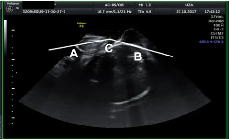
Figure 2 aOP is measured by drawing a line through the axis of the symphysis to the lowest point of the symphysis (A); next, from this point, a line is drawn at the anterior border of the skull (C); the angle between these two lines is measured (B) and it is the aOP.
Abbreviation: aOP, angle of progression.
International Journal of Women's Health downloaded from https://www.dovepress.com/ by 118.70.13.36 on 23-Aug-2020
Dovepress Intrapartum ultrasound: viewpoint
Acceptability of ultrasound examination for women in labor was tested by presenting statements to the patients, at least 2 hours after delivery. For each statement, the possible answers were as follows: strongly agree, agree, neutral, disagree and strongly disagree, resulting in a 5-point Likert score. For each ultrasound, a written informed consent was signed. Patients who entered neutral for VE did not remember if an explicit informed consent was asked, but they did not feel forced to undergo the examination in any way.
Statements were subdivided into categories, concerning VE (12 statements, eg, I experienced the vaginal examina-tion as being painful), translabial ultrasound (14 statements, eg, I experienced this ultrasound as painful) and the condi-tions under which the examination was performed (five statements, eg, during ultrasound examination, I prefer my partner to be in the same room). The questionnaire was based
on similar studies on vaginal ultrasound.12,13 In addition, a
Visual Analog Score for pain during the examination was performed. Pain scores were evaluated at rest (between uter-ine contractions) and were compared with the pain scores of previous VE (with or without epidural anesthesia).
A similar questionnaire was developed for the midwives including 18 statements on ultrasound technique (eg, the tech-nique was easy to learn for me) and 11 statements concerning vaginal examination versus translabial ultrasound (eg, vagi-nal examination provides more clear results than ultrasound).
The questionnaire was based on a similar example.14
SPSS 24.0 (IBM Corporation, Armonk, NY, USA) was used for statistical analysis. Interobserver and intraobserver variability was examined using intraclass correlation coef-ficient (ICC; two-way mixed absolute agreement single mea-sures). ICC0.70 is considered good correlation; ICC0.3 is reported as low correlation and ICC between 0.5 and 0.7 are
scored as moderate correlation.15 To visualize results, Bland–
Altman plots and limits of agreement (LA) have been used. To analyze acceptability, the 5-point Likert scale was coded in three groups: agree, neutral, not agree. Fisher’s exact test was performed with p0.05 accepted as significant. Wilcoxon signed-rank test was used for the comparison of Visual Analog Score results between groups.
Results
A total of 19 midwives received the training for intrapartum ultrasound, of these 11 midwives actually performed the examinations and filled in the questionnaire. Table 1 presents characteristics of this population. From the 33 included women, three were excluded because ultrasound imaging was not performed according to the protocol. It was possible to
measure AOP in every patient, and SHD was not measured in four patients because the fetal head did not yet reach to the under border of the symphysis.
Questionnaires were given to 33 women in labor, the questionnaires were always given after the ultrasound exami-nations and 28 women returned the questionnaire. All 19 midwives who received the training also received the ques-tionnaire for midwives, and 13 returned the quesques-tionnaire.
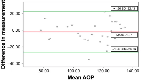
Table 2 presents the ICCs for interobserver variability, and Figures 3 and 4 illustrate Bland–Altman plots for inter-observer variability. Table 3 presents ICCs for intrainter-observer variability.
A total of 20 of the 28 patients (71.4%) preferred intra-partum ultrasound over VE; the other eight patients (28.6%) had no preference. Table 4 presents the results of the different statements concerning women in labor.
Table 5 presents the answers to the statements that were given to the midwives who have been performing ultra-sound for this study. The majority of midwives considered ultrasound as easy to use (63.7%) with an advantage for the patient. As to be expected, midwives who have been in
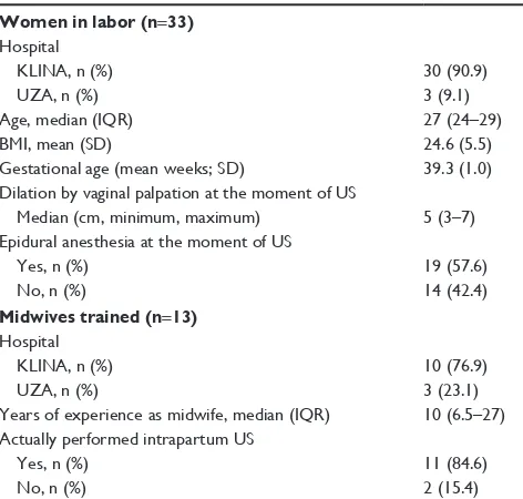
Table 1 characteristics of study population Women in labor (n=33)
Hospital
KlIna, n (%) 30 (90.9)
UZa, n (%) 3 (9.1)
age, median (IQr) 27 (24–29)
BMI, mean (sD) 24.6 (5.5)
gestational age (mean weeks; sD) 39.3 (1.0)
Dilation by vaginal palpation at the moment of Us
Median (cm, minimum, maximum) 5 (3–7)
epidural anesthesia at the moment of Us
Yes, n (%) 19 (57.6)
no, n (%) 14 (42.4)
Midwives trained (n=13) Hospital
KlIna, n (%) 10 (76.9)
UZa, n (%) 3 (23.1)
Years of experience as midwife, median (IQr) 10 (6.5–27) actually performed intrapartum Us
Yes, n (%) 11 (84.6)
no, n (%) 2 (15.4)
Abbreviations: BMI, body mass index; IQr, interquartile range; Us, ultrasound; UZa, Universitair Ziekenhuis antwerpen; KlIna, Klinieken noord antwerpen.
Table 2 Icc for interobserver variability
Measurement n ICC 95% CI p-value
sHD 26 0.603 0.285–0.801 0.001
aOP 30 0.755 0.549–0.875 0.001
Abbreviations: aOP, angle of progression; sHD, symphysis–head distance; Icc, intraclass correlation coefficient.
International Journal of Women's Health downloaded from https://www.dovepress.com/ by 118.70.13.36 on 23-Aug-2020
Dovepress
Van adrichem et al
practice for several years felt more at ease with VE as com-pared with intrapartum ultrasound after minimal training.
Discussion
Concerning interobserver variability after minimal training in intrapartum ultrasound as in this study, moderate agree-ment between measureagree-ments was reached for SHD. For AOP, a good agreement between different observers could be reached. The results that have been published in previous studies were conflicting and mainly obtained with observ-ers who had previous ultrasound experience. The study by
Duckelmann et al9 presented an ICC of 0.72, comparable to
our results. In the same study, it was examined whether a difference in previous ultrasound experience influenced vari-ability. Observers with more experience demonstrated larger ICC and a smaller confidence interval, and this was confirmed
in other studies.16 In addition, the presence of uterine
contrac-tions can influence differences in the ICC.8,17
Intraobserver variability in our study is good for both SHD and AOP, and the results are comparable to those from
other studies.16
When preparing this study, it became clear that mid-wives in the Belgian context of not receiving any formal ultrasound training during training are not very motivated to introduce intrapartum ultrasound in their practice. Although the majority of midwives declare not to be able to perform intrapartum ultrasound autonomously and correctly (72.7%); at the same moment, they considered the technique easy to use and perform. Almost all midwives have experienced learning intrapartum ultrasound as an agreeable expansion of their activities. Midwives do find ultrasound an advantage for the patients but not for themselves. This discrepancy and the lack of personal advantage can hinder the introduction of intrapartum ultrasound in clinical practice.
Women in labor seem to prefer the noninvasive transla-bial intrapartum ultrasound over VE; the difference in pain between both types of examination is remarkable resulting in a significantly different visual analog pain score. To the best of our knowledge, this is the first study that compares women’s experiences of digital VE with translabial ultra-sound; all previous studies have compared VE with
trans-vaginal ultrasound.12
A major bias of this study is that the technique that mid wives have been using for several years in daily work, and feel very much at ease with, is compared with something they have learned very recently and in a very short time. To introduce intrapartum ultrasound in daily midwifery practice, one needs more than a short training. Eventual advantages for women in labor have to be put against getting used to new technical skills.
Conclusion
This small study demonstrates acceptable intra- and interob-server variability for both SHD and AOP. Women in labor seem to prefer translabial ultrasound over VE; more: inten-sive training of midwives will allow for eventual introduction of intrapartum ultrasound in clinical practice. Intrapartum ultrasound for the evaluation of labor progress could be a supplementary tool for active management and may reduce the use of unnecessary VE during labor and improves obstet-ric and neonatal outcomes. This study can be considered as a preliminary study; a larger prospective study is needed to verify our results.
Table 3 Icc for intraobserver variability
Measurement n ICC 95% CI p-value
sHD 28 0.844 0.732–0.919 0.001
aOP 30 0.914 0.849–0.955 0.001
Abbreviations: aOP, angle of progression; sHD, symphysis–head distance; Icc, intraclass correlation coefficient.
±
±
±
0HDQ+6'
'LIIHUHQFHLQPHDVXUHPHQW
0HDQ± 6'
±6' ±
Figure 3 Bland–altman plot for interobserver variability between two observers for sHD.
Note: red line represents mean; green lines represent ±1.96 sD.
Abbreviation: sHD, symphysis–head distance.
Figure 4 Bland–altman plot for interobserver variability for aOP.
Note: red line represents mean; green lines represent ±1.96 sD.
Abbreviation: aOP, angle of progression.
±
±
0HDQ$23
'LIIHUHQFHLQPHDVXUHPHQW
0HDQ± 6'
±6' ±
International Journal of Women's Health downloaded from https://www.dovepress.com/ by 118.70.13.36 on 23-Aug-2020
Dovepress Intrapartum ultrasound: viewpoint
Table 4 answers by women in labor on statements concerning Ve and translabial ultrasound
Statement n=28 Opinion VE, n (%) Translabial
ultrasound, n (%)
p-value
I experienced this as painful agree
neutral not agree
19 (67.9) 1 (3.6) 8 (28.6)
28 (100) 0.004
I was well informed about what was going to happen agree neutral not agree
26 (92.9) 2 (7.1) 0
28 (100) 0.001
My consent was asked agree
neutral not agree
24 (85.7) 4 (14.3) 0
28 (100) 0.00
The test was performed with respect for me agree
neutral not agree
28 (100) 28 (100) 1.000
I felt my privacy was respected during the test agree
neutral not agree
27 (96.4) 1 (3.6) 0
28 (100) 0.001
This kind of examination makes me feel ashamed agree
neutral not agree
2 (7.1) 6 (21.4) 20 (71.4)
0 2 (7.1) 26 (92.9)
0.180
The test is “not” stressful agree
neutral not agree
11 (39.3) 7 (25) 10 (35.7)
25 (89.3) 1 (3.6) 2 (7.1)
0.787
This test gives me a feeling of anxiety agree
neutral not agree
1 (3.6) 3 (10.7) 23 (82.1)
1 (3.6) 0 27 (96.4)
0.984
The test was “less” comfortable than I expected agree
neutral not agree
12 (42.9) 7 (25) 9 (32.1)
1 (3.6) 0 27 (96.4)
0.501
The information that is given to me after the test makes me feel anxious agree neutral not agree
1 (3.6) 2 (7.1) 25 (89.3)
0 1 (3.6) 27 (96.4)
0.107
I experienced this test as rude agree
neutral not agree
4 (14.3) 5 (17.9) 19 (67.9)
28 (100) 0.001
Visual analog score for pain Median
IQr
4 2–6
0 0–0
0.001
Abbreviations: IQr, interquartile range; Ve, vaginal examination.
Table 5 answers to statements on Ve and translabial ultrasound by midwives
Statement n=11 Agree, n (%) Neutral, n (%) Not agree, n (%)
Ultrasound technique was easy to learn 6 (54.5) 3 (27.3) 2 (18.2)
Ultrasound was easy to use 8 (72.7) 2 (18.2) 1 (9.1)
as a user, I feel intrapartum ultrasound has advantages for me as compared with manual Ve 1 (9.1) 3 (27.3) 7 (63.6)
I think intrapartum ultrasound has advantages for the woman in labor 7 (63.6) 0 4 (36.4)
It is easy to recognize the anatomical landmarks 6 (54.5) 1 (9.1) 4 (36.4)
Introducing ultrasound in practice was easy 8 (72.7) 0 3 (27.3)
I feel able to do this ultrasound autonomously and correctly 0 3 (27.3) 8 (72.7)
I need more training to perform this ultrasound correctly 8 (72.7) 3 (27.3) 0
I want to continue using ultrasound in labor 6 (54.5) 3 (27.3) 2 (18.2)
I enjoyed learning a new technique 10 (90.9) 1 (9.1) 0
classic Ve still feels easier to perform than ultrasound 10 (90.9) 0 1 (9.1)
VE fits better in our workflow 10 (90.9) 1 (9.1) 0
For me as a midwife, vagina palpation has more advantages than ultrasound 10 (90.9) 1 (9.1) 0 By performing Ve, I am able to provide more information to the patient
(n=10, one blank answer)
8 (80) 2 (20) 0
I feel more comfortable when performing Ve than doing intrapartum ultrasound 11 (100) 0 0
I find it easier to find anatomical landmarks with VE 8 (72.7) 0 3 (27.3)
I feel able to “perform Ve” autonomously and correctly 11 (100) 0 0
Abbreviation: Ve, vaginal examination.
International Journal of Women's Health downloaded from https://www.dovepress.com/ by 118.70.13.36 on 23-Aug-2020
International Journal of Women’s Health
Publish your work in this journal
Submit your manuscript here: http://www.dovepress.com/international-journal-of-womens-health-journal
The International Journal of Women’s Health is an international, peer-reviewed open-access journal publishing original research, reports, editorials, reviews and commentaries on all aspects of women’s healthcare including gynecology, obstetrics, and breast cancer. The manuscript management system is completely online and includes
a very quick and fair peer-review system, which is all easy to use. Visit http://www.dovepress.com/testimonials.php to read real quotes from published authors.
Dovepress
Dove
press
Van adrichem et al
Disclosure
The authors report no conflicts of interest in this work.
References
1. Benediktsdottir S, Eggebo TM, Salvesen KA. Agreement between trans-perineal ultrasound measurements and digital examinations of cervical dilatation during labor. BMC Pregnancy Childbirth. 2015;15:273. 2. Hassan WA, Eggebo TM, Ferguson M, Lees C. Simple two-dimensional
ultrasound technique to assess intrapartum cervical dilatation: a pilot study. Ultrasound Obstet Gynecol. 2013;41(4):413–418.
3. Debska M, Kretowicz P, Debski R. Intrapartum sonography – eccentricity or necessity? J Ultrason. 2015;15(61):125–136.
4. Akmal S, Kametas N, Tsoi E, Hargreaves C, Nicolaides KH. Compari-son of transvaginal digital examination with intrapartum Compari-sonography to determine fetal head position before instrumental delivery. Ultrasound
Obstet Gynecol. 2003;21(5):437–440.
5. Malvasi A, Giacci F, Gustapane S, Sparic R, Barbera A, Tinelli A. Intrapartum sonographic signs: new diagnostic tools in malposi-tion and malrotamalposi-tion. J Matern Fetal Neonatal Med. 2016;29(15): 2408–2413.
6. Erlik U, Wolman I. Intrapartum sonographic assessment of labor. J Obstet
Gynaecol India. 2013;63(5):297–300.
7. Torkildsen EA, Salvesen KA, Eggebo TM. Prediction of delivery mode with transperineal ultrasound in women with prolonged first stage of labor. Ultrasound Obstet Gynecol. 2011;37(6):702–708.
8. Youssef A, Maroni E, Ragusa A, et al. Fetal head-symphysis distance: a simple and reliable ultrasound index of fetal head station in labor.
Ultrasound Obstet Gynecol. 2013;41(4):419–424.
9. Duckelmann AM, Bamberg C, Michaelis SA, et al. Measurement of fetal head descent using the ‘angle of progression’ on transperineal ultrasound imaging is reliable regardless of fetal head station or ultra-sound expertise. Ultraultra-sound Obstet Gynecol. 2010;35(2):216–222. 10. Chan YT, Ng VK, Yung WK, Lo TK, Leung WC, Lau WL.
Relation-ship between intrapartum transperineal ultrasound measurement of angle of progression and head-perineum distance with correlation to conventional clinical parameters of labor progress and time to delivery.
J Matern Fetal Neonatal Med. 2015;28(12):1476–1481.
11. Levy R, Zaks S, Ben-Arie A, et al. Can angle of progression in pregnant women before onset of labor predict mode of delivery? Ultrasound
Obstet Gynecol. 2012;40(3):332–337.
12. Bennett CC, Richards DS. Patient acceptance of endovaginal ultrasound.
Ultrasound Obstet Gynecol. 2000;15(1):52–55.
13. Lewin D, Fearon B, Hemmings V, Johnson G. Women’s experiences of vaginal examinations in labour. Midwifery. 2005;21(3):267–277. 14. de Veer AJE, Francke AL. Ervaringen van Verpleegkundigen en
Verzorgenden met Nieuwe Technologieën in de Zorg [Experiences of nurses with new technologies]. Utrecht: NIVEL; 2009. Dutch. 15. Alessi J, Verhagen A. Evidence Based Diagnostiek van het
Beweging-sapparaat [Evidence based diagnosis of the musculoskeletal system]. Springer, Berlin; 2014:178. Dutch.
16. Molina FS, Terra R, Carrillo MP, Puertas A, Nicolaides KH. What is the most reliable ultrasound parameter for assessment of fetal head descent? Ultrasound Obstet Gynecol. 2010;36(4):493–499.
17. Ghi T, Maroni E, Youssef A, et al. Sonographic pattern of fetal head descent: relationship with duration of active second stage of labor and occiput position at delivery. Ultrasound Obstet Gynecol. 2014;44(1): 82–89.
International Journal of Women's Health downloaded from https://www.dovepress.com/ by 118.70.13.36 on 23-Aug-2020