HIGHLIGHTED ARTICLE
| INVESTIGATION
Contrasting Levels of Molecular Evolution on the
Mouse X Chromosome
Erica L. Larson,* Dan Vanderpool,* Sara Keeble,*,†Meng Zhou,†Brice A. J. Sarver,* Andrew D. Smith,†
Matthew D. Dean,†and Jeffrey M. Good*,1 *Division of Biological Sciences, University of Montana, Missoula, Montana 59812 and†Molecular and Computational Biology, University of Southern California, Los Angeles, California 90089
ABSTRACT The mammalian X chromosome has unusual evolutionary dynamics compared to autosomes. Faster-X evolution of spermatogenic protein-coding genes is known to be most pronounced for genes expressed late in spermatogenesis, but it is unclear if these patterns extend to other forms of molecular divergence. We tested for faster-X evolution in mice spanning three different forms of molecular evolution—divergence in protein sequence, gene expression, and DNA methylation—across different developmental stages of spermatogenesis. We used FACS to isolate individual cell populations and then generated cell-specific transcriptome profiles across different stages of spermatogenesis in two subspecies of house mice (Mus musculus), thereby overcoming a fundamental limitation of previous studies on whole tissues. We found faster-X protein evolution at all stages of spermatogenesis and faster-late protein evolution for both X-linked and autosomal genes. In contrast, there was less expression divergence late in spermatogenesis (slower late) on the X chromosome and for autosomal genes expressed primarily in testis (testis-biased). We argue that slower-late expression divergence reflects strong regulatory constraints imposed during this critical stage of sperm development and that these constraints are particularly acute on the tightly regulated sex chromosomes. We also found slower-X DNA methylation divergence based on genome-wide bisulfite sequencing of sperm from two species of mice (M. musculusandM. spretus), although it is unclear whether slower-X DNA methylation reflects development constraints in sperm or other X-linked phenomena. Our study clarifies key differences in patterns of regulatory and protein evolution across spermatogenesis that are likely to have important consequences for mammalian sex chromosome evolution, male fertility, and speciation.
KEYWORDSfaster X evolution; gene expression; DNA methylation;fluorescence-activated cell sorting; postmeiotic sex chromosome repression (PSCR)
T
HE X chromosome plays a disproportionately large role in adaptation and speciation (Bachtroget al.2011; Ellegren 2011; Charlesworth 2013), but the underlying molecular and evolutionary drivers of these patterns remain unclear. On one hand, the X chromosome often shows strong signatures of evolutionary constraint. For example, the evolution of dosage compensation via epigenetic X-chromosome inactivation (XCI) in females (Lyon 1961, 1962) imposes regulatory con-straints that select for strong conservation of X-linked gene content in placental mammals (Ohno 1967; Kohn et al.2004). On the other hand, these inherent constraints are punctuated by strong specialization in X-linked gene content (Emersonet al.2004; Muelleret al.2008, 2013; Potrzebowski et al.2008; Zhanget al.2010; Sinet al.2012) and numerous examples of rapid X-linked evolution (Torgerson and Singh 2003, 2006; Baines and Harr 2007; Kousathanaset al.2014; Namet al.2015).
The X chromosome is predicted to evolve faster than the autosomes if beneficial mutations are on average recessive because selection will act more efficiently on X-linked muta-tions exposed in hemizygous males (Charlesworthet al.1987). Under this model, faster-X evolution should be most intense for male-specific genes (Rice 1984; Vicoso and Charlesworth 2009). Indeed, the strongest evidence for faster-X evolution comes from patterns of protein-coding evolution during spermatogenesis (Torgerson and Singh 2006; Baines et al. 2008; Grath and Parsch 2012; Sin et al. 2012; Kousathanas Copyright © 2016 by the Genetics Society of America
doi: 10.1534/genetics.116.186825
Manuscript received February 9, 2016; accepted for publication June 8, 2016; published Early Online June 14, 2016.
Supplemental material is available online atwww.genetics.org/lookup/suppl/doi:10. 1534/genetics.116.186825/-/DC1.
1Corresponding author: Division of Biological Sciences, University of Montana, 32 Campus
et al.2014). Molecular evolution on the X chromosome may also differ from the autosomes due to a smaller effective population size (i.e., 3/4 autosomal Ne assuming equal sex ratios)
(Vicoso and Charlesworth 2009; Mank et al.2010) or sex-linked differences in mutation rates (m) (Miyataet al.1987; Begun and Whitley 2000; Ellegren 2007). Differences inNe
andmaside, the theoretical expectations for faster-X evolu-tion should extend to other funcevolu-tional aspects of DNA sequence evolution. Gene regulation is usually measured through various biochemical phenotypes (e.g., transcript abundances, methylation patterns) that may not directly
re-flect linked sequence evolution, yet accelerated divergence of X-linked gene expression levels has been reported in fruit
flies (Kayseriliet al.2012; Meiselet al.2012a; Coolonet al. 2015; Llopart 2015), birds (Deanet al.2015), and mammals (Khaitovichet al.2005a; Zhanget al.2010; Brawand et al. 2011). The evolution of other regulatory phenotypes has not been widely considered and the extent to which different forms of molecular evolution show similar patterns of X-linked evolution remains to be seen.
A critical evaluation of molecular evolution in the male germ line depends on a few important details of spermato-genesis. Spermatogenesis is defined by progressive gene spe-cialization, with postmeiotic genes tending to be more narrowly expressed and functionally specific (Eddy 2002; Schultz et al.2003; Good and Nachman 2005). Expression breadth and specialization influence rates of protein-coding evolution (Liaoet al.2006; Ellegren and Parsch 2007; Meisel et al. 2012b) and genes expressed later in spermatogenesis show more rapid protein-coding evolution (Good and Nachman 2005; Sinet al.2012). Yet there remain relatively few examples where patterns of divergence have been eval-uated across different stages of spermatogenesis (Good and Nachman 2005; Kousathanas et al. 2014), and how different forms of molecular evolution change in this devel-opmental context is largely unknown. The sex chromosomes are also silenced during male meiosis through meiotic sex chromosome inactivation (MSCI) (Turner 2007). This pe-riod of inactivation results in strong selection against X-linked genes that need to be expressed during meiosis, while genes expressed prior to MSCI are enriched on the X chromosome (Wang et al.2001; Khilet al.2004). Gene ex-pression remains transcriptionally repressed in postmeiotic stages of spermatogenesis [postmeiotic sex chromosome repression (PSCR)], although several X-linked genes over-come PSCR and are highly expressed in round spermatids (Namekawaet al.2006; Muelleret al.2008; Sinet al.2012). In general, X-linked genes expressed during PSCR tend to be more specialized with narrower expression profiles and show more rapid protein evolution relative to coexpressed autoso-mal genes (Sinet al.2012; Kousathanaset al.2014).
While the context and timing of expression during sper-matogenesis play crucial roles in the interpretation of faster-X protein evolution, most comparative expression studies have focused on whole tissues. This experimental approach implic-itly assumes that differential gene expression between species
is not simply an artifact of differences in cell composition. Yet spermatogenesis is usually asynchronous and overlapping in mature testis, leading to age-dependent heterogeneity in the abundances of germ cell populations through time. Testis cellular composition (i.e., testis histology) may also evolve rapidly when selection from sperm competition results in allometric shifts toward more sperm-producing seminiferous tubules (Firmanet al.2015). Such technical issues confound patterns of gene expression measured from whole testis (Good et al.2010), especially when combined with differ-ences in stages of maturity, levels of fertility, or comparisons between species. For example, testis expression patterns in primates cluster more strongly with mating system than evo-lutionary relatedness (Brawand et al. 2011; Saglicanet al. 2014), indicating that testis transcriptomes are strongly infl u-enced by convergent shifts in cellular composition associated with different reproductive strategies. When combined with considerable variation in relative enrichment for or against X linkage across different stages of spermatogenesis (Khilet al. 2004), it is apparent that a rigorous examination of faster-X gene expression evolution requires a cell- or stage-specific approach. Here we report two experiments designed to evaluate three different forms of molecular evolution across sper-matogenesis in mice. First, we usedfluorescence-activated cell sorting (FACS) (Getunet al.2011) and RNA sequencing (RNA-seq) (Wang et al.2009) to generate transcriptomes from mitotic, meiotic, and postmeiotic spermatogenic cells in two subspecies of house mice (Mus musculus musculus and M. m. domesticus). We quantified genome-wide pat-terns of protein-coding and expression divergence across key developmental stages of spermatogenesis and tested for faster-X molecular evolution. Second, we performed whole genome bisulfite sequencing (BS-seq) (Frommer et al.1992) to quantify patterns of DNA methylation diver-gence between sperm from house mice (M. m. musculus) and the closely related Algerian mouse (M. spretus). This second experiment allowed us to quantify the evolution of a key regulatory phenotype on and off the X chromosome for thefirst time in mice. Collectively, these experiments allow us to quantify different molecular evolutionary patterns in light of specific stages of sperm development.
Materials and Methods
Experimental design and choice of mouse strains
et al.2004) and so we focused our FACS-based partitioning of gene expression across spermatogenesis on two subspe-cies of house mice (M. m. domesticusandM. m. musculus). We used four wild-derived inbred strains (purchased from The Jackson Laboratory, Bar Harbor, ME) to generate inter-strain F1’s for each subspecies (M. m. musculus: CZECHII/EiJ
females 3 PWK/PhJ males; M. m. domesticus: WSB/EiJ females 3LEWES/EiJ males). This crossing design reduces the impacts of inbreeding depression on basic reproductive phenotypes and follows previous studies on these mice (Good et al. 2008, 2010; Campbell et al. 2013). Less in known about the evolutionary tempo of DNA methylation divergence, but patterns of methylation can be highly con-served between species (Molaroet al.2011). Therefore, we focused our analysis of sperm DNA methylation on contrasts betweenM. spretusandM. m. musculusrepresented by four partially inbred strains of M. spretus (SFM and STF) and M. m. musculus (MPB and MBS) acquired from François Bonhomme (University of Montpellier, Montpellier, France).
Gene expression and DNA methylation experiments were initiated independently at the University of Montana (UM) (expression) and the University of Southern California (USC) (methylation) using different strains of mice. To confirm the expected evolutionary relationships among the mouse strains used in this study, we estimated a phylogeny using published whole genome data for WSB/EiJ and SPRET/EiJ (Keaneet al. 2011) and new whole exome data from all other strains. Whole exomes were enriched for Illumina sequencing using an in-solution NimbleGen SeqCap EZ Mouse (Roche) exome design targeting 54.3 Mb of exonic regions in the mouse genome (Fairfield et al. 2011). This in-solution platform performs well when used across different species of Mus with minimal biases in capture efficiency and sensitivity (B. A. J. Sarver and J. M. Good, unpublished data). Custom individually barcoded Illumina libraries (Meyer and Kircher 2010) were enriched and sequenced [100 bp, paired end (PE)] at the University of Utah Microarray and Genomic Analysis Core (HiSequation 2000), the University of Oregon Genomics and Cell Characterization Core Facility (HiSequation 2500), and the USC Epigenome Center (HiSequation 2000 and NextSequation 500). We mapped quality-filtered reads to the mouse reference genome (GRCm38) using Burrows– Wheeler Aligner v0.7.12 (Li and Durbin 2009), called variants using HaplotypeCaller in the Genome Analysis Toolkit v3.3.0 (McKennaet al.2010),filtered variants on a minimum quality score of 30 and depth of 10 reads, and combined variants using VCFtools v0.1.12b (Daneceket al.2011). The combined set of SNPs was filtered to include sites called across all samples. We then used a concatenated alignment of all variant genotypes to estimate a maximum likelihood phylogeny using RAxML v8.2.3 (Stamatakis 2014).
Animal use was approved by USC or UM Institutional Animal Care and Use Committees. Experimental males were weaned at21 days after birth and individually caged for at least 15 days to mitigate potential reproductive influences associated with dominance interactions (Snyder 1967). All
experimental males were killed between 60 and 90 days using CO2 followed by cervical dislocation (UM protocol
002-13) or only cervical dislocation (USC protocol 11394).
Transcriptome and methylome sample preparation and sequencing
We used FACS to enrich individual cell populations from across the developmental timeline of spermatogenesis fol-lowing Getunet al.(2011). Testes were dissected following euthanasia, decapsulated, and seminiferous tubules were digested. Cells were washed repeatedly, stained with Hoechst 33343 (Invitrogen) and propidium iodide, filtered twice through a 40-mm cell strainer, and kept on ice prior to sorting. Cell sorting was performed on a FACSAria IIu cell sorter (BD Biosciences) at the UM Center for Environmental Health Sci-ences Fluorescence Cytometry Core. Cell populations were sorted based on size, granularity, andfluorescence and col-lected in 15mlb-mercaptoethanol (Sigma, St. Louis, MO) per milliliter of RLT lysis buffer (Qiagen, Valencia, CA). RNA was extracted from each cell population using the Qiagen RNeasy kit and quantified using a Bioanalyzer 2000 (Agilent, Santa Clara, CA). Samples with RNA integrity (RIN)$8 were pre-pared for RNA-seq using the Illumina Truseq Sample Prep Kit v2 in a design that avoided batch effects between cell pop-ulations and genotypes. Libraries were sequenced [100 bp PE and 100 bp, single end (SE)] on Illumina machines at the QB3 Vincent J. Coates Genomics Sequencing Laboratory at University of California, Berkley (HiSequation 2000), the University of Utah Microarray and Genomic Analysis Core (HiSequation 2000), the University of Oregon Genomics and Cell Characterization Core Facility (HiSequation 2500), and the USC Epigenome Center (HiSequation 2000 and NextSequation 500).
For BS-seq, sperm were isolated from caudal epididymes by incubating diced tissue in 50 ml of equilibrated M199 for 40 min at 5% CO2at 37°. We removed tissue debris and
allowed sperm to settle for 20 min. We then collected 100ml of the top suspension and incubated the sample in 100ml of extraction buffer (20 mM Tris Cl pH 8.0, 20 mM EDTA, 200 mM NaCl, 80 mM DTT, 4% SDS, 250mg/ml Pro-teinase K) at 55°, with occasional mixing, until the sample was completely dissolved. We extracted DNA with the QIAamp DNA Mini Kit or the QIAamp DNA Blood Mini Kit (Qiagen) with afinal elution in 50ml of distilled H2O. Bisul-fite treatment, which converts unmethylated cytosines to thy-mine, and 100 bp PE Illumina sequencing was performed at Beijing Genomics Institute.
Illumina sequence read processing and mapping
C57BL/6J (Huanget al.2014). This approach leverages the extensive annotation developed for the mouse genome while minimizing mapping bias that favors reads matching the C57BL/6J reference, which is predominantly derived from M. m. domesticus(Yanget al.2011). We translated reads back into the GRCm38 coordinates using Lapels v1.0.5 (Holtet al. 2013; Huanget al. 2013) and counted the number of frag-ments uniquely mapping to protein-coding genes (GRCm38, Ensembl release 78) using featureCounts v1.4.4 (Q 20, C, primary). In addition, we counted multiple-mapped fragments within 203 multicopy/ampliconic X-linked genes (Mueller et al.2013) and 156 Y-linked genes.
For methylome sequence data, we trimmed adaptors and mapped PE reads using rmapbs-pe in the RMAP package (Smith et al.2008, 2009). We mappedM. m. musculusto GRCm38 andM. spretusto a custom pseudoreference generated with GATK v1.6 that combined the coordinates of GRCm38 with the sequence variation information (Sanger release version 1303) ofM. spretusstrain SPRET/EiJ (Keaneet al.2011).
Gene expression across spermatogenesis
All gene expression analyses were conducted using R v3.1.1(R Development Core Team 2014) and the Bioconductor v3.0 R package edgeR v3.12 (Robinsonet al.2010) withP-values adjusted to 5% false discovery rate (FDR) (Benjamini and Hochberg 1995). We restricted our analyses to protein-coding genes with greater than one fragment per kilobase of exon per million mapped reads (FPKM) in at least 4 of our 36 samples, and we also tested a range of expression thresholds (see Results). We evaluated library normaliza-tion using the weighted trimmed mean ofM-values (Robinson and Oshlack 2010) or the scaling factor method (R package DESeq v1.22, Anders and Huber 2010). For brevity, we only present results using scaling factor normalization. To visu-alize expression data, we normvisu-alized FPKM values so that the sum of squares equals one (R package vegan v2.3-1) (Oksanenet al.2015) or used variance-stabilizing transfor-mation (Anders and Huber 2010). We plotted normalized FPKM values using the R packages beanplot v1.2 (Kampstra 2008) and gplots v2.17.0 (Warneset al.2015).
We defined a gene as“expressed”in a particular cell type (e.g., spermatogonia, round spermatids) if it had an FPKM.1 in a minimum of four individuals for a given cell type. We defined a gene as“induced”in a particular cell type if the median expression in the focal cell was higher (.23) than the median expression of the other cell types combined. Sim-ilarly, we defined genes as induced at a particular stage of spermatogenesis (e.g., early, late) when expression was higher (.43) than the maximum expression of all other stages (e.g., Kousathanaset al.2014). We used Affymetrix microarray data across 96 tissues (Mouse Genome 430 2.0 Array) from the Mouse MOE430 Gene Atlas (Su et al. 2002; Lattin et al. 2008) to identify testis-biased genes (testis expression.23 median tissue expression) and to estimate tissue specificity (t) following the recommendations of Liao and Zhang (2006). Thet-value ranges from 0 to 1 with higher values indicative
of expression restricted to one or a few tissues (Yanaiet al. 2005; Liao and Zhang 2006; Liaoet al.2006).
Evolutionary analyses of protein-coding and expression divergence
We used codeml in PAML v4.7 (Yang 2007) to estimate the rate of nonsynonymous substitutions (dN), synonymous
substitutions (dS), and the dN/dS ratio (v). Annotated
protein-coding sequences were extracted from published whole-genome sequences ofM. m. musculus(PWK/PhJ), M. m. domesticus (WSB/EiJ), and M. spretus (SPRET/EiJ) (Keaneet al.2011). We used Biostrings v2.32.1 (Pageset al. 2008) to retrieve sequences from the longest transcript for each gene, excluding transcripts with internal stop codons. For genes that are induced early and late in spermatogenesis, we estimated pairwise and global (i.e., one rate) v from unrooted three-taxon alignments of (1) individual transcripts and (2) a concatenated set of transcripts for each chromo-some. Estimates of dNanddSfrom one-to-one orthologs
be-tween M. m. domesticus (C57BL/6J) and Rattus norvegicus were retrieved from Ensembl Biomart release 83 (Smedley et al. 2015). For analyses of individual genes, we discarded transcripts withdSvalues above the 95% quantile as afilter for
poor alignments and transcripts withdSestimates near zero,
which are uninformative and can artificially inflatev. To identify differentially expressed (DE) genes between M. m. musculusandM. m. domesticus, wefit our data with negative binomial generalized linear models with Cox–Reid tagwise dispersion estimates (McCarthy et al. 2012). Our model included species and cell type as a single factor and we constructed a design matrix that contrasted each unique combination (e.g.,M. m. musculusspermatogoniavs. M. m. domesticus spermatogonia). We then tested for DE using likelihood ratio tests, dropping one coefficient from the de-sign matrix (i.e., the “null” model) and comparing that to the full model. We also calculated the correlation (1 2Spearman’sr) in chromosome-wide expression di-vergence between subspecies using pairwise median FPKM values with C.I.’s generated by bootstrapping the data 1000 times.
Sperm methylation divergence
we randomly downsampled uniquely mapped autosomal reads that contained at least one CpG so that individual au-tosomes had average CpG coverage similar to the X-linked CpGs. HMRs and other basic statistics were called using the HMR program in MethPipe (Song et al. 2013). Genomic windows that were called HMR in one species but not the other were flagged as potential differentially methylated regions (DMRs). We then called DMRs using the DMR pro-gram in MethPipe, requiring each DMR to contain at least
five orthologous CpGs that were differentially methylated between the species based on an hypergeometric test of the four possible states (methylated vs.not methylated3 M. spretus vs. M. m. musculus).
We used three different null hypotheses to test whether the proportion of X-linked DMRs differed from expectations. First, the number of X-linked base pairs divided by the total number of base pairs [minus mitochondrial DNA (mtDNA) and chromosome Y (chrY); 171,032,299/2,633,777,672 = 0.065] in the sequenced mouse genome (GRCm38) pro-vided a crude expectation of the proportion of X-linked DMRs. Second, because hypomethylation tends to cluster near protein-coding genes, we calculated the null expecta-tion based on the proporexpecta-tion of protein-coding genes that are X linked (869/19,720 = 0.044). Third, to further account for potential differences in CpG sequencing coverage between the X chromosome and the autosomes, we calculated the X-to-autosome ratio of CpGs that were covered at least 33in one species and at least 43 in the other (the minimum number of reads necessary to detect a significantly differ-entially methylated CpG) among the downsampled reads (348,647/9,968,595 = 0.035).
Data availability
The data reported in this paper are available through the National Center for Biotechnology Information under ac-cession nos. SRP065082, SRP065034, SRP075865 and SRP077631.
Results
Phylogenetic relationship of the mice used in this study
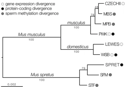
We estimated a maximum likelihood phylogeny using whole genome and targeted exome resequencing data from the mouse strains used in this study (Figure 1). The resulting tree conformed to the expected evolutionary relationships among strains of M. m. musculus, M. m. domesticus, and M. spretus. Strains within a given taxon are closely related and approximate levels of individual variation follow general expectations for these species and subspecies. Likewise,M. musculusandM. spretuswere separated by about twice as much genetic divergence as M. m. musculusandM. m. domesticus. Thus, the different strains of mice used within our studies of expression, DNA methylation, and protein-coding divergence represent closely related samples from these three lineages.
Stage-specific dissection of gene expression across mouse spermatogenesis
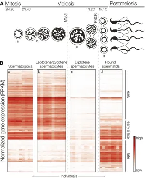
We used FACS to generate highly enriched populations of four cell types spanning three phases of spermatogenesis (Supple-mental Material,Figure S1, Figure 2A): (1) spermatogonia (mitosis), (2) leptotene/zygotene spermatocytes (meiosis, prior to MSCI), (3) diplotene spermatocytes (meiosis, after MSCI), and (4) round spermatids (postmeiosis). For each subspecies,M. m. musculusandM. m. domesticus, we mapped between 53 and 74 million unique sequencing fragments from each cell type to their respective genomes (Table S1). Each cell population had a distinct expression profile, there was strong clustering of transcription levels by cell type, and expression patterns of established cell- or stage-specific genes were specific to their cell type (Figure S2). Overall, these results indicate that our FACS experiments yielded highly enriched cell populations with low variance among individ-ual samples.
good overall agreement between functional annotations and developmental processes indicative of that stage of spermato-genesis (Table 1). We also observed very high gene-by-gene correspondence between our general groupings and previ-ously described patterns of X-linked expression during sper-matogenesis (Figure S3), which used different methods of cell enrichment and expression profiling (Namekawaet al. 2006). These results indicate that our FACS-based enrich-ment captures the dynamic changes in transcription across spermatogenesis.
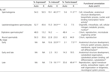
Next, we combined our data with published multitissue expression data and found that testis-biased genes made up a larger proportion of the genes induced late in spermato-genesis (early 4.0%, late 35.4%), consistent with increasing gene specificity during postmeiotic development (Schultz et al. 2003). The tissue specificity of gene expression (t) also increased late (median 6SE tearly, 0.83160.005; late, 0.90360.009, Wilcoxon testP#0.001). This pattern was particularly striking on the X chromosome where nearly half of late-induced genes were also testis-biased (Table 1). In addition, the X chromosome was enriched for genes induced early. These results are in agreement with previous studies indicating that X-linked gene content has been shaped by natural selection for spermatogenic genes
expressed before or after MSCI (Khil et al. 2004; Mueller et al.2008; Sinet al.2012).
Faster-late and faster-X protein evolution
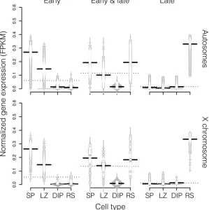
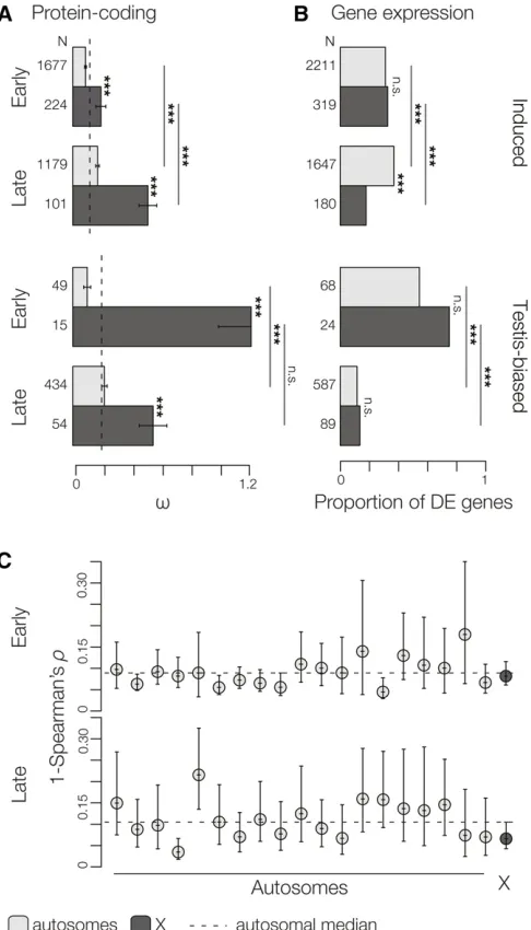
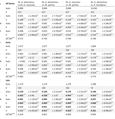
We calculated rates of protein-coding evolution (v) between M. m. musculus,M. m. domesticus, andM. spretusfor genes induced at different stages of spermatogenesis. These con-trasts revealed two striking patterns. First, late-induced autosomal and X-linked genes evolved more rapidly than early-induced genes (i.e., faster-late, Figure 4A, Table 2), confirming a previous finding of a positive correlation be-tween rates of protein evolution and timing of expression during spermatogenesis (Good and Nachman 2005). Second, X-linked genes showed significantly highervwhen compared to autosomal genes (i.e., faster-X, Figure 4A). These pat-terns of faster-late and faster-X protein evolution held when considering only pairwise differences within M. musculus (M. m. musculus vs. M. m. domesticus) (Table S2) or when con-sidering much more divergent contrasts between the house mouse and the Norwegian rat (Table 2). They were also robust to different expression level thresholds (Table S3).
Interpreting patterns of v requires consideration of the synonymous (dS) and nonsynonymous (dN) per site
substitu-tion rates in isolasubstitu-tion. The mammalian X chromosome has Figure 2X-linked expression across spermatogenesis in
a lower per-nucleotide mutation rate than the autosomes as a consequence of male-driven molecular evolution (Ellegren and Parsch 2007). Consistent with this, we observed lowerdS
for X-linked genes across all evolutionary contrasts (Table 2, Table S2) and for X-linked genes expressed early or late in spermatogenesis (Table S2). The X chromosome is also predicted to have a lower Ne relative to the autosomes
and thus shallower coalescent depths on average, which could have a particularly strong impact on the comparison of X vs. autosomal divergence between closely related lineages. Consequently, we observed the greatest differ-ence in X-linked vs. autosomal estimates of dS between M. m. musculus and M. m. domesticus (dSX/dSauto =
0.572vs. dSX/dSauto0.8 for contrasts involvingM. spretus
or rat). Finally, we found that the X chromosome shows higherdNcompared to the autosomes (Table 2), with the
greatest difference among genes induced late in spermato-genesis (Table S2). Thus, we found consistently faster-late and faster-X protein-coding evolution for mouse sper-matogenic genes across different levels of evolutionary divergence and when considering both relative and abso-lute rates of nonsynonymous divergence.
Slower-late and slower-X expression evolution
We observed strikingly different patterns of expression diver-gence relative to protein evolution. Although there was a general trend toward more DE genes induced late in sper-matogenesis (24% of spermatogonia genesvs.37% of round spermatids genes were DE) this trend was restricted to auto-somal genes and there were actually fewer X-linked DE genes induced late (i.e., slower-X, Figure 4, B and C). The slower-X pattern late in spermatogenesis was robust to different tem-poral classifications (Table S4), different expression thresh-olds (Table S5), different methods of accounting for X-linked ampliconic/multicopy genes (Figure S4), and was also appar-ent in per-chromosome comparisons (Figure 5). As discussed above, testis-biased genes tended to be expressed late in spermatogenesis (Table 1). When we restricted our analysis to testis-biased genes, we found that expression divergence was dramatically reduced late and was similarly constrained between the X chromosome and the autosomes (i.e., slower-late, Figure 4B). If we compare expression divergence on the autosomes and the X chromosome while excluding testis-biased genes, the pattern of slower-X late is even more Table 1 The X chromosome is enriched for genes induced before and after MSCI and for testis-biased genes
% Expresseda % Inducedb % Testis-biasedc
Functional annotation
Auto X Auto X Auto X Auto and X
By cell
Spermatogonia 54.3 50.5 19.1 40.3*** 5.6 11.3*** Cell, intracellular, anatomical structure development, biosynthetic process, nucleic acid binding transcription factor activityd
Leptotene/zygotene spermatocytes 52.7 45.6 15.3 34.0*** 5.2 7.5 Organelle, intracellular, cell, cellular nitrogen metabolic process, nucleusd
Diplotene spermatocytese 48.0 13.2 16.2 — 48.4 — Cilium, reproduction, microtubule organizing center
Round spermatids 50.3 35.6 22.8 23.0 42.5 44.9 Cilium, reproduction By stage
Early NA NA 10.6 33.9*** 3.1 7.5*** Anatomical structure development,
immune system process, plasma membrane, signal transduction, cell differentiationd
Early and late NA NA 2.3 9.3 14.3 19.5 Anatomical structure development, cytoskeletal organization, cytoskeletal protein binding, cell differentiation, cytoskeletond
Late NA NA 7.9 19.1*** 35.6 49.4*** Reproduction, signal transducer
activity, extracellular region, cell wall organization or biogenesis, neurological system process
Values represent the percentage of genes that meet a given expression threshold in each cell type and stage of spermatogenesis. Enrichment of X-linked genes are based on chromosome-wise hypergeometric tests, FDR-correctedP-values:***P#0.001.
aExpressed, genes with FPKM.1 in a minimum of four individuals for a given cell type out of the total genes detected (14,223 genes). bInduced, by cell: genes with a median FPKM.23the median FPKM of all other cell types combined out of the total genes expressed. By stage:
genes with a maximum FPKM.43the maximum FPKM of all other stages combined of the total genes expressed. (Note: a gene can be induced in more than one cell type, but can only be induced at a single stage).
cTestis-biased, genes with higher expression in the testes compared 96 other tissues from the Mouse MOE430 Gene Atlas out of the total genes
induced.
dOnly the topfive gene enrichment categories are listed.
apparent (Figure S5). Thus, slower-late expression evolution appears to be a general feature of X-linked spermatogenic genes, but was restricted to testis-biased genes on the autosomes.
These estimates of differential expression reflect diver-gence of a biochemical phenotype that do not account for the potential impacts of lower mutation rates and/or shallower coalescent times on the X chromosome. If we conservatively scale our expectation for X-linked differential expression by the X-to-autosome ratio of synonymous divergence (dSX/dSauto= 0.572 for the contrast betweenM. m. musculus
andM. m. domesticus) then wefind that the X chromosome becomes significantly enriched for DE genes induced early (P,0.001) and the slower-X pattern becomes nonsignifi -cant late (P= 0.38). Likewise, for testis-biased genes, the X chromosome is significantly enriched for DE genes early (P,0.001) but not late (P= 0.924). Thus, the strongest pattern in our data is that expression divergence changes considerably across spermatogenesis both on and off the X chromosome, but at each stage the rate of evolution of the X chromosome relative to the autosomes depends on the ex-tent to which mutation and effective population size infl u-ence evolution of this phenotype.
Overall patterns of protein divergence and expression di-vergence were qualitatively different across this timeline. Protein divergence increased late in spermatogenesis and was elevated on the X chromosome, while the opposite was true for expression divergence. Interpretation of X vs. autosomal expression divergence is clearly dependent on the assumptions
one makes with respect to null expectations. Given this, it is informative to focus on the relationships between gene expres-sion and protein evolution across spermatogenesis. We calcu-lated correlations and partial correlations between protein evolution (v), expression level (normalized FPKM), log2 fold change between subspecies (logFC), and tissue specificity (t) (Figure S6). There was a strong positive relationship between expression level and logFC on and off the X chro-mosome for genes induced early and late in spermatogene-sis. LogFC andtwere positively correlated on the autosomes early, indicating that tissue-specific genes tended to also show larger changes in expression levels between M. m. musculusandM. m. domesticus. In contrast, logFC was neg-atively correlated with bothtandvon the autosomes late in spermatogenesis, but only when testis-biased genes were included (Figure S6). These results support our general
finding of elevated protein-coding divergence coupled with more constrained gene expression for testis-biased genes late in spermatogenesis.
Slower-X sperm methylome evolution
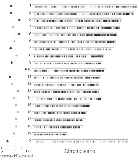
close to expectations based on the relative length of the X chromosome (6.4%). We defined methylome divergence by identifying DMRs as genomic regions with adequate coverage in both species but where an HMR was called in only one of the species. Only 2.3% of the 9580 DMRs betweenM. m. musculus andM. spretuswere X linked, a strongly significant reduction given the proportion of X-linked CpGs (x2= 23.2,P,1025;
Figure 6, Table S9). We observed considerable variation in
DMR enrichment among chromosomes (Figure 6), likely be-cause of the sensitivity of enrichment tests when sample sizes are large. Nine of 20 linkage groups showed significant skews in observedvs.expected DMRs but the X chromosome always showed the strongest de-enrichment of DMRs across a range of test configurations. We emphasize here that we downsampled autosomal reads to match X-linked coverage; therefore, our results cannot be explained by differential coverage. Our re-sults remained qualitatively identical across five different downsampling schemes. Thus, in mouse sperm, the X chromo-some shows less absolute divergence in sperm DNA methyl-ation status relative to the autosomes.
Finally, we evaluated the potential influence of lower X chromosome substitution rates on sperm methylome evolu-tion. Similar to our expression analyses above, we corrected our expectation for the number of X-linked DMRs by the X-to-autosome ratio of synonymous substitutions (dSX/dSauto=
0.77 for the contrast between M. m. musculus and M. m. spretus). Using this correction, the reduction in the frequency of DMRs on the X chromosome becomes nonsignificant (x2,P= 0.088).
Discussion
Forty years ago, King and Wilson argued that protein sequence and gene expression represent distinct levels of evolution (King and Wilson 1975). This influential paper popularized the ideas that evolution proceeds through different molecu-lar mechanisms along the transition from genotype to phe-notype and that gene expression may play a predominant role in organismal evolution. In our study we found striking contrasts in patterns of divergence between different forms of molecular evolution dependent on chromosome origin and developmental stage of spermatogenesis. Our cell-specific data yield new insights into the evolution of spermatogenesis and the critical role that developmental context plays in mo-lecular evolution on and off the sex chromosomes. Below we discuss the implications of our results for the evolution of spermatogenesis, the X chromosome, and speciation.
Protein evolution and spermatogenesis
Faster-X protein evolution has now been found across a broad range of taxa, including mammals (Hvilsom et al. 2012; Veeramah et al. 2014), birds (Mank et al. 2007), flies (Begun et al.2007; Langley et al.2012; Garriganet al. 2014), and other insects (Jaquieryet al. 2012; Sackton et al. 2014). Both higher rates of adaptive substitutions (Kousathanas et al. 2014) and relaxed constraint (Wright et al. 2015) likely contribute to these patterns. X-linked se-quence evolution is not always unusual (reviewed in Vicoso and Charlesworth 2006; Ellegren and Parsch 2007; Meisel and Connallon 2013), but there is strong support for faster-X protein evolution for genes expressed in male reproductive tissues (Torgerson and Singh 2003, 2006; Baineset al.2008; Grath and Parsch 2012; Sin et al.2012; Kousathanaset al. 2014). From these data, gene expression has emerged as a Figure 4 Evolutionary divergence in protein-coding sequence and
defining factor in faster-X protein evolution, with breadth of expression, tissue specificity, and degree of sex bias all strongly influencing evolutionary rates (Meisel 2011; Meiselet al.2012b).
Given these generalfindings, the details of spermatogen-esis should strongly dictate patterns of molecular evolution on and off the sex chromosomes, yet most evolutionary studies have not utilized a strong developmental framework (but see Kousathanaset al.2014). Our FACS data allowed us to par-tition genes across spermatogenesis, yielding strong support for faster-X protein evolution (Figure 4A) based on relative (v) and absolute (dN) estimates of amino acid divergence.
Faster-X protein evolution was most striking when consider-ing vestimates that useddSto approximate neutral
diver-gence. This assumption is violated in many species due to selection for biased codon usage. For example, the X chromo-some shows stronger codon usage bias inDrosophila(Singh et al.2005), which in turn may inflate X-linked estimates ofv (Camposet al.2013). There is also weak codon usage bias in mice, but unlikeDrosophila, it is significantly weaker on the X chromosome (Kessler and Dean 2014) and therefore conser-vative with respect to the observation of faster-X protein evo-lution. Further, both lower mutation rates and shallower coalescent times likely contribute to less nucleotide diver-gence on the mouse X chromosome (Table S2), making the observation of higher X-linkeddNconservative.
Some aspects of our faster-X protein-coding results are seemingly contrary to the basic dominance predictions of faster-X theory. Postmeiotic cells are haploid, leading to the prediction that faster-X evolution in the germ line should be restricted to genes expressed prior to meiosis (Kousathanas et al.2014). However, spermatids form a multicellular syn-cytium connected through intercellular bridges that enable functional equivalence through cytoplasmic exchange (Braun et al.1989; Caldwell and Handel 1991). Exchange of gene products between spermatids is likely a functional necessity given that many sex-linked genes are essential to the post-meiotic stages of spermatogenesis. Assuming exchange of
autosomal gene products maintains functional diploidy, then our finding of faster-X protein evolution across the diploid and haploid stages of spermatogenesis is generally consistent with the dominance predictions of faster-X theory (Charlesworthet al.1987; Vicoso and Charlesworth 2009).
Regulatory evolution and spermatogenesis
In contrast to protein evolution, X-linked postmeiotic genes (i.e., induced late) showed less differential expression than comparable autosomal genes (Figure 4). As with expression divergence, sperm methylome evolution also appeared slower on the X chromosome (Figure 5). Given the strong signature of faster-X protein evolution, why do wefind evi-dence for equivalent or slower-X gene expression and DNA methylation evolution across the same developmental time-line? One simple explanation is that one or more of the as-sumptions of the faster-X model sequence evolution do not hold for these complex biochemical traits. First, for the pre-dictions of faster-X theory to hold, these regulatory pheno-types must reflect linked-sequence evolution. For faster-X expression divergence, this would require that differences in transcript abundances are due to divergence incis -regula-tory elements and/or X-linked trans-regulatory elements (Kayseriliet al. 2012; Meiselet al.2012a; Meisel and Con-nallon 2013). Several studies suggest that divergence in tran-script abundances is largely determined by evolution incis (Wittkopp et al. 2004; Landry et al. 2005; Wittkopp et al. 2008; Graze et al.2009; Goncalves et al. 2012; Shi et al. 2012; Shenet al.2014; Macket al.2016), while other re-search indicates that trans-regulatory divergence is more common (Emerson et al. 2010; McManus et al. 2010; Schaefkeet al. 2013; Coolonet al.2014; Meiklejohn et al. 2014; Combeset al.2015) or that the inferred mode of reg-ulatory divergence is dependent on taxon and experimental design (Coolon and Wittkopp 2013; Guerrero et al.2016). The mechanisms underlying the evolution of DNA methyl-ation are even more poorly understood, but levels of DNA methylation within a given genomic region appear strongly Table 2 Protein-coding divergence on the X chromosome and the autosomes
Global estimates amongMusspecies M. m. domesticus vs. R. norvegicus
N
Auto 9898 10,565
X 416 311
v
Auto 0.11463.0031023 0.11961.6131023
X 0.235***62.8231022 0.192***61.3331022
dN
Auto 0.00365.4831025 0.02363.3831024
X 0.004***64.9931024 0.029***62.8231023
dS
Auto 0.02461.1031024 0.19665.7331024
X 0.018***64.9031024 0.156***63.5231023
dSX/dSauto 0.750 0.796
Estimates of median (6SE) omega (v), nonsynonymous substitution rate (dN), and synonymous substitution rate (dS) amongM. m. musculus,M. m. domesticus,M. spretus,
dependent on underlying (i.e., cis) genetic sequences (Hernando-Herraezet al.2015).
Faster-X sequence evolution also assumes that benefi -cial mutations are on average at least partially recessive (Charlesworthet al.1987), while theory (Gibson and Weir 2005) and several empirical studies (Lemos et al. 2008; McManus et al. 2010; Schaefke et al.2013) suggest that cis-regulatory elements should on average act additively. Nonetheless, these studies also find a substantial propor-tion ofcis-regulatory elements that act nonadditively or, in some cases, a greater overall proportion of nonadditivecis -regulatory elements (Coolon et al. 2014). Unfortunately, these issues remain largely unresolved on the X chromo-some because both dominance relationships and cis- vs. trans-regulatory evolution are usually assessed through allele-specific expression in F1 genotypes, which cannot
be evaluated for X-linked genes during spermatogenesis. It is also important to consider what fraction of X-linked substitutions that influence gene expression or DNA methyl-ation reflect new mutations that have been targeted by pos-itive directional selection. One formal possibility is that divergence of regulatory traits simply reflects neutral se-quence evolution. Both patterns of slower-X gene expression and methylation divergence become nonsignificant when expectations are corrected by lower synonymous substitution rates on the X chromosome. This simple correction is likely highly conservative, but does suggest that differences in mu-tation rates and/or the effective population sizes may partially account for patterns of divergence on the X chromosome. We still lack a strong theoretical framework for differentiating between adaptive and neutral divergence of expression phe-notypes (Khaitovichet al.2005b; Meisel and Connallon 2013). Recent genomic analyses of patterns of sequence polymor-phism and divergence in mice indicate widespread adaptive evolution of both coding (Halliganet al.2010) and noncoding regions (Halliganet al.2011, 2013). Most putatively adaptive autosomal substitutions in mouse genomes occur within can-didate regulatory regions (i.e., untranslated exons and con-served noncoding elements), although the individual fitness effects of amino acid substitutions appear to be much larger (Halliganet al.2013). These studies did not consider X-linked patterns directly but do suggest that a substantial fraction of substitutions within regulatory regions may be targeted by
positive selection. However, if selection on gene expression acts on standing genetic variation, as opposed to new muta-tions, then substitution rates are predicted to be lower on the X chromosome (Meisel and Connallon 2013), given lower levels of X-linked genetic diversity in mice (Geraldeset al.2008). The relative contribution of new mutations vs. standing genetic variation to adaptive regulatory divergence has not yet been explicitly addressed (Coolon and Wittkopp 2013).
In sum, it is clear that extending the faster-X model se-quence evolution to regulatory phenotypes depends on sev-eral assumptions that are both restrictive and remain largely unresolved. However, if regulatory divergence between sub-species of mice were simply a consequence of the genetic architecture of these traits, mutational processes, or the origin of adaptive variation, then we might reasonably expect pat-terns on and off the X chromosome to be consistent across different stages of spermatogenesis. Instead we found that X-linked expression divergence, both overall and relative to the autosomes, changed dynamically over the timeline of spermatogenesis with a strong signature of less divergence late (Figure 4B). Given thisfinding, we propose that slower-late regulatory evolution may be best explained by strong developmental constraints on gene expression phenotypes during the later stages of spermatogenesis that are particu-larly acute on the sex chromosomes.
increase in relative expression and subsequent counterse-lection for upregulation of the other gene group (Cocquet et al.2012). So while the underlying genomic architecture evolves rapidly via gene duplication (Elliset al.2011), ex-pression level phenotypes appear to be under strong stabi-lizing selection for a dosage equilibrium between sex-linked postmeiotic genes that if disrupted leads to male sterility. This is evidence for strong constraint on the regulatory phenotypes of X-linked genes that overcome PSCR to be expressed during this critical period of sperm develop-ment. Our data suggest that such constraints likely extend more broadly across the X chromosome and to testis-biased autosomal genes.
We also found that patterns of DNA methylation diverged more slowly on X chromosomes compared to the autosomes (Figure 5). Dynamic methylation plays a key role in mamma-lian genome regulation (Li et al.1992; Okano et al.1999; Reik et al.2001), yet very little is known about either the tempo or mechanisms of DNA methylome evolution. Sperm are thefirst mature cells following a major“erasure”; nearly the entire genome is demethylated in spermatogonia stem cells and then remethylated late in spermatogenesis (Reik et al.2001). A study involving primates found the methylomes of mature sperm to differ from those of somatic cells in several important ways. Most notably, the HMRs are more numerous
and extend for longer genomic intervals in sperm compared to somatic cells (Molaroet al.2011). We propose that the obser-vation of less X-linked sperm methylome divergence may be an extension of the same regulatory constraints that lead to slower gene expression divergence late in spermatogenesis, as well as other X-linked phenomena such as mutational envi-ronment. Comparable methylome data from other tissues and other stages in spermatogenesis will be helpful in resolving these outstanding questions.
Cis-regulatory evolution is thought to proceed with fewer pleiotropic constraints than protein-coding divergence (e.g., Carroll 2008). However, our results indicate that the stages of spermatogenesis that show the fastest rates of protein evolu-tion show less expression divergence for both X-linked and testis-biased genes. Protein evolution is strongly influenced by underlying patterns of gene expression; there are gener-ally higher rates of protein-coding divergence for genes that have lower expression (Nguyenet al.2015) and for narrowly expressed genes (Liaoet al.2006; Meisel 2011). Expression specificity increases late in spermatogenesis and the X chromosome is enriched for testis-biased postmeiotic genes. Therefore expression specificity may be the primary factor underlying faster-late and faster-X protein evolution. While divergence in gene expression (logFC) and tissue specificity (t) were also positively correlated early in spermatogenesis, they were negatively correlated for autosomal testis-biased genes expressed late (Figure S6). Thus, specificity may ac-tually constrain expression divergence late in spermatogen-esis, especially for postmeiotic testis-biased genes that presumably play critical roles in sperm development and maturation. There is typically a positive association between differential expression and tissue specificity (Meisel et al. 2012a), suggesting that our results reflect a unique feature of sperm development rather than a general molecular evo-lutionary trend.
Our observation of less X-linked expression divergence late in spermatogenesis differs some from studies in mammals reporting elevated (uncorrected) X-linked expression diver-gence primarily in testis (Khaitovichet al.2005a; Zhanget al. 2010; Brawand et al. 2011). These previous studies have focused on various whole tissue contrasts spanning different taxa and phylogenetic depths. However, Zhanget al.(2010) did report greater relative X-linked expression divergence in spermatids between mouse and rat, the exact cell population that we found to be the most conserved between subspecies of mice (Figure 4). While this could reflect changes in X-linked expression evolution over deeper evolutionary time-scales, this previous result was based on a metaanalysis of microarray data collected independently in mouse (Chalmel et al.2007) and rat (Johnstonet al.2008) using different cell-enrichment procedures. A full assessment of testis gene expression evolution between mouse and rat awaits the comparison of cell-specific transcriptomes that control for potential experimental artifacts.
How broadly the slower-late X and testis-biased autosomal patterns of regulatory evolution apply to other taxa remains Figure 6 Evolution of the mouse sperm methylome. Chromosomal
to be seen. Expression divergence has not been systematically evaluated in nonmammalian systems using cell- or stage-specific data, making the relative contributions of cellular composition and developmental constraint difficult to deter-mine. This technical limitation aside, the influence of devel-opmental timing on spermatogenic expression evolution will also be dependent on the existence or extent of MSCI/PSCR-like phenomena in other taxa. For example, faster-X expres-sion divergence has also been reported inDrosophila(Kayserili et al. 2012; Meiselet al.2012a; Coolonet al.2015; Llopart 2015) where the X chromosome appears to be transcription-ally repressed during spermatogenesis (Meiklejohn et al. 2011). Whether this repression reflects a process akin to MSCI has been contentious, but it does suggest that regula-tory constraints likely play an important role in sex chromo-some evolution (Vibranovski 2014).
Implications for speciation
Faster-X theory was originally proposed in part to explain two observations that invoke a large role for sex chromosomes in speciation (Charlesworth et al. 1987). First, when hybrids between recently diverged lineages show sex-specific sterility or inviability, it is usually the heterogametic sex (Haldane 1922). Second, heterogametic sterility is disproportionately linked to the X or Z chromosomes, a pattern known as the large-X effect (Coyne 1992). Faster-X evolution remains one of the predominant mechanisms invoked to explain the evo-lution of hybrid sterility (Kousathanaset al.2014), and the link between rapid evolution and speciation is intuitive. A long-standing alternative hypothesis is that spermatogenesis is an inherently sensitive process that may be easily disrupted in hybrids (Lifschytz and Lindsley 1972; Jablonka and Lamb 1991; Wu and Davis 1993). Disruptions of MSCI/PSCR are stronga prioricandidate regulatory mechanisms for the sen-sitivity of spermatogenesis (Lifschytz and Lindsley 1972). Studies in mice (Good et al. 2010; Bhattacharyya et al. 2013; Campbell et al.2013; Turneret al.2014) and other mammals (Daviset al.2015) have recently found a strong link between regulatory disruption of the X chromosome during spermatogenesis and the evolution of hybrid male sterility. Our results reveal that the same developmental stages that have disrupted gene expression in mouse hybrids (Good et al.2010; Bhattacharyyaet al.2013; Campbellet al.2013) show the strongest evolutionary constraints in gene expression levels (Figure 4). While these data do not discount an impor-tant role for faster-X protein-coding evolution in speciation, they do suggest that inherent developmental constraints on the regulation of gene expression late in spermatogenesis may play a central and underappreciated role in the evolu-tion of hybrid male sterility.
Acknowledgments
We thank Pamela K. Shaw, Irina Getun, Bivian Torres, Colin Callahan, and Brent Young for assistance with FACS and other sample preparations; Francois Bonhomme for mice;
and the staff of the University of Montana (UM) Laboratory Animal Research facility. Members of the J.M.G. and M.D.D. labs gave us helpful feedback on experimental results, and comments from Bret Payseur and multiple anonymous reviewers improved previous versions of this manuscript. We thank the UM Fluorescence Cytometry Core, supported by the National Institute of General Medical Sciences of the National Institutes of Health (NIH) (P30-GM103338) and the UM Genomics Core, supported by a grant from the M. J. Murdock Charitable Trust and the Vincent J. Coates Genomics Sequencing Laboratory at the University of California, Berkeley, supported by NIH S10 instrumentation grants S10-RR029668 and S10-RR027303. This research was supported by the Eunice Kennedy Shriver National Institute of Child Health and Human Development (R01-HD073439; to J.M.G.), the National Institute of General Medical Sciences (R01-GM098536; to M.D.D.), and the National Science Foundation (1146525; to M.D.D.).
Literature Cited
Anders, S., and W. Huber, 2010 Differential expression analysis for sequence count data. Genome Biol. 11: R106.
Bachtrog, D., M. Kirkpatrick, J. E. Mank, S. F. McDaniel, J. C. Pires et al., 2011 Are all sex chromosomes created equal? Trends Genet. 27: 350–357.
Baines, J. F., and B. Harr, 2007 Reduced X-linked diversity in de-rived populations of house mice. Genetics 175: 1911–1921. Baines, J. F., S. A. Sawyer, D. L. Hartl, and J. Parsch, 2008 Effects
of X-linkage and sex-biased gene expression on the rate of adap-tive protein evolution inDrosophila. Mol. Biol. Evol. 25: 1639– 1650.
Begun, D. J., and P. Whitley, 2000 Reduced X-linked nucleotide polymorphism inDrosophila simulans. Proc. Natl. Acad. Sci. USA 97: 5960–5965.
Begun, D. J., A. K. Holloway, K. Stevens, L. W. Hillier, Y.-P. Poh et al., 2007 Population genomics: whole-genome analysis of polymorphism and divergence in Drosophila simulans. PLoS Biol. 5: e310–e326.
Benjamini, Y., and Y. Hochberg, 1995 Controlling the false dis-covery rate: a practical and powerful approach to multiple test-ing. J.R. Stat. Soc. 57: 289–300.
Bhattacharyya, T., S. Gregorova, O. Mihola, M. Anger, J. Sebestova et al., 2013 Mechanistic basis of infertility of mouse intersub-specific hybrids. Proc. Natl. Acad. Sci. USA 110: E468–E477. Bolger, A. M., M. Lohse, and B. Usadel, 2014 Trimmomatic: a
flexible trimmer for Illumina sequence data. Bioinformatics 30: 2114–2120.
Braun, R. E., R. R. Behringer, J. J. Peschon, R. L. Brinster, and R. D. Palmiter, 1989 Genetically haploid spermatids are phenotypi-cally diploid. Nature 337: 373–376.
Brawand, D., M. Soumillon, A. Necsulea, P. Julien, G. Csárdiet al., 2011 The evolution of gene expression levels in mammalian organs. Nature 478: 343–348.
Caldwell, K. A., and M. A. Handel, 1991 Protamine transcript sharing among postmeiotic spermatids. Proc. Natl. Acad. Sci. USA 88: 2407–2411.
Campbell, P., J. M. Good, and M. W. Nachman, 2013 Meiotic sex chromosome inactivation is disrupted in sterile hybrid male house mice. Genetics 193: 819–828.
sizes on the X chromosome vs. the autosomes in Drosophila melanogaster. Mol. Biol. Evol. 30: 811–823.
Carroll, S. B., 2008 Evo-devo and an expanding evolutionary syn-thesis: a genetic theory of morphological evolution. Cell 134: 25–36.
Chalmel, F., A. D. Rolland, C. Niederhauser-Wiederkehr, S. S. W. Chung, P. Demouginet al., 2007 The conserved transcriptome in human and rodent male gametogenesis. Proc. Natl. Acad. Sci. USA 104: 8346–8351.
Charlesworth, B., J. A. Coyne, and N. H. Barton, 1987 The rela-tive rates of evolution of sex chromosomes and autosomes. Am. Nat. 130: 113–146.
Charlesworth, D., 2013 Plant sex chromosome evolution. J. Exp. Bot. 64: 405–420.
Cocquet, J., P. J. I. Ellis, Y. Yamauchi, S. K. Mahadevaiah, N. A. Affara et al., 2009 The multicopy geneSlyrepresses the sex chromosomes in the male mouse germline after meiosis. PLoS Biol. 7: e1000244.
Cocquet, J., P. J. I. Ellis, Y. Yamauchi, J. M. Riel, T. P. S. Karacs et al., 2010 Deficiency in the multicopy Sycp3-like X-linked genesSlxandSlxl1causes major defects in spermatid differen-tiation. Mol. Biol. Cell 21: 3497–3505.
Cocquet, J., P. J. I. Ellis, S. K. Mahadevaiah, N. A. Affara, D. Vaiman et al., 2012 A genetic basis for a postmeiotic Xvs.Y chromo-some intragenomic conflict in the mouse. PLoS Genet. 8: e1002900.
Combes, M. C., Y. Hueber, A. Dereeper, S. Rialle, J. C. Herreraet al., 2015 Regulatory divergence between parental alleles deter-mines gene expression patterns in hybrids. Genome Biol. Evol. 7: 1110–1121.
Coolon, J. D., and P. J. Wittkopp, 2013 cis- and trans-regulation in Drosophila interspecific hybrids, pp. 37–57 inPolyploid and Hybrid Genomics, edited by Z. J. Chen, and J. A. Birchler. John Wiley & Sons, New York.
Coolon, J. D., C. J. McManus, K. R. Stevenson, B. R. Graveley, and P. J. Wittkopp, 2014 Tempo and mode of regulatory evolution inDrosophila. Genome Res. 24: 797–808.
Coolon, J. D., K. R. Stevenson, C. J. McManus, B. Yang, B. R. Graveleyet al., 2015 Molecular mechanisms and evolutionary processes contributing to accelerated divergence of gene expres-sion on the Drosophila X chromosome. Mol. Biol. Evol. 32: 2605–2615.
Coyne, J. A., 1992 Genetics and speciation. Nature 355: 511–515. Danecek, P., A Auton, G Abecasis, C. AE. Albers, and Bankset al., 2011 The variant call format and VCFtools. Bioinformatics 27: 2156–2158.
Davis, B. W., C. M. Seabury, W. A. Brashear, G. Li, M. Roelke-Parker et al., 2015 Mechanisms underlying mammalian hybrid steril-ity in two feline interspecies models. Mol. Biol. Evol. 32: 2534– 2546.
Dean, R., P. W. Harrison, A. E. Wright, F. Zimmer, and J. E. Mank, 2015 Positive selection underlies faster-Z evolution of gene expression in birds. Mol. Biol. Evol. 32: 2646–2656.
Eddy, E. M., 2002 Male germ cell gene expression. Recent Prog. Horm. Res. 57: 103–128.
Ellegren, H., 2007 Characteristics, causes and evolutionary con-sequences of male-biased mutation. Proc. Biol. Sci. 274: 1–10. Ellegren, H., 2011 Sex-chromosome evolution: recent progress
and the influence of male and female heterogamety. Nat. Rev. Genet. 12: 157–166.
Ellegren, H., and J. Parsch, 2007 The evolution of sex-biased genes and sex-biased gene expression. Nat. Rev. Genet. 8: 689–698.
Ellis, P. J. I., J. Bacon, and N. A. Affara, 2011 Association ofSly with sex-linked gene amplification during mouse evolution: a side effect of genomic conflict in spermatids? Hum. Mol. Genet. 20: 3010–3021.
Emerson, J. J., H. Kaessmann, E. Betrán, and M. Long, 2004 Extensive gene traffic on the mammalian X chromosome. Science 303: 537–540.
Emerson, J. J., L.-C. Hsieh, H.-M. Sung, T.-Y. Wang, C.-J. Huang et al., 2010 Natural selection oncis andtrans regulation in yeasts. Genome Res. 20: 826–836.
Fairfield, H., G. J. Gilbert, M. Barter, R. R. Corrigan, M. Curtain et al., 2011 Mutation discovery in mice by whole exome se-quencing. Genome Biol. 12: R86.
Firman, R. C., F. Garcia-Gonzalez, E. Thyer, S. Wheeler, Z. Yamin et al., 2015 Evolutionary change in testes tissue composition among experimental populations of house mice. Evolution 69: 848–855.
Frommer, M., L. E. McDonald, D. S. Millar, C. M. Collis, F. Watt et al., 1992 A genomic sequencing protocol that yields a pos-itive display of 5-methylcytosine residues in individual DNA strands. Proc. Natl. Acad. Sci. USA 89: 1827–1831.
Garrigan, D., S. B. Kingan, A. J. Geneva, J. P. Vedanayagam, and D. C. Presgraves, 2014 Genome diversity and divergence in Drosophila mauritiana: multiple signatures of faster X evolution. Genome Biol. Evol. 6: 2444–2458.
Geraldes, A., P. Basset, B. Gibson, K. L. Smith, B. Harr et al., 2008 Inferring the history of speciation in house mice from autosomal, X-linked, Y-linked and mitochondrial genes. Mol. Ecol. 17: 5349–5363.
Getun, I. V., B. Torres, and P. R. J. Bois, 2011 Flow cytometry purification of mouse meiotic cells. J. Vis. Exp. 50: e2602. Gibson, G., and B. Weir, 2005 The quantitative genetics of
tran-scription. Trends Genet. 21: 616–623.
Goncalves, A., S. Leigh-Brown, D. Thybert, K. Stefflova, E. Turro et al., 2012 Extensive compensatorycis-transregulation in the evolution of mouse gene expression. Genome Res. 22: 2376–2384. Good, J. M., and M. W. Nachman, 2005 Rates of protein evolution are positively correlated with developmental timing of expres-sion during mouse spermatogenesis. Mol. Biol. Evol. 22: 1044– 1052.
Good, J. M., M. D. Dean, and M. W. Nachman, 2008 A complex genetic basis to X-linked hybrid male sterility between two spe-cies of house mice. Genetics 179: 2213–2228.
Good, J. M., T. Giger, M. D. Dean, and M. W. Nachman, 2010 Widespread over-expression of the X chromosome in sterile F1 hybrid mice. PLoS Genet. 6: e1001148.
Grath, S., and J. Parsch, 2012 Rate of amino acid substitution is influenced by the degree and conservation of male-biased tran-scription over 50 myr of Drosophilaevolution. Genome Biol. Evol. 4: 346–359.
Graze, R. M., L. M. McIntyre, B. J. Main, M. L. Wayne, and S. V. Nuzhdin, 2009 Regulatory divergence inDrosophila melanogaster andD. simulans, a genomewide analysis of allele-specific ex-pression. Genetics 183: 547–561.
Guerrero, R. F., A. L. Posto, L. C. Moyle, and M. W. Hahn, 2016 Genome-wide patterns of regulatory divergence re-vealed by introgression lines. Evolution 70: 696–706.
Haldane, J. B. S., 1922 Sex ratio and unisexual sterility in hybrid animals. J. Genet. 12: 101–109.
Halligan, D. L., F. Oliver, A. Eyre-Walker, B. Harr, and P. D. Keightley, 2010 Evidence for pervasive adaptive protein evolution in wild mice. PLoS Genet. 6: e1000825–e1000829.
Halligan, D. L., F. Oliver, J. Guthrie, K. C. Stemshorn, B. Harret al., 2011 Positive and negative selection in murine ultraconserved noncoding elements. Mol. Biol. Evol. 28: 2651–2660.
Halligan, D. L., A. Kousathanas, R. W. Ness, B. Harr, L. Eöryet al., 2013 Contributions of protein-coding and regulatory change to adaptive molecular evolution in murid rodents. PLoS Genet. 9: e1003995.
methylation and sequence divergence in recent human evolu-tion. Nucleic Acids Res. 43: 8204–8214.
Holt, J., S. Huang, L. McMillan, and W. Wang, 2013 Read anno-tation pipeline for high-throughput sequencing data. BCB 13: 605–612.
Hu, Y. C., and S. H. Namekawa, 2015 Functional significance of the sex chromosomes during spermatogenesis. Reproduction 149: R265–R277.
Huang, S., C.-Y. Kao, L. McMillan, and W. Wang, 2013 Transforming genomes using MODfiles with applications. BCB 13: 595–604. Huang, S., J. Holt, C.-Y. Kao, L. McMillan, and W. Wang, 2014 A
novel multi-alignment pipeline for high-throughput sequencing data. Database 2014: bau057.
Hvilsom, C., Y. Qian, T. Bataillon, Y. Li, T. Mailund et al., 2012 Extensive X-linked adaptive evolution in central chim-panzees. Proc. Natl. Acad. Sci. USA 109: 2054–2059.
Jablonka, E., and M. J. Lamb, 1991 Sex chromosomes and speci-ation. Proc. Biol. Sci. 243: 203–208.
Jaquiery, J., S. Stoeckel, C. Rispe, L. Mieuzet, F. Legeai et al., 2012 Accelerated evolution of sex chromosomes in aphids, an X0 system. Mol. Biol. Evol. 29: 837–847.
Johnston, D. S., W. W. Wright, P. Dicandeloro, E. Wilson, G. S. Kopf et al., 2008 Stage-specific gene expression is a fundamental characteristic of rat spermatogenic cells and Sertoli cells. Proc. Natl. Acad. Sci. USA 105: 8315–8320.
Kampstra, P., 2008 Beanplot: a boxplot alternative for visual com-parison of distributions. J. Stat. Softw. 28: 1–9.
Kayserili, M. A., D. T. Gerrard, P. Tomancak, and A. T. Kalinka, 2012 An excess of gene expression divergence on the X chro-mosome in Drosophila embryos: implications for the faster-X hypothesis. PLoS Genet. 8: e1003200.
Keane, T. M., L. Goodstadt, P. Danecek, M. A. White, K. Wonget al., 2011 Mouse genomic variation and its effect on phenotypes and gene regulation. Nature 477: 289–294.
Kessler, M. D., and M. D. Dean, 2014 Effective population size does not predict codon usage bias in mammals. Ecol. Evol. 4: 3887–3900.
Khaitovich, P., I. Hellmann, W. Enard, K. Nowick, M. Leinweber et al., 2005a Parallel patterns of evolution in the genomes and transcriptomes of humans and chimpanzees. Science 309: 1850–1854.
Khaitovich, P., S. Pääbo, and G. Weiss, 2005b Toward a neutral evolutionary model of gene expression. Genetics 170: 929–939. Khil, P. P., N. A. Smirnova, P. J. Romanienko, and R. D. Camerini-Otero, 2004 The mouse X chromosome is enriched for sex-biased genes not subject to selection by meiotic sex chromosome inactivation. Nat. Genet. 36: 642–646.
Kim, D., G. Pertea, C. Trapnell, H. Pimentel, R. Kelley et al., 2013 TopHat2: accurate alignment of transcriptomes in the presence of insertions, deletions and gene fusions. Genome Biol. 14: R36.
King, M. C., and A. C. Wilson, 1975 Evolution at two levels in humans and chimpanzees. Science 188: 107–116.
Kohn, M., H. Kehrer-Sawatzki, W. Vogel, J. A. M. Graves, and H. Hameister, 2004 Wide genome comparisons reveal the ori-gins of the human X chromosome. Trends Genet. 20: 598– 603.
Kousathanas, A., D. L. Halligan, and P. D. Keightley, 2014 Faster-X adaptive protein evolution in house mice. Genetics 196: 1131– 1143.
Landry, C. R., P. J. Wittkopp, C. H. Taubes, J. M. Ranz, A. G. Clarket al., 2005 Compensatorycis-transevolution and the dysregulation of gene expression in interspecific hybrids of Drosophila. Genetics 171: 1813–1822.
Langley, C. H., K. Stevens, C. Cardeno, Y. C. G. Lee, D. R. Schrider et al., 2012 Genomic variation in natural populations of Drosophila melanogaster. Genetics 192: 533–598.
Lattin, J. E., K. Schroder, A. I. Su, J. R. Walker, J. Zhang et al., 2008 Expression analysis of G Protein-Coupled Receptors in mouse macrophages. Immunome Res. 4: 5–13.
Lemos, B., C. D. Meiklejohn, M. Cáceres, and D. L. Hartl, 2004 Rates of divergence in gene expression profiles of pri-mates, mice andflies: stabilizing selection and variability among functional categories. Evolution 59: 126–137.
Lemos, B., L. O. Araripe, P. Fontanillas, and D. L. Hartl, 2008 Dominance and the evolutionary accumulation of cis -and trans-effects on gene expression. Proc. Natl. Acad. Sci. USA 105: 14471–14476.
Li, E., T. H. Bestor, and R. Jaenisch, 1992 Targeted mutation of the DNA methyltransferase gene results in embryonic lethality. Cell 69: 915–926.
Li, H., and R. Durbin, 2009 Fast and accurate short read align-ment with Burrows-Wheeler transform. Bioinformatics 25: 1754–1760.
Liao, B. Y., and J. Zhang, 2006 Low rates of expression profile divergence in highly expressed genes and tissue-specific genes during mammalian evolution. Mol. Biol. Evol. 23: 1119–1128. Liao, B.-Y., N. M. Scott, and J. Zhang, 2006 Impacts of gene
es-sentiality, expression pattern, and gene compactness on the evo-lutionary rate of mammalian proteins. Mol. Biol. Evol. 23: 2072–2080.
Lifschytz, E., and D. L. Lindsley, 1972 The role of X-chromosome inactivation during spermatogenesis. Proc. Natl. Acad. Sci. USA 69: 182–186.
Llopart, A., 2015 Parallel faster-X evolution of gene expression and protein sequences inDrosophila: beyond differences in ex-pression properties and protein interactions. PLoS One 10: e0116829.
Lyon, M. F., 1961 Gene action in the X-chromosome of the mouse (Mus musculusL.). Nature 190: 372–373.
Lyon, M. F., 1962 Sex chromatin and gene action in the mamma-lian X-chromosome. Am. J. Hum. Genet. 14: 135–148. Mack, K. L., P. Campbell, and M. W. Nachman, 2016 Gene
regu-lation and speciation in house mice. Genome Res. 26: 451–461. Mank, J. E., E. Axelsson, and H. Ellegren, 2007 Fast-X on the Z: rapid evolution of sex-linked genes in birds. Genome Res. 17: 618–624.
Mank, J. E., B. Vicoso, S. Berlin, and B. Charlesworth, 2010 Effective population size and the faster-X effect: empirical results and their interpretation. Evolution 64: 663–674.
McCarthy, D. J., Y. Chen, and G. K. Smyth, 2012 Differential expression analysis of multifactor RNA-Seq experiments with respect to biological variation. Nucleic Acids Res. 40: 4288– 4297.
McKenna, A., M. Hanna, E. Banks, A. Sivachenko, K. Cibulskiset al., 2010 The Genome Analysis Toolkit: a MapReduce framework for analyzing next-generation DNA sequencing data. Genome Res. 20: 1297–1303.
McManus, C. J., J. D. Coolon, M. O. Duff, J. Eipper-Mains, B. R. Graveleyet al., 2010 Regulatory divergence inDrosophila re-vealed by mRNA-seq. Genome Res. 20: 816–825.
Meiklejohn, C. D., E. L. Landeen, J. M. Cook, S. B. Kingan, and D. C. Presgraves, 2011 Sex chromosome-specific regulation in the Drosophilamale germline but little evidence for chromosomal dosage compensation or meiotic inactivation. PLoS Biol. 9: e1001126.
Meiklejohn, C. D., J. D. Coolon, D. L. Hartl, and P. J. Wittkopp, 2014 The roles ofcis- andtrans-regulation in the evolution of regulatory incompatibilities and sexually dimorphic gene ex-pression. Genome Res. 24: 84–95.