| INVESTIGATION
An Evolutionarily Conserved Transcriptional
Activator-Repressor Module Controls Expression of
Genes for D-Galacturonic Acid Utilization in
Aspergillus niger
Jing Niu,* Ebru Alazi,* Ian D. Reid,†Mark Arentshorst,* Peter J. Punt,*,‡Jaap Visser,* Adrian Tsang,† and Arthur F. J. Ram*,1 *Molecular Microbiology and Biotechnology, Leiden University, Institute of Biology Leiden, Leiden, The Netherlands,†Centre for Structural and Functional Genomics, Concordia University, Quebec, Canada H4B1R6, and‡Dutch DNA Biotech, Utrecht, The Netherlands
ABSTRACT The expression of genes encoding extracellular polymer-degrading enzymes and the metabolic pathways required for carbon utilization in fungi are tightly controlled. The control is mediated by transcription factors that are activated by the presence of specific inducers, which are often monomers or monomeric derivatives of the polymers. A D-galacturonic acid-specific transcription factor named GaaR was recently identified and shown to be an activator for the expression of genes involved in galacturonic acid
utilization inBotrytis cinereaandAspergillus niger. Using a forward genetic screen, we isolatedA. nigermutants that constitutively
express GaaR-controlled genes. Reasoning that mutations in thegaaRgene would lead to a constitutively activated transcription factor,
thegaaRgene in 11 of the constitutive mutants was sequenced, but no mutations ingaaRwere found. Full genome sequencing offive
constitutive mutants revealed allelic mutations in one particular gene encoding a previously uncharacterized protein (NRRL3_08194).
The protein encoded by NRRL3_08194 shows homology to the repressor of the quinate utilization pathway identified previously in
Neurospora crassa (qa-1S)andAspergillus nidulans(QutR). Deletion of NRRL3_08194 in combination with RNA-seq analysis showed that the NRRL3_08194 deletion mutant constitutively expresses genes involved in galacturonic acid utilization. Interestingly,
NRRL3_08194 is located next to gaaR(NRRL3_08195) in the genome. The homology to the quinate repressor, the chromosomal
clustering, and the constitutive phenotype of the isolated mutants suggest that NRRL3_08194 is likely to encode a repressor, which we
name GaaX. The GaaR–GaaX module and its chromosomal organization is conserved among ascomycetes filamentous fungi,
re-sembling the quinate utilization activator-repressor module in amino acid sequence and chromosomal organization.
KEYWORDSgene regulation; galacturonic acid; repressor protein; genomics; transcriptomics; pectin
T
HEfilamentous fungusAspergillus nigeris an important producer of pectin-degrading enzymes that are used in industrial applications including in food and feed processing (Kashyapet al.2001; Khanet al.2013). In nature,A. nigeris a saprotrophic fungus that feeds on organic matter from decay-ing plants. The major carbon sources in plant cells are thestorage polysaccharides starch, and less frequently inulin, as well as the cell wall polymers cellulose, hemicelluloses, and pectin. Of the different plant polysaccharides, pectin has the most complex structure. Pectin is made up of four substruc-tures, including homogalacturonan, xylogalacturonan, rham-nogalacturonan I, and rhamrham-nogalacturonan II. The abundance of each substructure varies with plant species, but typically homogalacturonan is the most abundant polysaccharide in pectin (65%), followed by rhamnogalacturonan I (25–30%). Xylogalacturonan and rhamnogalacturonan II comprise,10% of the total pectin (Mohnen 2008).
Utilization of plant polysaccharides by fungi, including
A. niger, is accomplished by tightly controlled secretion of extracellular enzymes that degrade the polymers into
Copyright © 2017 by the Genetics Society of America doi: 10.1534/genetics.116.194050
Manuscript received October 29, 2016; accepted for publication November 5, 2016; published Early Online November 9, 2016.
Supplemental material is available online atwww.genetics.org/lookup/suppl/doi:10. 1534/genetics.116.194050/-/DC1.
monosaccharides or oligosaccharides that are taken up and catabolised by the fungus. The controlled regulation is not only confined to the expression of genes encoding extracel-lular proteins. It also includes the controlled expression of genes encoding specific sugar transporters to guarantee efficient uptake of the liberated sugars and the intracellular catabolic pathway enzymes. The precise induction of the net-work of genes encoding substrate-specific enzymes, transporters, and catabolic pathway enzymes has so far been shown to be mediated via Zn(II)2Cys6transcription factors. Specific transcrip-tion factors inA. nigerhave been characterized that regulate the utilization of the major polysaccharides. They include AmyR, the regulator for starch utilization (Petersenet al.1999; Yuanet al.
2008a; vanKuyket al.2012); InuR for inulin (Yuanet al.2008b); ManR, ClrA, and ClrB for cellulose (Rauloet al.2016); XlnR for xylan (Van Peij et al. 1998; Battaglia et al. 2014); AraR for arabinan (Battaglia et al.2014); RhaR for rhamnose (Gruben
et al.2014); and GaaR for polygalacturonic acid (PGA) (Alazi
et al.2016). These transcription factors exert coordinated regu-lation of the target genes by interacting with conserved binding sites that are located upstream of the target genes. Computa-tional analysis has been used to identify the galacturonic acid (GA) responsive element (GARE) of GA-induced genes (Martens-Uzunova and Schaap 2008). The predicted sequence (CCNCCAA) was shown to be required for the induction of GA-responsive genes in A. niger (Niu et al. 2015) and
Botrytis cinerea(Zhanget al.2016). Furthermore, using the yeast one-hybrid method, it was shown inB. cinerea that the GaaR transcription factor interacts specifically with the GARE (Zhanget al.2016).
Phenotypic characterization of mutants lacking the GA-regulator in bothB. cinereaandA. nigerhas shown that GaaR is required for growth on GA (Alazi et al.2016; Zhanget al.
2016). Expression analysis in both fungi confirmed that GaaR is required for the induced expression of GA-responsive genes. On complex pectins, growth ofB. cinereaandA. niger gaaR
deletion mutants was severely reduced and genome-wide expression analysis in A. niger revealed that the residual growth on pectin is likely due to the GaaR-independent expression of pectinases acting on arabinofuranosyl- and galactopyranosyl-containing side chains in rhamnogalacturonan (Alaziet al.2016).
In addition to the transcription regulation via GaaR, GA-responsive genes are also under carbon catabolite repression (CCR) control (de Vrieset al.1999; de Vries et al.2002). In
filamentous fungi, CreA mediates CCR (Dowzer et al. 1991; Ruijter and Visser 1997). In A. niger, CreA also exerts CCR control on GA-responsive genes (de Vries et al. 1999; Niu
et al.2015). Using anin vivo reporter construct consisting of the promoter of the GA-induciblepgaXgene (PpgaX) and the acetamidase (amdS) gene as a reporter, both the specific induc-tion ofpgaXon GA as well as the carbon repression ofpgaXvia CreA had been demonstrated (Niuet al.2015). In this study, we have used the PpgaX-amdS reporter strain to isolate mutants displaying constitutive expression of GA-responsive genes. Analysis of the mutants resulted in the identification of a protein
that likely acts as a repressor that specifically inhibits GaaR transcription activation activity under noninducing conditions.
Materials and Methods
Strains, media, and growth conditions
All strains in this study are listed in Table 1. Strains were grown in liquid or solidified (1.5% agar) minimal medium (MM) containing 7 mM KCl, 8 mM KH2PO4, 70 mM NaNO3, and 2 mM MgSO4(pH adjusted to 5.5) as described (Bennett and Lasure 1991). MM was supplemented with 50 mM glu-cose, 50 mM D-galacturonic acid, 50 mM fructose, or 50 mM sorbitol as carbon source. Complete medium (CM) was also used and consists of MM supplemented with 0.1% casamino acids and 0.5% w/v yeast extract and 50 mM glucose. MM agar plates containing acetamide as sole nitrogen source were made as described previously (Arentshorstet al.2012).
Isolation of mutants with constitutive expression of genes involved in PGA utilization
A. nigerstrain JN29.2 (Table 1) was used for the selection of mutants with constitutive expression of genes involved in PGA utilization. Spontaneous mutants were obtained by plat-ing out freshly harvested and myracloth-filtered conidia (13 104conidia per plate) on MM glucose/acetamide plates and incubated at 30° for 5 days. In addition, mutants were obtained after mild UV mutagenesis (80% survival) as de-scribed (Damveldet al. 2008). Individual mutants growing on the primary MM-glucose/acetamide selection plates were purified twice on the MM-glucose/acetamide agar plates. In total, 14 spontaneous mutants and 59 UV-mutants were iso-lated that grew well on MM-glucose/acetamide agar plates and they were potential mutants with constitutive expression of genes involved in PGA utilization. To identify mutants constitutively producing PGA-degrading enzymes, all 73 mutants were grown by inoculating 5 3 107 spores in 50 ml MM-glucose medium for 36 hr at 30° with shaking at 150 rpm. Supernatant of each culture was harvested by
filtration. The extracellular culture fluid and the mycelia were stored at280°for enzymatic assays and RNA extrac-tion, respectively. A total of 10ml supernatant of each sample was spotted on PGA plates made by dissolving 0.2% PGA (Sigma) in NaAc buffer (pH 4.2) with 1% agarose (Sphaero). The PGA plate assay was modified from the protocol used to detect cellulase activity (Teather and Wood 1982). Plates were incubated at 37° for 17 hr after spotting. PGA was stained byflooding the plates with afilter-sterile 0.05% so-lution of Congo Red (Sigma) dissolved in Milli-Q water for 15 min. The Congo Red solution was then poured off and the plates were washed with Milli-Q water and further treated by
flooding with 1 M NaCl for 15 min. The formation of a clear zone of hydrolysis indicated PGA degradation.
reagent (Invitrogen, Carlsbad, CA). Quantification and purity assessment of total RNA was done by spectrophotometric method (NanoDrop 2000; Thermo Scientific). Total RNA
(3.5mg) was loaded per sample and blotted to a Hybond-N+
nylon membrane (Amersham, GE Healthcare) followed by hy-bridization with [a-32P]-dCTP–labeled probes (Rediprime II kit; Amersham, GE Healthcare). Probes were PCR-amplified using the N402 genomic DNA and the primer pairs are listed in Sup-plemental Material, Table S1. Standard molecular techniques were applied as described (Sambrook and Russell 2001).
DNA sequencing and data analysis
Sequencing of the gaaRgene from 11 constitutive mutants was performed by PCR amplification of thegaaRgene, in-cluding 137 bp upstream and 152 bp downstream sequences, using genomic DNA of the mutants as template and primers
gaaRP7f andgaaRP8r (Table S1). Genomic DNA was iso-lated as described (Arentshorstet al.2012). The PCR frag-ment (2765 bp in size) was sequenced in both directions using gaaR sequencing primers (Table S1). Sequencing was performed by Macrogen Europe (Amsterdam, The Netherlands).
Genomic DNA of three spontaneous mutants and two UV mutants was isolated as described (Arentshorstet al.
2012), and was further purified with DNA Isolation Kit (MO BIO Laboratories) for whole-genome DNA sequencing. The mutant genomes were sequenced at the McGill Univer-sity Génome Québec Innovation Centre (QC, Canada) using the Illumina HiSeq platform to50-fold coverage. The DNA reads were aligned to the NRRL3 genome with Bowtie2 (Langmead and Salzberg 2012) and sequence differ-ences were detected with Freebayes (Garrison and Marth 2012).
Deletion of gaaX gene
Deletion of the gaaX gene (NRRL3_08194) in the JC1.5, JN29.2, and MA323.1 backgrounds (Table 1) was carried out using the split marker approach (Arentshorst et al.
2015). The 869 bp 59-flank and 870 bp 39-flank regions were PCR amplified with the primers listed inTable S1, using N402 genomic DNA as template. These PCR fragments were used in fusion PCRs with hygromycin, phleomycin resistance genes, or thenicBgene (Niuet al.2016) to generate the split marker fragments. After amplification, the 59flank-hyg and 39flank-hyg fragments were transformed to the recipient strain JC1.5, the 59flank-phleoand 39flank-phleofragments were transformed to the recipient strain JN29.2, and the 59flank-nicB and 39flank-nicB fragments were transformed to the recipient strain MA323.1. Putative gaaXdisruption strains were purified by two consecutive single colony streaks. Genomic DNA was isolated as described (Arentshorst et al.
2012) and Southern blot hybridizations, using PCR-amplified fragments generated with primers listed inTable S1as probes, were performed to confirm proper deletion and to exclude additional integrations.
Bioreactor cultivation
Controlled bioreactor cultivations for A. nigerstrains MA234.1 and JN123.1 were performed in 6.6-L BioFlo3000 bioreac-tors (New Brunswick Scientific) as previously described (Jørgensenet al.2010). Briefly, autoclaved bioreactor vessels were filled with 5 liter of sterile MM with 0.75% fructose. During cultivation at 30°, the controller was set to maintain pH 3 by addition of titrants (2 M NaOH or 1 M HCl). Sterile air was supplied at a rate of 1 liter/min. Prior to inoculation, 1.5 ml of 10% (w/v)filter-sterilized yeast extract was added to enhance conidial germination. Cultures were inoculated with freshly harvested spores at a concentration of 7.03108 conidia per liter. To reduce the loss of hydrophobic conidia during germination, the stirrer speed was set to 250 rpm and the culture was aerated via the headspace during the
first 6 hr after inoculation. Subsequently, the stirrer speed
Table 1 Aspergillus nigerstrains used in this study
Name Genotype/description Reference/source
N402 cspA1, derivative of N400 Boset al.(1988) AB4.1 pyrG-, derivative of N402 van Hartingsveldt
et al.(1987) MA234.1 Dku70::DR_amdS_DRin
MA169.4
Alaziet al.(2016)
MA70.15 Dku70::amdSin AB4.1 Meyeret al.(2007)
MA299.2 Dku70in MA70.15 Niuet al.(2015)
MA323.1 Dku70::amdS,DnicB-,pyrG- Niuet al.(2016)
JC1.5 pgaX-amdSin MA299.2,
pyrG+
Niuet al.(2015)
JN29.2 DcreA::hygBin JC1.5 Niuet al.(2015)
JN38 Spontaneous mutation S1
in JN29.2
This study
JN39 Spontaneous mutation S2
in JN29.2
This study
JN42 Spontaneous mutation S5
in JN29.2
This study
JN44 Spontaneous mutation S7
in JN29.2
This study
JN52 UV1 in JN29.2 This study
JN53 UV2 in JN29.2 This study
JN54 UV3 in JN29.2 This study
JN55 UV4 in JN29.2 This study
JN56 UV5 in JN29.2 This study
JN57 UV6 in JN29.2 This study
JN58 UV7 in JN29.2 This study
JN59 UV8 in JN29.2 This study
JN60 UV9 in JN29.2 This study
JN61 UV10 in JN29.2 This study
JN62 UV11 in JN29.2 This study
JN63 UV12 in JN29.2 This study
JN64 UV13 in JN29.2 This study
JN122.1, JN122.2 and JN122.3
DgaaX::phleo in JN29.2
This study
JN123.1, JN123.2, JN123.3
DgaaX::hygB in JC1.5
This study
JN125.1 DgaaX::nicBin MA323.1
This study
JN126.2, JN126.5, JN126.6
PgaaX-gaaX::GFP-TgaaX in JN125.1
This study
JN127.1, JN127.2, JN127.3
PgaaX-GFP::GaaX-TgaaX in JN125.1
was increased to 750 rpm, 0.5 ml of polypropyleneglycol P2000 was added as an antifoam agent, and air was sup-plied via the sparger. Cultures broth was harvested at reg-ular intervals from batch cultures and mycelial biomass was retained by vacuumfiltration using glass microfiber
filters (Whatman). Both biomass andfiltrate were quickly frozen in liquid nitrogen and subsequently stored at280°. Dry biomass concentrations were gravimetrically deter-mined from lyophilized mycelia originating from a known mass of culture broth.
Transcriptome analysis
Mycelia grown in bioreactors to midexponential phase were used to isolate RNA using TRIzol reagent (Invitrogen) and purified with NucleoSpin RNA Clean-up kit (Macherey-Nagel) with DNase treatment. Quantity and quality of the RNA samples were determined with a NanoDrop 2000 spectropho-tometer and by RNA gel electrophoresis, respectively. RNA sequencing was conducted by Genome scan (Leiden, the Netherlands). Briefly, messenger RNA was isolated from the total RNA using NEBNext Ultra Directional RNA Library Prep Kit for Illumina, according to the manufacturer’s protocol. After fragmentation of the messenger RNA, complementary DNA was synthesized using random primers; after a second-strand complementary DNA synthesis reaction, fragments were ligated to the sequencing adapters. Clustering and DNA sequencing was performed using the Illumina NextSeq-uation 500 SR75. We refer toA. nigergene IDs based on the most up-to-date and accurate annotation of the A. niger
NRRL3 genome (http://genome.fungalgenomics.ca/). The RNA-seq reads were cleaned by correcting sequencing errors with Rcorrector (Song and Florea 2015), trimming sequenc-ing adapters and low quality sequences with Skewer (Jiang
et al.2014), and removing ribosomal RNA with SortMeRNA (Kopylovaet al. 2012). The cleaned reads were mapped to NRRL3 transcripts and counted with Salmon (Patro et al.
2016), and the read counts were analyzed for differences in transcript expression between genotypes with DESeq2 (Love
et al.2014).
Construction of strains expressing GaaX-GFP or GFP-GaaX fusion proteins
To construct fusions of GFP to the N-terminus or C-terminus of GaaX, PgaaX_GFP::GaaX_TgaaX and PgaaX_GaaX::GFP_TgaaX constructs were generated using a fusion-PCR approach in which N402 genomic DNA, as well as plasmid PagsA_eGFP_TtrpC (Damveldet al. 2008), were used as template DNA. For con-structing the PgaaX_GFP::GaaX_TgaaX construct, the promoter region ofgaaXwas PCR amplified using primers
PgaaX_P7f-NotI and PgaaX_P11r, GFP was PCR amplified from plasmid PagsA_eGFP_TtrpC using primers GFP_P1f and GFP_P3r,gaaX
and the terminator region of gaaXwas PCR amplified using primers gaaX_P12f and TgaaX_P10r-NotI, and the three frag-ments were combined together in a two-step fusion PCR. Two amino acids (Gly-Ala) were introduced as spacer between GFP and GaaX. Subsequently, the fusion fragment was cloned into
vector pJet1.2 to give plasmid pJN34. For the PgaaX_GaaX:: GFP_TgaaX construct, gaaX with the promoter region of
gaaX was PCR amplified using primers PgaaX_P7f-NotI and gaaX_P8r, GFP was PCR amplified using primers GFP_P1f and GFP_P2r, the terminator region ofgaaXwas PCR
ampli-fied using primers gaaX_P9f and TgaaX_P10r-NotI, and the three fragments were combined together in a two-step fusion PCR. Again, a Gly-Ala spacer was introduced between GaaX and GFP. Subsequently, the fusion fragment was cloned into vector pJet1.2 to give plasmid pJN35.
Plasmids pJN34 and pJN35 were digested by NotI, and the fragments containing PgaaX_GFP::GaaX_TgaaX and PgaaX_GaaX::GFP_TgaaX were cloned into pMA334 (Arentshorstet al.2015) to generate pJN36 and pJN37, re-spectively. The pMA334 plasmid was designed such that the reporter constructs are targeted to thepyrG locus. Plasmids pJN36 and pJN37 were linearized byAscI digestion and
puri-fied from gel before transformation toA. nigerstrain JN125.1. Proper integration of the PgaaX_GFP::GaaX_TgaaX or PgaaX:: GaaX_GFP_TgaaX fragments at thepyrGlocus was confirmed by Southern blot analysis using PCR-amplified fragments gen-erated with primers listed inTable S1as probes.
Microscopy
For microscopic analysis, conidia of strains MA323.1, JN125.1, JN126.2, and JN127.3 were inoculated on cover-slips in Petri dishes. Liquid MM supplemented with 50 mM GA or 50 mM fructose as the carbon source was used. After incubation at 30° for 16 hr, the coverslips with adherent germlings were mounted upside down on glass slides and observed under a confocal laser scanning microscope (Zeiss Imager; Zeiss, Jena, Germany), equipped with an LSM 5 ex-citer using 633objectives. Images were processed by ImageJ with the exact same brightness and contrast adjustments and the medianfilter (radius 1.0).
Data availability
Strains are listed in Table 1 and are available upon request.
Table S2contains SNPs and indels detected in genomes of
mutants. Table S3A contains transcript per million (TPM) values of NRRL3 gene models in wild type and the gaaX
mutant, andTable S3B contains their DEseq2 analysis. The DNA reads described in this study are deposited in the Short Read Archive under accession number SRP078415. The RNA reads described in this study are deposited in the Short Read Archive under accession number SRP078485.
Results
Mutants constitutively expressing genes related to galacturonic acid utilization
pgaX gene is specifically induced by GA, PGA, and pectin, allowing the reporter PpgaX-amdS strain to grow on acet-amide as a nitrogen source when GA, PGA, or pectin is pre-sent as a carbon source (Niuet al.2015). We also showed that deletion of the CCR protein (CreA) did not result in growth of the PpgaX-reporter strain on glucose, indicating that derepres-sion viacreAdeletion was not sufficient to drive PpgaX-amdS
expression to sustain growth. For the mutant screen, we used the PpgaX-amdS reporter strain in theDcreA background (JN29.2) to prevent interference with possible CreA pathway-related repression mechanisms. Spores ofA. nigerstrain JN29.2 were UV-mutagenized and surviving spores (80%) were plated on MM-glucose-acetamide plates. After mutagenesis, 59 mu-tants were isolated based on growth on acetamide. In addi-tion to UV-generated mutants, 14 spontaneous mutants were isolated, resulting in a total of 73 mutants that could grow on glucose/acetamide plates.
To determine whether mutations werecis- ortrans-acting, mutants were cultured in glucose medium for 36 hr and the medium was analyzed for polygalacturonase activity. Initial experiments showed that cultivation of JN29.2 in glucose medium resulted in very low polygalacturonase levels and no halo was formed on Congo Red-stained PGA plates when culture medium was spotted on a PGA plate (Figure 1A). We rea-soned that if the mutation istrans-acting, the medium should con-tain increased levels of both exo- and endo-polygalacturonases, leading to the formation of a halo. On the other hand, if the mutation iscis-acting, thereby only affecting the PpgaX-amdS
reporter construct, it would not result in a halo on a PGA plate. Based on this assay, we concluded that the mutations of 65 out of 73 mutants aretrans-acting, while the remaining eight mu-tants carry presumedcis-acting mutations.
To further demonstrate that the presumed trans-acting mutations indeed affected expression of multiple genes re-lated to GA utilization and belonging to the GA-induced genes (Martens-Uzunova and Schaap 2008; Niuet al.2015; Alaziet al.2016), the expression of three GA-induced genes (pgaX,gatA, andgaaB)was examined by Northern blot anal-ysis in a subset of mutants after growth on glucose. ThepgaX,
gatA, and gaaBgenes encode an exo-polygalacturonase, a GA-specific transporter, and the L-galactonic acid dehydra-tase involved in the GA release from PGA, the uptake of GA, and subsequent metabolism of GA, respectively. As shown in Figure 1B, expression of these genes was not de-tected in the wild type (N402) and the parent JN29.2 (DcreA, PpgaX-amdS) strains, whereas these genes were expressed in the constitutive mutants that displayed increased polygalac-turonase activity. The presumedcis-acting mutants from the plate assay (UV2, UV7, UV9, UV10, UV11, and S2) did not constitutively expresspgaX,gatA, andgaaB, and showed a small halo on PGA plates, indicating the halo assay can be used to discriminate betweencis- andtrans-acting mutants. To determine whether acis-acting mutation in thepgaX pro-moter in front of theamdSgene was responsible for the abil-ity of this class of mutants to grow on acetamide, the pgaX
promoter in front of the amdS gene of all eight cis-acting
mutants was PCR amplified using pyrG- andamdS-specific primers (Table S1). This analysis revealed no mutations in the pgaX promoter region of any of the eight presumed
cis-acting mutants. Hence, the nature of the mutation(s) in these strains that allows growth on acetamide remains un-known. A possibility could be the activation of expression of endogenousamdSgenes, as at least fouramdS-like genes are present in the genome ofA. niger.
Identification of mutations responsible for the constitutive expression of the galacturonic acid utilization genes
A possible explanation for the constitutive expression of GA utilization genes in the mutants is that they carry mutations in the recently identified GaaR transcriptional activator (Alazi
et al.2016). We therefore PCR amplified and sequenced the
gaaR locus of eight constitutive mutants obtained after UV mutagenesis (UV1, UV3, UV4, UV5, UV6, UV8, UV12, and UV13) and three spontaneous mutants (S1, S5, and S7). ThegaaRcoding regions as well as 300 bpflanking regions were sequenced, but no mutation in thegaaRgene in any of these eleven mutants was found (data not shown).
To determine whether the constitutive expression of GA-induced genes in these mutants involves a functioning GaaR transcription factor, we deleted thegaaRgene in seven of the constitutive mutants (UV1, UV3, UV4, UV5, UV6, UV8, and S1) and analyzed constitutive expression using the PpgaX-amdSreporter. All seven mutants were unable to grow on glucose/acetamide plates (data not shown), indicating that the constitutive expression of the GA-induced genes re-quires a functionalgaaRgene.
To identify mutation(s) in the gene(s) responsible for the constitutive phenotype, the genomes offive mutants (UV1, UV8, S1, S5, and S7) and the parental strain JN29.2 were sequenced. Table 2 summarizes the number of SNPs and indels detected in thefive mutant strains andTable S2lists positions and type of all SNPs and indels detected. Sponta-neous mutant S7 contains only 11 SNPs or indels, of which 10 are located in intergenic regions and only one SNP mu-tated a gene (NRRL3_08194). Remarkably, in all four other mutants a mutation was found in the same gene. Two of the mutants carry nonsense mutations (UV1 and S5) and one carries a frame-shift mutation (S3), all leading to premature stop codons and predicted to result in truncated proteins (Table 2). Mutants S7 and UV8 have missense mutations in the C-terminal part of the protein. These results strongly sug-gest that the constitutive expression of genes encoding pectin-degrading enzymes in thefive mutants is caused by a loss of function of the protein encoded by NRRL3_08194.
Deletion of NRRL3_08194 results in the constitutive expression of genes required for PGA breakdown and GA catabolism
deleted this gene in thepgaX-amdSreporter strains JC5.1 and JN29.2 (DcreA), as well in a parental background without reporter constructs (MA323.1) (Table 1). The deletion mutants were purified and deletion of NRRL3_08194 was confirmed by Southern blot analysis for JC5.1, JN29.2, and MA323.1 (Figure S1 andFigure S2). The verified deletion mutants were tested for growth on acetamide plates contain-ing different carbon sources (Figure 2), as well as for consti-tutive expression of polygalacturonases (see section GaaX is induced on galacturonic acid and localized in the cytosol). Figure 2 shows that deletion of NRRL3_08194 in theDcreA
background (JN122) resulted in the ability to grow on glu-cose/acetamide, fructose/acetamide, and sorbitol/acetamide. The colony size on all three different carbon sources was sim-ilar, indicating that theamdSgene was expressed regardless of the carbon source used. However, deletion of NRRL3_08194 in the JC1.5 reporter strain (JN123) resulted in similar growth on fructose/acetamide and sorbitol/acetamide plates as JN122, but reduced growth on glucose. This indicates that glucose-mediated CCR repressed PpgaX-drivenamdSexpression even in the absence of NRRL3_08194. The ability of theDNRRL3_08194 strain to grow on acetamide plates strongly suggests that a loss of function of NRRL3_08194 results in constitutive expression of
pgaXand other pectinolytic genes. Furthermore, deletion ofgaaX
did not result in an altered growth behavior on GA, PGA, apple pectin, glucose, fructose, sorbitol, xylose, and arabinose (data not shown). These results are most easily explained by proposing that NRRL3_08194 encodes a repressor protein, which we name GaaX, that represses the activity of the GaaR transcription factor in the absence of GA. Interestingly, GaaX shows sequence simi-larity to a previously identified repressor protein, QutR (Grant
et al. 1988). Moreover, the transcriptional activator (GaaR, NRRL3_08195) and the repressor (GaaX, NRRL3_08194) are clustered in the genome, similar to the quinic acid utilization transcriptional activator (QutA/Qa-1F) and repressor (QutR/ Qa-1S) in Aspergillus nidulansand Neurospora crassa, respec-tively (Geeveret al.1989; Levesleyet al.1996).
The GaaR–GaaX target gene regulon
We posit that the regulation of GA-responsive genes is likely to be negatively controlled by the repressor protein GaaX. A
possible mode of action is that the repressor GaaX inhibits the activity of the transcriptional activator GaaR in the absence of an inducer. This would imply that deletion of the repressor or activation of the transcription factor by growth on GA would result in activation of the same set of genes. To show that the loss of function of the repressor activates the GA regulon and to identify the genes repressed by GaaX under noninducing conditions, RNA-seq profiles of theDgaaXand its parental strain (MA234.1) were compared after growth on fructose, a nonrepressing carbon source. Controlled cultivations in bio-reactors showed that the growth rates [mmaxparental strain 0.21460.007 g dry wt/kg/hr (n= 3);mmaxDgaaX0.2236 0.004 g dry wt/kg/hr (n= 2)] as well as biomass yields [Ymax parental strain 4.1560.13 g dry wt/kg (n= 3);YmaxDgaaX 4.29 60.19 g dry wt/kg (n= 2)] of the two strains were highly comparable, indicating thegaaXdeletion did not re-sult in major physiological changes affecting the growth and biomass yield.
To identify differentially expressed genes in the DgaaX
strain as compared to its parental strain, RNA-seq was per-formed on RNA isolated from exponentially growing cells at the time point at which about 75–80% of the maximum bio-mass yield was reached. RNA-seq reads were mapped to the NRRL3/N400 genome as this is the parent of the laboratory strain N402 and derivatives used in this study. TPM values were calculated using Salmon (Patroet al.2016) (Table S3). Analysis of differential gene expression, based on a stringent false discovery rate (FDR) ,0.001 and a fold change
(FC).4.0, identified 37 upregulated genes (Table 3). Gene
Ontology (GO) enrichment analysis using FetGOat (Nitsche
et al.2012) and manual inspection of the genes upregulated in theDgaaXmutant indicated that genes involved in pectin catabolism were highly enriched. Of the 37 genes, 16 are predicted to encode extracellular enzymes acting on the GA backbone of pectin or acting on pectin side chains (Table 3). Nine genes in the group of 37 upregulated genes in theDgaaX
strain are predicted to encode intracellular proteins. Four of these nine genes (gaaA–gaaD) are required for the conver-sion of GA into pyruvate and glycerol (Martens-Uzunova and Schaap 2008). The exact role of the otherfive genes and their possible role in GA catabolism is currently unknown. The
Figure 1 Enzymatic and RNA blot
group of 37 upregulated genes also includes seven genes predicted to encode sugar transporter proteins. Of these seven transporter-encoding genes, only GatA has been stud-ied in detail and shown to be able to transport GA (Sloothaak
et al. 2014). Apart from the genes encoding extracellular enzymes (16), transporters (seven), and enzymes possibly involved in GA catabolism (nine), the remainingfive genes in this group encode proteins with unknown functions or with similarities to known proteins that, for now, cannot be directly linked to GA metabolism. The deletion ofgaaXhas the most profound effect on the transcript levels of the genes encoding thefirst three steps of the GA utilization pathway (gaaA,gab, andgaaC) and on the expression ofgatA. Deletion ofgaaXresulted in a 1.24-fold (P= 0.000035) increase in
gaaR gene activity. Since the upregulation of gaaR in the DgaaXmutant is modest, it is likely that the repressing activ-ity of GaaX is mediated at the protein level (e.g., by interact-ing with GaaR) rather than by transcriptional control ofgaaR. Seventeen of the 37 genes upregulated in the DgaaX mu-tant were previously identified as part of the GA regulon (Martens-Uzunova and Schaap 2008; Alaziet al.2016) (Fig-ure 3 and Table 3). Sixteen of the 17 genes found in common with previous studies are predicted or demonstrated to en-code extracellular pectin-degrading enzymes. These results indicate that loss of function ofgaaXaffects the expression of the GA regulon. The other 20 genes were identified as signif-icantly upregulated in thegaaXmutant, but these were not identified previously as being part of the GA regulon (Figure 3 and Table 3). A re-examination of the expression of these 20 genes in the RNA-seq data published earlier (Alaziet al.
2016) indicated that 10 of the genes (indicated in Table 3 by the asterisk,Table S4) were also GA-induced or GaaR-dependent for induction in this previous study. On the other hand, 15 genes identified to be GA-induced in a GaaR-dependent manner in the previous study (Alaziet al.2016) were not significantly upregulated in thegaaXdeletion strain (Figure 3). These re-sults therefore suggest that full induction of GA-inducible genes requires more than the loss of GaaX activity, and that an additional induction mechanism plays a role.
An additional GA-induced gene identified in the study of Martens-Uzunova and Schaap (2008) but missing in the GaaR study isgaaXitself. Expression ofgaaXwas not exam-ined in the Alaziet al.(2016) study as its function was not yet directly linked to GA utilization. However, re-evaluation of
the dataset revealed that the induced expression ofgaaXon GA is dependent on GaaR (FC of wild typevs.DgaaR: 18.7;
P= 0.003;Table S4). Combining the expression data of the DgaaXmutant (this study), theDgaaRmutant (Alaziet al.
2016) and the genes induced on GA (Martens-Uzunova and Schaap 2008), we propose a panregulon of 53 GaaR–GaaX controlled genes and a core GaaR–GaaX regulon of at least 27 genes (Figure 3, Table 3, andTable S4). These 27 genes include 11 genes present in the intersection of all three data sets, six genes present in the intersection of theDgaaXdata and theDgaaRdata (Alaziet al.2016), nine genes identified by examining the gaaX dataset with supporting evidence from previous studies, andgaaX(Figure 3). Of these 27 genes, all except NRRL3_00660 (carboxyesterase), NRRL3_10865 (a-N-arabinofuranosidase), NRRL3_03342 (short-chain dehydrogenase/reductase), NRRL3_08833 (hypothetical protein), and NRRL3_02479 (b-galactosidase), have at least one predicted GARE motif in the upstream regions of the cod-ing region (Table 3). It is interestcod-ing to note that among the genes listed in Figure 3 and Table 3 that are upregulated in theDgaaX, some of them (NRRL3_00957 and NRRL3_00958; NRRL3_09862 and NRRL3_09863; NRRL3_03291 and NRRL3_03292) are clustered. Except for NRRL3_00958, which encodes a GA-specific transporter (Sloothaaket al.
2014), the possible role of these genes in pectin degrada-tion is currently unknown.
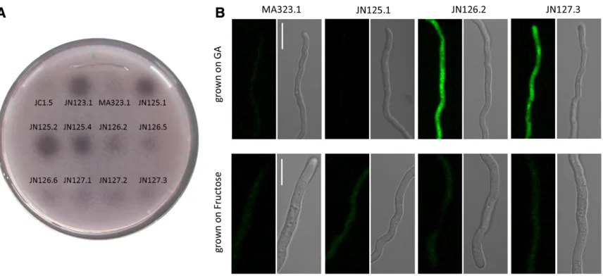
GaaX is induced on galacturonic acid and localized in the cytosol
GaaX was previously identified as a GA-induced gene with unknown function (Martens-Uzunova and Schaap 2008). To monitor the induction of GaaX and to localize the GaaX protein in the cell, GaaX was fused to GFP at either the N-or C-terminal part of GaaX and expressed from the endoge-nous GaaX promoter. Fusion constructs were targeted to the
pyrGlocus ofA. nigerin a strain lacking endogenousgaaX
(JN125.1) to be able to test complementation of the GFP-GaaX and GFP-GaaX-GFP fusion proteins (Figure S3). As shown in Figure 4A, JN125.1 (DgaaX::nicB) constitutively expressed pectinases indicated by the halo on PGA plates, while both the C-terminally tagged as well as the N-terminally tagged versions of GaaX (JN126.2 and JN127.3, respectively) com-plemented the constitutive expression phenotype, indicating that both fusion proteins are functional. Confocalfluorescent
Table 2 Mutations in the constitutive mutants as compared to the parental strain JN29.2
Strain
Total number of SNPs and indels
SNPs or indels in coding region
Mutation in NRRL3_08194
Position of mutation relative to ATG of
NRRL3_08194
Mutation in codon (bold)
Amino acid change
Predicted protein length (full length
protein is 697 amino acids)
JN29.2_UV1 40 19 G-T 1372 GAG-TAG E to Stop 457 aa
JN29.2_UV8 21 4 G-A 1577 GGA-GAA G to E (526) 697 aa
JN29.2_S1 68 34 Extra G 1958 GTT-GGT V to G out of
frame
663 aa
JN29.2_S5 48 11 C-T 1105 CAA-TAA Q to Stop 368 aa
microscopy was performed on GFP-tagged strains to localize GaaX (Figure 4B). Spores were germinated either on GA or on fructose (a nonrepressing carbon source) and afl uores-cent signal was only detectable in the GFP-labeled strains after growth on GA. This observation confirms the results from the expression data that indicate that GaaX is lowly expressed under noninducing conditions and is induced on GA. The expression of GaaX is low on fructose and no GFP signal above the background level was detected on fructose. Based on thefluorescent pictures, GaaX is likely to be local-ized in the cytosol.
Discussion
The forward genetic screen with a positive selection strategy for the isolation ofA. nigermutants with constitutive expres-sion of genes involved in PGA degradation resulted in the identification of a repressor protein (NRRL3_08194), which we named GaaX. Both the genome sequencing offive inde-pendently obtained mutants, as well as the analysis of a tar-geted deletion mutant (DgaaX), showed that the loss of function of gaaX leads to constitutive expression of genes previously identified as GA-induced genes (Martens-Uzunova and Schaap 2008) and genes encoding pectinolytic enzymes that are activated via the transcription factor GaaR (Alaziet al.
2016). Deletion ofgaaXdid not result in a growth alteration on any carbon source tested (Figure 2 and data not shown). Tran-scriptome analysis (Table S3) strongly suggests that deletion of gaaX only affects the expression of genes related to the degradation and metabolism of (poly)galacturonic acid. Genes
encoding enzymes involved in the hydrolysis of nonpectin polysaccharides are not differentially regulated inDgaaX. In addition, GO enrichment analysis of DgaaX transcriptome shows a strong correlation only between the activity of GaaX and the expression of GA-induced genes. In agreement with these observations, the phenotype of thegaaRdeletion mutant was specific for (poly)galacturonic acid, with no growth defect observed on other substrates tested (glucuronic acid, rham-nose, xylose, and arabinose) (Alazi et al. 2016). Taken to-gether, these findings indicate that GaaR and GaaX are specifically involved in the regulation of pectin catabolism.
Interestingly, thegaaXgene is located next to the recently identified GA-specific transcriptional activator gaaR
(NRRL3_08195). The GaaR transcriptional activator is con-served in 19 out of the 20Aspergillusspecies for which geno-mic sequences are available via Aspergillus Genome Database (AspGD), and only absent inAspergillus glaucus(Alaziet al.
2016), which corresponds with the inability ofA. glaucusto grow on GA (http://www.fung-growth.org/). In all 19Aspergillus spe-cies containing GaaR, a GaaX ortholog could be identified ad-jacent to GaaR. Only in Aspergillus fumigatus(Figure 5) and
Aspergillus wentii (data not shown) were ORFs predicted to be present in betweengaaXandgaaR. The ORFs betweengaaX
andgaaR inA. fumigatusare Afu4g06430 and Afu4g06450. Afu4g06430 is predicted to encode a 128-aa long protein that has no ortholog in other aspergilli. According to available ex-pression data (Lindet al.2015), this gene is not expressed, and it is questionable whether this predicted gene actually encodes a protein. Afu4g06450 is predicted to encode a Tan1-related transposase of the DDE family. This type of transposase is
Figure 2 Regulation of thepgaXexpression is controlled by GaaX and by CreA-mediated glucose repression. Growth ofpgaX-amdSreporter strains in
found in bothA. nidulansandA. niger, as well as in many other organisms. This transposase is also lowly expressed inA. fumigatus(Lindet al.2015).
LikegaaR,gaaXis also missing inA. glaucus. BLASTP and synteny analysis between A. niger and A. glaucus revealed that the GaaR/GaaX encoding genes have been excised, as surrounding genes are conserved. Despite the loss of GaaX and GaaR,A. glaucusstill possesses the GA-specific catabolic genes gaaA (Aspgl1_0124049), gaaB (Aspgl1_0091535), andgaaC(Aspgl1_0065497).
The GaaR transcriptional activator has previously been reported to be conserved in other Ascomycetes belonging to the Pezizomycotina subdivision, including members of the Eurotiomycetes (Penicillium,Talaromycesspp), Leotiomycetes (Botrytis,Oidiodendron), Sordariomycetes (Neurospora, Myce-liophthora, Magnaporthe, Trichoderma, and Fusarium spp.), and Dothideomycetes [Zymoseptoria(Mycosphaerella), Aureo-basidium, andCochliobolusspp.] (Zhanget al.2016). Synteny analysis of 17 species belonging to four classes of Pezizomy-cetes (EurotiomyPezizomy-cetes, LeotiomyPezizomy-cetes, SordariomyPezizomy-cetes, and Dothideomycetes) revealed a strong conservation of the geno-mic clustering ofgaaRandgaaXorthologs (Figure 5 andTable
S5). For most fungal species analyzed,gaaRandgaaXare next
to each other on the chromosome or close to each other and separated by one to five genes (Figure 5). The head to tail orientation ofgaaR–gaaXdriving expression ofgaaRandgaaX
from different promoters is conserved in all species except in
Oidiodendron maius. Like GaaR, GaaX was found only in the Pezizomycotina and not in ascomycete yeasts, zygomycetes, or basidiomycetes.
The strategy to identify the responsible mutation by sequencing five independently obtained mutants has been
successful and efficient. Clearly, sequencing only a limited number of mutants leads only to successful identification when the mutants isolated in the screen all belong to a single complementation group. If more complementation groups are involved, more mutants would need to be sequenced. It is interesting to note that in addition to mutations in gaaX
which were present in allfive mutants, we noticed that two mutants (S1 and UV1) also contained allelic mutations in NRRL3_06175 (Table S2). The protein encoded by this gene is predicted to encode a cocaine esterase and belongs to a protein subfamily of hydrolases that included cocaine ester-ase (CocE), several glutaryl-7-ACA acylester-ases, and the putative diester hydrolase NonD ofStreptomyces griseus. This family shows extensive, low-level similarity to a family of Xaa-Pro dipeptidyl-peptidases. Whether this gene also contributes to the constitutive expression of GA-dependent genes remains to be determined, but this is unlikely as mutants without mutations in this gene display essentially the same constitu-tive phenotype.
Previous studies have identified genes specifically induced by GA (Martens-Uzunova and Schaap 2008) and pectinolytic genes that were dependent on the GaaR transcriptional acti-vator for induction by GA (Alaziet al.2016). Eleven of the 15 GA-induced genes identified by Martens-Uzunova and Schaap were upregulated in thegaaXmutant (Figure 3 and Table 3). The three genes that are considered GA-inducible but not detected as differentially expressed in thegaaX mu-tant are predicted to encode a transporter (NRRL3_04281), an exo-polygalacturonase (NRRL3_09810,pgxA), and a pec-tin lyase (NRRL3_00965,pelA). These three genes were not classified as differentially expressed according to the strin-gent statistical settings in our current study. The fourth gene
Figure 3 Venn diagram showing the
over-laps between upregulated genes in the wt_fructosevs.wt_GA study (Martens-Uzunova and Schaap 2008), the upregulated genes be-tweenDgaaR-GAvs.wt-GA (Alaziet al.2016), and the upregulated genes in wt_fructosevs.
induced on GA in the study of Martens-Uzunova and Schaap (2008), but missing in our study, isgaaXitself.
In our recent study on the GaaR transcriptional activator, we identified 32 pectinolytic genes whose expression on GA was dependent on GaaR (Alazi et al. 2016). These genes overlap largely with the previously identified GA-responsive genes (Martens-Uzunova and Schaap 2008) (Figure 3 and Table 3), but also include 18 new potential GaaR target genes. Six of these genes [including NRRL3_02479 (lacB), NRRL3_05252 (pmeC), NRRL3_08325 (pmeA), NRRL3_07470 (pmeB), NRRL3_10559 (rgxC), and NRRL3_01237 (pelD)] were also found to be significantly upregulated inDgaaX (Fig-ure 3 and Table 3) and are therefore considered to be part of the core GA regulon. The remaining 12 genes identified as being GaaR dependent for induction on GA (Alazi et al.
2016) were not identified as differentially expressed based on the stringent settings in this study. Whether these genes are indeed directly controlled by GaaR and GaaX, and therefore part of the core GA regulon, awaits further study.
The GaaX protein is predicted to be 697 aa long and displays significant similarity to the last three domains in the C-terminal half of the AROM protein. AROM is a large (1586 aa in A. niger) pentafunctional protein composed of
five domains and the individual domains are involved infive different enzymatic steps representing the prechorismate shikimate pathway, which is required for aromatic amino acid biosynthesis (Duncanet al.1987; Hawkins and Smith 1991). The last three domains of the AROM protein encode the shikimate kinase, 3-dehydroquinate dehydratase, and shiki-mate dehydrogenase and are homologous to the respective bacterial enzymes (aroL,aroD, andaroE) (Lambet al.1996). The AROM protein is present in fungi, including yeasts, and Euglena. The evolutionary origin of AROM is likely to be bacte-rial and it has been suggested that the AROM protein is the
result of gene fusion events (Richardset al.2006). Sequence alignment and BLASTP searches showed that the GaaX pro-tein has significant sequence homology with the last three domains of the AROM protein. The observation of a transcrip-tional activator (GaaR) located next to a possible repressor protein (GaaX) that displays significant homology to AROM is analogous to the clustered transcriptional activator/repressor module regulating quinic acid utilization (Geeveret al.1989; Lamb et al.1990). Like GaaX, the quinate repressor protein shows significant sequence similarities with the last three C-terminal domains of AROM (Lambet al.1996).
The regulation of metabolic enzymes required for quinic acid utilization has been a classical example of gene regulation both inN. crassaandA. nidulans(Geeveret al.1989; Leversley
et al.1996). InA. nidulansandN. crassa, the transcriptional activator and repressor are located in a gene cluster which consists of the activator and repressor and other genes in-volved in quinic acid catabolism and transport (Geever
et al.1989; Lambet al.1990). A. nigeralso has a quinic acid gene cluster that includes, besides the qutA gene (NRRL3_11038) andqutRgene (NRRL3_11039), a catabolic 3-dehydroquinase (NRRL3_11037) and an Major facilitator superfamily (MFS) transporter possibly involved in quinate uptake (NRRL3_11036). In contrast to the quinic acid gene cluster in which the regulatory genes (activator and repressor) are clustered with structural genes, no structural genes involved in GA utilization were clustered with GaaR and GaaX. Deletion of the qutA transcription factor (NRRL3_11038) inA. nigerresults in a quinate nonutilizing mutant (M. Arentshorst and A. F. J. Ram, unpublished re-sults). Both in A. nidulans andN. crassa, the regulation of genes involved in quinic acid metabolism has been studied in detail and is characterized by the presence of a transcrip-tional activator (named QutA in A. nidulans, and qa-1Fin
Figure 4 (A) Complementation analysis of GaaX-GFP fusions. Polygalacturonase activities ofgaaXdeletion strains,gaaX-GFP and GFP-gaaX
N. crassa) located next to a repressor protein (QutR in A. nidulans, andqa-1SinN. crassa). Loss of function of quinic acid repressor qutRor qa-1S in A. nidulans and N. crassa, respectively, leads to constitutive expression of quinic acid utilization genes (Giles et al.1985; Lambet al.1996), very similar to the effect observed for the loss of function of GaaX, resulting in constitutive expression of GA utilization genes. Based on the phenotype of thegaaXmutant and the analogy to the organization of the quinic acid utilization gene cluster, our current working hypothesis is that gaaXencodes a re-pressor protein which is required to keep the transcriptional activator GaaR in an inactive form in the absence of the in-ducer molecule.
As noted earlier,gaaX(NRRL3_08194) was identified as an upregulated gene when anA. nigerculture pregrown for 18 hr with 2% fructose was transferred to a medium contain-ing 1% GA as the sole carbon source (Martens-Uzunova and Schaap 2008). The expression of a functional GFP-tagged version of GaaX confirmed the induced expression and showed cytosolic localization of GaaX in the presence of GA (Figure 4). In the promoter region ofgaaX, a GA-responsive element (GARE) was found, suggesting that activation of the
transcription factor results in increased levels of repressor protein. Although this might seem contradictory at thefirst glance, it could be an elegant mechanism to ensure that the expression of GA-induced genes is tightly controlled and quickly responds to the presence or absence of GA. The induction of the expression of the repressor is partially anal-ogous the activation/repression system of the qa cluster in
N. crassa. InN. crassait has been shown that both the acti-vator (qa-1F) and the repressor (qa-1S) are transcriptionally induced in the presence of quinic acid (Patelet al.1981; Giles
et al.1991). In the GA regulation system ofA. niger, only the repressor protein is induced and not the activator. It should be noted that in almost all of 17 species analyzed, thegaaX
andgaaRgenes do not share the same promoter region (head to tail orientation; Figure 5), while theqa-1Sandqa-1Fgenes of N. crassa share the same promoter region, which might function as a bidirectional promoter.
As a working model (Figure 6), we postulate that in the presence of GA, the inducer molecule, which could be GA or a derivative of GA, binds in the cytosol to repressor protein GaaX. Binding of the inducer to the GaaX repressor is posited
Figure 5 Schematic overview of the conservation of thegaaX-gaaRgene
pair in 17Pezizomycotinaspecies. GaaX orthologs (green), GaaR ortho-logs (yellow), and ORFs between gaaXand gaaR(gray) are indicated. Arrow heads indicate the direction of transcription.
Figure 6 Model for the regulation of GA-induced gene expression inA.
to result in the activation of the transcription factor GaaR. Active GaaR is expected to induce the expression of GA-responsive genes involved in GA release, uptake and me-tabolism, but also induces the expression of repressor pro-tein. As long as the inducer is present in sufficient amounts, the GaaX repressor is predicted to be inactive as a repressor and thereby the GaaR transcription factor remains active. When the concentration of inducer decreases, it is reasonable to suggest that repressor proteins lacking bound inducer could inactivate the GaaR transcriptional activator, thereby restraining the expression of GA-responsive genes. Thus, high expression of the repressor could serve as a sensitive system to ensure that, when intracellular GA levels decrease, the cell can tightly turn off expression of GA-responsive genes. More-over, this mechanism also ensures the rapid response to the presence of GA as it does not requirede novo synthesis of GaaR. Induction simply requires the binding of inducer to the repressor and subsequent activation of GaaR via post-translational mechanisms, as the expression of GaaR is not dramatically induced by GA (Alaziet al.2016) or in thegaaX
mutant (this study). The expression of GA-induced genes is also controlled via CreA mediated CCR (de Vrieset al.2002; Niu et al. 2015). The analysis of the PpgaX-amdSreporter strain (Figure 2) suggests that the expression of pgaX is carbon catabolite repressed even in the DgaaXstrain. This suggests that CreA directly represses pgaX expression via CreA binding sites in the pgaX promoter, independent of GaaX repression (Figure 6).
The proposed model for the mechanism by which GaaR and GaaX regulate gene expression resembles in some aspects the Gal3/Gal4/Gal80 module of Saccharomyces cerevisiae, but shows at least two important differences. Whereas the Gal4 regulatory system consists of three proteins (Gal4 as the tran-scriptional activator, Gal80 as the repressor, and Gal3 as pos-sible galactose sensor), we have identified two genes/ proteins involved in GA regulation and no evidence for a third member. Also in the regulation of quinate metabolism, no third regulatory gene has been identified even though satu-rating mutant screens have been performed. These observa-tions do not exclude the possibility that a third factor is involved in the GA or quinic acid regulation, but it is unlikely with the available evidence. Whereas the sensor (Gal3)/ repressor (Gal80) function is mediated via two different pro-teins in the Gal regulatory system inS. cerevisiae, in the GA and quinic acid regulatory systems, the sensor/repressor function might well be performed by a single protein, GaaX and QutR, respectively. Another important difference is that GaaX and QutR do not show homology to Gal80 or Gal3, nor do Gal80 or Gal3 display homology to AROM. Based on these observations, we suggest that the GAL repressor module has evolved independently from that of GaaX/QutR.
In addition to GaaX (NRRL3_08194) and QutR (NRRL3_11039), we identified two additional paralogues in theA. nigergenome (NRRL3_08276 and NRRL3_07605). All four paralogues showed significant homology to the A. niger AROM protein, as well as limited homology towards
each other. Both NRRL3_08276 and NRRL3_07605 are also located next to predicted Zn(II)2Cys6 domain transcription factors, NRRL3_08275 and NRRL3_07604, respectively. Whereas the function of the GaaR/GaaX and QutA/QutR modules are related to GA and quinic acid metabolism, re-spectively, the function of the two other pairs that are present inA. nigerremains to be elucidated. The sequence similarity of NRRL3_08276 and NRRL3_07605 to QutR and GaaX and their genome clustering with predicted transcription factors suggest that the proposed activator/repressor modules ob-served for GaaR–GaaX and QutA-QutR is an evolutionarily conserved mechanism to control gene expression infi lamen-tous ascomycete fungi. The number of similar activator/ repressor modules varies among Pezizomycotina species (Figure S4andFigure S5). Most Pezizomycotina species con-tain the galacturonic acid and quinic acid related transcrip-tional activator/repressor modules. It is interesting to note that some fungi, e.g.,Talaromyces stipitatus andB. cinerea, seem to have lost the quinic acid specific repressor, which suggests they might have lost the capacity to utilize quinic acid. The GaaR/GaaX and QutA/QutR activator/repressor modules and their variants are specific for Pezizomyco-tina and missing in ascomycete yeasts, zycomycetes, and basidiomycetes.
Acknowledgments
We thank Cees van den Hondel for helpful discussions and Tim Knetsch and Jos Reijngoud for assistance with bio-reactor cultivations. J.N. is supported through a grant from the China Scholarship Council. E.A. is supported by a grant from BE-Basic within flagship 10 Advanced Microbial Bio-fuel and chemicals production (AMBIC). This work was in part supported by Genome Canada and Génome Québec.
Literature Cited
Alazi, E., J. Niu, J. E. Kowalczyk, M. Peng, M. V. Aguilar Pontes
et al., 2016 The transcriptional activator GaaR of Aspergillus
niger is required for release and utilization of D-galacturonic
acid from pectin. FEBS Lett. 590: 1804–1815.
Arentshorst, M., A. F. Ram, and V. Meyer, 2012 Using non-homologous end-joining-deficient strains for functional gene analyses infi lamen-tous fungi. Methods Mol. Biol. 835: 133–150.
Arentshorst, M., J. Niu, and A. F. Ram, 2015 Efficient Generation
of Aspergillus niger knock out Strains by combining NHEJ
mutants and a split marker approach, pp. 263–272 inGenetic
Transformation Systems in Fungi, Vol. 1, edited by M. A. van den
Berg, and K. Maruthachalam. Springer International Publishing, New York.
Battaglia, E., M. Zhou, and R. P. de Vries, 2014 The transcrip-tional activators AraR and XlnR fromAspergillus nigerregulate expression of pentose catabolic and pentose phosphate pathway genes. Res. Microbiol. 165: 531–540.
Bennett, J. W., and L. Lasure, 1991 Growth media, pp. 441–458
inMore Gene Manipulations in Fungi, edited by J. W. Bennett,
and L. L. Lasure. Academic Press, San Diego.
for assignment of genes to six linkage groups inAspergillus niger. Curr. Genet. 14: 437–443.
Damveld, R. A., A. Franken, M. Arentshorst, P. J. Punt, F. M. Klis
et al., 2008 A novel screening method for cell wall mutants in
Aspergillus niger identifies UDP-galactopyranose mutase as an
important protein in fungal cell wall biosynthesis. Genetics 178: 873–881.
de Vries, R. P., J. Visser, and L. H. de Graaff, 1999 CreA modulates the XlnR-induced expression on xylose of Aspergillus niger
genes involved in xylan degradation. Res. Microbiol. 150: 281–285.
de Vries, R. P., J. Jansen, G. Aguilar, L. Parenicova, V. Joostenet al., 2002 Expression profiling of pectinolytic genes from
Aspergil-lus niger. FEBS Lett. 530: 41–47.
Dowzer, C. E., and J. M. Kelly, 1991 Analysis of thecreAgene, a regulator of carbon catabolite repression inAspergillus nidulans. Mol. Cell. Biol. 11: 5701–5709.
Duncan, K., R. M. Edwards, and J. R. Coggins, 1987 The penta-functional arom enzyme ofSaccharomyces cerevisiaeis a mosaic of monofunctional domains. Biochem. J. 246: 375–386. Garrison, E., and G. Marth, 2012 Haplotype-based variant
detec-tion from short-read sequencing. ArXiv 1207.3907 [q-bio.GN]. Geever, R. F., L. Huiet, J. A. Baum, B. M. Tyler, V. B. Patelet al.,
1989 DNA sequence, organization and regulation of the qa gene cluster ofNeurospora crassa. J. Mol. Biol. 207: 15–34. Giles, N. H., M. E. Case, J. Baum, R. Geever, L. Huiet et al.,
1985 Gene organization and regulation in the qa (quinic acid) gene cluster ofNeurospora crassa. Microbiol. Rev. 49: 338–358. Giles, N. H., R. F. Geever, D. K. Asch, J. Avalos, and M. E. Case, 1991 The Wilhelmine E. Key 1989 invitational lecture. Organi-zation and regulation of the qa (quinic acid) genes in Neurospora crassa and other fungi. J. Hered. 82: 1–7.
Grant, S., C.F. Roberts, H. Lamb, M. Stout, and A.R. Hawkins 1988 Genetic regulation of the quinic acid utilization (QUT) gene cluster in Aspergillus nidulans. J. Gen. Microbiol. 134: 347–358.
Gruben, B. S., M. Zhou, A. Wiebenga, J. Ballering, K. M. Overkamp
et al., 2014 Aspergillus niger RhaR, a regulator involved in
L-rhamnose release and catabolism. Appl. Microbiol. Biotechnol. 98: 5531–5540.
Hawkins, A. R., and M. Smith, 1991 Domain structure and inter-action within the pentafunctional arom polypeptide. Eur. J. Biochem. 196: 717–724.
Jiang, H., R. Lei, S. Ding, and S. Zhu, 2014 Skewer: a fast and accurate adapter trimmer for next-generation sequencing paired-end reads. BMC Bioinformatics 15: 182.
Jørgensen, T. R., B. M. Nitsche, G. E. Lamers, M. Arentshorst, C. A. van den Hondelet al., 2010 Transcriptomic insights into the physiology of Aspergillus niger approaching a specific growth rate of zero. Appl. Environ. Microbiol. 76: 5344–5355. Kashyap, D. R., P. K. Vohra, S. Chopra, and R. Tewari,
2001 Applications of pectinases in the commercial sector: a review. Bioresour. Technol. 77: 215–227.
Khan, M., E. Nakkeeran, and S. Umesh-Kumar, 2013 Potential application of pectinase in developing functional foods. Annu. Rev. Food Sci. Technol. 4: 21–34.
Kopylova, E., L. Noé, and H. Touzet, 2012 SortMeRNA: fast and accuratefiltering of ribosomal RNAs in metatranscriptomic data. Bioinformatics 28: 3211–3217.
Lamb, H. K., A. R. Hawkins, M. Smith, I. J. Harvey, J. Brownet al., 1990 Spatial and biological characterisation of the complete quinic acid utilisation gene cluster inAspergillus nidulans. Mol. Gen. Genet. 223: 17–23.
Lamb, H. K., J. D. Moore, J. H. Lakey, L. J. Levett, K. A. Wheeler
et al., 1996 Comparative analysis of the QUTR transcription
re-pressor protein and the three C-terminal domains of the penta-functional AROM enzyme. Biochem. J. 313(Pt 3): 941–950.
Langmead, B., and S. Salzberg, 2012 Fast gapped-read alignment with Bowtie2. Nat. Methods 9: 357–359.
Levesley, I., G. H. Newton, H. K. Lamb, E. van Schothorst, R. W. Dalgleish et al., 1996 Domain structure and function within the QUTA protein of Aspergillus nidulans: implications for the control of transcription. Microbiology 142(Pt 1): 87–98. Love, M. I., W. Huber, and S. Anders, 2014 Moderated estimation
of fold change and dispersion for RNA-seq data with DESeq2. Genome Biol. 15: 550.
Martens-Uzunova, E. S., and P. J. Schaap, 2008 An evolutionary conserved D-galacturonic acid metabolic pathway operates acrossfilamentous fungi capable of pectin degradation. Fungal Genet. Biol. 45: 1449–1457.
Meyer, V., M. Arentshorst, A. El-Ghezal, A. C. Drews, R. Kooistra
et al., 2007 Highly efficient gene targeting in theAspergillus
niger kusAmutant. J. Biotechnol. 10: 770–775.
Mohnen, D., 2008 Pectin structure and biosynthesis. Curr. Opin. Plant Biol. 11: 266–277.
Nitsche, B. M., T. R. Jørgensen, M. Akeroyd, V. Meyer, and A. F. Ram, 2012 The carbon starvation response ofAspergillus niger
during submerged cultivation: insights from the transcriptome and secretome. BMC Genomics 13: 380.
Niu, J., T. G. Homan, M. Arentshorst, R. P. de Vries, J. Visseret al., 2015 The interaction of induction and repression mechanisms in the regulation of galacturonic acid-induced genes in
Aspergil-lus niger. Fungal Genet. Biol. 82: 32–42.
Niu, J., M. Arentshorst, F. Seelinger, A. F. Ram, and J. P. Oue-draogo, 2016 A set of isogenic auxotrophic strains for con-structing multiple gene deletion mutants and parasexual crossings inAspergillus niger. Arch. Microbiol. 198: 861–868. Patel, V. B., M. Schweizer, C. C. Dykstra, S. R. Kushner, and N. H.
Giles, 1981 Genetic organization and transcriptional regula-tion in the qa gene cluster of Neurospora crassa. Proc. Natl. Acad. Sci. USA 78: 5783–5787.
Patro, R., G. Duggal, M. I. Love, R. A. Irizarry, and C. Kingsford, 2016 Salmon provides accurate, fast, and bias-aware tran-script expression estimates using dual-phase inference. bioRxiv 021592.
Petersen, K. L., J. Lehmbeck, and T. Christensen, 1999 A new transcriptional activator for amylase genes in Aspergillus. Mol. Gen. Genet. 262: 668–676.
Raulo, R., M. Kokolski, and D. B. Archer, 2016 The roles of the zinc finger transcription factors XlnR, ClrA and ClrB in the breakdown of lignocellulose by Aspergillus niger. AMB Express 6: 5.
Richards, T. A., J. B. Dacks, J. M. Jenkinson, C. R. Thornton, and N. J. Talbot, 2006 Evolution offilamentous plant pathogens: gene exchange across eukaryotic kingdoms. Curr. Biol. 16: 1857–1864.
Ruijter, G. J. G., and J. Visser, 1997 Carbon repression in
Asper-gilli. FEMS Microbiol. Lett. 151: 103–114.
Sambrook, J., and D. W. Russell, 2001 Molecular Cloning: A
Lab-oratory Manual, 3rd Ed. Cold Spring Harbor Press, New York.
Sloothaak, J., M. Schilders, P. J. Schaap, and L. H. de Graaff, 2014 Overexpression of theAspergillus nigerGatA transporter leads to preferential use of D-galacturonic acid over D-xylose. AMB Express 4: 66.
Song, L., and L. Florea, 2015 Rcorrector: efficient and accurate error correction for Illumina RNA-seq reads. Gigascience 4: 14. Teather, R. M., and P. J. Wood, 1982 Use of Congo red-polysaccharide interactions in enumeration and characterization of cellulolytic bacteria from the bovine rumen. Appl. Environ. Microbiol. 43: 777–780.
van Hartingsveldt, W., I. E. Mattern, C. M. van Zeijl, P. H. Pouwels, and C. A. van den Hondel, 1987 Development of a homolo-gous transformation system for Aspergillus nigerbased on the
van Peij, N. N., J. Visser, and L. H. de Graaff, 1998 Isolation and analysis of xlnR, encoding a transcriptional activator co-ordinating xylanolytic expression inAspergillus niger. Mol. Microbiol. 27: 131–142.
vanKuyk, P. A., J. A. Benen, H. A. Wösten, J. Visser, and R. P. de Vries, 2012 A broader role for AmyR in Aspergillus niger: regulation of the utilisation of D-glucose or D-galactose contain-ing oligo- and polysaccharides. Appl. Microbiol. Biotechnol. 93: 285–293.
Yuan, X. L., R. M. van der Kaaij, C. A. van den Hondel, P. J. Punt, M. J. van der Maarel et al., 2008a Aspergillus niger genome-wide analysis reveals a large number of novel alpha-glucan acting
enzymes with unexpected expression profiles. Mol. Genet. Genomics 279: 545–561.
Yuan, X. L., J. A. Roubos, C. A. van den Hondel, and A. F. Ram, 2008b Identification of InuR, a new Zn(II)2Cys6 transcrip-tional activator involved in the regulation of inulinolytic genes
inAspergillus niger. Mol. Genet. Genomics 279: 11–26.
Zhang, L., R. J. Lubbers, A. Simon, J. H. Stassen, P. R. Vargas Ribera
et al., 2016 A novel Zn2Cys6 transcription factor BcGaaR
reg-ulates D-galacturonic acid utilization in Botrytis cinerea. Mol. Microbiol. 100: 247–262.
Suppl. Figure 1
A
A
A
A
sp
er
gi
llu
s
n
ig
er
A
sp
er
gi
llu
s
fu
m
ig
at
u
s
GaaX
E
NRRL3_07605
QutR
NRRL3_08276
E
E
E
E
L
L
S
A
sp
er
gi
llu
s
n
id
u
la
n
s
Ta
la
ro
m
yc
es
s
ti
pt
at
u
s
Pe
n
ic
ili
u
m
r
u
b
en
s
B
o
tr
yt
is
ci
n
er
ea
O
id
io
d
en
d
ro
n
m
ai
u
s
N
eu
ro
sp
o
ra
c
ra
ss
a
S
S
S
S
S D D D
M
yc
el
io
p
ht
h
o
ra
t
h
er
m
o
p
h
ila
M
ag
n
ep
o
rt
h
e
gr
is
ae
Ti
ch
o
d
er
m
a
re
es
ei
Fu
sa
ri
u
m
gr
am
in
ea
ru
m
Fu
sa
ri
u
m
o
xys
p
o
ru
m
Zy
m
o
se
pt
o
ri
a
tr
ici
ti
A
u
re
o
p
u
llu
la
n
s
C
o
ch
lio
b
o
lu
s
h
et
er
o
st
ro
p
h
u
s
Sa
Zy
Ba
Sa
cc
h
ar
o
m
yc
es
c
er
ev
is
ia
e
U
si
la
go
m
ay
d
is
E
A
sp
er
gi
llu
s
o
ry
za
e
2
3
M
u
co
r
ci
rc
in
el
lo
id
es
Supplemental Table 1 Primers used in this study. Overlapping sequences for fusion PCR are written in bold.
Primer name Sequence (5’ to 3’) Used for
gaaRP7f TCCTCTTCGGCTTCTGCTTC amplification and sequencing of gaaR
gaaRP8r CATGTGATCATTTCGTGGCCT amplification and sequencing of gaaR
gaaRP9r GCATACTCCAGGCTCCCTTG sequencing of gaaR
gaaRP10f CCTACATGGGAAGCCCTATTGG sequencing of gaaR
gaaRP11r CGATAGAGCAGACAGTGCACGA sequencing of gaaR
gaaRP12f CCCTGTTGGTCGACATTACGC sequencing of gaaR
gaaRP13r AAATCGGGTCGGTCCAGTG sequencing of gaaR
gaaRP14f AGGACTTCCTGGACCGGTAT sequencing of gaaR
gaaXP1f (P1) TGAGAAATGAGGATGGAAGAGG amplification of gaaX 5’flank
gaaXP2r (P2) CAATTCCAGCAGCGGCTTCTCATACTTGAATGCGACCGTT amplification of gaaX 5’flank
gaaXP3f (P3) ACACGGCACAATTATCCATCGAAAAGCAATCACGGCAACTGG amplification of gaaX 3’flank
gaaXP4r (P4) ACAGCTGGGACGTAACCGAT amplification of gaaX 3’flank
gaaXP5f CGTTGGTTCGATGTAAATGGG diagnostic PCR to confirm deletion of gaaX
gaaXP6r GTAAGTGTTCGGGGTTGGAG diagnostic PCR to confirm deletion of gaaX
hygP6f (P5) AAGCCGCTGCTGGAATTGGGCTCTGAGGTGCAGTGGAT amplification of hygB marker
hygP9r (P6) GGCGTCGGTTTCCACTATC amplification of hygB marker
hygP8f (P7) AAAGTTCGACAGCGTCTCC amplification of hygB marker
hygP7r (P8) CGATGGATAATTGTGCCGTGTTGGGTGTTACGGAGCATTC amplification of hygB marker
phleoP4f (P9) AAGCCGCTGCTGGAATTGCTCTTTCTGGCATGCGGAG amplification of phleomycin marker
phleoP7r (P10) CACGAAGTGCACGCAGTTG amplification of phleomycin marker
phleoP6f (P11) AAGTTGACCAGTGCCGTTCC amplification of phleomycin marker
phleoP5r (P12) CGATGGATAATTGTGCCGTGTGGAGCATTCACTAGGCAACCA amplification of phleomycin marker
PgaaX-P7f-NotI ATAAGAATGCGGCCGCCGTTGGTTCGATGTAAATGGG amplification of the promoter of gaaX
PgaaX-P11r TCCTCGCCCTTGCTCACCATCTCATTGCATACGCAGTATA amplification of the promoter of gaaX
GFP-P1f ATGGTGAGCAAGGGCGAGGA amplification of GFP
GFP-P3r TGAAGGGCTGTCTGCATCATGGCGCCCTTGTACAGCTCGTCCATGC amplification of GFP
gaaXP12f ATGATGCAGACAGCCCTTCA amplification of gaaX and the terminator of gaaX
TgaaX-P10r-NotI ATAGTTTAGCGGCCGCGTAAGTGTTCGGGGTTGGAG amplification of gaaX and the terminator of gaaX
gaaX-P8r TCCTCGCCCTTGCTCACCATGGCGCCAAACACCCGTCCCGATGCCA amplification of gaaX and the promoter of gaaX
GFP-P2r TATACTATATAAGAAAAATCCTACTTGTACAGCTCGTCCATGC amplification of GFP
ABpyrGP11f AAGGAAAAAAGCGGCCGCCGTCGCGTGATAAGGGTTG amplification of pyrG 3’flank for probe
ABpyrGP13r CGAGCTCGGCGCGCCTCGGGTCAATTTCCTCTGTTG amplification of pyrG 3’flank for probe