| WORMBOOK CELL AND ORGANELLE BIOLOGY
Mitotic Cell Division in
Caenorhabditis elegans
Lionel Pintard*,1and Bruce Bowerman†,1 *Equipe labellisée Ligue contre le Cancer, Institut Jacques Monod, Team Cell Cycle and Development UMR7592, Centre National de la Recherche Scientifique–Université Paris Diderot, Sorbonne Paris Cité, 75013 Paris, France,†Institute of Molecular Biology, University of Oregon, Eugene, OR 97403 ORCID IDs: 0000-0003-0286-4630 (L.P.); 0000-0002-6479-8707 (B.B.)
ABSTRACT Mitotic cell divisions increase cell number while faithfully distributing the replicated genome at each division. The
Caenorhabditis elegansembryo is a powerful model for eukaryotic cell division. Nearly all of the genes that regulate cell division in
C. elegansare conserved across metazoan species, including humans. TheC. eleganspathways tend to be streamlined, facilitating dissection of the more redundant human pathways. Here, we summarize the virtues of C. elegansas a model system and review our current understanding of centriole duplication, the acquisition of pericentriolar material by centrioles to form centrosomes, the assembly of kinetochores and the mitotic spindle, chromosome segregation, and cytokinesis.
KEYWORDSCell division; Centrosome; Cytokinesis; Kinetochore; Mitosis; Spindle assembly; WormBook
TABLE OF CONTENTS
Abstract 35
Centriole Duplication and Centrosome Maturation: Ensuring Fidelity in Bipolar Mitotic Spindle Assembly 37
Centriole disengagement and the initiation of centriole duplication 39
The centriole assembly pathway 39
Centriole assembly: a higher resolution view 42
Limiting centriole duplication by controlling the levels of centriole components 43
PCM assembly dynamics and structure 43
In vitroreconstitution of PCM assembly 45
Kinetochore Assembly, Function, and Regulation 45
Molecular architecture of the C. eleganskinetochore 47
Inner kinetochore proteins: connecting with chromosomal DNA 47
Outer kinetochore proteins: connecting with microtubules 47
From lateral to end-on microtubule attachment: cross-talk between RZZ-Spindly and the Ndc80 complex 49
Microtubule attachments and the SAC 49
The SAC 49
Hierarchy of SAC assembly during checkpoint activation 50
Checkpoint inactivation 51
Kinetochores direct CDC-20 to the mitotic accelerator (APC/C) or brake (SAC) 51
Continued
Copyright © 2019 by the Genetics Society of America doi:https://doi.org/10.1534/genetics.118.301367
Manuscript received July 16, 2018; accepted for publication October 24, 2018
1Corresponding authors: Institut Jacques Monod, 15 rue helene Brion, 75013 Paris, France. E-mail: [email protected]; and 1229 University of Oregon, 1318 Franklin Blvd.,
CONTENTS,continued
Mitotic Spindle Assembly and Chromosome Segregation 52
Centrosome-based spindle assembly in the early embryo 52
Centrosome-localized factors regulating mitotic spindle assembly 53
Kinetochore proteins and central spindle assembly 54
Chromosome segregation 54
Cytokinesis: The Last Chapter in Cell Division 55
Contractile ring components 55
Contractile ring assembly and dynamics 57
Two mitotic spindle signals influence contractile ring assembly 59
Molecular pathways that mediate furrow signaling from the spindle poles 60
NOP-1: a nematode-specific regulator of cytokinesis contributes to spindle pole signaling 60
Centralspindlin and cytokinesis: the role of the CYK-4 GAP domain 61
Further contributions from the spindle midzone to the regulation of cytokinesis 62
Cell polarity and cytokinesis 63
Abscission: the last chapter in cytokinesis 63
Concluding remarks and perspectives 64
C
ELL division in the earlyCaenorhabditis elegansembryo alternates between rapid S phases and mitosis without intervening gap phases, a common feature during early ani-mal embryogenesis; most studies of cell division inC. elegans have focused on the one-cell zygote (Figure 1). During fertil-ization, the sperm brings not only a haploid genome, but also a pair of centrioles into the oocyte, which lacks centrioles. Subsequently, the two sperm-derived centrioles recruit peri-centriolar material (PCM) from the oocyte cytoplasm to form centrosomes that can nucleate microtubules and separate to be positioned on either side of the paternal pronucleus. The oocyte and sperm-derived pronuclei are initially located at opposite ends of the embryo, but then migrate toward each other as chromosomes condense, meeting near the posterior pole. After the pronuclei meet, the nuclear–centrosome com-plex moves to the center while rotating to align with the embryo’s long axis (Figure 1). The nuclear envelope then breaks down as the mitotic spindle assembles, with asymmet-ric elongation during anaphase contributing to a posterior displacement of the spindle. Thus, when the cleavage furrow bisects the mitotic spindle during cytokinesis, an asymmetric division results (Figure 1).C. elegansoffers compelling advantages for dissecting the molecular mechanisms governing mitotic cell division. It is easy to cultivate, its short life cycle (only 3 days at 25°) makes for fast genetics, and its transparency and the large size (503
20mm) and rapid division (20 min) of the one-cell stage
embryo make it amenable to live (Gönczyet al.1999) and
fixed imaging, as well as correlative light microscopy and electron tomography (O’Toole et al. 2003; Pelletier et al. 2006; Sraykoet al.2006; Schlaitzet al.2007; Labandet al. 2017; Redemann et al. 2017). Moreover, cell divisions in
C. elegans are highly stereotyped, providing a rich context for quantitative analysis.
degron-tagged proteins in transgenic animals has expanded conditional loss-of-function approaches (Zhanget al.2015). Finally, genome editing approaches including MosSCI (Frøkjaer-Jensenet al.2008) and CRISPR/Cas9 (Dickinson and Goldstein 2016; Paix et al. 2016), in conjunction with RNAi- or degron-mediated depletion of endogenous wild-type gene products, have enabled powerful gene re-placement strategies that facilitate functional probing of specific domains and residues within evolutionarily con-served proteins. All of this information about genome se-quence, gene structure and function, RNAi phenotypes, the availability of TS alleles, and more are available at the WormBase database (www.wormbase.org) (R. Y. N. Lee et al.2018).
Some cell division traits in C. elegans are unusual com-pared to other model systems. In particular,C. eleganshas holocentric chromosomes. Rather than being restricted to a limited region of the chromosome (the centromere), the ki-netochores that mediate attachment to spindle microtubules run along the entire length of each sister chromatid (Dernburg 2001; Melterset al.2012). Other distinctive
prop-erties include relatively small and simple centrioles that nucleate the microtubule-organizing centers called centro-somes, incomplete nuclear envelope breakdown during mitosis, and a nearly exclusive reliance on centrosome-me-diated nucleation of microtubules during mitotic spindle assembly. Nevertheless, work in C. elegans continues to in-form our general understanding of animal cell division.
Centriole Duplication and Centrosome Maturation: Ensuring Fidelity in Bipolar Mitotic Spindle Assembly
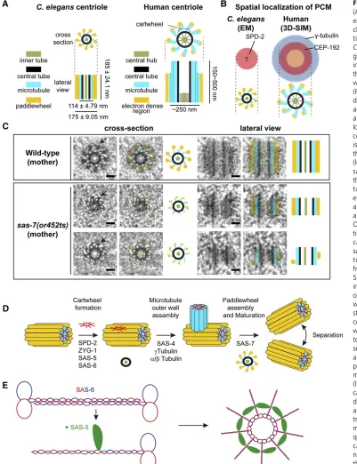
Mitotic spindle assembly requires a collaboration between two spindle poles and the chromosomes that are ultimately segregated into daughter cells. The spindle poles are orga-nized by two centrosomes, microtubule-organizing centers that each have a pair of orthogonally oriented centrioles at their core (Figure 2). Centrioles direct the assembly of an attached matrix of PCM that nucleates and anchors microtu-bules. Across species, centrioles are cylindrical structures roughly 100–400 nm long and 100–250 nm wide (Figure 2A). Their distinguishing feature is a ninefold symmetric Figure 1 Mitotic cell division in the earlyC. elegans embryo. (A) Differential interference contrast (DIC) im-age of an adult worm. The positions of the two most mature oocytes relative to the spermatheca are indi-cated by white arrows and labeled -1 and -2; embryos within the uterus are also indicated. Bar, 100mm. (B) Live image of embryos in the uterus of an adult worm expressing GFP-taggedb-tubulin (green) and mCherry-tagged H2B (red) to visualize oocyte meiotic and em-bryonic mitotic spindles. (C) Frames from in vivolive imaging of mitotic embryos expressing GFP-tagged
array of stabilized microtubules. Depending on the species and tissue, the microtubule blades that make up the centriole outer wall can contain singlet, doublet, or triplet microtu-bules [reviewed in Carvalho-Santos et al. (2011), Bornens (2012), Gönczy (2012), Fırat-Karalar and Stearns (2014), Conduitet al.(2015), Fuet al.(2015)]. During interphase, centrioles are surrounded by a thin highly ordered layer of
PCM. Upon entry to mitosis, centrosomes undergo a mitotic maturation that prepares them for spindle assembly. During this process, the amount of PCM and its microtubule nucle-ation capacity increases 5- to 10-fold. In addition to being larger, the mitotic PCM matrix also appears less organized than its interphase counterpart [reviewed in Mennellaet al. (2014), Woodruffet al.(2014)].
C. eleganscentrioles are relatively small and simple com-pared to those in other model systems, being roughly 200 nm long and 100 nm wide (Figure 2A). Rather than doublets or triplets, centrioles in theC. elegansembryo have singlet mi-crotubules and lack the appendages that decorate the distal ends of mature mother centrioles in vertebrate cells.C. ele-ganscentrioles are similar in size and structure to those in the early Drosophilaembryo (Moritzet al. 1995), which has a similarly short cell-cycle time, and to the early intermediates observed during centriole assembly in mammalian cultured cells (Vorobjev and Nadezhdina 1987), suggesting that they represent a minimal form of the centriole that can assemble quickly to provide centrosome function in a rapidly develop-ing embryo. Nevertheless, centrioles in later-stage embryos appear to be even smaller, roughly 100 nm in both length and width (Feldman and Priess 2012).
Centriole disengagement and the initiation of centriole duplication
Centriole duplication begins after the two centrioles at each spindle pole disengage during anaphase, a step that licenses their subsequent duplication (Fırat-Karalar and Stearns 2014; Lu and Roy 2014). Intriguingly, chromatin factors required for the cohesion of duplicated chromosomes during meiosis and mitosis also regulate centriole disengagement, although their requirements vary among species and at different de-velopmental times. In C. elegans, these cohesion factor re-quirements are most apparent during spermatogenesis, when the two centrioles at each spindle pole remain associ-ated throughout sperm maturation after the completion of meiosis II (Schvarzsteinet al.2013). Before disengagement, HORMA domain proteins that initially form axial elements to promote homologous chromosome pairing and cohesion, and the meiosis-specific cohesin REC-8, prevent centriole disen-gagement, until cell-cycle regulation leads to activation of the cysteine protease SEP-1/separase, cleavage of REC-8, and centriole disengagement. Loss of the HORMA domain pro-teins or REC-8 leads to premature disengagement and over-duplication during spermatogenesis (Schvarzstein et al. 2013).
After fertilization of the oocyte and the completion of meiosis I and II, the two centrioles contributed by the sperm normally disengage and separate, and then each assembles a daughter centriole. Analysis of separase-depleted embryos revealed that the centrioles disengage but fail to separate further or duplicate. The defects in centriole separation and duplication resulting from lack of separase could both be rescued by perturbations that enhance PCM or microtubule assembly (Cabralet al.2013). These results suggest that at the meiosis-to-mitosis transition physical proximity of centri-oles, rather than engagementper se, is sufficient to suppress duplication; that separase promotes separation of the sperm centrioles, rather than their disengagement; and that en-hanced microtubule-dependent forces acting on centrosomes can substitute for the role of separase in promoting centriole separation.
In contrast to their critical role in controlling centriole cohesion and separation during the meiosis to mitosis tran-sition, cohesion and separase do not seem to control centriole dynamics during subsequent mitotic divisions. However, pre-mature disruption of the PCM in these later divisions, by upshifting a fast-acting TS allele of the PCM component SPD-5, resulted in premature centriole separation and over-duplication (Cabralet al.2013), suggesting that the mother and daughter centrioles may be held together by the PCM matrix that forms around the mother. Disassembly of this matrix would then release the daughter and allow the two centrioles to separate and duplicate.
The centriole assembly pathway
After disengagement, a daughter centriole grows out from a single spot near the proximal end of each mother centriole, initially forming an electron-dense central tube that subse-quently becomes decorated with a ninefold symmetric array of singlet microtubules (the outer centriole wall) (Pelletieret al. 2006) (Figure 2C). The core components required for centri-ole assembly were discovered in C. elegansand are widely conserved. Indeed, identification of the centriole assembly pathway is a premier C. elegans contribution to our under-standing of fundamental biological processes.
SixC. eleganscentriole assembly genes have been
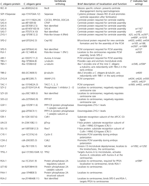
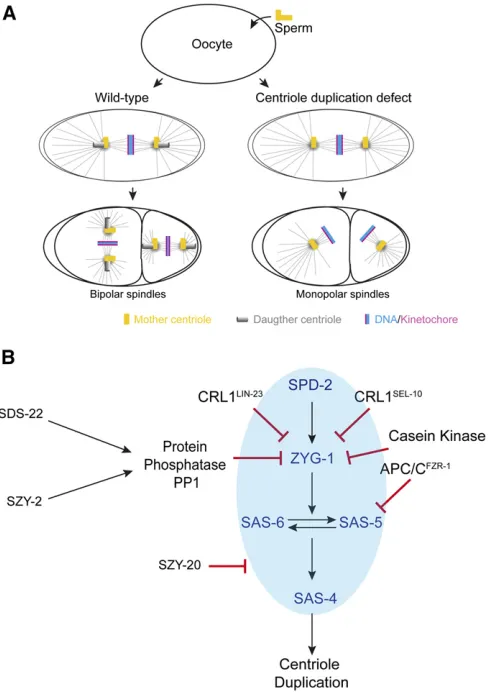
identi-fied through chemical mutagenesis and genome-wide RNAi knockdown screens:sas-4, -5, -6, and-7,spd-2, andzyg-1(sas for spindle assembly abnormal,spdfor spindle defective, and zygfor zygote-defective; Figure 2D) (O’Connellet al.2001; Kirkhamet al.2003; Leidel and Gönczy 2003; Dammermann et al.2004; Delattreet al.2004; Kempet al.2004; Pelletier et al.2004; Leidelet al.2005; Sugiokaet al.2017). Four of these genes encode the conserved coiled-coil proteins SAS-4/ CPAP, SAS-5/STIL, SAS-6/SAS6, and SPD-2/Cep192, while zyg-1 encodes the C. elegans ortholog of mammalian Plk4 (Table 1). When any one of these genes is reduced in function in the oocyte, the sperm still brings a pair of centrioles into the egg during fertilization to support mitotic spindle assem-bly. However, the mutant oocyte cytoplasm fails to support duplication of the sperm-derived centrioles, and the two daughters each inherit a single centriole and form monopolar spindles (Figure 3A). Thus, inhibition of the four genes that are specifically required for centriole assembly (zyg-1,sas-4, sas- 5, andsas-6) leads to a signature phenotype in which the
Table 1 Centrosomes and spindle assembly proteins
C. elegansprotein C. elegansgene
Vertebrate
ortholog Brief description of localization and function
TS alleles (*indicates fast
acting)
REC-8 rec-8(W02A2.6) Rec8 Meiosis specific cohesin: prevents centriole
disengagement during spermatogenesis SEP-1/Separase sep-1(Y47G6A.12) Separase Cysteine protease: cleaves REC-8 and promotes
centriole separation after fertilization SAS-1 sas-1(Y111B2A.24) C2CD3, RPH3A, DOC2A Centriole protein required for centriole stability
SAS-4 sas-4(F10E9.8) CPAP Centriole protein required for centriole assembly
SAS-5 sas-5(F35B12.5) Not identified Centriole protein required for centriole assembly
SAS-6 sas-6(Y45F10D.9) SAS-6 Centriole protein required for centriole assembly or1167
SAS-7 sas-7(T07C4.10) Not identified Centriole protein required for centriole assembly or452
ZYG-1 zyg-1(F59E12.2) Polo-like kinase 4 (Plk4) Centriole protein required for centriole assembly it25,or278,or297*, or409*,or1018
SPD-2 spd-2(F32H2.3) Cep192 Bifunctional protein required for new centriole
formation and for the assembly of the PCM
or655,or493,or454, or183,or188, or293*,or1089
SPD-5 spd-5(F56A3.4) Not identified PCM component required for PCM assembly or213*
PLK-1 plk-1(C14B9.4) Polo-like kinase 1 (Plk1) Localizes to the centrosomes regulates PCM assembly; binds SPD-2
or683
AIR-1 air-1(K07C11.2) Aurora A kinase PCM component required for PCM assembly
TBG-1 tbg-1(F58A4.8) g-tubulin Provides caps and anchors microtubule ends
TBA-1 tba-1(F26E4.8) a-tubulin tba-1encodes one of the nineC. elegans
a-tubulins; acts redundantly with TBA-2 in the early embryo
or346,or594*
TBB-2 tbb-2(C36E8.5) b-tubulin tbb-2encodes aC. elegansb-tubulin; acts
redundantly with TBB-1 in the early embryo
or600
ZYG-9 zyg-9(F22B5.7) XMAP-215 PCM component or634,or628,or593,
or635,or623*
TAC-1 tac-1(Y54E2A.3) TACC-1, 2, 3 PCM component or369,or402,or455
SZY-2 szy-2(Y32H12A.4) Phosphatase 1 inhibitor 2 - I2 Localizes to centrosomes; negatively regulates centrosome size
SZY-20 szy-20(C18E9.3) Not identified Localizes to centrosomes; negatively regulates centrosome size
SDS-22 sds-22(T09A5.9) PPP1R7 Localizes to centrosomes; negatively regulates
centrosome size
GSP-1 gsp-1(F29F11.6) PPP1CB (protein phosphatase
1 catalytic subunitb)
Downregulates ZYG-1 levels
GSP-2 gsp-2(F56C9.1) PPP1CA (protein phosphatase
1 catalytic subunita)
Downregulates ZYG-1 levels
FZR-1 fzr-1(ZK1307.6) Cdh1 Substrate recognition subunit of the APC/C
E3-ligase
LIN-23 lin-23(K10B2.1) bTrcp F-box protein: substrate recognition subunit of
Cullin 1-RING E3-ligases (CRL1)
SEL-10 sel-10(F55B12.3) Fbw7 F-box protein: substrate recognition subunit of
Cullin 1-RING E3-ligases (CRL1)
CYE-1 cye-1(C37A2.4) Cyclin E Promotes PCM assembly during embryo
polarization
CDK-2 cdk-2(K03E5.3) Cdk2 Promotes PCM assembly during embryo
polarization
KLP-7 klp-7(K11D9.1) MCAK Kinesin-13 microtubule depolymerase; localizes to
the kinetochores, the centrosomes.
or1092,or1292
TPXL-1 tpxl-1(Y39G10AR.12) TPX2 Targets Aurora A to microtubules, activates
Aurora A; colocalizes with Aurora A at the centrosomes
RSA-1 rsa-1(C25A1.9) Protein phosphatase 2A
regulatory subunit
Localizes to centrosomes, required for PP2A localization to centrosomes; binds RSA-1
or598*
LET-92 let-92(F38H4.9) Protein phosphatase 2A
catalytic subunit
Localizes to centrosomes
PAA-1 paa-1(F48E8.5) Protein phosphatase 2A
structural subunit
Localizes to centrosomes
sperm, new daughter centrioles can recruit mitotic PCM and assemble bipolar spindles during thefirst and second mitotic divisions. However, the new daughter centrioles cannot re-produce, resulting in cells with monopolar spindles during the third cell division.
Investigation of these six genes has identified a conserved centriole assembly pathway (Figure 2D) (Hodgeset al.2010; Carvalho-Santos et al. 2011). The order in which they act has been determined by taking advantage of the initial con-tribution of centrioles to the one-cell stage zygote by the sperm, with oocytes lacking centrioles due to their earlier elimination during oogenesis (Albertson and Thomson 1993; Mikeladze-Dvaliet al.2012). Maternally expressed centriolar
proteins present in the oocyte are required to assemble the new daughter centrioles adjacent to each of the sperm-pro-vided parent centrioles. A sequence of centriolar protein as-sembly has been established using immunofluorescence in
fixed embryos to detect native sperm-donated centriolar pro-teins and maternal centriolar propro-teins, marked by translational fusions to green fluorescent protein (GFP) (Dammer mann et al.2004; Delattreet al.2006; Pelletieret al.2006).
In brief, such studies have shown that SPD-2 is recruited to the sperm centrioles early in meiosis I and is the centriolar receptor for ZYG-1/Plk4, the master regulatory kinase that controls centriole assembly (Delattre et al. 2006; Pelletier et al. 2006). ZYG-1/Plk4 is in turn required for the recruit-ment of SAS-5 and SAS-6, which, together with ZYG-1/Plk4, direct formation of thefirst intermediate in centriole assembly, called the central tube or cartwheel (Figure 2E) (Dammermann et al.2004; Delattreet al.2004; Leidelet al.2005; Delattre et al.2006; Pelletieret al.2006)
Downstream of cartwheel assembly, SAS-4 controls forma-tion of the microtubule-containing centriole outer wall (Fig-ure 2D) (Kirkham et al. 2003; Leidel and Gönczy 2003; Pelletieret al.2006). Live imaging of SAS-4 recruitment fol-lowing fertilization indicates that it is recruited to daughter centrioles at the same time as SAS-6. Based on recovery after photobleaching, centriolar SAS-4 initially exists in a form that can rapidly exchange with SAS-4 in the cytoplasm, and sub-sequently converts to a more stable form that does not un-dergo rapid exchange. The conversion of SAS-4 to a stably associated form requiresg-tubulin and microtubule assembly (Dammermannet al.2008). These results suggest that SAS-4 is stably incorporated into the centriole outer wall as the centriolar microtubules are formed. After assembly of the microtubule-containing outer wall, the coiled-coil protein SAS-7 directs assembly of an additional ninefold symmetric structure, termed the paddlewheel, that runs along the length of the centriolar microtubules (Figure 2D) (Sugioka et al.2017). The paddlewheel forms concurrent with centri-ole maturation, the acquisition by the daughter centricentri-ole of the ability to reproduce, and SAS-7 is thefirstC. elegans pro-tein known to be required for centriole maturation. Consis-tent with a role in maturation, SAS-7 binds to SPD-2 in yeast two-hybrid assays (Boxemet al.2008; Sugiokaet al.2017), localizes to daughter centrioles independently of SPD-2, and is at least partially required for SPD-2 to localize to centrioles (Sugiokaet al.2017). Thus, in addition to being required for addition of the paddlewheel to the outer wall that completes formation of the daughter centriole, SAS-7 is also the earliest acting protein in centriole assembly through its role in recruiting SPD-2. SAS-7 shows some weak homology to the centriole-localized Chibby family of proteins, but thus far these potential homologs have not been found to have roles in centriole duplication.
Finally, SAS-1 (a C2 domain protein that binds to and stabilizes microtubulesin vitro) is a centriole-localized pro-tein that appears to promote normal centriole structure and stability, but may not be required for centriole duplication Figure 3 Pathways regulatingC. eleganscentriole duplication and
(Table 1) (von Tobelet al.2014). Centriole proteins them-selves inC. elegansare remarkably stable: when introduced by fertilization into wild-type hermaphrodites expressing un-marked centriolar proteins, GFP fusions to some sperm cen-triole proteins persist throughout embryogenesis (Balestra et al.2015).
Centriole assembly: a higher resolution view
From a structural perspective, the best-understood centriole component is SAS-6 [reviewed in Gönczy (2012), Hirono (2014), Dong (2015)]. This widely conserved protein in-cludes an unstructured C-terminal region, a long central coiled-coil domain that mediates homodimerization, and a globular N-terminal domain (Figure 2E). Interactions be-tween the globular head domains of SAS-6 dimers oligomer-izes them to form a remarkable ring-shaped structure called the cartwheel, with ninefold radial symmetry that beautifully explains the ninefold symmetric organization of the microtu-bules that assemble along the length of each centriole (Figure 2E) (Kitagawa et al. 2011b; van Breugelet al. 2011). The N-terminal globular domains form the hub of this cartwheel, with the coiled-coil dimerized spokes projecting outward (Figure 2E). In other organisms, the SAS-6 cartwheels appear to be stacked upon each other with aligned spokes, forming an electron-dense central tube and spoke structure visible in electron micrographs (Figure 2). InC. elegans, it has been suggested that SAS-6 may instead form a spiral in which the spokes are not fully aligned (Hilbertet al.2013), which could explain the lack of prominent electron-dense spokes in electron micrographs (Figure 2) (Pelletier et al. 2006; Sugioka et al. 2017). How the termini of the coiled-coil spokes promote centriole microtubule assembly is not known, but the process requires SAS-4 (Pelletier et al. 2006), which resides at the centriole perimeter near the spoke termini (Kirkhamet al.2003).
Mechanisms that promote assembly of the SAS-6– containing cartwheel have begun to emerge. Across species, ZYG-1/Plk4 forms a homodimer through the antiparallel as-sociation of its cryptic polo box domain (Park et al. 2014; Shimanovskayaet al.2014). Dimerization of the cryptic polo box domain of ZYG-1/Plk4 generates a basic patch that in-teracts with an acidic region at the N-terminus of SPD-2, to recruit Plk4/ZYG-1 to the mother centriole (Shimanovskaya et al.2014). The recruitment of SAS-6 to the assembly site on the mother centriole requires ZYG-1 and SAS-5, which inter-act with adjacent regions on the SAS-6 coiled-coil (Qiaoet al. 2012; Lettmanet al.2013). The kinase activity of ZYG-1/Plk4 is not required to recruit SAS-6 to the vicinity of the mother centriole, but is required for its subsequent oligomerization to form the cartwheel (Lettmanet al.2013). The critical func-tion of the kinase activity of ZYG-1/Plk4 in promoting SAS-6 oligomerization is not known, although the phosphorylated target is unlikely to be SAS-6 (Kitagawaet al.2009; Lettman et al.2013). SAS-5 forms homo-oligomers through two dis-tinct domains, and mutations that disrupt oligomerization prevent centriole duplication (Qiao et al. 2012; Rogala
et al.2015), although the function of SAS-5 oligomerization in centriole assembly remains unclear.
The PCM also has a role in centriole assembly. While not absolutely required for centriole duplication, RNAi knock-down of the coiled-coil PCM scaffolding protein SPD-5 pre-vents centriole duplication in roughly half of the mutant embryos based on both light microscopy (Dammermann et al.2008) and electron tomography studies (O’Tooleet al. 2012). This requirement reflects a role for the SPD-5 depen-dent accumulation of PCM-associatedg-tubulin in centriole duplication, as g-tubulin knockdown also partially compro-mises centriole duplication but does not affect SPD-5 accu-mulation. As noted earlier, at least one role for g-tubulin appears to involve conversion of the initially exchangeable centriolar SAS-4 into a form that cannot rapidly exchange with cytoplasmic pools, likely by promoting assembly of the microtubules that make up the outer centriole wall (Dammermannet al.2008).
Further evidence for a link between PCM assembly and centriole duplication has come from the identification of SZY-20, a conserved protein with RNA binding domains that negatively regulates centrosome maturation (Song et al.2008). Theszy-20gene was identified in a mutagenesis screen for suppressors of a conditional zyg-1allele (Kemp et al.2007), and reducing SZY-20 function can restore cen-triole duplication in bothzyg-1andspd-2mutants. Normally, SZY-20 limits the accumulation of ZYG-1, SPD-2, and other PCM components to centrosomes, and loss of SZY-20 results in abnormally large centrosomes and ensuing defects in spin-dle position and function that can be suppressed by reducing ZYG-1 levels. Thus, reducing SZY-20 function might restore centriole duplication inzyg-1andspd-2mutants by increas-ing the accumulation of partially functional ZYG-1 and SPD-2 mutant proteins, or alternatively by increasing the PCM levels
ofg-tubulin (Dammermannet al.2004; Songet al.2008).
SAS-4–containing outer wall that forms may depend on the size of the cartwheel and its ability to nucleate outer wall microtubules.
Intriguingly, electron tomography has shown that in the absence of eitherg-tubulin or SPD-5, both of which result in the production of daughter centrioles with abnormally low levels of SAS-4 (Dammermannet al.2004), centriolar micro-tubules often extend well beyond the ends of both the mother and daughter central tubes (O’Tooleet al.2012). It is surpris-ing that lower levels of SAS-4, which is required for centriolar microtubule assembly, and ofg-tubulin, which can nucleate microtubule assembly, result in abnormally long centriolar microtubules. However, it is possible that reducing the num-ber of PCM-associated microtubule polymers might increase local tubulin levels and thereby promote centriolar microtu-bule growth. Alternatively, PCM disruption in these mutants might interfere with factors that normally limit centriolar microtubule length.
Limiting centriole duplication by controlling the levels of centriole components
How does a mother produce only a single daughter? Studies in multiple systems indicate that tight regulation of Plk4 levels is critical (Nigg and Stearns 2011). Indeed, Plk4 is a suicide kinase regulated by autophosphorylation on an internal phosphodegron; disruption of the phophodegron increases Plk4 levels and produces centriolar rosettes with multiple daughters per mother centriole (Guderian et al. 2010; Holland et al. 2010; Cunha-Ferreira et al. 2013; Klebba et al. 2013). Furthermore, superresolution microscopy has shown that Plk4 initially forms a ring around one end of the mother centriole that subsequently resolves into a single spot to establish the site of daughter centriole assembly (Mennella et al.2014). Presumably, tight regulation of Plk4 levels is im-portant for resolving its localization to a single spot.
While genetic studies inC. eleganshave focused on positive regulation of centriole duplication, a screen for suppressors of a TSzyg-1mutant (Kempet al.2007) has identified neg-ative regulators of centriole duplication that provide insight into how ZYG-1/Plk4 levels are regulated (Figure 3B). Two suppressors of TS zyg-1 embryonic lethality and centriole duplication,szy-2/I-2andsds-22, reduce the function of the protein phosphatase PP1 (Peel et al. 2017). Reducing PP1 function, or its positive regulators SZY-2/I-2 and SDS-22, increases early embryo ZYG-1 levels and results in the pro-duction of extra centrioles, and ZYG-1 overexpression in lar-val neuroectodermal stem cell lineages also results in centriole amplification (Wolfet al.2018). Live-cell and super-resolution imaging suggest that the extra centrioles in early embryos result from the simultaneous assembly of two or more daughters from one mother centriole (Peel et al. 2017). Thus, increased ZYG-1 levels may interfere with res-olution of the ZYG-1/Plk4 ring into a single focus to produce only one daughter centriole, although whether ZYG-1 ini-tially forms a ring at one end of the mother centriole has yet to be determined.
Additional inputs serve to further limit ZYG-1 levels dur-ing centriole duplication (Figure 3B). In addition to PP1 negatively regulating ZYG-1 levels, ubiquitin-dependent proteasome degradation, mediated by the Cullin 1 family member CUL-1 as an E3 ligase scaffold and two F-box sub-strate adaptor proteins LIN-23/bTrcp and SEL-10/Fbw7/ Cdc4, also negatively regulates ZYG-1 levels, although loss of this regulation has not been shown to result in centriole overduplication (Peelet al.2012). More recently, Casein ki-nase II also has been shown to negatively regulate ZYG-1 levels (Medleyet al.2017b). While it remains unclear how these different negative regulators cooperate, precise regula-tion of ZYG-1 levels appears to be critical for limiting centri-ole duplication to produce a single daughter.
Other factors also influence centriole assembly by regulat-ing the levels of centriole components. The phosphatase PP2A influences the SAS-5–SAS-6 complex by inhibiting both ZYG-1 and SAS-5 degradation (Kitagawa et al.2011a; Songet al. 2011), while the transcriptional regulator E2F/DP1 nega-tively regulates SAS-6 expression and posinega-tively regulates ZYG-1 and other centriole duplication factors. Reducing E2F/DP1 can rescue reduced ZYG-1 function through the resulting increased SAS-6 levels (Milleret al.2016). Finally, negative regulation of SAS-5 levels also is important for the control of centriole duplication. The same screen that led to the identification of a role for PP1 in limiting ZYG-1 levels also recovered two loss-of-function alleles of the FZR-1/Cdh1 subunit of the anaphase promoting complex/cyclosome (APC/C) E3 ligase as suppressors of a TSzyg-1centriole du-plication defect (Medleyet al.2017a). Further investigation showed that FZR-1/Cdh1 and other APC/C subunits are re-quired to limit SAS-5 levels (Figure 3B). The higher SAS-5 levels are at least partially responsible for restoring centri-ole duplication in the zyg-1 mutant background, although reducing APC/C function was not found to result in overduplication.
PCM assembly dynamics and structure
is detectable (Pelletieret al.2004), andg-tubulin is detect-able soon after fertilization (Hannaket al.2001), the order of appearance of these different PCM components and their limits of detection using available reagents remain unclear.
The amount of PCM surrounding the centrioles subse-quently increases coincident with the onset of polarity estab-lishment via a process that depends on Cyclin E/CYE-1 and its partner CDK-2 (Cowan and Hyman 2006). We refer to this process as interphase PCM assembly, to distinguish it from the mitotic maturation of the PCM described below. Inter-phase PCM assembly is required for the proper temporal spec-ification of the anterior–posterior axis of zygote polarity, which depends on signal(s) from the maturing centrosome (O’Connell et al. 2000; Wallenfang and Seydoux 2000; Cowan and Hyman 2006). In the absence of Cyclin E/CDK-2, PCM assembly is delayed but ultimately recovers and reaches wild-type levels during mitosis (Cowan and Hyman 2006). Assembly of the interphase PCM also has recently been shown to require SAS-7, which interacts with SPD-2 and is required for assembly of the paddlewheel that runs along the length of centriolar microtubules (Sugiokaet al.2017), rais-ing the possibility that interphase PCM assembly involves recruitment of SPD-2 to the paddlewheel.
As the embryo enters mitosis, the amount of PCM surround-ing the centrioles dramatically increases. Dursurround-ing thefirst cell division in the C. elegans embryo, both the volume of the centrosome and the amount of g-tubulin increase 5- to 10-fold during this mitotic PCM assembly process (Hannak et al.2001; Deckeret al.2011). The amount of PCM that the centrosomes acquire during mitotic maturation depends on the size of the cell and presumably the amount of maternally loaded centrosome components that it contains. This propor-tionality is thought to contribute to the scaling process that reduces the size of the spindles as cell size decreases during development (Greenanet al.2010; Deckeret al.2011).
In-terestingly, SAS-7 is required for the recruitment of inter-phase PCM and polarity establishment but may not be required for assembly of the mitotic PCM, suggesting that these two processes are distinct (Sugiokaet al.2017). How-ever, complete loss of sas-7results in adult sterility due to failed germline proliferation, and the reduction of SAS-7 function in mutant zygotes is likely incomplete. Thus it re-mains possible that a complete loss of SAS-7 might result in more severe mitotic PCM assembly defects.
SPD-2 and SPD-5 are required for all PCM assembly in the embryo including mitotic PCM maturation (O’Connellet al. 2000; Hamillet al.2002; Kempet al.2004), which is con-trolled by the mitotic kinases PLK-1 and Aurora A (Hannak et al. 2001; Woodruffet al.2015). SPD-2 and SPD-5 have each been shown to exist primarily as monomers within the cytoplasm, and thus any interactions between them appear to occur only within the PCM (Wuesekeet al.2014). The polo-like kinase PLK-1 docks onto SPD-2 via an interaction that is essential for mitotic PCM assembly (Deckeret al.2011), and like the interaction between SPD-2 and SPD-5, their interac-tion is thought to occur only on the PCM and not in the cytoplasm (Wuesekeet al.2014). These studies have estab-lished that in C. elegans, SPD-2, SPD-5, PLK-1, and Aurora A/AIR-1 are key players required for assembly of the PCM that serves as a docking site for other centrosomal proteins, including the microtubule nucleating and anchoring protein g-tubulin and the microtubule polymerase ZYG-9/XMAP215 (O’Connellet al. 2001; Hamillet al. 2002; Brouhardet al. 2008). How SPD-2 and SPD-5 interact during the mitotic maturation of the PCM and how they recruit other PCM com-ponents remain important areas for further investigation.
PCM is much less ordered (Mennellaet al.2014; Woodruff et al.2014). In contrast to prior work examining PCM incor-poration of centrosomin in Drosophila embryos (Conduit et al.2010), where it was suggested that new PCM is added near the centriole and then moves outward, Fluorescence recovery after photobleaching (FRAP) experiments examin-ing theC. elegansPCM matrix component SPD-5 suggest it is incorporated uniformly throughout the PCM during mitotic maturation (Laoset al.2015). Nevertheless, light microscopy and electron tomography have suggested that theC. elegans PCM at metaphase may also have substantial internal struc-ture, with microtubule density forming a ring around a core
ofg-tubulin density and microtubule ends embedded at this
interface (O’Tooleet al.2012). Knockdown ofg-tubulin re-sults in loss of this internal structure and a more random distribution of microtubule ends throughout the PCM. More-over, most of the microtubule ends within the PCM no longer appear capped and blunt, but frayed, suggesting that centro-somal microtubules in wild-type embryos are oriented with minus ends capped by g-tubulin complexes anchored within the PCM at the g-tubulin/microtubule interface. Knockdown of SPD-5 also results in a more random distribu-tion of microtubule ends within the PCM and a loss of blunt ends, perhaps due not only to the loss ofg-tubulin, but also to a disorganization of other factors that normally regulate mi-crotubule nucleation and organization. Superresolution im-aging of multiple PCM components, together with electron tomography studies, both during polarity establishment and mitosis, promise to further improve our understanding of PCM organization inC. elegans.
In vitro reconstitution of PCM assembly
Reconstitution of PCM assembly in vitrohas recently shed new light on the mitotic maturation of the centrosomes and highlights the central roles played by SPD-2, SPD-5, and the polo-like kinase PLK-1. First, purified SPD-5 was shown to assemble into large scaffolds in vitro, with both SPD-2 and PLK-1 accelerating this process (Woodruffet al.2015). Mu-tant SPD-5 protein that cannot be phosphorylated by PLK-1 can still form scaffolds, but at a reduced rate (Wuesekeet al. 2016). Most recently, SPD-5 was shown to form liquid drop-lets, in a phase separation process, when the crowding re-agent glycol was added to thein vitroassays (Woodruffet al. 2017). Addition of PLK-1 and the tubulin dimer-binding TOG domain protein ZYG-9/XMAP215 to these assembly reactions led to the assembly of liquid droplets that concentrate tubulin dimers and promote microtubule nucleation in the absence of g-tubulin. Moreover, computational modeling indicates that phase separation can account for the scaling of cell size and centrosome size, and for PCM defects in centriole-defective mutants (Zwickeret al.2014). Thus phase separation may play a role in PCM assembly, with locally increased tubulin dimer concentration promoting microtubule nucleation and assembly. These results, and electron tomography analysis (O’Tooleet al.2012), raise the possibility that microtubule nucleation may be, at least in part, g-tubulin–independent,
and that g-tubulin ring complexes may play more of a cap-ping and anchoring role, in addition to or instead of a nucle-ation role. How AIR-1 might interface with such a phase separation process is not clear, and it remains to be shown that PCMin vivoexhibits liquid droplet-like properties. Fur-thermore, recent studies in Xenopusextracts and infission yeast indicate that XMAP215/ZYG-9 family members and g-tubulin cooperate to promote microtubule nucleation (Flor-Parra et al.2018; Thawani et al.2018). Future work on phase separation, both in vivoand in vitroand with C. elegansand mammalian proteins, may provide further insight into PCM assembly and function.
The past 20 years have witnessed remarkable advances in our understanding of centriole duplication and centrosome maturation, with studies inC. elegansbeing of particular im-portance for identifying a widely conserved pathway of cen-triole assembly. Future research promises to further advance our understanding of the initiation of centriole duplication, the link between central tube formation and centriolar mi-crotubule assembly, centriole length control, and the role of the PCM in nucleating and organizing mitotic spindle microtubules.
Kinetochore Assembly, Function, and Regulation
Mitosis equally partitions a replicated genome between two daughter cells. Essential to this process is a large, multi-sub-unit protein complex called the kinetochore that connects chromosomes to spindle microtubules (Cheeseman 2014; Musacchio and Desai 2017). The kinetochore is composed of numerous polypeptides (Table 2) that assemble on chro-mosomes in a cell-cycle–dependent manner and coordinate multiple functions. The most apparent functions are to me-diate microtubule/chromosome attachment and to generate force and tension required for chromosome movement. Ki-netochores also must sense microtubule attachments, correct any errors, and signal the cell-cycle machinery to delay ana-phase onset until all chromosomes achieve proper bipolar attachments.
C. eleganschromosomes are unusual in being holocentric, with diffuse kinetochores that form along their entire length, but their kinetochore structure and molecular composition are similar to those in other metazoans (Maddox et al. 2004; Musacchio and Desai 2017). ThefirstC. elegans kinet-ochore proteins were identified by homology and their uni-form localization along chromosomes, leading to the term hcp forholocentricproteins (Moore et al.1999; Moore and Roth 2001; Kitagawa 2009). Later, essential kinetochore proteins were identified through RNAi-based genome-wide
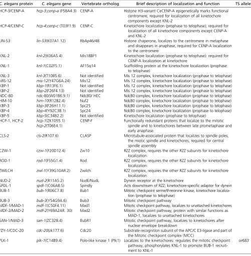
screens for the KNL phenotype (Desai et al. 2003; P. S. Maddoxet al.2007; Gassmannet al.2008), and by tandem mass spectrometry interrogation of affinity purified KNL com-plexes (Cheeseman et al.2004). Immunofluorescence com-bined with RNAi-mediated depletion revealed a hierarchy of kinetochore protein assembly (Cheeseman et al. 2004; Oegema and Hyman 2006). Subsequent in vitro recon-stitution using purified components identified various subcomplexes and microtubule-binding activities within the kinetochore, and regulatory mechanisms controlling Table 2 Kinetochore proteins
C. elegansprotein C. elegansgene Vertebrate ortholog Brief description of localization and function TS alleles HCP-3/CENP-A hcp-3;cenp-a(F58A4.3) CENP-A Histone H3-variant CeCENP-A epigenetically marks functional
centromere, required for localization of all kinetochore components except KNL-2
HCP-4/CENP-C hcp-4;cenp-c(T03F1.9) CENP-C Kinetochore localization (prophase to telophase); required for localization of all kinetochore components except CENP-A and KNL-2
LIN-53 lin-53(K07A1.12) RbAp46/48 Histone chaperone, localizes to the centromere in metaphase and disappears in anaphase, required for CENP-A localization to the centromere
KNL-2 knl-2(K06A5.4) Mis18BP1 Kinetochore localization (prophase to telophase): required for CENP-A localization at kinetochore
KNL-1 knl-1(C02F5.1) AF15q14 Scaffolding protein at the kinetochore localization (prophase
to telophase)
KNL-3 knl-3(T10B5.6) Not identified Mis 12 complex, kinetochore localization (prophase to telophase) MIS-12 mis-12(Y47G6A.24) Mis12 Mis 12 complex, kinetochore localization (prophase to telophase) KBP-1 kbp-1(R13F6.1) Not identified Mis 12 complex, kinetochore localization (prophase to telophase) KBP-2 kbp-2(F26F4.13) Not identified Mis 12 complex, kinetochore localization (prophase to telophase) NDC-80 ndc-80(W01B6.9.1) Ndc80/ HEC Ndc80 complex, kinetochore localization (prophase to telophase)
HIM-10 him-10(R12B2.4) Nuf2 Ndc80 complex, kinetochore localization (prophase to telophase)
KBP-3 kbp-3(F26H11.1) Spc25 Ndc80 complex, kinetochore localization (prophase to telophase)
KBP-4 kbp-4(Y92C3B.1) Spc24 Ndc80 complex, kinetochore localization (prophase to telophase)
KBP-5 kbp-5(C34B2.2) Not identified Kinetochore localization (prophase to telophase) HCP-1, HCP-2 hcp-1(ZK1055.1)
hcp-2(T06E4.1)
CENP-F Functionally redundant proteins that localize to the mitotic spindle and to kinetochores between late prometaphase and early anaphase.
CLS-2 cls-2(R107.6) CLASP Microtubule-associated protein that localizes to spindle poles,
the miotic spindle and kinetochores, required for central spindle assembly
CZW-1 czw-1(F20D12.4) Zw10 RZZ complex, requires the other RZZ subunits for kinetochore
localization
ROD-1 rod-1(F55G1.4) Rod RZZ complex, requires the other RZZ subunits for kinetochore
localization
ZWILCH zwl-1(Y39G10AR.2) Zwilch RZZ complex, requires the other RZZ subunits for kinetochore localization
NUD-2 nud-2(R11A5.2) NudE/NudL Dynein receptor at the kinetochore
SPDL-1 spdl-1(C06A8.5) Spindly Acts downstream of RZZ; kinetochore-specific adaptor for dynein
BUB-1 bub-1(R06C7.8) Bub1 Mitotic checkpoint serine/threonine kinase, kinetochore
localiza-tion (prophase to telophase)
BUB-3 bub-3(Y54G9A.6) Bub3 Mitotic checkpoint pathway
MDF-1/MAD-1 mdf-1(C50F4.11) Mad1 Mitotic checkpoint pathway, localizes to unattached kinetochores MDF-2/MAD-2 mdf-2(Y69A2AR. 30) Mad2 Mitotic checkpoint pathway, protein with similar functions as
MAD-1, localizes to unattached kinetochores
SAN-1/MAD-3 san-1(ZC328.4) BubR1 Mitotic checkpoint pathway, localizes to kinetochores after nuclear envelope breakdown
FZY-1/CDC-20 cdc-20(zk177.6) Cdc20 Substrate recognition subunit of the APC/C E3-ligase and part of the Mitotic checkpoint complex (MCC)
PLK-1 plk-1(C14B9.4) Polo-like kinase 1 (Plk1) Localizes to the kinetochores: regulates the mitotic checkpoint pathway; phosphorylates KNL-1 to promote BUB-1 recruit-ment to KNL-1
kinetochore-microtubule attachments (Cheeseman et al. 2006). Kinetochore proteins coordinate multiple functions, and the generation and analysis of“separation-of-function” mutants via genome editing has led to an impressive under-standing of kinetochore protein functions.
Molecular architecture of the C. elegans kinetochore
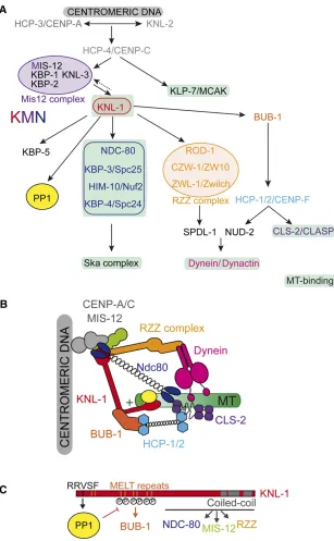
Inner kinetochore proteins: connecting with chromosomal DNA:The kinetochore assembles on a specific chromosomal region called the centromere (Figure 5), which includes spe-cialized nucleosomes containing the histone H3 variant HCP-3/ CENP-A (Buchwitz et al. 1999) that epigenetically marks functional centromeres (Musacchio and Desai 2017). The histone chaperone LIN-53/RbAp46/48 is required for HCP-3/CENP-A localization to holocentricC. eleganscentromeres and for accurate chromosome segregation, suggesting that LIN-53/RbAp46/48 mediates HCP-3/CENP-A deposition (Leeet al.2016). To date, one other factor modulates HCP-3/ CENP-A deposition at holocentromeres: the myb/SANT-domain protein KNL-2 (P. S. Maddox et al. 2007). Down-stream of KNL-2 and HCP-3/CENP-A is HCP-4/CENP-C (Moore and Roth 2001; Oegema et al.2001). These three inner kinetochore proteins connect chromosomal DNA to the outer kinetochore inC. elegans(Figure 6). In humans, the inner kinetochores are built by additional CENP subunits, which form the constitutive centromere-associated network (CCAN). With the notable exception of HCP-4/CENP-C,
CCAN subunits have not been found to date in some spe-cies, including C. elegans and Drosophila [for review see Musacchio and Desai (2017), van Hooffet al.(2017)].
Outer kinetochore proteins: connecting with microtubules: RNAi screens for KNL phenotypes initially identified KNL-1 and KNL-3, in addition to the inner kinetochore proteins HCP-3/CENP-A, HCP-4/CENP-C, and KNL-2 (Desai et al. 2003; P. S. Maddoxet al. 2007) (Figure 6A). Subsequent mass spectrometry analysis of KNL-1–and KNL-3–interacting proteins identified a network of 10 proteins: KNL-1, MIS-12, KNL-3, KBP-1, KBP-2, NDC-80, HIM-10/Nuf2, KBP-3/Spc24, KBP-4, and KBP-5 (Cheesemanet al.2004). Analysis of their assembly hierarchy revealed that KNL-1, which acts down-stream of KNL-3, is required for the localization of all known outer kinetochore proteins (Cheesemanet al.2006) (Figure 6A). Biochemical reconstitution and characterization of this network from purified components identified three interacting and conserved units: KNL-1 and the Mis12 and Ndc80 com-plexes (together known as KMN), bearing distinct microtubule-binding activities in the Ndc80 complex and KNL-1 (Cheeseman et al.2006). The Ndc80 complex forms load-bearing attach-ments to dynamic microtubules (Cheeseman et al. 2006), while the KNL-1 microtubule-binding domain is required for checkpoint signaling, but dispensable for chromosome segre-gation (Espeutet al.2012).
heterodimers—NDC-80 and HIM-10/Nuf2, and KBP-3/ Spc25 and KBP-4/Spc24—joined by overlapping a-helical coiled-coiled domains located at the C and N termini of the NDC-80-HIM-10/Nuf2 and KBP-3/Spc25-KBP-4/Spc24 het-erodimers, respectively (Figure 6B) (Cheesemanet al.2006; DeLuca et al. 2006; Wei et al. 2007; Ciferri et al. 2008). Therefore MT-binding, mediated by the N-terminal globular domain of NDC-80 and HIM-10 subunits, and kinetochore-targeting, mediated by the C-terminal regions of KBP-3 and KBP-4 subunits, are located at opposite ends of the complex. The microtubule-binding region of the Ndc80 complex con-sists of a basic, unstructured N-terminal tail of the NDC-80 subunit and a pair of calponin-homology (CH) domains in the
protein CLS-2/CLASP (Figure 6A). In turn, CLS-2/CLASP controls kinetochore-bound microtubule dynamics to pro-mote chromosome biorientation (Cheesemanet al.2005).
From lateral to end-on microtubule attachment: cross-talk between RZZ-Spindly and the Ndc80 complex
Chromosomes are initially captured along microtubule sides (lateral attachments) and then tethered to microtubule ends, with end-on attachments that can resist the forces that pull on chromosomes during anaphase (Cheerambathur and Desai 2014). Furthermore, the two sister chromatids must attach to spindle microtubules emanating from opposite spindle poles to become bioriented. A lack of chromosome attach-ment, or incorrect ones such as merotelic attachments (a single sister kinetochore is simultaneously attached to micro-tubules from both spindle poles), must be corrected before anaphase onset. Proper orientation of sister kinetochores be-fore stable microtubule attachment is of critical importance in C. elegans, with large holocentric kinetochores prone to mer-otely. The nonkinetochore motor protein KLP-19 discourages merotelic attachments by providing a polar ejection force that favors an orientation in which sister chromatid kinetochores face opposite spindle poles (Powerset al.2004). The spindle assembly checkpoint (SAC) also promotes proper segregation by delaying sister chromatid separation until all chromo-somes are bioriented and under tension (Musacchio and Salmon 2007) (discussed below).
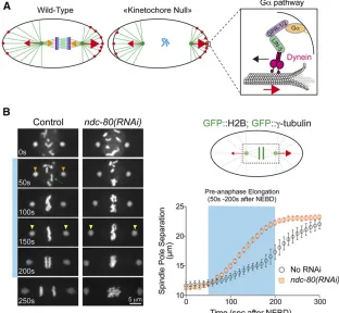
In addition, regulatory mechanisms ensure that robust end-on microtubule attachments are achieved after transient, lateral kinetochore-microtubule attachments (Gassmann et al. 2008; Cheerambathur et al. 2013; Cheerambathur et al. 2017; Gamaet al.2017; Simõeset al. 2018).spdl-1, theC. elegansortholog of the conserved dynein adaptor Spin-dly, also results in a kinetochore null phenotype when inacti-vated. Removal of SPDL-1 is nearly equivalent to removal of NDC-80 (Gassmann et al. 2008). SPDL-1 interacts with ROD-1/rough deal, CZW-1/Zeste-white, and ZWL-1/Zwilch, which form the evolutionarily conserved RZZ complex, itself recruited to the kinetochore by KNL-1 (Figure 6A) (Gassmannet al.2008). Paradoxically, although RZZ recruits SPDL-1 to kinetochores,spdl-1inactivation results in a much more severe phenotype than depletion of upstream RZZ com-plex subunits (Gassmannet al.2008). The reason is that in the absence of SPDL-1, RZZ binds to and inhibits the Ndc80 complex, preventing end-on microtubule attachments (Cheerambathuret al.2013). Codepleting RZZ relieves this inhibition and ameliorates the more severe defect resulting fromspdl-1depletion alone.
SPDL-1 recruits dynein-dynactin to kinetochores (Griffis et al. 2007; Gassmann et al.2008; Yamamoto et al.2008; Chanet al.2009), which contributes to initial lateral micro-tubule capture, accelerates stable end-on attachment by the Ndc80 complex, and relieves the inhibition of RZZ on Ndc80 by an as yet unknown mechanism (Cheerambathur et al. 2013). Cross-talk between Ndc80 and dynein-dynactin thus ensures accurate chromosome segregation. SPDL-1 recruits
dynein-dynactin to kinetochores via two conserved motifs, the N-terminally located CC1 box and the spindly motif, through which SPDL-1 binds dynein light-intermediate chains and the dynactin pointed complex, respectively (Gamaet al.2017). Specific SPDL-1 point mutations, or de-letion of the dynactin-pointed complex subunit DNC-6/p27, abrogate the interaction between SPDL-1 and the dynein-dynactin complex and phenocopy loss of spdl-1 function, demonstrating the importance of dynein recruitment to ki-netochores for chromosome segregation (Cheerambathur et al.2013; Gamaet al.2017).
The recruitment of dynein at kinetochores also depends on the dynein cofactor NUD-2/NudE/NudL, which is recruited to kinetochores by HCP-1/2 (Figure 6A) (Simõeset al.2018). Loss of NUD-2 delays the formation of load-bearing microtu-bule attachments and causes chromatin bridges during ana-phase. This phenotype is identical to that resulting from inactivation of RZZ complex subunits. However, loss of NUD-2 reduces dynein levels at kinetochores by only 50%, whereas RZZ depletion completely removes dynein from ki-netochores, indicating that high levels of dynein at kineto-chores are required for proper kinetochore function (Simões et al.2018). To summarize, SPDL-1 and NUD-2 function as dynein receptors at unattached kinetochores to promote ini-tial lateral microtubule capture and facilitate their matura-tion into end-on Ndc80 complex attachments.
Once proper end-on kinetochore-microtubule attachments are achieved, they must be stabilized to dampen chromosome motion before segregation. Dephosphorylation of the NDC-80 N-tail and/or tension on the attachments generated by GPR-1/2–dependent cortical pulling forces, promotes a microtu-bule-bound conformation of NDC-80 that is recognized by the conserved microtubule-binding SKA complex that stabi-lizes kinetochore-MT attachments and dampens chromo-some motions (Cheerambathuret al.2017).
Together, these sophisticated regulatory events ensure that chromosomes are bioriented and under tension before chromo-some segregation begins. In addition, these steps are monitored by the SAC, which prevents anaphase onset until all sister chromatid pairs have attached to spindle microtubules emanat-ing from opposite spindle poles (Musacchio and Salmon 2007).
Microtubule attachments and the SAC
The earlyC. elegansembryo mounts a checkpoint response that delays mitotic entry upon exposure to microtubule depo-lymerizing drugs such as nocodazole (Encaladaet al.2005), and robust genetic assays have been developed to monitor spindle checkpoint signaling (Essexet al. 2009). One assay uses two-cell stage embryos with monopolar spindles caused by inactivation of centriolar duplication proteins (Essexet al. 2009). Monopolar spindles, which contain unattached kinet-ochores and attached kinetkinet-ochores that are not under ten-sion, elicit a robust and quantifiable checkpoint-mediated cell-cycle delay that is suppressed by inactivation of check-point components (Essex et al. 2009). Another assay uses genetic interactions with a TS mutation in the essential APC/C subunit MAT-3/Apc8, mat-3(or344ts) (Rappleye et al. 2002; Kim et al. 2017). As the checkpoint prevents cell-cycle progression by inhibiting the APC/C, compromising the APC/C enhances a checkpoint defect (Bezler and Gönczy 2010; Kimet al.2017).
Most SAC components, including MDF-1/MAD-1, MDF-2/ MAD-2, BUB-1/Bub1, BUB-3/Bub3, and SAN-1/MAD-3/ Mad3 (known as BubR1 in vertebrates andDrosophila), are conserved inC. elegans(Table 2), with the noticeable
excep-tion of the Mps1 kinase (Kitagawa and Rose 1999; Essexet al. 2009; Vleugel et al. 2012; Espeut et al. 2015). Depleting these proteins does not affect cell-cycle timing in basal con-ditions, except for BUB-1/BUB-3 (Kimet al.2015), but all are required to delay cell-cycle progression by roughly 60% in the presence of monopolar spindles (Essex et al.2009). In this section, for clarity and consistency with the cell-cyclefield, we use the gene namesmad-1,mad-2, andmad-3instead of mdf-1,mdf-2, andsan-1.
(Figure 7). In vertebrates, the BubR1 subunit of the MCC binds to a Cdc20 subunit physically associated with the APC/C and prevents it from binding substrates (Izawa and Pines 2015; Alfieriet al. 2016); this inhibitory mechanism likely is conserved inC. elegans(Figure 7).
The mechanism by which the MAD-1–MAD-2 complex is recruited to kinetochores is emerging. MAD-1 physically in-teracts with the BUB-1 kinase (Moyleet al.2014), which is at the top of the checkpoint protein kinetochore localization hierarchy from yeast to human (Musacchio and Salmon 2007). BUB-1 acts in a complex with its binding partner, the WD40-fold protein BUB-3 (Kim et al. 2015), to recruit MAD-1–MAD-2 to unattached kinetochores (Moyle et al. 2014). MAD-1 mutants defective in binding to BUB-1 do not localize to unattached kinetochores and are defective in checkpoint signaling (Moyleet al.2014).
BUB-1 itself is recruited to kinetochores by KNL-1, an interaction regulated by phosphorylation (Figure 6B, bottom panel). In most organisms, the Mps1 kinase phosphorylates KNL-1 MELT repeats (Met-Glu-Leu-Thr) to create phospho-docking sites for Bub1/Bub3 and initiate checkpoint signal-ing. However, Mps1 is absent inC. elegansand instead, the polo-like kinase PLK-1 phosphorylates KNL-1 to direct BUB-1 recruitment to kinetochores (Espeutet al.2015). How PLK-1 gets recruited to kinetochores inC. elegansis not understood (Cheerambathur and Desai 2014). Other kinetochore com-ponents are also required for the localization of MAD-1– MAD-2 at unattached kinetochores, in particular the RZZ complex, SPDL-1, and NDC-80 (Gassmann et al. 2008; Yamamotoet al.2008; Essexet al.2009), but the molecular mechanisms are unclear.
Checkpoint inactivation:Once all kinetochores are properly attached to microtubules and all chromosomes are bi-oriented, the SAC must be inactivated. The RZZ-spindly-Dynein pathway plays a prominent role in checkpoint inacti-vation by shedding MAD-1—MAD-2 from kinetochores to the spindle poles (Howell et al.2001; Wojciket al. 2001). The N-terminus of KNL-1 also senses microtubules attachment to kinetochores and recruits the PP1 phosphatase that contrib-utes to checkpoint silencing (Espeutet al.2012). In particu-lar, PP1 dephosphorylates KNL-1 on the MELT repeats to disrupt the KNL-1—BUB-1 interaction (Espeutet al.2012), with higher levels of BUB-1 at kinetochores in a KNL-1 PP1-binding mutant (Matonet al.2015; Kimet al.2017).
Kinetochores direct CDC-20 to the mitotic accelerator (APC/C) or brake (SAC)
While the spindle checkpoint pathway is conserved inC. ele-gansand delays mitosis in the presence of unattached kinet-ochores, its inhibition in unperturbed one-cell embryos does not lead to significant defects in chromosome segregation nor accelerated mitotic progression (Essex et al.2009). More-over, in embryos depleted of MAD-2, cyclin B degradation occurs with normal kinetics (Kim et al. 2017), indicating the existence of additional mechanisms restraining APC/
CCdc20activity. A key factor that controls the timing of mitosis in one-cell embryos is BUB-1, which not only directs the SAC proteins to inhibit the APC/C in the presence of unattached kinetochores, but also promotes APC/C activation. BUB-1 thus plays a dual role, inhibiting and activating anaphase onset, with the balance between these potentially being mod-ulated by microtubule attachment status (Figure 7). A role for BUB-1 in promoting anaphase onset emerged from the observation that inactivation of BUB-1, or its binding partner BUB-3, increased the time between NEBD and anaphase on-set independently of the SAC, as this delay is still observed in BUB-1; MAD-2 or BUB-1; MAD-3 double depletions (Kim et al. 2015; Kim et al. 2017). A similar delay in anaphase onset is observed when BUB-1 recruitment to the kineto-chores is prevented, indicating that BUB-1 is required at ki-netochores for the normal timing of anaphase onset.
How might BUB-1 influence anaphase onset? In other systems, Bub1 and BubR1, which share a common Cdc20 interaction motif known as the ABBA motif (Di Fioreet al. 2015) or the Phe box (Diaz-Martinez et al. 2015), recruit Cdc20 via these motifs to kinetochores. However, the func-tional significance of Cdc20 recruitment to the kinetochore for its role in APC/C activation and anaphase onset has remained unclear. In C. elegans, BUB-1 similarly recruits CDC-20 to kinetochores through an ABBA motif (Kimet al. 2017). Mutation of the ABBA motif prevents CDC-20 recruit-ment to kinetochores and delays anaphase onset, demon-strating that CDC-20 recruitment to kinetochores is required for BUB-1 to promote anaphase onset. FRAP exper-iments showed that the residence time of CDC-20 at the ki-netochores is only 1.2 sec, suggesting that it is dynamically
fluxing through kinetochores (Figure 7).
BUB-1, and this delay is suppressed in the presence of CDC-20 nonphosphorylatable mutants (Kimet al.2017). Overall, these data suggest that in embryos, CDC-20 is inhibited by Cdk-mediated phosphorylation and upon mitotic entry, CDC-20 recruitment to kinetochores through BUB-1 promotes its dephosphorylation by PP1c docked onto KNL-1 and its sub-sequent activation of the APC/C. Preventing CDC-20 recruit-ment to kinetochores delays anaphase onset but does not block it, possibly because CDC-20 is also dephosphorylated in the cytoplasm (Kimet al.2017).
Strikingly, in the presence of unattached kinetochores, the BUB-1 ABBA motif mutant is also defective in checkpoint signaling. Thus, in situations where kinetochore-microtubule attachments are defective, CDC-20 recruited to the kineto-chores by the BUB-1 ABBA motif also has the ability to delay anaphase onset (Kimet al.2017). Therefore, CDC-20 dynam-ically fluxing through kinetochores via the ABBA motif of BUB-1 is either incorporated into the inhibitory checkpoint complex with MAD-2 (MCC) to inhibit the APC/C, or is dephosphorylated by PP1 to promote APC/C activation and anaphase onset (Figure 7). Whether and how microtubule attachment status influences the two fates of CDC-20 remains an open and important question. In principle, microtubule attachments trigger dynein-dependent MAD-1/2 removal from the kinetochores, and dephosphorylation of the MELT repeats of KNL-1 also promote BUB-1/BUB-3 removal. How-ever, microtubule attachments remove MAD-1–MAD-2 but leave behind a substantial pool of BUB-1/BUB-3 (Kimet al. 2015), such that CDC-20 at attached kinetochores is more likely to be dephosphorylated (Figure 7). Microtubule attach-ments may also modulate PP1 activity and/or localization to the kinetochores. Further work is required to determine the relationship between microtubule attachment at kineto-chores and alterations in the balance between the two fates of CDC-20fluxing through kinetochores.
Tremendous progress has been made in understanding kinetochore assembly and function in C. elegans. However, despite considerable progress, important issues still need to be addressed. In particular, how do tension and mechan-ical forces stabilize kinetochore-microtubule attachments (Akiyoshiet al. 2010), and how do kinetochores sense and signal microtubule attachments to dephosphorylate NDC-80 and recruit the SKA complex to trigger anaphase onset? Interestingly, we have seen that dephosphorylation events trigger both stabilization of kinetochore-microtubule attach-ments and anaphase onset, further highlighting the impor-tance of protein phosphatases in mitotic progression.
Mitotic Spindle Assembly and Chromosome Segregation
Mitotic spindle microtubules are organized and regulated by microtubule-associated proteins (MAPs), microtubule mo-tors, and other associated proteins to generate three distinct sets of microtubules: astral microtubules emanating from the centrosomes and contacting the cortex, kinetochore microtu-bules (KMTs) that connect centrosomes to kinetochores, and
central spindle microtubules that assemble between segre-gating sister chromatids after anaphase onset. KMTs mediate chromosome dynamics during mitosis. The central spindle, an array of antiparallel microtubules with their plus-ends over-lapping at the center, coordinates chromosome segregation with furrow ingression during cytokinesis. Astral microtu-bules influence spindle position, contribute to chromosome segregation, and promote contractile ring assembly during cytokinesis.
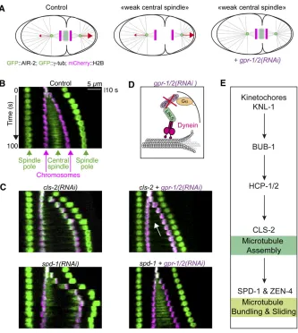
Depending on the organism, different microtubule nu-cleation pathways support spindle assembly (Heald and Khodjakov 2015; Prosser and Pelletier 2017). InDrosophila and vertebrates, spindle assembly depends on three comple-mentary mechanisms: (i) Ran-GTP produced in the vicinity of the chromatin binds to importin-band thereby releases spin-dle assembly factors, (ii) centrosome-nucleated microtubules search and capture chromosomes, and (iii) Augmin-depen-dent recruitment of the microtubule nucleator gTuRC for branched microtubule nucleation. In the earlyC. elegans em-bryo, the Augmin pathway is absent and mitotic spindle as-sembly depends largely, if not exclusively on centrosomes. In this section, we review mitotic spindle assembly inC. elegans and discuss recent data highlighting the role of a subset of kinetochore proteins in the initial stages of central spindle assembly. Finally, we consider the mechanisms that medi-ate chromosome segregation, including a possible contribu-tion from the central spindle via an inside-out pushing mechanism.
Centrosome-based spindle assembly in the early embryo
The one-cell stage mitotic spindle is large (14mm from pole to pole), with prominent centrosomal spindle poles that nucle-ate microtubules (Figure 4A). Spindles fail to form inC. ele-gansembryos that lack functional centrosomes (Hamillet al. 2002; Kempet al.2004; Pelletieret al.2004; Nahabooet al. 2015), whereas half-spindles assemble in embryos that have one centrosome instead of two (O’Connell et al. 2001). Whether this strict requirement for centrosomes in spindle assembly is also true in later-stage embryos or larvae awaits further investigation. Fast-acting TS alleles of core centro-some components (e.g.,spd-5ts) can address this issue.
The centrosome-based mitotic spindle assembles rapidly. Early on, both cortical and sperm pronuclear envelope local-ized dynein promote centrosome separation in the one-cell zygote (De Simoneet al.2016). Subsequently, concomitant with nuclear envelope break down, centrosomal microtu-bules penetrate the nuclear space to capture chromosomes (Figure 4A), with further assembly also requiring functional kinetochores (Oegemaet al.2001). Spindle assembly is com-pleted in the subsequent 2–3 min between nuclear envelope breakdown and anaphase onset, with astral microtubules at-tached to the cortex subject to pulling forces that position the spindle and promote chromosome segregation (Figure 4A) [for review see Kotak and Gönczy 2013, McNally 2013)].
(O’Tooleet al.2003; Ozlüet al.2005; Schlaitzet al.2007; Redemannet al.2017; Müller-Reichertet al.2018). Tomo-graphic reconstruction of metaphase and anaphase mitotic spindles in early embryos from electron micrographs com-bined with live-imaging confirmed that C. elegans mitotic spindles consist exclusively of microtubules radiating from the centrosomes (Figure 4, B and C) (O’Toole et al.2003; Redemannet al.2017). At metaphase, the mitotic spindle is composed of roughly 20,000 individual microtubules among which only 500 (2.5%) are KMTs, with only 6–50 KMTs attaching to each of the 12 chromosomes per pole-facing side (Redemann et al.2017). These KMTs do not form bundles that resemble the kinetochorefibers observed in vertebrate andDrosophilacells, probably due to the holocentric nature ofC. eleganschromosomes.
Do 6–50 KMTs directly connecting centrosomes to kinet-ochores provide a sufficiently strong mechanical connection between chromosomes and centrosomes? One possibility is that the number of microtubules directly connecting centro-somes to chromocentro-somes might be underestimated, owing to the difficulty in tomography reconstructions and the density of microtubules emanating from the poles in all directions. Alternatively, one or two KMTs might be sufficient for chro-mosome segregation, as in budding yeast (Wineyet al.1995). Or, as supported by tomography reconstructions, chromo-somes might be indirectly connected to centrochromo-somes by an-choring into a spindle network, which raises questions about how KMTs form. Mathematical modeling of KMTs formation suggests that microtubules grow out from centrosomes and eventually either depolymerize or become KMTs upon attachment to a kinetochore. These KMTs might then se-lectively detach from the centrosomes and shrink from their minus ends. This model, supported by FRAP experi-ments, suggests that KMTs are short-lived relative to the timescale of mitosis (Redemannet al.2017).
Because of the transient nature of KMTs, astral microtu-bules must rapidly find kinetochores. Given that chromo-somes are holocentric, microtubules can bind at any point of the surface, and mitosis is semiclosed in the early embryo. The nuclear envelope breaks down in close proximity to the centrosomes, but remnants persist around the mitotic spindle until the metaphase-to-anaphase transition (Leeet al.2000). The persistence of the nuclear envelope limits the space through which centrosome-based microtubules search and capture the chromosomes. Finally, a spindle matrix may con-centrate free tubulin and other factors to favor assembly in a way that promotes kinetochore capture (Figure 4C) (Hayashi et al.2012). Accordingly, during early spindle assembly, the density of EBP-2 foci, which decorate polymerizing microtu-bule plus-ends, is higher within the spindle (Srayko et al. 2005), suggesting that microtubules preferentially grow to-ward chromatin. The GTPase RAN-1, which is essential for nuclear import and spindle assembly (Askjaer et al. 2002; Bambaet al.2002), contributes to this bias in microtubule outgrowth toward chromosomes, a function that is indepen-dent of kinetochores (Srayko et al. 2005). However, the
mechanism by which RAN influences the direction of micro-tubule outgrowth toward chromatin is unclear (Sraykoet al. 2005). Altogether, these traits likely contribute to the effi -cient capture of the chromosomes by astral microtubules, facilitating rapid spindle assembly.
Centrosome-localized factors regulating mitotic spindle assembly
Several centrosome-localized factors are required for mitotic spindle assembly (Figure 4D) (Sönnichsenet al.2005; Srayko et al.2005; Oegema and Hyman 2006; Müller-Reichertet al. 2010). Microtubule nucleation requires core centrosomal components, including the microtubule nucleator g-tubulin and the AIR-1/Aurora A kinase, as discussed in the centro-some section. Other centrocentro-some-localized factors further regulate microtubule dynamics (Sraykoet al.2005). Micro-tubule plus-end growth requires the evolutionarily conserved microtubule polymerase ZYG-9/XMAP215 and its activator TAC-1/TACC1 (Transforming and acidic coiled-coiled pro-tein) (Srayko et al. 2005; Bellanger et al. 2007; Brouhard et al.2008). The number of microtubules that grow out from centrosomes is controlled and limited by the microtubule depolymerizing kinesin KLP-7 (Sraykoet al.2005). Regula-tion of KMT length depends on the AIR-1/Aurora A kinase and its binding partner TPXL-1, the worm ortholog of the well-characterized microtubule-stabilizing protein TPX2 (Grusset al.2001; Schatzet al.2003). Intpxl-1(RNAi) em-bryos, just after nuclear envelope breakdown, the assembling spindle collapses and centrosomes are pulled toward the chromatin (Ozlüet al.2005); the rate of microtubule assem-bly is not affected (Sraykoet al.2005), but the microtubules are unstable. TPX-2 activates and targets AIR-1 to microtubules (Ozlüet al.2005), which likely facilitates Aurora A-dependent phosphorylation of downstream substrates required for mi-crotubule stability. For example, the tpxl-1(RNAi) pheno-type might result at least in part from a defect in NDC-80 phosphorylation. As discussed in the kinetochore section, phosphorylation of the N-terminal NDC-80 tail prevents pre-mature recruitment of the SKA complex to kinetochores such that microtubule attachments remain dynamic, allowing for tension-coupled polymerization. This phosphorylation is likely mediated by Aurora A (Cheerambathur et al.2017), and preventing N-tail phosphorylation gives a short spindle phenotype that, although not as severe, is reminiscent of the tpxl-1phenotype.