GENETICS | INVESTIGATION
Chromatin Remodeling Factors Isw2 and Ino80
Regulate Checkpoint Activity and Chromatin
Structure in S Phase
Laura Lee,*,†Jairo Rodriguez,* and Toshio Tsukiyama*,1 *Basic Sciences Division, Fred Hutchinson Cancer Research Center, Seattle, Washington 98109, and†Molecular and Cellular Biology Program and Fred Hutchinson Cancer Research Center, University of Washington, Seattle, Washington 98195
ABSTRACTWhen cells undergo replication stress, proper checkpoint activation and deactivation are critical for genomic stability and cell survival and therefore must be highly regulated. Although mechanisms of checkpoint activation are well studied, mechanisms of checkpoint deactivation are far less understood. Previously, we reported that chromatin remodeling factorsIsw2andIno80attenuate the S-phase checkpoint activity inSaccharomyces cerevisiae, especially during recovery from hydroxyurea. In this study, we found that Isw2 and Ino80have a more pronounced role in attenuating checkpoint activity during late S phase in the presence of methyl methanesulfonate (MMS). We therefore screened for checkpoint factors required forIsw2andIno80checkpoint attenuation in the presence of MMS. Here we demonstrate thatIsw2andIno80antagonize checkpoint activators and attenuate checkpoint activity in S phase in MMS either through a currently unknown pathway or through RPA. Unexpectedly, we found thatIsw2andIno80increase chromatin accessibility around replicating regions in the presence of MMS through a novel mechanism. Furthermore, through growth assays, we provide additional evidence thatIsw2andIno80partially counteract checkpoint activators specifically in the presence of MMS. Based on these results, we propose thatIsw2andIno80attenuate S-phase checkpoint activity through a novel mechanism.
KEYWORDSIsw2; Ino80; chromatin accessibility; S-phase checkpoint
F
OR proper propagation of cells, DNA must be faithfully copied during replication in S phase . However, during replication, cells are prone to encountering nucleotide depletion or DNA damage, which generates replication stress and induces replication fork stalling. Upon replication stress, cells trigger a mechanism called the S-phase checkpoint to allow for proper completion of replication before exit out of S phase. Because this pathway is essential for genomic stability, as evidenced by the development of cancer or cell death in checkpoint mutants (Zeman and Cimprich 2014) and strict conservation in eukary-otic cells, mechanisms of S-phase checkpoint activation have been studied extensively (Branzei and Foiani 2009).When replication forks stall in budding yeast cells, excess single-stranded DNA (ssDNA) is produced that is bound by replication protein A (RPA). Accumulation of the ssDNA-RPA
complex recruits a central kinase Mec1[ataxia telangiectasia and Rad3-related kinase (ATR) in mammalian cells] with its interacting partner Ddc2 [ATR interacting protein (ATRIP)], which activates the downstream effector kinases Rad53 (Chk2) and Chk1(Chk1) to stabilize replication forks, up-regulate damage-inducible genes, and slow S-phase progression through the delay of late originfiring until DNA is replicated correctly (Rouse and Jackson 2002; Zou and Elledge 2003; Friedelet al.2009). The S-phase checkpoint can be activated via two different pathways, the DNA damage checkpoint (DDC) and/or the DNA replication checkpoint (DRC), depend-ing on the type of damage recognized by the cell (Crabbeet al.
2010). In the DDC of budding yeast, the clamp loader RFCRad24 (RFCRad17) and subsequently the PCNA-like 9-1-1
clamp Rad17-Ddc1-Mec3 (Rad9-Hus1-Rad1) are recruited to ssDNA–double-stranded DNA (dsDNA) junctions to facilitate ac-tivation of the checkpoint along with Dpb11 (TopBP1) and Rad9 (Gilbert et al. 2001; Kondo et al. 2001; Majka et al.
2006; Navadgi-Patil and Burgers 2008). In the DRC, Tof1 (TIM), Mrc1 (claspin), and Dna2 are recruited to replication forks to facilitate activation of the checkpoint (Katou et al.
2003; Kumar and Burgers 2013). Copyright © 2015 by the Genetics Society of America
doi: 10.1534/genetics.115.174730
Manuscript received January 19, 2015; accepted for publication February 13, 2015; published Early Online February 19, 2015.
Supporting information is available online athttp://www.genetics.org/lookup/suppl/ doi:10.1534/genetics.115.174730/-/DC1
In contrast to S-phase checkpoint activation mechanisms, mechanisms that regulate checkpoint deactivation are far less understood. The best-characterized mechanism of checkpoint deactivation is viaRad53phosphatasesPph3 andPtc2. Dele-tion ofPph3andPtc2results in elevated sensitivity to replica-tion inhibitors and complete replicareplica-tion fork arrest (Szyjka
et al. 2008). This result demonstrates that proper control of the amplitude and inactivation of the S-phase checkpoint is also essential for DNA replication control in the presence of replication stress. It also has been shown that other modes of checkpoint deactivation exist that have yet to be identified, highlighting the need for more studies on checkpoint deactiva-tion mechanisms (Travesaet al.2008).
We have recently proposed that the highly conserved chromatin remodeling factorsIsw2andIno80play roles in S-phase checkpoint attenuation (Auet al.2011). Chromatin remodeling factors use the energy of ATP hydrolysis to alter chromatin structure and have been implicated in many DNA-dependent processes, including transcription, DNA replication, DNA repair, and checkpoint regulation (Morrison and Shen 2009). Isw2 has been demonstrated to slide nucleosomes
in vivo(Fazzio and Tsukiyama 2003). Functionally, Isw2has been shown to repress transcription of genes by sliding nucle-osomes over transcriptional start sites (Goldmarket al.2000; Whitehouseet al.2007). In the mean time,Ino80can replace the histone variant Htz1within nucleosomes with canonical histone H2A (Papamichos-Chronakiset al.2011).Ino80is re-quired for proper DNA damage response after DNA double-strand breaks (Shen et al.2000; van Attikumet al.2004), as well as replication stress responses such as maintaining fork stability and promoting recovery of stalled forks (Papamichos-Chronakis and Peterson 2008; Shimadaet al.2008). However, how this complex performs these functions remains largely unknown.
We previously reported thatIsw2andNhp10, a subunit of the Ino80 complex, function together to attenuate the S-phase checkpoint after transient exposure to the DNA replica-tion inhibitor hydroxyurea (HU) (Auet al.2011). In this study, we show thatIsw2andNhp10exhibit a much stronger effect in attenuating checkpoint activity in the presence of the DNA alkylating agent methyl methanesulfonate (MMS) throughout late S phase. Through systematic genetic tests, we show that Isw2 and Nhp10antagonize checkpoint activators and facili-tate checkpoint attenuation in MMS either through a currently unknown pathway or through the ssDNA binding protein RPA. In investigating changes in chromatin structure inisw2 nhp10 cells during replication stress, we unexpectedly found that Isw2andIno80increase chromatin accessibility around repli-cating regions through a mechanism unrelated to their known biochemical activities. Consistent with checkpoint activity con-trol, growth assays in the presence of MMS reveal that Isw2 andIno80partially oppose checkpoint activators specifically in the presence of MMS. Based on these results, we propose that Isw2andIno80alter chromatin structure at replicating regions and attenuate the S-phase checkpoint through currently un-known mechanism(s).
Materials and Methods
Yeast strains and culture
All yeast strains are MATaand congenic to W303-1a with a correction for the weak rad5 allele in the original W303 (Thomas and Rothstein 1989; Zhao et al. 1998). Strains used for stable isotope labeling by amino acids in cell culture (SILAC) mass spectrometry were in a Dlys2Darg4 CAN1+ background. Strains were constructed using standard gene knockout protocols and genetic crosses (Table 1).
Cell synchronization andflow cytometry (FACS)
All yeast strains were grown at 30°to early log phase to a density of OD660= 0.2–0.25 and then arrested in G1phase with a final concentration of 5 mg/ml afactor. Cells were
filtered on a 0.45-mm nitrocellulose membrane, washed twice with YPD, and released into half the volume of pre-warmed YPD containing 0.02% MMS. Cells were harvested at each time point, stored in afinal concentration of 66.7% (v/v) ethanol, and then processed for flow cytometry as described previously (Vincentet al.2008).
Rad53 in situ autophosphorylation assay (ISA) and Rad53 Western blotting
Cells were arrested in G1byafactor (5mg/ml) and released into rich medium (YPD) containing 0.02% MMS, as described earlier. In the initial protein preparation method,25 ml of cells was harvested for each time point. However, in the mod-ified method accounting for cell density, 108 cells were har-vested at each time point. Cells were washed once with water and then with 20% trichloroacetic acid (TCA). Protein samples were prepared as described previously and run on an 8% SDS– polyacrylamide gel (Pellicioli et al. 1999). Rad53 auto-phosphorylation was normalized by autoauto-phosphorylation of nonspecific proteins within the same sample that served as a loading control. For Rad53 Western blotting, goat polyclonal Rad53 (yC-19) antibody (sc-6749, Santa Cruz Biotech, Santa Cruz, CA) was used.
Chromatin fractionation and SILAC
Strains were grown in yeast culture (YC) medium with either 15 mg/literL-arginine and 30 mg/literL-lysine or 15 mg/liter [13C6]L-arginine and 30 mg/liter [13C6,15N2]L-lysine. Then 43109cells were harvested at mid-S phase in 0.02% MMS, and chromatin was prepared as described previously (Kubota
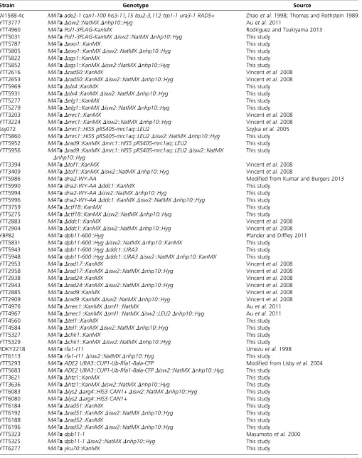
Table 1 Summary of Yeast Strains Used in This Study
Strain Genotype Source
W1588-4c MATaade2-1 can1-100 his3-11,15 leu2-3,112 trp1-1 ura3-1 RAD5+ Zhaoet al.1998; Thomas and Rothstein 1989 YTT3777 MATaDisw2::NatMXDnhp10::Hyg Auet al.2011
YTT4960 MATaPol1-3FLAG-KanMX Rodriguez and Tsukiyama 2013
YTT5031 MATaPol1-3FLAG-KanMXDisw2::NatMXDnhp10::Hyg This study
YTT5787 MATaDexo1::KanMX This study
YTT5805 MATaDexo1::KanMXDisw2::NatMXDnhp10::Hyg This study
YTT5822 MATaDsgs1::KanMX This study
YTT5852 MATaDsgs1::KanMXDisw2::NatMXDnhp10::Hyg This study
YTT2616 MATaDrad50::KanMX Vincentet al.2008
YTT2653 MATaDrad50::KanMXDisw2::NatMXDnhp10::Hyg Vincentet al.2008
YTT5969 MATaDslx4::KanMX This study
YTT5931 MATaDslx4::KanMXDisw2::NatMXDnhp10::Hyg This study
YTT5277 MATaDelg1::KanMX This study
YTT5279 MATaDelg1::KanMXDisw2::NatMXDnhp10::Hyg This study
YTT3203 MATaDmrc1::KanMX Vincentet al.2008
YTT3224 MATaDmrc1::KanMXDisw2::NatMXDnhp10::Hyg Vincentet al.2008 Ssy072 MATaDmrc1::HIS5 pRS405-mrc1aq::LEU2 Szyjkaet al.2005 YTT5860 MATaDmrc1::HIS5 pRS405-mrc1aq::LEU2Disw2::NatMXDnhp10::Hyg This study YTT5952 MATaDrad9::KanMXDmrc1::HIS5 pRS405-mrc1aq::LEU2 This study YTT5956 MATaDrad9::KanMXDmrc1::HIS5 pRS405-mrc1aq::LEU2Disw2::NatMX
Dnhp10::Hyg
This study
YTT3394 MATaDtof1::KanMX Vincentet al.2008
YTT3409 MATaDtof1::KanMXDisw2::NatMXDnhp10::Hyg Vincentet al.2008
YTT5986 MATadna2-WY-AA Modified from Kumar and Burgers 2013
YTT5990 MATadna2-WY-AADddc1::KanMX This study
YTT5994 MATadna2-WY-AADisw2::NatMXDnhp10::Hyg This study YTT5996 MATadna2-WY-AADddc1::KanMXDisw2::NatMXDnhp10::Hyg This study
YTT3759 MATaDctf18::KanMX This study
YTT5275 MATaDctf18::KanMXDisw2::NatMXDnhp10::Hyg This study
YTT2883 MATaDddc1::KanMX Vincentet al.2008
YTT2904 MATaDddc1::KanMXDisw2::NatMXDnhp10::Hyg Vincentet al.2008
YBP82 MATadpb11-600::Hyg Pfander and Diffley 2011
YTT5831 MATadpb11-600::HygDisw2::NatMXDnhp10::KanMX This study YTT5943 MATadpb11-600::HygDddc1::URA3 This study YTT5948 MATadpb11-600::HygDddc1::URA3Disw2::NatMXDnhp10::KanMX This study
YTT2953 MATaDrad17::KanMX Vincentet al.2008
YTT2958 MATaDrad17::KanMXDisw2::NatMXDnhp10::Hyg Vincentet al.2008
YTT2938 MATaDrad24::KanMX Vincentet al.2008
YTT2943 MATaDrad24::KanMXDisw2::NatMXDnhp10::Hyg Vincentet al.2008
YTT2885 MATaDrad9::KanMX Vincentet al.2008
YTT2909 MATaDrad9::KanMXDisw2::NatMXDnhp10::Hyg Vincentet al.2008 YTT4976 MATaDmec1::KanMXDsml1::NatMX Auet al.2011 YTT4967 MATaDmec1::KanMXDsml1::NatMXDisw2::LEU2Dnhp10::Hyg Auet al.2011
YTT4560 MATaDtel1::KanMX This study
YTT4584 MATaDtel1::KanMXDisw2::NatMXDnhp10::Hyg This study
YTT5327 MATaDchk1::KanMX This study
YTT5329 MATaDchk1::KanMXDisw2::NatMXDnhp10::Hyg This study
RDKY2218 MATarfa1-t11 Umezuet al.1998
YTT6113 MATarfa1-t11Disw2::NatMXDnhp10::Hyg This study
YTT5293 MATaADE2 URA3::CUP1-Ub-Rfa1-8ala-CFP Modified from Lisbyet al.2004 YTT5683 MATaADE2 URA3::CUP1-Ub-Rfa1-8ala-CFPDisw2::NatMXDnhp10::Hyg This study
YTT3621 MATaDhtz1::KanMX This study
YTT3636 MATaDhtz1::KanMXDisw2::NatMXDnhp10::Hyg This study YTT6083 MATaDlys2Darg4::HIS3 CAN1+Disw2::NatMXDnhp10::Hyg This study YTT6080 MATaDlys2Darg4::HIS3 CAN1+ This study
YTT6184 MATaDrad51::KanMX This study
YTT6192 MATaDrad51::KanMXDisw2::NatMXDnhp10::Hyg This study
YTT6188 MATaDrad52::KanMX This study
YTT6196 MATaDrad52::KanMXDisw2::NatMXDnhp10::Hyg This study
YTT5323 MATadpb11-1 Masumotoet al.2000
YTT5325 MATadpb11-1Disw2::NatMXDnhp10::Hyg This study
YTT6277 MATayku70::KanMX This study
Liquid chromatography–tandem mass spectrometry and data analysis
The 10 gel pieces were destained with 50% methanol, 5% acetic acid and washed consecutively with water twice and 50% acetonitrile. After rinsing once more with 100 mM ammonium bicarbonate in water, the gel pieces were dehy-drated using acetonitrile. The protein was digested overnight at 37° with 12.5 ng/ml trypsin (Promega, Madison, WI) in 50 mM ammonium bicarbonate. After transferring excess sol-utions to separate vials, the peptides werefirst extracted using 0.1% trifluoroacetic acid in water for 30 min, and then aceto-nitrile was added to make 50% acetoaceto-nitrile, 0.1% trifl uoroace-tic acid. The pooled extracts were dried in a speed vacuum and cleaned using ZipTip C18 (Millipore, Bedford, MA) before the subsequent MS analysis following the manufacture’s protocols. Liquid chromatography–tandem mass spectrometry (LC-MS/ MS) analysis was performed with an Easy-nLC 1000 (Thermo Fisher Scientific) coupled to an Orbitrap Elite mass spectrom-eter (Thermo Scientific). The LC system configured in a vented format (Licklideret al.2002) consisted of a fused-silica nano-spray needle (PicoTip emitter, 50 mm ID, New Objective, Woburn, MA) packed in-house with Magic C18 AQ 100 Å reverse-phase medium (25 cm, Michrom BioResources, Au-burn, CA) and a trap (IntegraFrit Capillary, 100mm ID, New Objective) containing Magic C18 AQ 200 Å (2 cm). The pep-tide sample was diluted in 10ml of 2% acetonitrile and 0.1% formic acid in water, and 8ml was loaded onto the column and separated using a two-mobile-phase system consisting of 0.1% formic acid in water (A) and 0.1% acetic acid in acetonitrile (B). A 90-min gradient from 7 to 35% acetonitrile in 0.1% formic acid at a flow rate of 400 nl/min was used for chro-matographic separations. The mass spectrometer was operated in a data-dependent MS/MS mode over the m/z range of 400– 1800. The mass resolution was set at 120,000. For each cycle, the 15 most abundant ions from the scan were selected for MS/MS analysis using 35% normalized collision energy. Se-lected ions were dynamically excluded for 30 sec. Data analysis was performed using Proteome Discoverer 1.4 (Thermo Fisher Scientific). The data were searched againstSaccharomyces
cerevisiaestrain S288C protein sequences that were updated on February 3, 2011, from the Saccharomyces Genome Database (SGD, http://www.yeastgenome.org/), which included com-mon contaminants. Trypsin was set as the enzyme, with max-imum missed cleavages set to 2. The precursor ion tolerance was set to 10 ppm, and the fragment ion tolerance was set to 0.6 Da. Variable modifications were set to methionine oxida-tion (+15.995 Da), 6313C (+6.020 Da) on arginine, and 63 13C and 2315N on lysine. Sequest (Enget al.1994) was used for search, and search results were run through Percolator (Kall et al.2007) for scoring. The ratio between heavy and light amino acids was normalized to an average of all histone proteins detected by MS.
Genomic DNA profiles, microarray hybridization, and data analysis
DNA samples were amplified, fragmented, and labeled as described previously (Rodriguez and Tsukiyama 2013). S-phase DNA samples were competitively hybridized to G1 DNA for replication profiling to custom-tiled arrays, as described previ-ously (Rodriguez and Tsukiyama 2013). The data were smoothed using a previously established pseudomedian method (Royceet al.2007) with a 50-bp moving window. Relative RPA levels were ranked based on increasing amounts of genomic DNA content for both wild-type (WT) andisw2 nhp10samples, where the average amounts of RPA were further smoothed with a 100-bp sliding window. The ribosomal DNA (rDNA) locus and outliers from either extremely low or saturated DNA signals were left out of the analysis.
Normalized chromatin accessibility to MNase (NCAM) assay
Cells were harvested from G1 and at an early time point S phase in 0.02% MMS, where FACS profiles were similar be-tween WT andisw2 nhp10cells. Sample harvest, preparation, and analysis were performed as described previously (Rodriguez and Tsukiyama 2013; Rodriguezet al.2014).Z-score normal-ization was performed as described previously (Rodriguez and Tsukiyama 2013) but based on digestion patterns at the +2 to +6 nucleosomes within ORFs genome-wide rather Table 1,continued
Strain Genotype Source
YTT6282 MATayku70::KanMXDisw2::NatMXDnhp10::Hyg This study
YTT6115 MATarad52-21 Mileset al.2013
YTT6117 MATarad52-21Disw2::NatMXDnhp10::Hyg This study
YTT6355 MATaorc2-1 This study
YTT6357 MATaorc2-1Disw2::NatMXDnhp10::KanMX This study
YTT2612 MATaDswr1::Hyg This study
YTT6363 MATaDswr1::HygDnhp10::KanMX This study YTT6365 MATaDswr1::HygDisw2::NatMX This study YTT6367 MATaDswr1::HygDisw2::NatMXDnhp10::KanMX This study
YTT3122 MATaDhtz1::Hyg This study
YTT2189 MATaDisw2::NatMXDies5::KanMX This study YTT6371 MATaDisw2::NatMXDies5::KanMXDhtz1::Hyg This study
06328 MATapol2-12 Liet al.2008
than transcriptional start sites (TSSs) because promoter regions tend to be targets forIsw2andIno80remodeling (Whitehouse
et al. 2007; Papamichos-Chronakis et al. 2011). The NCAM graphs were generated by integrating the signal over 40-kb regions surrounding three classes of origin: early high (n = 11), early low (n = 12), and late (n = 37). To determine S-phase-specific changes, NCAM from G1was subtracted from S phase. NCAM was averaged over a 40-kb region surrounding the origin or a 3-kb region surrounding TSSs to quantitate rel-ative NCAM signal.
Results
Isw2 and Ino80 attenuate the checkpoint on MMS treatment in middle-late S phase
S. cerevisiaeIsw2pis the ATPase subunit of theIsw2chromatin remodeling factor involved in gene repression (Goldmarket al.
2000; Whitehouseet al.2007).Nhp10pis a subunit ofIno80 that is known to exist exclusively within the Ino80 complex and facilitates interactions with chromatin (Morrison et al.
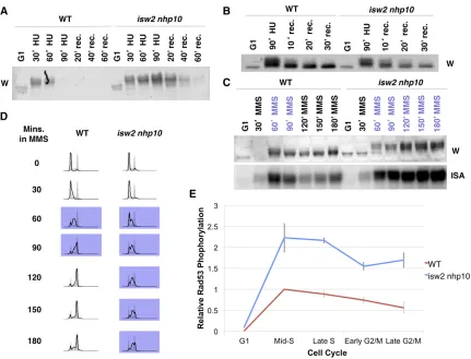
2004). We previously reported that the isw2 nhp10mutant exhibits two phenotypes in the presence of replication inhib-itors: (1) slow growth due to a prolonged S phase without an increase in cell death and (2) overactivation of the checkpoint during S phase (Vincentet al.2008; Auet al.2011). Previously, we reported thatisw2 nhp10elevates and prolongs checkpoint activity during S phase in the presence of replication inhibitors, especially after transient exposure to HU (Auet al.2011). Our Rad53 ISA revealed rapid loss of checkpoint activity in WT cells but not inisw2 nhp10cells after transient treatment with HU (Auet al.2011). However, on examiningRad53checkpoint activity by Western blot, we noticed that the Rad53 protein could no longer be detected in later time points when samples were prepared according to the protocol used in our previous report (Auet al.2011) (Figure 1A). Importantly,Rad53protein was lost at much earlier time points in WT than inisw2 nhp10 cells (Figure 1A). We found that the loss ofRad53protein was attributed to an increase in cell density as the time course went on, which we did not adjust for previously, causing inefficient TCA precipitation of proteins. Because of the slower S-phase progression in isw2 nhp10 cells, Rad53 is lost later in isw2 nhp10compared with WT cells (Figure 1A). Thus the apparent checkpoint hyperactivity in isw2 nhp10 cells after HU treat-ment detected by ISA could be attributed to inefficient recov-ery of Rad53 protein in WT cells. We therefore prepared protein samples adjusting for the differences in cell numbers between WT andisw2 nhp10cells (seeMaterials and Methods
for details) and repeated the ISA. This revealed that although Rad53 protein is still slightly more phosphorylated based on Western blotting inisw2 nhp10compared with WT cells after HU treatment (Figure 1B), the difference in phosphorylation is not to the same degree as what we previously reported using ISA (Figure 1A).
These results prompted us to reexamine the checkpoint activity of WT andisw2 nhp10cells under various conditions
using both Western blotting andRad53ISA while accounting for cell density. These tests revealed that the difference in Rad53checkpoint activity between WT andisw2 nhp10cells is much more robust upon MMS treatment than HU treatment (Figure 1, B and C). To investigate howIsw2andIno80 atten-uate checkpoint activity in MMS, we sought to determine when, during S phase,Isw2andIno80perform their roles by defining the kinetics of checkpoint activity. To this end, we performed a time-course experiment in which cells were arrested in G1and released into S phase in rich medium con-taining MMS, and we monitored cell cycle progression and checkpoint activity by FACS andRad53ISA, respectively (Fig-ure 1D and Supporting Information, Figure S1). Consistent with our previous report (Vincentet al.2008),isw2 nhp10cells have a delay in S-phase progression that is not a result of incomplete replication (Figure 1D andFigure S1B). The delay is associated with stronger and prolonged checkpoint activity in the presence of MMS (Figure 1E). This experiment revealed that the biggest difference in checkpoint activity occurs during middle-late S phase (Figure 1E). Consistent with the fact that early origin firing is not greatly affected in isw2 nhp10cells (Vincentet al.2008), these results suggest thatIsw2andIno80 function after replication has already been initiated. Once checkpoint activity is more strongly triggered in S phase, acti-vation remains elevated via transition into G2/M phase, which coincides with the presence of a subpopulation of cells that continue to remain in S phase in isw2 nhp10cells (Figure 1, C and D). Taken together, our results suggest thatIsw2 and Ino80 prevent hyperactivation of the checkpoint activity in middle to late S phase in the presence of MMS to allow for proper replication progression and transition into G2/M phase.
Isw2 and Ino80 function outside of DNA repair or fork protection pathways
Because checkpoint activity is much more strongly activated in isw2 nhp10 cells by MMS than by HU, we next tested whetherISW2andNHP10could function through DNA repair or replication fork protection pathways. We have shown pre-viously, by genetic tests, thatISW2andNHP10do not function through several genes involved in replication fork protection and DNA repair pathways (Vincentet al.2008), leading to the conclusion that they do not function via these pathways (Au
et al. 2011). However, these tests were not very quantitative, and additional genes involved in DNA damage response and replication fork protection were identified after our previous tests (Crabbeet al.2010; Engelset al.2011; Hegnaueret al.
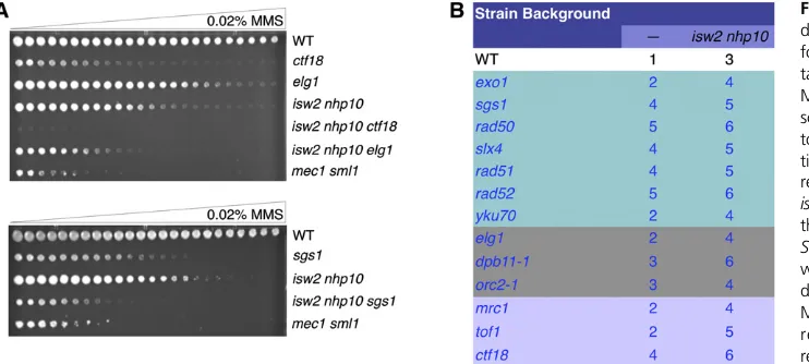
sensitivity. In contrast, if ISW2and NHP10function indepen-dently of a DNA damage response or replication fork protection gene, the phenotypes should be additive, and the triple mutant should be more sensitive than the single mutant.CTF18,ELG1, andSGS1all influence checkpoint activity (Kanelliset al.2003; Crabbe et al. 2010; Hegnauer et al. 2012). All the triple mutants within these mutant backgrounds show additive MMS sensitivity, suggesting that these genes are not required for ISW2 andNHP10 function (Figure 2A). Other genes in-volved in fork protection (lavender), replication (gray), and different DNA damage response pathways (teal) also show that Isw2and Ino80act independently of these pathways (Figure 2B). DNA polymerasee(Pole) associates withDpb11to facil-itate DNA replication and the S-phase checkpoint (Masumoto
et al.2000), and theDpb4subunit of Polealso exists in the Isw2 complex (Iida and Araki 2004; Mcconnellet al.2004). Therefore, to test genetic interactions of PolewithIsw2and Nhp10, we used a pol2-12 mutant that attenuates S-phase checkpoint activity (Li et al. 2008). However, the pol2-12
isw2 nhp10 mutants exhibited extreme growth defects and readily picked up suppressor mutations (data not shown). While we were not able to measure MMS sensitivity of this mutant, the additive effect even in the absence of replication stress suggests thatIsw2andNhp10have functions indepen-dent of Pole. Together these results reinforce the conclusion thatIsw2 andIno80do not function through DNA repair or replication fork protection pathways.
Isw2 and Ino80 antagonize checkpoint activators and attenuate checkpoint activity independent of any single checkpoint factor downstream of RPA
The preceding results are consistent with our model that Isw2andIno80play a direct role in attenuating the S-phase checkpoint activity. Thus, to gain insight into the mechanism of checkpoint attenuation byIsw2andIno80, we performed sys-tematic genetic tests to screen for checkpoint factors required forIsw2andIno80checkpoint function using theRad53ISA. Based on a similar reasoning as earlier, if a checkpoint protein Figure 1 isw2 nhp10cells display stronger checkpoint activity throughout S phase and into G2/M phase in the presence of MMS. (A) Rad53 Western blot of WT andisw2 nhp10cells using the protein preparation methods in our previous report (Auet al.2011). Cells were arrested in G1withafactor for 90 min and released into 200 mM HU for 90 min, and then HU was washed out. rec, recovery time in minutes. (B) Rad53 Western blot of protein preparations that used the amount of TCA normalized for cell volume. (C) A Western blot (W) andin situautophosphorylation assay (ISA) of Rad53 of samples taken from indicated time points. Time points indicated in purple represent when cells are in S phase. (D) Cell cycle profiles of WT andisw2 nhp10
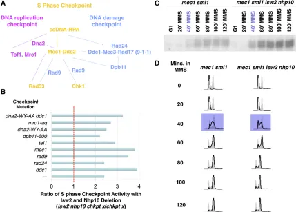
is required forIsw2andIno80to attenuate checkpoint activity, we would expect deletion ofISW2andNHP10to not increase checkpoint activity in the relevant mutant background. Alter-natively, if a given checkpoint protein is not required forIsw2 and Ino80 function, then deletion of ISW2 and NHP10 still would increase the deficient checkpoint activity in the check-point mutants. Through these analyses, we aimed to identify the most upstream checkpoint factor by whichIsw2andIno80 attenuate checkpoint activity. We screened all major point factors known to be required for robust S-phase check-point activity (Figure 3A). Unless otherwise indicated, the checkpoint mutants tested were null mutations. The S-phase checkpoint cascade has two partially overlapping pathways (Figure 3A), the DRC and the DDC, that both function to activate the S-phase checkpoint. Although the DDC (blue) is activated primarily in the presence of MMS, the DRC (pink) can redundantly function to partially activate the S-phase checkpoint in the absence of DDC factors (Branzei and Foiani 2009). Some Checkpoint components (yellow) function in both the DDC and the DRC and thus are most critical for proper activation. Complete deletion of a DRC component resulted in severe MMS sensitivity because such components are also involved in DNA replication, in addition to the DRC (Figure 2B). Therefore, for checkpoint factors that also have roles in DNA replication, we usedmrc1-aq(Szyjkaet al.2005), dpb11-600 (Pfander and Diffley 2011), and dna2-WY-AA
(Kumar and Burgers 2013), which are alleles that were shown previously to be specifically deficient in checkpoint activation. Unexpectedly, we found that theisw2 nhp10mutation partially rescued checkpoint activity in all the checkpoint-deficient mutants tested, for both DRC and DDC mutants, as indicated by a checkpoint activity ratio that was significantly higher than one on ISW2andNHP10deletion (Figure 3B). These results suggest thatIsw2andIno80antagonize checkpoint activators in affecting S-phase checkpoint activity.Mec1 plays a central role in S-phase checkpoint activation, and the absence of the Mec1kinase severely abrogates the S-phase checkpoint (Branzei and Foiani 2010). Perhaps most surprisingly, even inmec1 sml1
cells, where checkpoint activity is mostly depleted, deletion of ISW2andNHP10still results in higher checkpoint activity dur-ing S phase, indicatdur-ing thatIsw2andIno80function indepen-dent ofMec1(Figure 3, C and D). Residual checkpoint activity in the mec1 sml1 mutant is due to the partially redundant function of Tel1 (Mantiero et al. 2007). The mode of Tel1 activation in S phase is not well understood, but in the absence of Mec1, Tel1 can activate the checkpoint to a much lesser degree (Zou 2013). We attempted to examine the effects of Isw2 and Ino80 when the S-phase checkpoint is completely abolished by making a mec1 tel1 sml1 isw2 nhp10 mutant. However, tel1 deletion alone causes strong synthetic growth defects with theisw2 nhp10mutation independent of S-phase checkpoint function (Figure 6A). Because of this genetic inter-action, the quintuple mutant had extreme growth defects and readily picked up suppressors (data not shown), making phe-notype analyses impossible. The dna2-WY-AA ddc1 mutant, which abolishes Mec1 activation through both the DRC and DDC pathways (Kumar and Burgers 2013), also displayed higher checkpoint activity onISW2andNHP10 deletion (Figure 3B). This result is consistent with the partial rescue of the checkpoint activity of the mec1 mutant byisw2 nhp10. These
findings suggest that Isw2 and Ino80 deactivate checkpoint activity independent of both the DRC and DDC pathways. We were unable to test RPA because of the lack of anrpamutant allele that solely affects checkpoint activity. Because RPA is es-sential for viability and has various roles in DNA replication, checkpoint activation, DNA damage response, and recombina-tion, deleting ISW2 and NHP10 in rpa mutant backgrounds rfa1-degron and rfa1-t11 (Umezu et al. 1998; Lisby et al.
2004) causes severe growth defects (data not shown). While the strong synthetic growth defects of therpaisw2 nhp10 mu-tant were not unexpected, they made measuring the checkpoint activity in the mutant impossible. Nevertheless, our data dem-onstrate that no known S-phase checkpoint factor downstream of RPA is required forIsw2andIno80to attenuate checkpoint activity. These results collectively suggest thatIsw2andIno80 function either through currently unidentified checkpoint Figure 2 Isw2 and Ino80 function is in-dependent of DNA damage response or fork protection pathways. (A) Represen-tative results of the spot tests on 0.02% MMS gradient plates performed as de-scribed previously (Auet al.2011). The top panel shows that deletion of replica-tion components such asELG1orCTF18
results in additive MMS sensitivity with
isw2 nhp10. The bottom panel shows that deletion of a DNA resection factor
activator(s) or through RPA, which feeds into multiple inde-pendent checkpoint activation pathways. For MRC1, DPB11, andDNA2, we cannot rule out the possibility that the alleles we used did not inactivate their relevant checkpoint activation function.
Isw2 and Ino80 do not affect the abundance of checkpoint factors
We envision thatIsw2andIno80attenuate checkpoint activity via at least two possible mechanisms. One possibility is that Isw2andIno80may facilitate removal of a checkpoint protein from replication forks. Alternatively, they may down-regulate the activity of a checkpoint protein. We first tested whether Isw2andIno80alter the abundance of chromatin-bound RPA by chromatin fractionation from cells in G1and S phase with 0.02% MMS (Figure 4A), followed by Western blotting. This analysis revealed that relative levels of chromatin-bound RPA were similar in both WT and isw2 nhp10 cells in G1 and S phase, indicating that higher checkpoint activity in the mutant is not a result of RPA accumulation on bulk chromatin (Figure 4B). Consistent with this result, overexpression ofRad51and Rad52, proteins involved in displacement of RPA from double-strand break (DSB) sites (Sugiyama and Kowalczykowski
previously to have a function in the S-phase checkpoint. No other Rad53phosphatases were found to have different pro-tein abundances in the SILAC data, and because of the redun-dancy of the Rad53 phosphatases, Ptc2 alone plays only a minor role in checkpoint recovery after MMS treatment (Szyjka et al.2008). Furthermore, we have shown previously thatIsw2andIno80clearly function independent of theRad53 phosphatases (Au et al. 2011). Taken together, our results show thatIsw2- andIno80-dependent checkpoint attenuation cannot be explained by changes in the abundance of chromatin-bound proteins, at least at the bulk level.
Chromatin accessibility around replication forks is decreased in isw2 nhp10 cells in the presence of MMS
Previously, we (Vincent et al.2008) and others (Papamichos-Chronakis and Peterson 2008; Shimada et al. 2008) have shown that bothIsw2andIno80are enriched at stalled repli-cation forks. Therefore, we considered the possibility thatIsw2 andIno80may affect chromatin structure around replication forks, thereby altering checkpoint activity.Ino80functions in opposition to Swr1 and exchanges nucleosomal H2A.Z/H2B dimers for H2A/H2B dimers (Papamichos-Chronakis et al.
2006, 2011). Meanwhile,Isw2 alters chromatin structure by sliding nucleosomes away from coding regions or toward gene promoters (Fazzio and Tsukiyama 2003; Yadonet al.2010). We sought to determine whether these known biochemical activities might be required for the S-phase functions ofIsw2 andIno80. If the important role ofNhp10is inHtz1exchange, we can expect to see partial rescue of the MMS sensitivity ofisw2 nhp10cells by deletingHTZ1. However,htz1mutation not only showed no rescue but also increased the MMS sensitivity ofisw2 nhp10 cells, suggesting thatHtz1exchange is not the relevant activity ofIsw2andNhp10in MMS (Figure 5A). To confirm that the genetic interaction withHtz1was representative of theIno80
complex rather thanNhp10alone, we tested another subunit ofIno80,Ies5. Theisw2 ies5 htz1cells displayed an even stron-ger additive growth defect in the presence of MMS (Figure S2A). Deletingswr1inisw2 nhp10cells also resulted in a stron-ger growth defect in the presence of MMS compared with eitherswr1orisw2 nhp10(Figure S2B). These results collec-tively suggest thatHtz1deposition is not the relevant biochem-ical activity for growth in MMS.
We also mapped nucleosome positions around replicating regions in WT andisw2 nhp10cells in both G1and S phase in MMS. For this experiment, harvesting cells in early S phase was necessary to achieve the best possible synchrony of repli-cation forks. We first confirmed that nucleosome positions at known Isw2 targets shift inisw2 nhp10cells, as anticipated. However, we did not detect significant changes in nucleosome positioning at replicating regions in either G1or early S phase in MMS (data not shown). Although this negative result has caveats that replication forks are not completely synchronous within cell population and that the resolution of our nucleo-some mapping may not be high enough, it does not support the idea that a model that regulates nucleosome positions accounts for the mechanism behind checkpoint regulation byIsw2and Ino80.
We next tested whether chromatin accessibility might be altered inisw2 nhp10cells around replication forks. We have recently developed a method to measure chromatin accessi-bility to micrococcal nuclease (MNase) in a way normalized for nucleosome occupancy callednormalized chromatin acces-sibility to MNase (NCAM) (Rodriguez and Tsukiyama 2013; Rodriguez et al.2014). To measure NCAM, we digest chro-matin by MNase and purify monoucleosomal DNA, followed by hybridization of nucleosomal DNA to high-density tiled microarrays. We then quantify nucleosome signals, which can be affected by both nucleosome occupancy and the sensitivity of Figure 4Bulk levels of chromatin-bound checkpoint proteins are largely unchanged between WT and
isw2 nhp10 cells. (A) FACS profiles of samples used
nucleosomes to MNase at any given site (Weineret al.2010; Rodriguez and Tsukiyama 2013). At the same time, we perform chromatin immunoprecipitation using anti-histone H3 antibody to measure nucleosome occupancy. By normalizing the strength of MNase signals to nucleosome occupancy, we can calculate NCAM, which measures the relative amount of MNase digestion per nucleosome (Rodriguez and Tsukiyama 2013). As expected for both WT andisw2 nhp10cells, NCAM increases at regions of
isw2 nhp10cells exhibit similar levels of DNA synthesis around early-firing origins at this time point (Figure 5B). In contrast, nonreplicating regions, which are represented by chromatin surrounding late origins that have notfired at this time point, are overall less accessible than the rest of the genome, as expected (Figure 5, B and C). We did detect minor decreases in NCAM inisw2 nhp10compared with WT cells at these non-replicating regions, which is most likely attributed to passive replication occurring at these sites (Figure 5C). Taken together, these results suggest that Isw2 and Ino80 complexes affect NCAM around actively replicating regions. Furthermore, the reduction of NCAM inisw2 nhp10cells is not a result of global reduction of the NCAM signal because NCAM is very similar or even slightly increased in the mutant cells at transcriptional start sites in both G1and S phase in MMS (Figure 5D). These results collectively show thatIsw2andIno80increase chroma-tin accessibility specifically around replication forks in the pres-ence of MMS. Although the decrease in NCAM inisw2 nhp10 cells at actively replicating regions can be attributed in part to differences in nucleosome occupancy, the changes are mainly driven by a decrease in nucleosome signal (Figure S2, C and D). We have confirmed the genome-wide decrease in nucleosome signals around replication forks in isw2 nhp10 cells by deep sequencing nucleosomal DNA (data not shown).
isw2 nhp10 partially rescues MMS sensitivity of severe checkpoint-deficient mutants
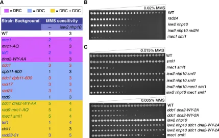
Because isw2 nhp10can stimulate checkpoint activity even in the absence of the known checkpoint activators, we sought to determine whether deletion of isw2 nhp10in checkpoint-deficient mutants also could rescue cell growth in the presence of MMS. The DRC checkpoint-specific mutations yielded WT-like MMS sensitivity, consistent with previous reports that the DRC checkpoint components are dispensable for the check-point in MMS (Alcasabas et al. 2001; Crabbe et al. 2010) (Figure 6A). Deletion of ISW2 and NHP10 in these back-grounds did not cause detectable genetic interactions. How-ever, the DDC mutants were more sensitive to MMS than the WT cells, as expected (Hustedtet al.2013), and unexpectedly, deletion ofISW2andNHP10did not yield a rescue in growth despite a rescue in checkpoint activation, as shown earlier (Figure 6, A and B). These results show, for thefirst time, that checkpoint overactivation and growth in MMS are genetically separable because checkpoint overactivation does take place without affecting MMS sensitivity in DDC mutants (Figure 6, A and B). However, in the mutants that could not activate either the DRC or the DDC in S phase, isw2 nhp10partially rescued MMS sensitivity, which is consistent with the partial rescue in checkpoint activity (Figure 6, A and C). We previ-ously failed to detect the rescue of MMS sensitivity of mec1 mutants byisw2 nhp10mutation (Auet al.2011) because the rescue is slight and can be detected only after an extended period of time owing to very slow growth of the mutant. While the rescue of themec1phenotype byisw2 nhp10was modest, we saw a more prominent rescue in ddc1 dna2-WY-AA cells (Figure 6, A and C). Because sensitivity of mec1 to MMS is
a result of both an abrogated checkpoint and a replication fork protection defect, it is not surprising that isw2 nhp10 only slightly rescues MMS sensitivity. In contrast, isw2 nhp10 dra-matically rescues ddc1 dna2-WY-AA cells, which have been shown to be sensitive to replication inhibitors solely because of their defect in activating the checkpoint. To determine whether the rescue was specific to MMS, we also tested genetic interactions on other replication inhibitors, including HU and camptothecin (CPT) (Figure S3, A and B). Both CPT and HU exposure yielded either minimal or additive sensitivity when ISW2 and NHP10 were deleted in the checkpoint-deficient mutants (Figure S3, A and B). These results are consistent with the fact thatIsw2andIno80only minimally affect checkpoint activity on HU treatment (Figure 1B). While growth sensitivity to MMS does not always directly reflect the changes in check-point regulation, a partial rescue of the severe MMS sensitivity of these checkpoint-deficient mutants byisw2 nhp10is consis-tent with the notion thatIsw2andIno80oppose checkpoint activators in the presence of MMS.
Discussion
We have shown previously thatIsw2andIno80are important for attenuation of the S-phase checkpoint, especially after tran-sient HU treatment (Auet al.2011). However, in this study, we discovered that the previous method of protein preparation resulted in the loss of Rad53 protein. Using a method that can efficiently retainRad53protein, we found thatIsw2and Ino80 play much bigger roles in attenuating S-phase check-point activity in the presence of MMS than in the presence of HU. Consistent with thisfinding, growth assays also show that Isw2 and Ino80 antagonize checkpoint activators more pro-foundly in the presence of MMS compared with other replica-tion inhibitors. Althoughisw2 nhp10cells are sensitive to other replication inhibitors (Vincentet al.2008), our results suggest thatIsw2andIno80may play distinct roles in the presence of different replication inhibitors.
Based on previousfindings (Vincentet al.2008; Auet al.
2011) and this study, we believe thatisw2 nhp10cells do not have increased replication fork problems or DNA damage response defects, leading to our model that Isw2 andIno80 directly attenuate checkpoint activity. We cannot completely exclude the possibility that there is an increased uncoupling of the replicative helicase to DNA polymerase (Byun et al.
2005) inisw2 nhp10cells in MMS, which could be the cause of stronger checkpoint activation and slower S-phase progres-sion. However, we do not favor this model. As we have shown previously (Auet al.2011) and here, the dissociation of repli-cative helicase and DNA polymerases in the presence of MMS is detrimental in the absence of replication fork protection by the S-phase checkpoint. For example, we previously reported that deletion of CTF4orTOF1, which are implicated in cou-pling MCM helicase with DNA polymerase during replication (Katou et al. 2003; Tanaka et al. 2009), in the mec1 back-ground leads to strongly additive MMS sensitivity (Au et al.
partially rescue the MMS sensitivity of severe checkpoint mutants such asmec1andddc1 dna2-WY-AA.
How doIsw2andIno80complexes function? We do not believe thatIsw2andIno80function by affecting transcription because there were no significant differences in transcription between MMS-treated WT andisw2 nhp10cells (Vincentet al., 2008, data not shown). Our checkpoint activity assays suggest thatIsw2andIno80attenuate checkpoint activity independent of all major checkpoint activators except RPA. Therefore, we propose that they function either through a currently uniden-tified checkpoint regulator(s) or through RPA. We show here thatIsw2andIno80do not affect the amounts of chromatin-bound checkpoint proteins including RPA at the bulk level, but it is possible that they affect the location of checkpoint proteins on chromatin. Isw2 has the ability to translocate on ssDNA (Fischeret al.2009). Furthermore, chromatin remodeling fac-tors have been shown to interact with nonhistone proteins, and Mot1, an ATPase that is highly related to chromatin remodel-ing factors, can displaceTBP, a nonhistone protein, from DNA (Aubleet al.1994; Moreauet al.2003; Auet al.2011; Sugimoto
et al.2011). BecauseIsw2and Ino80physically interact with RPA (Auet al.2011), we initially considered the possibility that they may be able to directly remove RPA from the ssDNA tem-plate using DNA translocase activity. However, the results of the
direct test for this possibilityin vitrohave been negative so far (data not shown). We also have attempted RPA chromatin immunoprecipitation with DNA microarray (ChIP-chip) and chromatin immunoprecipitation with massively parallel DNA sequencing (ChIP-seq), which have yet to yield any conclusive results because of the difficulty of capturing RPA peaks on moving replication forks with imperfect synchrony (data not shown). While it is still possible thatIsw2andIno80could be affecting RPA localization at sites of stalled replication, we currently do not have adequate tools to address this scenario. Although it seems unlikely based on biochemical activities of chromatin remodeling factors, it is also possible thatIsw2and Ino80directly affect the activity of a checkpoint kinase rather than localization of checkpoint proteins.
Most intriguingly, we demonstrate for the first time that Isw2andIno80promote chromatin accessibility around actively replicating regions. Together with the fact that bothIsw2and Ino80 are enriched at stalled replication forks (Vincent et al.
2008), this is consistent with a model suggesting that the chro-matin remodeling factors function at stalled replication forks. Multiple reports have suggested that chromatin remodeling fac-tors can alter chromatin structure to facilitate replication fork progression (Fyodorov et al.2004; Pootet al. 2004; Shimada
previously that chromatin accessibility increases around replica-tion forks to promote replicareplica-tion (Rodriguez and Tsukiyama 2013). Therefore, one possibility is that Isw2andIno80 may attenuate checkpoint activity by changing the way checkpoint protein(s) interact with chromatin at stalled forks, which, in turn, affects replication progression and checkpoint dynamics. Because we found that Isw2andIno80likely do not function through their known biochemical activities, we speculate that Isw2andIno80function in a currently unknown manner. This can be either by modifying how DNA and core histones interact or by changing histone turnover rate, which may influence other factors to bind and potentially affect checkpoint factor localiza-tion or activity. For example, weakening histone-DNA interac-tions around stalled replication forks could cause an increase in accessibility to recruit checkpoint repressors to facilitate removal of checkpoint factor(s) and promote resumption of DNA repli-cation. Indeed, another ATP-dependent chromatin remodeling factor, Fun30, and its mammalian counterpart, SMARCAD1, were both found to be recruited to sites of DSBs to promote resection presumably by removingRad9from DSB ends (Chen
et al. 2012; Costelloe et al. 2012). Alternatively, checkpoint hyperactivation in the chromatin-remodeling-factor mutant may be the cause of the observed decrease in chromatin acces-sibility. Interestingly, we have shown previously that a mutation inmec1also results in decreased chromatin accessibility at rep-licating regions in S phase in HU (Rodriguez and Tsukiyama 2013). However, becauseMec1facilitates chromatin accessibility around stalled replication forks (Rodriguez and Tsukiyama 2013), it is difficult to explain the increased checkpoint activity inisw2 nhp10cells as the direct cause of the decreased chroma-tin accessibility in the mutant. Whether these changes in chro-matin structure are related to the phenotypes ofisw2 nhp10cells in MMS remains to be explored and will be investigated in future work. Finally, it is also possible that the changes in chro-matin accessibility are separable from checkpoint activation.
Ourfindings suggest thatIsw2andIno80likely function through currently unknown mechanisms to regulate both chro-matin accessibility and S-phase checkpoint attenuation. In the future, understanding the molecular mechanisms underlying Isw2- and Ino80-dependent chromatin accessibility at replica-tion forks will be crucial in defining new biochemical activities of chromatin remodeling factors as well as elucidating a cur-rently unknown mechanism of checkpoint control.
Acknowledgments
We thank Steve Elledge, Peter Burgers, Dave Toczyski, John Diffley, Hiroyuki Araki, Jac Nickoloff, Sue Biggins, Linda Breeden, Keiko Umezu, Rodney Rothstein, and Oscar Aparicio for plasmids and strains used in this study. We also thank Anne Donaldson for her advice on SILAC protocols and Phil Gafken for his help with mass spectrometry and data analysis. We are grateful to Tsukiyama Laboratory members and Sue Biggins for discussion and comments on this manuscript. This work is supported by Molecular and Cellular Biology Training Grant GM-07270 to L.L. and Grant R-01GM058465 to T.T.
Literature Cited
Alcasabas, A. A., A. J. Osborn, J. Bachant, F. Hu, P. J. Werleret al.,
2001 Mrc1 transduces signals of DNA replication stress to
ac-tivate Rad53. Nat. Cell Biol. 3: 958–965.
Au, T. J., J. Rodriguez, J. A. Vincent, and T. Tsukiyama,
2011 ATP-dependent chromatin remodeling factors tune S
phase checkpoint activity. Mol. Cell. Biol. 31: 4454–4463.
Auble, D. T., K. E. Hansen, C. G. Mueller, W. S. Lane, J. Thorner
et al., 1994 Mot1, a global repressor of RNA polymerase II transcription, inhibits TBP binding to DNA by an ATP-dependent
mechanism. Genes Dev. 8: 1920–1934.
Branzei, D., and M. Foiani, 2009 The checkpoint response to
rep-lication stress. DNA Repair 8: 1038–1046.
Branzei, D., and M. Foiani, 2010 Maintaining genome stability at
the replication fork. Nat. Rev. Mol. Cell Biol. 11: 208–219.
Byun, T. S., M. Pacek, M. C. Yee, J. C. Walter, and K. A. Cimprich,
2005 Functional uncoupling of MCM helicase and DNA
poly-merase activities activates the ATR-dependent checkpoint.
Genes Dev. 19: 1040–1052.
Chen, X., D. Cui, A. Papusha, X. Zhang, C. D. Chuet al., 2012 The
Fun30 nucleosome remodeller promotes resection of DNA
double-strand break ends. Nature 489: 576–580.
Costelloe, T., R. Louge, N. Tomimatsu, B. Mukherjee, E. Martini
et al., 2012 The yeast Fun30 and human SMARCAD1 chromatin
remodellers promote DNA end resection. Nature 489: 581–584.
Crabbe, L., A. Thomas, V. Pantesco, J. De Vos, P. Pasero et al.,
2010 Analysis of replication profiles reveals key role of
RFC-Ctf18 in yeast replication stress response. Nat. Struct. Mol. Biol.
17: 1391–1397.
Eng, J. K., A. L. McCormack, and J. R. Yates, 1994 An approach to
correlate tandem mass spectral data of peptides with amino acid sequences in a protein database. J. Am. Soc. Mass Spectrom. 5:
976–989.
Engels, K., M. Giannattasio, M. Muzi-Falconi, M. Lopes, and S.
Ferrari, 2011 14-3-3 Proteins regulate exonuclease 1–dependent
processing of stalled replication forks. PLoS Genet. 7: e1001367.
Fazzio, T. G., and T. Tsukiyama, 2003 Chromatin remodeling
in vivo: evidence for a nucleosome sliding mechanism. Mol. Cell
12: 1333–1340.
Fischer, C. J., K. Yamada, and D. J. Fitzgerald, 2009 Kinetic
mech-anism for single-stranded DNA binding and translocation by
S. cerevisiaeIsw2. Biochemistry 48: 2960–2968.
Friedel, A. M., B. L. Pike, and S. M. Gasser, 2009 ATR/Mec1:
co-ordinating fork stability and repair. Curr. Opin. Cell Biol. 21:
237–244.
Fyodorov, D. V., M. D. Blower, G. H. Karpen, and J. T. Kadonaga,
2004 Acf1 confers unique activities to ACF/CHRAC and
pro-motes the formation rather than disruption of chromatin in vivo.
Genes Dev. 18: 170–183.
Gilbert, C. S., C. M. Green, and N. F. Lowndes, 2001 Budding
yeast Rad9 is an ATP-dependent Rad53 activating machine.
Mol. Cell 8: 129–136.
Goldmark, J. P., T. G. Fazzio, P. W. Estep, G. M. Church, and T.
Tsukiyama, 2000 The Isw2 chromatin remodeling complex
re-presses early meiotic genes upon recruitment by Ume6p. Cell
103: 423–433.
Gonzalez-Prieto, R., A. M. Munoz-Cabello, M. J. Cabello-Lobato,
and F. Prado, 2013 Rad51 replication fork recruitment is
re-quired for DNA damage tolerance. EMBO J. 32: 1307–1321.
Hegnauer, A. M., N. Hustedt, K. Shimada, B. L. Pike, M. Vogelet al.,
2012 An N-terminal acidic region of Sgs1 interacts with Rpa70
and recruits Rad53 kinase to stalled forks. EMBO J. 31: 3768–
3783.
Hustedt, N., S. M. Gasser, and K. Shimada, 2013 Replication
checkpoint: tuning and coordination of replication forks in S
Iida, T., and H. Araki, 2004 Noncompetitive counteractions of DNA polymerase epsilon and ISW2/yCHRAC for epigenetic in-heritance of telomere position effect in Saccharomyces
cerevi-siae. Mol. Cell. Biol. 24: 217–227.
Kall, L., J. D. Canterbury, J. Weston, W. S. Noble, and M. J.
MacCoss, 2007 Semi-supervised learning for peptide
iden-tification from shotgun proteomics datasets. Nat. Methods 4:
923–925.
Kanellis, P., R. Agyei, and D. Durocher, 2003 Elg1 forms an
alter-native PCNA-interacting RFC complex required to maintain
ge-nome stability. Curr. Biol. 13: 1583–1595.
Katou, Y., Y. Kanoh, M. Bando, H. Noguchi, H. Tanaka et al.,
2003 S-phase checkpoint proteins Tof1 and Mrc1 form a stable
replication-pausing complex. Nature 424: 1078–1083.
Kondo, T., T. Wakayama, T. Naiki, K. Matsumoto, and K. Sugimoto,
2001 Recruitment of Mec1 and Ddc1 checkpoint proteins to
double-strand breaks through distinct mechanisms. Science
294: 867–870.
Kubota, T., S. Hiraga, K. Yamada, A. I. Lamond, and A. D. Donaldson,
2011 Quantitative proteomic analysis of chromatin reveals that
Ctf18 acts in the DNA replication checkpoint. Mol Cell Proteomics 10: M110.005561.
Kubota, T., K. Nishimura, M. T. Kanemaki, and A. D. Donaldson,
2013 The Elg1 replication factor C-like complex functions
in PCNA unloading during DNA replication. Mol. Cell 50:
273–280.
Kubota, T., D. A. Stead, S. Hiraga, S. ten Have, and A. D. Donaldson,
2012 Quantitative proteomic analysis of yeast DNA replication
proteins. Methods 57: 196–202.
Kumar, S., and P. M. Burgers, 2013 Lagging strand maturation
factor Dna2 is a component of the replication checkpoint
initi-ation machinery. Genes Dev. 27: 313–321.
Li, J. M., M. T. Tetzlaff, and S. J. Elledge, 2008 Identification of
MSA1, a cell cycle-regulated, dosage suppressor of drc1/sld2
and dpb11 mutants. Cell Cycle 7: 3388–3398.
Licklider, L. J., C. C. Thoreen, J. Peng, and S. P. Gygi, 2002
Au-tomation of nanoscale microcapillary liquid chromatography–
tandem mass spectrometry with a vented column. Anal. Chem.
74: 3076–3083.
Lisby, M., J. H. Barlow, R. C. Burgess, and R. Rothstein,
2004 Choreography of the DNA damage response:
spatiotem-poral relationships among checkpoint and repair proteins. Cell
118: 699–713.
Majka, J., A. Niedziela-Majka, and P. M. Burgers, 2006 The
check-point clamp activates Mec1 kinase during initiation of the DNA
damage checkpoint. Mol. Cell 24: 891–901.
Mantiero, D., M. Clerici, G. Lucchini, and M. P. Longhese,
2007 Dual role forSaccharomyces cerevisiaeTel1 in the
check-point response to double-strand breaks. EMBO Rep. 8: 380–387.
Masumoto, H., A. Sugino, and H. Araki, 2000 Dpb11 controls the
association between DNA polymerases alpha and epsilon and the autonomously replicating sequence region of budding yeast.
Mol. Cell. Biol. 20: 2809–2817.
McConnell, A. D., M. E. Gelbart, and T. Tsukiyama, 2004 Histone
fold protein Dls1p is required for Isw2-dependent chromatin
remodeling in vivo. Mol. Cell. Biol. 24: 2605–2613.
Moreau, J. L., M. Lee, N. Mahachi, J. Vary, J. Mellor et al.,
2003 Regulated displacement of TBP from the PHO8 promoter
in vivo requires Cbf1 and the Isw1 chromatin remodeling
com-plex. Mol. Cell 11: 1609–1620.
Morrison, A. J., J. Highland, N. J. Krogan, A. Arbel-Eden, J. F.
Greenblatt et al., 2004 INO80 andg-H2AX interaction links
ATP-dependent chromatin remodeling to DNA damage repair.
Cell 119: 767–775.
Morrison, A. J., and X. Shen, 2009 Chromatin remodelling
be-yond transcription: the INO80 and SWR1 complexes. Nat. Rev.
Mol. Cell Biol. 10: 373–384.
Navadgi-Patil, V. M., and P. M. Burgers, 2008 Yeast DNA
replica-tion protein Dpb11 activates the Mec1/ATR checkpoint kinase.
J. Biol. Chem. 283: 35853–35859.
Papamichos-Chronakis, M., J. E. Krebs, and C. L. Peterson,
2006 Interplay between Ino80 and Swr1 chromatin
remodel-ing enzymes regulates cell cycle checkpoint adaptation in
re-sponse to DNA damage. Genes Dev. 20: 2437–2449.
Papamichos-Chronakis, M., and C. L. Peterson, 2008 The Ino80
chromatin-remodeling enzyme regulates replisome function and
stability. Nat. Struct. Mol. Biol. 15: 338–345.
Papamichos-Chronakis, M., S. Watanabe, O. J. Rando, and C. L.
Peterson, 2011 Global regulation of H2A.Z localization by
the INO80 chromatin-remodeling enzyme is essential for
ge-nome integrity. Cell 144: 200–213.
Pellicioli, A., C. Lucca, G. Liberi, F. Marini, M. Lopes et al.,
1999 Activation of Rad53 kinase in response to DNA damage
and its effect in modulating phosphorylation of the lagging
strand DNA polymerase. EMBO J. 18: 6561–6572.
Pfander, B., and J. F. Diffley, 2011 Dpb11 coordinates Mec1
ki-nase activation with cell cycle-regulated Rad9 recruitment.
EMBO J. 30: 4897–4907.
Poot, R. A., L. Bozhenok, D. L. van den Berg, S. Steffensen, F.
Ferreiraet al., 2004 The Williams syndrome transcription
fac-tor interacts with PCNA to target chromatin remodelling by
ISWI to replication foci. Nat. Cell Biol. 6: 1236–1244.
Rodriguez, J., J. N. McKnight, and T. Tsukiyama, 2014
Genome-wide analysis of nucleosome positions, occupancy, and accessi-bility in yeast: nucleosome mapping, high-resolution histone
ChIP, and NCAM. Curr. Protoc. Mol. Biol. 108: 28.21:28.21.1–
21.28.16.
Rodriguez, J., and T. Tsukiyama, 2013 ATR-like kinase Mec1
fa-cilitates both chromatin accessibility at DNA replication forks and replication fork progression during replication stress. Genes
Dev. 27: 74–86.
Rouse, J., and S. P. Jackson, 2002 Lcd1p recruits Mec1p to DNA
lesions in vitro and in vivo. Mol. Cell 9: 857–869.
Royce, T. E., N. J. Carriero, and M. B. Gerstein, 2007 An efficient
pseudomedianfilter for tiling microrrays. BMC Bioinformatics 8: 186.
Shen, X., G. Mizuguchi, A. Hamiche, and C. Wu, 2000 A
chroma-tin remodelling complex involved in transcription and DNA
pro-cessing. Nature 406: 541–544.
Shimada, K., Y. Oma, T. Schleker, K. Kugou, K. Ohta et al.,
2008 Ino80 chromatin remodeling complex promotes
recov-ery of stalled replication forks. Curr. Biol. 18: 566–575.
Sugimoto, N., T. Yugawa, M. Iizuka, T. Kiyono, and M. Fujita,
2011 Chromatin remodeler sucrose nonfermenting 2 homolog
(SNF2H) is recruited onto DNA replication origins through in-teraction with Cdc10 protein-dependent transcript 1 (Cdt1) and promotes pre-replication complex formation. J. Biol. Chem. 286:
39200–39210.
Sugiyama, T., and S. C. Kowalczykowski, 2002 Rad52 protein
associates with replication protein A (RPA)-single-stranded DNA to accelerate Rad51-mediated displacement of RPA and
presynap-tic complex formation. J. Biol. Chem. 277: 31663–31672.
Szyjka, S. J., J. G. Aparicio, C. J. Viggiani, S. Knott, W. Xuet al.,
2008 Rad53 regulates replication fork restart after DNA
dam-age in Saccharomyces cerevisiae. Genes Dev. 22: 1906–1920.
Szyjka, S. J., C. J. Viggiani, and O. M. Aparicio, 2005 Mrc1 is
required for normal progression of replication forks throughout
chromatin in S. cerevisiae. Mol. Cell 19: 691–697.
Tanaka, H., Y. Katou, M. Yagura, K. Saitoh, T. Itoh et al.,
2009 Ctf4 coordinates the progression of helicase and DNA
polymerase alpha. Genes Cells 14: 807–820.
Thomas, B. J., and R. Rothstein, 1989 The genetic control of
direct-repeat recombination inSaccharomyces: the effect of rad52 and
rad1 on mitotic recombination at GAL10, a transcriptionally
Tkach, J. M., A. Yimit, A. Y. Lee, M. Riffle, M. Costanzo et al.,
2012 Dissecting DNA damage response pathways by analysing
protein localization and abundance changes during DNA
repli-cation stress. Nat. Cell Biol. 14: 966–976.
Travesa, A., A. Duch, and D. G. Quintana, 2008 Distinct
phospha-tases mediate the deactivation of the DNA damage checkpoint
kinase Rad53. J. Biol. Chem. 283: 17123–17130.
Umezu, K., N. Sugawara, C. Chen, J. E. Haber, and R. D.
Kolodner, 1998 Genetic analysis of yeast RPA1 reveals
its multiple functions in DNA metabolism. Genetics 148:
989–1005.
van Attikum, H., O. Fritsch, B. Hohn, and S. M. Gasser,
2004 Recruitment of the INO80 complex by H2A
phosphory-lation links ATP-dependent chromatin remodeling with DNA
double-strand break repair. Cell 119: 777–788.
Vincent, J. A., T. J. Kwong, and T. Tsukiyama, 2008 ATP-dependent
chromatin remodeling shapes the DNA replication landscape.
Nat. Struct. Mol. Biol. 15: 477–484.
Weiner, A., A. Hughes, M. Yassour, O. J. Rando, and N. Friedman,
2010 High-resolution nucleosome mapping reveals
tran-scription-dependent promoter packaging. Genome Res. 20:
90–100.
Whitehouse, I., O. J. Rando, J. Delrow, and T. Tsukiyama,
2007 Chromatin remodelling at promoters suppresses
anti-sense transcription. Nature 450: 1031–1035.
Yadon, A. N., D. Van de Mark, R. Basom, J. Delrow, I. Whitehouse
et al., 2010 Chromatin remodeling around nucleosome-free regions leads to repression of noncoding RNA transcription.
Mol. Cell. Biol. 30: 5110–5122.
Zeman, M. K., and K. A. Cimprich, 2014 Causes and consequences
of replication stress. Nat. Cell Biol. 16: 2–9.
Zhao, X., E. G. Muller, and R. Rothstein, 1998 A suppressor of two
essential checkpoint genes identifies a novel protein that
nega-tively affects dNTP pools. Mol. Cell 2: 329–340.
Zou, L., 2013 Four pillars of the S-phase checkpoint. Genes Dev.
27: 227–233.
Zou, L., and S. J. Elledge, 2003 Sensing DNA damage through ATRIP
recognition of RPA-ssDNA complexes. Science 300: 1542–1548.
GENETICS
Supporting Information http://www.genetics.org/lookup/suppl/doi:10.1534/genetics.115.174730/-/DC1
Chromatin Remodeling Factors Isw2 and Ino80
Regulate Checkpoint Activity and Chromatin
Structure in S Phase
Laura Lee, Jairo Rodriguez, and Toshio Tsukiyama
Figure S1 Checkpoint hyperactivation is not a result of insufficient replication in isw2 nhp10. (A) The Rad53 ISA
shown in Figure 1C with nonspecific bands. WT and isw2 nhp10 were arrested in G1 and released into S phase in the
presence of 0.02% MMS. Phosphorylation of Rad53 is depicted by the arrow and phosphorylation of two nonspecific
bands are indicated by the asterisks. The nonspecific bands were used as loading controls to normalize and quantitate
Rad53 phosphorylation. The times in purple denote times in S phase. (B) A snap shot of DNA profiles from
Chromosome XII for WT (bottom) and isw2 nhp10 (top) in S phase as shown in blue. Cells with similar FACs profiles
were taken at a late time point in S phase, 90 min. in WT cells and 150 min. in isw2 nhp10 cells, but at this time point
the checkpoint was more highly activated in isw2 nhp10 as shown in A. The green boxes indicate origins of replication
Figure S2 Isw2 and Ino80 function in S phase using novel mechanisms. (A) A spot assay of htz1 and htz1 isw2 ies5 on
YPD and YPD with 0.015% MMS. (B) Spot assay of swr1 and isw2 nhp10 swr1 on YPD and YPD with 0.01% MMS. (C)
Graphs represent Z‐score normalized MNase and H3 ChIP profiles from cells in G1 over a 40kb window from three
classes of origins on chromosomes III, VI and XII: early high (n=11), early low (n=12) and late (n=37). WT is shown in
blue and isw2 nhp10 is shown in red, with the dashed line indicating the origin midpoint. D) Z‐score normalized
Figure S3 Checkpoint deficient mutants show additive MMS sensitivity with isw2 nhp10. (A) Gradient plates
comparing mec1 sml1 and mec1 sml1 isw2 nhp10 with increasing amounts of 10mg/mL CPT and 5mM HU. (B)
Gradient plates comparing ddc1 dna2‐WY‐AA and ddc1 dna2‐WY‐AA isw2 nhp10 with increasing amounts of 10mg/mL