der Tierärztlichen Fakultät der Ludwig-Maximilians-Universität München
Arbeit angefertigt unter der Leitung von Univ.-Prof. Dr. E. Wolf
Inducible expression of RANKL in
transgenic pigs under the control of the
Tet-On system
Inaugural-Dissertation
zur Erlangung der tiermedizinischen Doktorwürde der Tierärztlichen Fakultät
der Ludwig-Maximilians-Universität München
von
Eleonore Schilling aus Herten
der Ludwig-Maximilians-Universität München
Dekan: Univ.-Prof. Dr. Braun
Berichterstatter: Univ.-Prof. Dr. Wolf
Korreferent/en: Univ.-Prof. Dr. Dr. habil. Heinritzi
T
ABLE OFC
ONTENTSTABLE OF CONTENTS ... IV
ABBREVIATIONS ... VIII
I. INTRODUCTION ... 1
II. LITERATURE ... 3
1. Osteoporosis in general ... 3
1.1. Pathophysiology of osteoporosis ... 3
1.1.1. Definition ... 3
1.1.2. Bone remodeling ... 3
1.1.3. Clinical aspects ... 3
1.1.4. Classification ... 4
1.1.5. Fragility fractures and health risk ... 4
1.2. Economic burden ... 5
2. Large animal models for osteoporosis... 5
2.1. Need for large animal models ... 5
2.2. Sheep as animal models ... 6
2.3. Dog as animal models ... 7
2.4. Nonhuman primates as animal models ... 8
2.5. Pig as animal models ... 9
2.6. Comparing different large animal models ... 10
3. Relevance of RANKL in health and disease ... 12
3.1. Role of RANKL/RANK/OPG system in bone remodeling ... 12
3.1.1. RANKL/RANK/OPG signaling ... 12
3.1.2. RANKL ... 12
3.1.3. RANK ... 13
3.1.4. OPG ... 13
3.2. RANKL/RANK/OPG axis in the state of disease ... 15
3.3. sRANKL transgenic mice ... 16
4. Gene regulation by a tetracycline inducible system ... 16
4.1. Inducible gene regulation ... 16
4.3. Tet-On (rtTA) ... 18
4.4. Doxycycline ... 19
5. Techniques of transgenesis in pigs ... 19
5.1. DNA Microinjection ... 19
5.2. Sperm mediated gene transfer ... 20
5.3. Lentiviral gene transfer ... 22
5.4. Somatic cell nuclear transfer (SCNT) ... 22
6. Examples of genetically modified pigs ... 24
6.1. Swine in biomedical research ... 24
6.2. Transgenic human disease models ... 24
6.2.1. Human heart disease model ... 24
6.2.2. Cystic fibrosis model ... 24
6.2.3. Alzheimer´s disease model ... 25
6.2.4. Diabetes mellitus type 3 model ... 25
6.3. Xenotransplantation ... 26
7. Embryo transfer (ET) in the pig ... 27
7.1. Factors influencing ET success ... 27
7.2. Pregnancy rates ... 28
7.3. Endoscopic embryo transfer ... 28
III. MATERIALS AND METHODS ... 29
1. Equipment and expendable items ... 29
1.1. In vitro works ... 29
1.2. Embryo transfer ... 30
2. Used media and stock solutions ... 30
2.1. Stock solutions ... 30
2.2. Cell culture ... 31
2.3. Nuclear transfer ... 32
3. Establishment of transgenic cell lines ... 34
3.1. Cells ... 34
3.2. Transfection of cells ... 35
3.3. Mass cell selection ... 35
4.1. Lentiviral vectors ... 35
4.2. Conventional vectors ... 37
5. Procedure of somatic cell nuclear transfer ... 38
5.1. In vitro maturation of oocytes ... 38
5.1.1. Ovary collection ... 38
5.1.2. Oocyte collection ... 38
5.1.3. Selection of oocytes ... 38
5.1.4. Oocyte maturation ... 39
5.1.5. Denudation of matured oocytes ... 39
5.2. Nuclear transfer in vivo experiment ... 39
5.2.1. Enucleation ... 39
5.2.2. Donor cell preparation ... 40
5.2.3. Donor cell injection ... 40
5.2.4. Fusion ... 41
5.2.5. Activation ... 41
5.2.6. In vitro culture of reconstructed embryos ... 41
5.3. Nuclear transfer in vitro experiment ... 41
5.4. Embryo transfer ... 42
5.4.1. Estrus synchronization ... 42
5.4.2. Endoscopic embryo transfer ... 42
6. Pregnancy control and birth ... 43
6.1. Pregnancy control ... 43
6.2. Induction of labor ... 43
7. Overview: transgenic pig production via SCNT ... 43
8. In vivo doxycycline stimulation ... 44
9. Statistical analysis ... 44
IV. RESULTS... 46
1. Assessment of SCNT and ET ... 46
1.1. Overview of the years 2006 to 2009 ... 46
1.2. Outcome of in vivo SCNT procedure ... 48
1.3. Evaluation in vivo SCNT data... 49
1.3.1. Impact of seasonal change ... 49
1.3.3. Different time periods of embryo culture ... 53
1.3.4. Number of transferred NT embryos per recipient ... 55
2. Production of cloned RANKL transgenic pig ... 57
2.1. In vitro SCNT embryo development competence of different cell lines ... 57
2.2. In vivo RANKL and Tet-On SCNT experiments ... 58
2.2.1. Recovery of Tet-On+RANKL+Neo fetuses ... 60
2.2.2. Recloning of Fetus 3 ... 60
2.2.3. Birth of Tet-On+CAG piglets ... 61
2.2.4. Birth of Tet-On 9894 + TARE RANKL piglets (two step strategy) ... 62
V. DISCUSSION... 65
1. SCNT over the years 2006 – 2009 ... 65
2. Outcome of transgenic porcine SCNT embryo transfers ... 65
3. Statistical analysis ... 66
3.1. Experimental setup ... 66
3.2. Seasons ... 66
3.3. Different treatment of donor cells ... 67
3.4. Different time span of in vitro culture of SCNT embryos ... 69
3.5. Number of transferred SCNT embryos ... 69
4. Production of transgenic Tet-On and RANKL pigs ... 70
5. Conclusion ... 71
VI. SUMMARY... 73
VII. ZUSAMMENFASSUNG ... 75
VIII. REFERENCES ... 77
IX. LIST OF FIGURES ... 95
X. LIST OF TABLES ... 97
A
BBREVIATIONS°C degree Celsius
µg microgram
µl microliter
µm micrometer
AC alternating current
AD Alzheimer’s disease
AHXR acute humoral xenograft rejection ALP alkaline phosphatase
APP amyloid precursor protein gene
bGH bovine growth hormone
Bla blasticidin
BMD bone mass density
BSA bovine serum albumin
BSD blasticidin-S deaminase
CAG CMV early enhancer and chicken beta-actin promoter
CB cytochalasin B
CF cystic fibrosis
CFTR CF transmembrane conductance regulator (CFTR)
CMV cytomegalovirus
COCs Cumulus-oocyte complexes
DC direct current
DNA deoxyribonucleic acid
e.g. for example
eCG equine chorionic gonadotropin EGF epidermal growth factor END early neonatal death
eNOS endothelial cell nitric oxide synthase et al. and others
ET embryo transfer
ExperiMed Experimental Surgery and Regenerative Medicine
FBS fetal bovine serum
Fig. Figure
FSH follicle-stimulating hormone
g gram
GFP green fluorescent protein
h hour(s)
HAR hyperacute rejection
hCG human chorionic gonadotropin ICSI intracytoplasmatic sperm injection
IU international unit
IVC in vitro culture IVF in vitro fertilization IVM in vitro maturation JNK c-jun N-terminal kinase
l liter
LH luteinizing hormone
LTR long terminal repeat
mg milligram
min minute(s)
ml milliliter
mm millimeter
mM millimolar
MODY3 maturity-onset diabetes of the young type 3
mRNA messenger RNA
MSC mesenchymal stem cell
NCSU-23 North Carolina State University medium-23 NF-kappaB nuclear factor kappa beta
NO nitric oxide
NT nuclear transfer
OPG osteoprotegerin
ORF open reading frame
OVX ovariectomy
pA (polyA) polyadenylation signal PBS phosphate buffered saline
Pcmv minimum promoter from human cytomegalovirus PEF porcine ear fibroblast
PFF porcine fetal fibroblast
PGE2 prostaglandin E2
PKC porcine kidney cell
pmol picomolar
PSEN presenilin gene PTH parathyroid hormone
PVA polyvinylalcohol
PVP polyvinylpyrrolidon
PZM porcine zygote medium
RANK receptor-activator of nuclear factor kappa beta RANKL receptor-activator of nuclear factor kappa beta ligand
RNA ribonucleic acid
RRE Rev-responsive element
rtTA reverse tetracycline-controlled transcriptional activator (Tet-On) SCNT somatic cell nuclear transfer
SD standard deviation
SMGT sperm mediated gene transfer
sRANKL soluble RANKL
Src sarcoma (proto-oncogenic tyrosine kinases) TA transcriptional activator
TALP Tyrode´s albumin lactate pyruvate
TARE Tet-advanced transactivator response element (TRE-Tight)
Tet tetracycline
TetO tetracycline operator TetR tetracycline repressor
TG transgenic
TGF transforming growth factor TNF tumor necrosis factor
TRE tetracycline response element
tTA tetracycline-controlled transcriptional activator (Tet-Off) TU Trächtigkeitsuntersuchung (pregnancy control)
vs. versus
WHO World Health Organization
WT wild-type
αGal α-1,3-Galactosyl
I.
I
NTRODUCTIONII.
L
ITERATURE1.
Osteoporosis in general
1.1. Pathophysiology of osteoporosis
1.1.1. Definition
The chronic skeletal disorder osteoporosis is described by low bone mass and microarchitectural deterioration of bone tissue, which leads to an increased fracture risk (REINWALD & BURR, 2008; RAHMANI & MORIN, 2009). The World Health Organization (WHO) defines osteoporosis in humans as a bone mineral density that is 2.5 standard deviations (SD) below the mean value of healthy young adults (T-score) (EGERMANN et al., 2005; SIPOS et al., 2009). Additionally, osteoporosis changes the course of fracture healing in terms of diminished amount and speed of callus formation (EGERMANN et al., 2005). The homeostasis of bone formation is disturbed through either too much absorption or too less build-up of bone tissue.
1.1.2. Bone remodeling
The most important cell types of bone are osteoblasts and osteoclasts. In the organ system bone, there is a life-long state of repairing, adapting bone constitution and maintaining calcium and phosphorus level (KEARNS et al., 2008; WRIGHT et al., 2009). Bone homeostasis is maintained by a balance between bone-forming cells, the osteoblasts, and bone-resorbing cells, the osteoclasts (NANES & KALLEN, 2009). Osteoclasts are derived from hematopoietic progenitors, whereas osteoblasts differentiate from mesenchymal stem cells. In a physiological state there is a balance between activity of osteoblast and osteoclast cells, but in osteoporotic patients this balance is disturbed. This balance is regulated through various hormones and cytokines. Complete osteoclastogenesis and osteoclast activity can only be performed if RANKL is present (YAVROPOULOU & YOVOS, 2008).
1.1.3. Clinical aspects
deficiency (RAHMANI & MORIN, 2009; SIPOS et al., 2009). Typically the disease proceeds without any clinical symptoms and doesn’t become apparent until a bone fracture occurs. Screening for individual fracture risk is still not feasible in usual clinical settings. To estimate the progression of the disease, the following factors have to be taken into account: bone architecture and geometry, mineralization, microdamage accumulation, and properties of the collagen and mineral matrix (NANES & KALLEN, 2009). Around the age of 30 years adult people reach their peak bone mass, and then bone density continues to fall in both sexes, with loss accelerating in women after menopause. The rate of loss is similar in older men and postmenopausal women (NANES & KALLEN, 2009). Physical activity has a major impact on incidence of osteoporosis. Weight and bone mass index are strongly aligned to bone mineral density in women and elderly men. Additionally weight affects bone mineral density as a load factor (FELSON et al., 1993). Dietary patterns and especially protein intake as well have an impact on bone mineral density (HANNAN et al., 2000; TUCKER et al., 2002).
1.1.4. Classification
There is a basic classification into primary and secondary osteoporosis. The primary form shows in a decrease of bone mineral density during the course of aging and the subsequent sex hormone changes. Bone loss in primary osteoporosis is caused by estrogen deficiency in postmenopausal women and aging men (senile osteoporosis) (SIPOS et al., 2009). Most frequently the cause of secondary osteoporosis is glucocorticoid medication. But there are also a variety of medical conditions and other medications that can affect bone turnover. Prevalently primary osteoporosis holds the biggest fraction of all cases (BONURA, 2009).
1.1.5. Fragility fractures and health risk
even after minor fractures with older age. Mortality risk was highest in the first 5 years following all types of fractures (BLIUC et al., 2009). Especially hip fractures had a fatal follow-up, which displayed in mobility loss that most of the times will never fully reach previous status and finally ended in the requirement of long term care (BONURA, 2009).
1.2. Economic burden
The prominence of osteoporosis is expected to increase rapidly in the close future, because of the demographic changes. The group of Häussler et al. examined cases of osteoporosis during the year 2003 in Germany. They calculated the total direct cost resulting from osteoporosis and the outcome was 5.4 billion Euros. They also estimated the average cost of an osteoporotic patient with a fracture to 9962 Euro and to 281 Euro without a fracture. The Central Bureau of Statistics in Germany figured that about 4 to 6 million people are affected by osteoporosis. It was predicted that in the year 2013 the number of patients will grow up to 990 million persons. The study shows that osteoporosis imposes a considerable economic burden for the healthcare system in Germany (HAUSSLER et al., 2007). In the United States costs to the health care system aligned to osteoporosis and related fractures were estimated at $17 billion in the year 2005 (BURGE et al., 2007; BONURA, 2009). This setting is probably due to impaired fracture healing in the osteoporotic bone, leading to an increased failure rate of implant fixation. There have been reported failure rates of up to 50% for osteoporotic patients, mostly due to a pull-out or cut-through phenomenon (EGERMANN et al., 2005).
2.
Large animal models for osteoporosis
2.1. Need for large animal models
(EGERMANN et al., 2005). One obstacle to the understanding of postmenopausal osteoporosis is that this disease is restricted to humans and does not naturally occur in other species. Due to the chronically progressive nature of the disease it is necessary to employ long-time studies of several years duration, resulting in a protracted progress in research. Accordingly, collection of data costs a lot of time and preservation of study groups can be very difficult (TURNER, 2001). Individual behavior like smoking, alcoholism, inadequate nutrition, and insufficient physical activity were reported to have an effect on the incidence of osteoporosis (BONURA, 2009). As a consequence creating a homogenous study group is problematic and data is impaired by relatively high variance. The gain of using an animal model is the availability of more uniform experimental material and the possibility of extensive testing of potential therapies. Large animal models provide also the possibility of testing new prosthetic devices which than can be optimized to fulfill all requirements of the human osteoporotic bone. Drug therapy trials and orthopedic implant testing in large animal models can be accomplished at a level of experimental control impossible in human clinical research (TURNER, 2001). Regulatory guidelines for preclinical evaluation of new experimental drug therapies to treat or prevent postmenopausal osteoporosis require the use of two species to assess bone safety: the rat, because it is well characterized, and a second large long-lived animal model with intracortical bone remodeling (THOMPSON et al., 1995; SMITH et al., 2009). The often used rodent models have the disadvantage of dissimilar anatomy and physiology of bone compared to human, limitation of size, and their short life-span. In consequence it is impossible to test human implants, gather greater amount of tissue samples, and to perform long-time studies of several years duration (PEARCE et al., 2007). Rodents show no intracortical bone remodeling, they lack structures as Haversian canals, and they have a different form of fracture healing when compared with humans (REINWALD & BURR, 2008).
2.2. Sheep as animal models
Due to their body size it is possible to implant prosthetics and to obtain large amounts of blood and urine samples as well as bone biopsies (SIGRIST et al., 2007). Older sheep display Haversian bone remodeling (ZARRINKALAM et al., 2009). Sexual cycles of different sheep breeds are generally seasonally polyestrous, but some breeds have an almost continues cycles (e.g. Merino). Seasonal changes in the bone density have been observed in the sheep, which could be caused by the periods of anestrous linked to the changing photoperiods throughout the year (TURNER, 2001). The sheep is a quadruped and herbivore animal, which leads to a different physiology when compared with humans (REINWALD & BURR, 2008). Aged sheep showed 6 months after ovariectomy an 8-10% loss of cortical bone and <1% loss of cancellous bone at the distal tibia (LILL et al., 2000; REINWALD & BURR, 2008). Thus in the sheep a maximum of 10% bone mass reduction could be achieved through estradiol withdrawal and aging, whereas in osteoporotic humans this value is at least fourfold higher (ROSS, 1996; SIGRIST et al., 2007). When osteoporosis was induced with ovariectomy, additional low calcium diet and steroid injection over 6 months, the bone mineral density decreased by more than 25% (ZARRINKALAM et al., 2009). To study the long-term effects of ovariectomy on bone metabolism, the group of Sigrist et al. operated six ewes and observed them over a period of 18 months. They could show that most of the bone loss (-13%) occurred during the first 4 months. Between 7 and 9 months the bone mass appeared to stabilize at the mentioned osteopenic level. Afterwards the bone mass returned to pre-ovariectomy levels and remained at this value for the rest of the study. Further on their studies revealed that there was a significant drop of systemic estrogen levels after ovariectomy, followed by increasing values until they finally reach again the basic level same as the control group. Therefore, it appears that the destructive effect of ovariectomy on sheep bone metabolism is a reversible process and the standard bone parameters are reestablished within 6 months after surgery (SIGRIST et al., 2007). Another approach to reduce the bone volume of sheep was achieved through intracerebroventricularly leptin injection. This report elucidated that bone remodeling is also regulated by the central nervous system (POGODA et al., 2006).
2.3. Dog as animal models
size and shape of canine bones in comparison to human bones. While adult human bone has a secondary osteonal structure, the canine bone is found to have a mixed microstructure of secondary osteonal bone and plexiform bone. Canine bone has a significantly higher mineral density than human bone. The rate of bone remodeling is different compared with humans (PEARCE et al., 2007). Dogs have already been used in studies modeling the human skeletal conditions like for example fracture healing, effects of immobilization, long-term effects of certain bone-active agents and allografts (TURNER, 2001). Dogs are like the human monogastric, but in their sexual cycle they are quite different. Female dogs ovulate twice a year and are therefore diestrus. So it appears that dogs have unchanged hormone levels during their long lasting periods of anestrus (TURNER, 2001). Despite some similarities between human and dog bone, a number of researchers remain uncertain of the potential of ovariectomy to induce significant bone loss in dogs. Results among laboratories have either differed or have been entirely encouraging (REINWALD & BURR, 2008). There is a report of beagles showing a 8-10% loss of bone density in the vertebrae in 8 to 12 months post ovariectomy (DREZNER & NESBITT, 1990). Ovariectomy in dogs leaded to either decreased or unchanged cancellous bone volume, whereas bone mineral density (BMD) was mostly unchanged. Additionally the fracture rate among ovariohysterectomized pet dogs, even in older age, is not comparable to those of postmenopausal women (EGERMANN et al., 2005). Although in the period of anestrus the levels of estrogen are extremely low, spontaneous fractures in dogs are almost unknown. Additionally the routinely removal of both ovaries and uterus appealingly does not lead to a major bone loss (TURNER, 2001).
2.4. Nonhuman primates as animal models
The long life-span of nonhuman primates is also important in bone studies, but on the other hand also challenging since cynomolgus monkeys are typically at least 9 years, and rhesus monkeys at least 12 years old, reflecting the reported age that they reach peak bone mass (SMITH et al., 2009). Osteoporosis only occurs naturally in humans and nonhuman primates. In a study, rhesus monkeys showed a decline in bone density with advancing age and several aged individuals developed vertebral wedge fractures (CERRONI et al., 2000, 2003; EGERMANN et al., 2005). This report is the only one describing natural occurring osteoporosis and spontaneous fractures in animals. Ovariectomy in cynomolgus monkeys lead to a bone loss of -1.4 SD compared with healthy control animals (EGERMANN et al., 2005). In humans osteoporosis is defined as a BMD 2.5 SD or more under the mean value of healthy young adults. In other studies nonhuman primates showed a loss of BMD of 3% to 7% from the baseline value 16 to 18 months after ovariectomy. They lost bone mass after OVX, but the extent of the bone loss cannot be described as osteoporotic (SMITH et al., 2009). Additional strategies, like steroid medication or/and calcium low diet, are therefore necessary to induce significant bone loss comparable to human osteoporosis patients (EGERMANN et al., 2005).
2.5. Pig as animal models
therefore have different weight distributions. Their size permits the introduction of prosthetic implants and also it is possible to apply repetitive bone biopsies and to obtain large amounts of blood samples (NEWMAN et al., 1995). With regard to bone anatomy, morphology, healing and remodeling, the pig bone is considered to be closely representative of human bone. While having a denser trabecular network, the pig has a human like lamellar bone structure. Pigs have a similar rate of bone regeneration and cortical bone mineralization to humans (PEARCE et al., 2007). There is a close similarity of bone remodeling rates of pigs and humans and an overlap in Haversian canal diameters and secondary osteon dimensions among humans and pigs (REINWALD & BURR, 2008). Swine reach their peak bone mass at the age of 2.5 to 3 years, thereafter their bone mineral density begins to drop (TURNER, 2001). They are one of the few species where spontaneous vertebral fractures have been reported. These fractures occurred after pregnancy and during the lactation period, a disease that could be termed ―porcine lactational osteoporosis‖ (SPENCER, 1979). Ovariectomy of sows did not significantly affect the variables of bone chemical and histological analyses (SCHOLZ-AHRENS et al., 1996). In Sinclair minipigs a (0.75%) calcium restricted diet in combination with ovariectomy resulted in a 7-10% reduction in spinal bone mineral density (BOYCE et al., 1995). Glucocorticosteroid-induced osteoporosis in adult Göttingen miniature pigs lead to bone mineral density depression of -10% from baseline. In the long term a slight further decline of 1% was observed (SCHOLZ-AHRENS et al., 2007).
2.6. Comparing different large animal models
porcine large animal model (see table below) (PEARCE et al., 2007; REINWALD & BURR, 2008). Dogs or monkeys provoke emotional attachments and are individualized unlike farm animals as pigs or sheep (TURNER, 2001). Although osteoporosis can be induced in several animal models, spontaneous fractures without adequate trauma were only found in nonhuman primates. Ovariectomy alone seems to be adequate to reduce the mechanical properties of bone but does not reduce bone mineral density to levels comparable with those seen in osteoporotic human patients. Models using steroid medication combined with ovariectomy and calcium-wasting diet show the most severe bone loss (EGERMANN et al., 2005). Another limitation of osteoporosis animal models is that commonly female individuals were utilized for trials and the osteoporotic conditions in the senile male are overlooked. All of the animal models presented above have certain differences from humans and it is not yet proven that they accurately resemble the situation found in osteoporotic patients (TURNER, 2001). Today there is no truly satisfying large animal model for osteoporosis at hand (REINWALD & BURR, 2008). Future investigations must address the finding that the elimination of ovarian function does not always lead to osteoporosis and a high risk of fracture (EGERMANN et al., 2005).
Table 1: Summary of four key attributes in terms of similarity between animal and human bone (PEARCE et al., 2007)
Canine Sheep/Goat Pig Rabbit
Macrostructure ++ +++ ++ +
Microstructure ++ + ++ +
Bone
Composition +++ ++ +++ ++
Bone
Remodelling ++ ++ +++ +
3.
Relevance of RANKL in health and disease
3.1. Role of RANKL/RANK/OPG system in bone remodeling
3.1.1. RANKL/RANK/OPG signaling
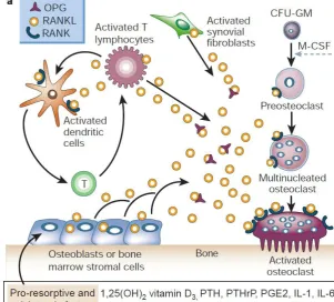
The discovery of the RANKL/RANK/OPG system in the late 1990s greatly improved the understanding of bone remodeling and maintenance of the skeletal structure (BOYCE & XING, 2008; WRIGHT et al., 2009). Three key proteins are responsible for the regulation process of bone resorption: RANK (receptor-activator of nuclear factor kappa beta), its ligand RANKL (receptor-(receptor-activator of nuclear factor kappa beta ligand) and a decoy receptor OPG (osteoprotegerin). This system is controlled by many osteotropic hormones (parathyroid hormone (PTH), 1,25dihydroxyvitaminD3 (Vitamin D3a), prostaglandin E2 (PG E2), estrogen, testosterone and prolactin) and cytokines (inflammatory cytokines e.g. interleukines: IL-1α, IL-1β, IL-6b, IL-11 and IL-17), tumor necrosis factor alpha and beta (TNFα and TNFβ) and transforming growth factor beta (TGF-β)) (WRIGHT et al., 2009). Osteoclasts’ number and activity are closely dependent on the ratio of RANKL/OPG and every change in this ratio will have an effect on bone turnover (BOYCE & XING, 2008). The RANK/RANKL/OPG signaling system is essential for skeletal homeostasis and plays a major role in most animal models of bone diseases characterized by increased resorption (BOYCE & XING, 2008). RANK-RANKL signaling not only activates a variety of downstream signaling pathways required for osteoclast development, but also fine-tunes bone homeostasis both in normal physiology and disease via crosstalk with other signaling pathways. Interestingly all factors that inhibit or enhance bone resorption by osteoclasts also positively or negatively influence RANKL and OPG mRNA levels as well as protein levels (LEIBBRANDT & PENNINGER, 2008).
3.1.2. RANKL
(BOYCE & XING, 2008). Membrane bound RANKL ensures cell-cell contact allowing only microenvironmental function, whereas soluble RANKL can diffuse to the target cells leading to a systemic function. When more RANKL is released by osteoblasts, bone resorption will be increased through osteoclast differentiation, activation and survival (WRIGHT et al., 2009).
3.1.3. RANK
RANK is a type I transmembrane glycoprotein, expressed on the surface of osteoclast precursors, mature osteoclasts, dendritic cells, mammary gland epithelial cells and cancer cells. When RANKL binds to its receptor RANK a signaling cascade is initiated and in this activation process the following factors are involved: cytoplasmic adaptor protein TRAFs, followed by the downstream pathways including NF-kappaB, c-jun N-terminal kinase (JNK) or Src (proto-oncogenic tyrosine kinases) pathways. This leads to the expression of various genes, which facilitate the differentiation of monocytes into osteoclasts and also the activation of mature osteoclasts as well as their enhanced survival (WRIGHT et al., 2009).
3.1.4. OPG
Figure 1: RANKL/RANK/OPG axis. RANKL expression is induced in osteoblasts and bone marrow stromal cells, and subsequently binds to its specific membrane-bound receptor RANK, that promotes osteoclast
differentiation, activation and survival. OPG binds and neutralizes RANKL (BOYLE et al., 2003)
a) Pro-resorptive and calcitropic status
3.2. RANKL/RANK/OPG axis in the state of disease
3.3. sRANKL transgenic mice
The group of Mizuno et al. generated two types of transgenic mice overexpressing soluble RANKL via DNA microinjection. One transgenic line CAG-promoter-sRANKL overexpressed CAG-promoter-sRANKL ubiquitously from an early developmental stage on, the other transgenic mouse line SG2-sRANKL overexpressed sRANKL only in the liver after birth. Unexpectedly, in the CAG-sRANKL line ubiquitous overexpression in the fetal stage resulted in a lethal phenotype. Also in the other line SG2-sRANKL some of the fetuses died at birth, but most of them grew up to adults and were fertile. As conclusion it is impossible to obtain living sRANKL transgenic progeny when the overexpression takes place uncontrolled during fetal development stage. In SG2-sRANKL mice at the age of 7-8 months the bone mineral density of femurs was significantly lower than in control mice. Additionally, the histological analyses revealed that trabecular bone mass was rapidly reduced with aging. Bone strength and stiffness were also markedly decreased. Bone resorption was increased by enhanced osteoclastogenesis and osteoclast activation (MIZUNO et al., 2002). These sRANKL overexpressing mice show a similar osteoporotic phenotype as OPG-deficient mice. The main difference between these both are that OPG-deficient mice have a low body weight and deformed skeletons, even before they are weaned. In contrast sRANKL transgenic mice have a normal body weight through their whole lifetime and there is no bone deformation in infants. Additionally, sRANKL transgenic mice exhibit a milder osteoporotic phenotype and lower serum ALP levels than OPG-deficient mice (BUCAY et al., 1998; MIZUNO et al., 1998; MIZUNO et al., 2002).
4.
Gene regulation by a tetracycline inducible system
4.1. Inducible gene regulation
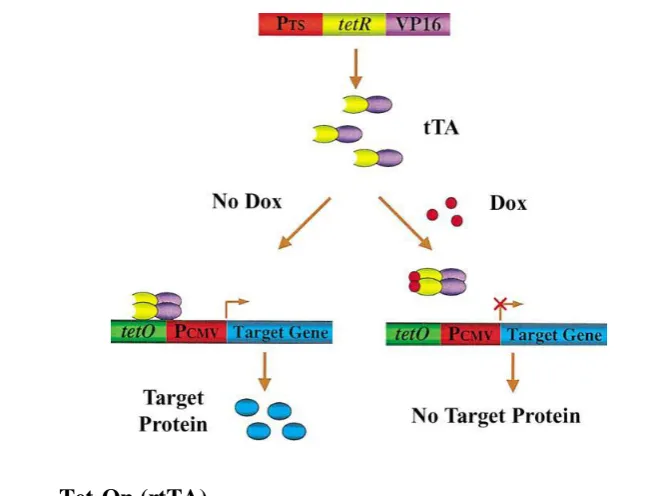
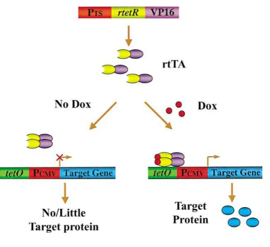
limitations different inducible transgenic modeling systems were established (ZHU et al., 2002). The most prominent and widely-accepted inducible systems so far are based on the tetracycline-controlled transcriptional regulator developed by Gossen and co-workers (GOSSEN & BUJARD, 1992; GOSSEN et al., 1995; STIEGER et al., 2009). To generate credible transgenic animal models to precisely mimic human disease states, it is critical to tightly regulate gene expression in the animals in a conditional manner. The ability to turn gene expression ―on‖ or ―off‖ in restricted cells or tissues at specific time points opens up a new level of research (SUN et al., 2007). Tetracycline (Tet) regulatable systems are based on the E. coli tetracycline resistance operon, which consists of the Tet repressor (TetR) protein and the Tet operator (TetO) DNA sequence (WISSMANN et al., 1986). In the absence of tetracycline or its derivate doxycycline (Dox), the TetR protein gets attached to the TetO DNA sequence, while in the presence of the drug, TetR changes its conformation to detaching from the DNA (ORTH et al., 1998). There are two basic variants of the tetracycline-inducible system: if transgene expression is allowed only in the absence of doxycycline, the system is called Tet-Off, whereas if transgene expression is allowed only in the presence of Dox, the system is called Tet-On (SUN et al., 2007; STIEGER et al., 2009).
4.2. Tet-Off (tTA)
These shortcomings limited the use of the Tet-Off system and leaded to the development of a new tetracycline depend system.
Figure 2: Tetracycline-controlled transcriptional activator (tTA) system: “Tet-Off” (ZHU et al., 2002)
4.3. Tet-On (rtTA)
Figure 3: Reverse tetracycline-controlled transcriptional activator (rtTA) system: “Tet-On” (ZHU et al., 2002)
4.4. Doxycycline
The inducer drug doxycycline, an analogue of tetracycline, is a well-documented antibiotic drug that has been used in the clinics for more than 30 years. Therefore it is considered as a save agent that can be used without major concern. The bioavailability of Dox after oral administration compared to intravenous administration is almost 100% and the serum half-life has been calculated to 14– 22 h. The tissue penetration is excellent and includes the brain. Concentrations are the highest in liver, kidney and digestive tract, as it is eliminated primarily via urine and faeces (AGWUH & MACGOWAN, 2006; STIEGER et al., 2009). In Tet-inducible system transgenic mouse models doxycycline is administrated dissolved in the drinking water at ad libitum supply (KISTNER et al., 1996; RAO & MONKS, 2009). The Tet-based systems are steadily evolving towards an ideal inducible transgenic system, especially because they are coupled with a simple, well-understood, inexpensive, and easy-to-use inducing agent, doxycycline (ZHU et al., 2002).
5.
Techniques of transgenesis in pigs
5.1. DNA Microinjection
cell stage (HOUDEBINE, 2005). This method was first established in the mouse (GORDON & RUDDLE, 1981) before being applied to various other mammalian species including the pig (BREM et al., 1985; HAMMER et al., 1985). Generally the efficiency of DNA microinjection is low. In the mouse the efficiency is 1-3% transgenic animals per microinjected embryos. In the pig the success of obtaining transgenic progeny is even lower, the same as in other animals like rabbits, rats and ruminants (HOUDEBINE, 2005). This inefficiency required microinjection and transfer of thousands of embryos to produce few transgenic offspring. In the end it is very costly to produce one single transgenic animal by DNA microinjection (ROBL et al., 2007). If integration of exogenous DNA occurs after embryonic cleavage begins, mosaic offspring can be obtained. For the species mouse the random integration of microinjected DNA has shown to bear a risk of insertional mutagenesis (RIJKERS et al., 1994). However, for pig, pathological side effects which could putatively be associated with insertional mutagenesis after DNA microinjection have not been reported so far (DEPPENMEIER et al., 2006). Nevertheless, expression levels of the transgene can differ due to position effects and variable numbers of integrated copies. An approach to reduce cost and labor of pronuclear microinjection would be the use of porcine embryos produced by in vitro fertilization (NAGASHIMA et al., 2003). Ovaries collected from slaughtered gilts are a source for oocytes, which undergo in vitro maturation (IVM) and in vitro fertilization (IVF). Resulting embryos would subsequently undergo DNA microinjection before being transferred to recipient sows. In vitro production systems yielding viable pig embryos and healthy piglets have been established (KIKUCHI et al., 2006), but still remain to be optimized (ALMINANA et al., 2008; GIL et al., 2008; KIKUCHI et al., 2008). Despite the overall low efficiency, most of the transgenic pig lines existing so far have been established by pronuclear microinjection technique. However other techniques of transgenesis have gained importance due to their higher efficiency and the potential to introduce targeted modification in the pig genome.
5.2. Sperm mediated gene transfer
Clearly, more work is needed before SMGT is widely applied (ROBL et al., 2007).
5.3. Lentiviral gene transfer
The principle of lentiviral gene transfer is based on infection of porcine zygotes with retroviral vectors carrying transgenes. Lentiviruses belong to the family of retroviruses. Irrespective of cell cycle they can reach the host genome, because they can pass the nuclear membrane. So they are capable of infecting quiescent and embryonic cells (HOUDEBINE, 2005; ROBL et al., 2007), which reduces the formation of mosaics. Based on these advantages, lentiviral vectors are actually considered to be the most efficient method of viral transgenesis (ROBL et al., 2007). The generation of transgenic mice and rats using lentiviral gene transfer was first reported in 2002 (LOIS et al., 2002; PFEIFER et al., 2002). In the pig, lentiviral vectors based on the human immunodeficiency virus-1 (HIV-1) and on the equine infectious anemia virus (EIAV) were used for gene transfer. The recombinant lentiviruses were injected under the zona pellucida of zygotes, which were transferred to synchronized recipients later on. The overall efficiency of generating transgenic pigs by lentiviral gene transfer ranged at 13% (transgenic offspring per infected and transferred embryo) (HOFMANN et al., 2003; WHITELAW et al., 2004). A high proportion of transgenic G0 animals (94%) showed transgene expression, also over a long time (6 months) (PFEIFER et al., 2004). However, lentiviral vectors are limited to a DNA uptake only between 8-10 kb of foreign DNA, and they lead to varying gene expression in the transgenic pigs (HOUDEBINE, 2005; ROBL et al., 2007) and their progenies. The Hofmann group reported about low expression levels and hypermethylation in one third of G1 offspring, due to multiple integration sites in the founder animals, which segregated in the following generations (HOFMANN et al., 2006).
5.4. Somatic cell nuclear transfer (SCNT)
et al., 2007). Future research should focus on improving the health and production efficiency of somatic cell porcine clones (MATSUNARI & NAGASHIMA, 2009).
6.
Examples of genetically modified pigs
6.1. Swine in biomedical research
Transgenic pigs are generated for biomedical research purposes, i.e. as animal model in medical research, as donor animal for xenotransplantation, as bioreactors for gene farming and as highly efficient productive livestock in agriculture. Among various possibilities, the established somatic cell nuclear transfer system with genetically engineered donor cells is an efficient and reliable approach to produce transgenic pigs (VAJTA et al., 2007). Various human disease models and donor pigs for xenotransplantation have been produced via SCNT.
6.2. Transgenic human disease models
6.2.1. Human heart disease model
Endothelial cell nitric oxide synthase (eNOS) over-expressing piglets were generated by nuclear transfer to study the role of nitric oxide metabolism in cardiac and skeletal muscle. The final goal was to generate large animal model for human heart disease (HAO et al., 2006). Nitric oxide (NO) is a messenger molecule, which modulates vascular function, structure and homeostasis. The enzyme endothelial cell nitric oxide synthase (eNOS) releases NO into the blood stream, where it plays an important role in the metabolism of cardiac and skeletal muscle. The created pigs carried the Tie2-eNOS transgene and also showed expression of the Tie2-eNOS in the endothelial cells of placental vasculature simultaneously to the endogenous eNOS (HAO et al., 2006).
6.2.2. Cystic fibrosis model
al., 2007). Due to many anatomical and physiological similarities, pigs were considered as a more suitable animal model. CFTR knockout pigs developed meconium ileus, exocrine pancreatic destruction, and focal biliary cirrhosis, replicating abnormalities seen in newborn patients with CF (ROGERS et al., 2008). Additional the CF pigs showed few months after birth already typical lung symptoms like airway inflammation, mucus accumulation and infection (STOLTZ et al., 2010).
6.2.3. Alzheimer´s disease model
Disease-causing mutations for Alzheimer’s disease (AD) were identified in the amyloid precursor protein gene (APP) and the presenilin 1 and presenilin 2 genes (PSEN1 and PSEN2). These mutations are associated with accumulation of the Aß peptide in the brain. Transgenic pigs were produced expressing the neuronal variant of the human amyloid precursor protein based on the Swedish APP mutation (KRAGH et al., 2009). APP695sw transgene expression, including protein, was observed in brain tissue and not in heart or liver tissues. Mutant protein accumulation will approximately take 1-2 years, and it’s expected that after this time the transgenic pigs show the desired phenotype.
6.2.4. Diabetes mellitus type 3 model
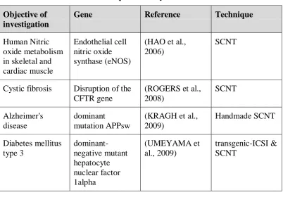
Table 2: Human disease models produced by SCNT
Objective of investigation
Gene Reference Technique
Human Nitric oxide metabolism in skeletal and cardiac muscle
Endothelial cell nitric oxide synthase (eNOS)
(HAO et al., 2006)
SCNT
Cystic fibrosis Disruption of the CFTR gene
(ROGERS et al., 2008) SCNT Alzheimer's disease dominant mutation APPsw
(KRAGH et al., 2009) Handmade SCNT Diabetes mellitus type 3 dominant-negative mutant hepatocyte nuclear factor 1alpha (UMEYAMA et al., 2009) transgenic-ICSI & SCNT 6.3. Xenotransplantation
homozygous Knockout of α-1,3-galactosyltransferase (αGalT) piglets (DAI et al., 2002; LAI et al., 2002; PHELPS et al., 2003; RAMSOONDAR et al., 2003; WATT et al., 2006; FUJIMURA et al., 2008), from that day on it was proven that gene targeting in the pig is possible (KLYMIUK et al., 2010). Development of αGal deficient pigs has reduced or eliminated the significance of αGal antigen in xenograft rejection. When αGal deficient pig organs are used for Pig-to-Primate Cardiac Xenotransplantation, xenograft rejection remains associated with antibody deposition, variable complement activation and microvascular thrombosis. Nevertheless organs from αGal knock out pigs are widely considered to be central for clinical xenotransplantation of solid organs (BYRNE et al., 2008).
7.
Embryo transfer (ET) in the pig
7.1. Factors influencing ET success
7.2. Pregnancy rates
First-estrus gilts are contraindicated for donating embryos, but they are quite acceptable as recipients. No difference in pregnancy rate (67.5% vs. 60%) or embryonic survival (69% vs. 75%) was reported in first-estrus versus third-estrus recipient gilts, respectively (ARCHIBONG et al., 1992). No evidence of uterine crowding adversely affecting litter size at Day 25 of gestation in females having 7 or fewer embryos present per uterine horn has been observed, suggesting that a minimum of 14 embryos should be transferred to each recipient (DZIUK, 1968). Pregnancy rates of 71% and 100% and embryo survival rates of 57% and 68% in gilts receiving 12 or 24 embryos, respectively, were reported (POPE et al., 1972). It was also reported that at least four viable embryos were necessary in pigs to maintain pregnancy in the early phases of embryo maternal communication (POLGE et al., 1966). Average pregnancy rates of 60% and embryonic survival rates (in pregnant recipients) of 60% are standard. So from all transferred embryos 35-40% survive and will result in living piglets. In all these experiments in vivo derived embryos were used and the success rate surely is lower, when dealing with manipulated embryos (YOUNGS, 2001).
7.3. Endoscopic embryo transfer
III.
M
ATERIALS AND METHODS1.
Equipment and expendable items
1.1. In vitro works
Petri dishes: 35x10 mm, Becton Dickinson Labware 50x9 mm, Becton Dickinson Labware 60x15 mm, Nunc® Brand Products
Glassware: Schott, Brand, Wertheim
Pipettes: Eppendorf, Hamburg
Pipette filter tips: Eppendorf, Hamburg Neubauer counting chamber: Brand, Wertheim Centrifuge tubes: 15 ml, Greiner
50 ml, Falcon®, Becton Dickinson Labware Glass cover slips: Marienfeld, Lauda-Koenigshofen
Reaction tubes: 0.5 ml / 1.5 ml / 2 ml, Eppendorf Sterile benches: Lamin Air®, HB 2472, Heraeus
KR-130 BW, Kojair
Sterile filters: Millipore Express®, 0.22μm, Millex® GP Centrifuges: Biofuge pico, Heraeus
Rotanta 96, Hettich Zentrifugen
Incubator: Type B 5060, Heraeus
Model 500M, MMM Group
Microscopes: Leitz Periplan, Leica
Micromanipulator: Eclipse TE 2000-U, Nikon Eclipse TE 300, Nikon
Warming plates: HT 200, Minitube
Electro activation: Multiporator, Eppendorf Electro cell fusion: Model LF 101, Nepa Gene
Cell transfection: Nucleofector® II, Amaxa biosystems
1.2. Embryo transfer
Endoscopic instruments: Karl Storz Endoskope
Ultrasonic device: Echo camera, SSD-500, Aloka
2.
Used media and stock solutions
2.1. Stock solutions
PBS (Phosphate-buffered saline)
8.00 g NaCl (Sigma)
0.20 g KCl (Sigma)
1.15 g Na2HPO4 x 2 H2O (Fluka, Neu-Ulm) 0.20 g KH2PO4 (Merck, Darmstadt)
0.10 g CaCl2 (Sigma)
0.10 g MgCl2 x 6 H2O (Sigma) 1.0 l Milli-Q water
sterile filtered, stored at room temperature
PBS− (Phosphate-buffered saline without calcium and magnesium) 8.00 g NaCl
0.20 g KCl
1.0 l Milli-Q water
sterile filtered, stored at room temperature
PBS P/S (Phosphate-buffered saline with 2 % Penicillin/Streptomycin) 98 ml PBS
2 ml Pen/Strep stock solution freshly prepared before use Pen/Strep stock solution
0.65 g Penicillin (Seromed, Berlin) 1.33 g Streptomycin (Seromed)
100 ml Milli-Q water
(contains 100 U/ml penicillin and 100 µg/ml active streptomycin) sterile filtered, stored at -20°C
2.2. Cell culture
Culture media for porcine fibroblasts and kidney cells:
DMEM (high glucose, without sodium pyruvate) (GIBCO)
1% non-essential amino acids (GIBCO)
1% sodium pyruvate (GIBCO)
0.1 mM Mercaptoethanol (7 µl/10 ml PBS 1%) (Sigma) 1% L-Glutamin 200 mM + 1% Pen/Strep (PAA)
10-15% FKS (GIBCO)
Serum Starvation media
DMEM (high glucose, without sodium pyruvate) (GIBCO)
1% non-essential amino acids (GIBCO)
1% sodium pyruvate (GIBCO)
0.5 % FKS (GIBCO) Trypsin/EDTA-solution for cell culture
Trypsin 0.25% (Difco)
EDTA 0.02% (Sigma)
PBS- (without calcium and magnesium) sterile filtered, stored at -20°C
G418 selection Media
Porcine fibroblast culture medium (see above)
G418 (Geneticin) (GIBCO)
Porcine kidney cells: 1.2 mg/ml
Porcine fetal fibroblast cells: 0.6 mg/ml
2.3. Nuclear transfer
NCSU-23 Stock A
NaCl 6.355 g (Sigma)
KCl 0.356 g (Sigma)
MgSO4 x 7 H2O 0.293 g (Sigma)
KH2PO4 0.162 g (Sigma)
Milli-Q water restocked up to 100 ml sterile filtered, stored at 4°C
NCSU-23 Stock B
CaCl2 x 2 H2O 0.5 g (Sigma) Milli-Q water restocked up to 20 ml
Ready to use NSCU-23
NaHCO3 0.421 g (Sigma)
Glucose 0.2 g (Sigma)
PenicillinG 0.013 g (Sigma)
Streptomycin 0.010 g (Sigma)
Taurine 0.175 g (Sigma)
Stock A 20 ml
Stock B 2 ml
Milli-Q water restocked up to 200 ml sterile filtered, stored at 4°C
Hepes-TaLP-PVP Stock A
NaCl 6.66 g (Sigma)
KCl 0.24 g (Sigma)
MgCl2 x 6 H2O 0.1 g (Sigma)
NaH2PO4 0.042 g (Sigma)
Na Lactate (60%) 1.85 ml (Sigma)
Phenol red 2 ml (Sigma)
Milli-Q water restocked up to 100 ml sterile filtered, stored at 4°C
Hepes-TaLP-PVP Stock B
CaCl2 x 2 H2O 0.58 g (Sigma) Milli-Q water restocked up to 20 ml
Hepes-TaLP-PVP Stock C
Hepes 2.4 g (Sigma)
Milli-Q water restocked up to 100 ml sterile filtered, stored at 4°C
Ready to use Hepes-TaLP-PVP
Glucose 0.45 g (Sigma)
Sorbitol 4 g (Sigma)
PVP 1.5 g (Sigma)
Streptomycin 0.025 g (Sigma)
PenicillinG 0.033 g (Sigma)
NaHCO3 0.085 g (Sigma)
Stock A 50 ml
Stock B 5 ml
Stock C 50 ml
Milli-Q water restocked up to 500 ml sterile filtered, stored at 4°C
Porcine zygote medium
PZM-5 Institute for the functional Peptide, Yamagata, Japan
3.
Establishment of transgenic cell lines
3.1. Cells
3.2. Transfection of cells
To establish a stable cell line expressing the desired construct plus antibiotic resistance, wild type fetal fibroblasts were electroporated by the Nucleofector® system. The foreign DNA was introduced into the cell by an electro current. Cells were harvested after passage three and counted in a Neubauer counting chamber. 5 μl DNA-solution (3.3 μg) containing the chosen gene construct and 100μl human dermal fibroblast Nucleofector® solution were mixed with 5 x 10⁵ PFF cells. Then electroporation with the program U12 took place. After this treatment cells were pipetted with an Amaxa plastic pipette in 1 ml medium to a cell culture dish. The next day media was exchanged.
3.3. Mass cell selection
Three days after electroporation cells were confluent (around 90%) and were harvested. For selection of cells, they were seated with medium containing Geneticin (G418; 1.0 mg/ml) on a new cell culture dish. Every two days selection medium was changed. After one week of culture all cells had died, which did not integrate the antibiotic resistance gene; these cells were floating in the supernatant. Successfully transfected cells were able to survive selection and formed colonies. The cells were cultured until 90% confluence and then harvested and cryopreserved until SCNT was performed.
4.
Vector design
4.1. Lentiviral vectors
Figure 4: Overview of lentiviral vectors
1) pLenti6-CMV-pRANKL (Bla+)
2) pLenti6-CMV-TetON (Bla+)
3) pLenti6-CMV-TetON-ΔBla (Bla-)
4) pLenti6-CMV-TetON-TREtight-pRANKL (Bla+)
5) CMV-TetOn-TREtight-pRANKL (Neo+)
These figures were a donation of PD Dr. med Wolfgang Böcker. BSD: blasticidin-S deaminase (Basticidin
resistance gene)
LTR: long terminal repeat Pcmv: cytomegalovirus promoter pRANKL: porcine receptor-activator of nuclear factor kappa beta ligand
pTRE tight: Tet-Advanced transactivator response element promoter (TARE) RRE: Rev-responsive element rtTA: reverse tetracycline-controlled transcriptional activator (Tet-On) NEO: neomycin resistance
Neomycin resistance gene. These assembly changes were applied to adapt the lentiviral vector to the Amaxa Nucleofector system. With the donated Tet-On-RANKL-Neo (5) vector porcine fetal fibroblast cells were transfected in our laboratory.
4.2. Conventional vectors
The vectors 6 and 7 were designed and constructed by Dr. rer. nat. Nikolai Klymiuk in our institute. The Tet-On and RANKL gene were integrated under the control of the CAG and the TARE (pTRE tight) promoter. Neomycin and blasticidin antibiotic resistance and the polyadenylation signal of the bGH gene was added. The poly adenosine tail contributes to the nuclear export, translation and stability of mRNA. The antibiotic resistance is needed for further selection of cells with stable integration of the desired genes. To avoid possible negative side effects of antibiotic resistance genes, loxP sites were introduced. With the enzyme Cre recombinase it is feasible to cut out specifically the loxP sites and the gene in between is deleted. Both gene constructs were used for cell transfection in our laboratory.
Figure 5: Overview of conventional vectors
6) CAG-rtTA: lox2 neo® – CAGpr – rtTA ORF – bGHpA
7) TARE-RANKL: TAREpr – RANKL ORF – bGHpA – lox2 bla®
These figures were a donation of Dr. rer. nat. Nikolai Klymiuk. bGH: bovine growth hormone gene
bla: blasticidin resistance
CAG: CMV early enhancer / chicken beta actin promoter
lox: locus of X-over P1 (lox P site) neo: neomycin resistance
ORF: open reading frame
pA: (PolyA) polyadenylation signal
PR: promoter
RANKL: porcine receptor-activator of nuclear factor kappa beta ligand
rtTA: reverse tetracycline-controlled transcriptional activator (Tet-On)
5.
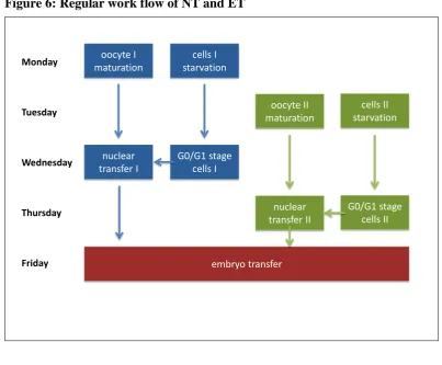
Procedure of somatic cell nuclear transfer
Per week, two nuclear transfer experiments were scheduled, one performed on Wednesday and the other one on Thursday. Oocyte maturation and cell preparation must be exactly timed to fit in the procedure. All produced NT embryos were equally mixed and transferred to one or two recipients on Friday. Figure 6: Regular work flow of NT and ET
oocyte I maturation
cells I starvation
nuclear transfer II
G0/G1 stage cells II
Monday
Tuesday
Wednesday
Thursday
Friday
oocyte II maturation
cells II starvation
nuclear transfer I
G0/G1 stage cells I
embryo transfer
5.1. In vitro maturation of oocytes
5.1.1. Ovary collection
Sixty to 90 ovaries per day were collected at a local abattoir and transported to the laboratory in phosphate buffered saline (PBS) containing 75 µg/ml potassium penicillin G, 50 µg/ml streptomycin sulfate and 0.1% (w/v) polyvinylalcohol (PVA) in a warming box under the stable temperature of 36°C to 37°C degrees.
5.1.2. Oocyte collection
Cumulus-oocyte complexes (COCs) were collected by aspiration with a needle and syringe from ovarian antral follicles of 3.0-6.0 mm in diameter.
5.1.3. Selection of oocytes
albumin lactate pyruvate (TALP) solution. Only COCs displaying over 3 layers of compacted cumulus cells with even cytoplasm were selected and used for further experiments. The remaining poor quality and altered COCs were discarded.
5.1.4. Oocyte maturation
120 to 180 first grade COCs per day were subsequently cultured in North Carolina State University medium-23 (NCSU-23) supplemented with 0.6 mM cysteine, 10 ng/ml epidermal growth factor (EGF), 10% (v/v) porcine follicular fluid, 75 µg/ml potassium penicillin G, 50 µg/ml streptomycin sulfate, 10 IU/ml equine chorionic gonadotropin (eCG; Intergonan, Intervet, Germany) and 10 IU/ml human chorionic gonadotropin (hCG; Ovogest, Intervet, Germany). The first 22 h they were cultured with the hormones eCG and hCG, and then for 20 h without these hormones in a humidified atmosphere of 5% CO2 and 95% air at 38.5 °C.
5.1.5. Denudation of matured oocytes
After 42 hours culture, the in vitro matured (IVM) oocytes with expanded cumulus cells were treated with 1 mg/ml hyaluronidase dissolved in TALP medium supplemented with 10 mM Hepes and 0.3% (w/v) polyvinylpyrrolidone (PVP) (Hepes-TALP-PVP) and were denuded of cumulus cells by gentle pipetting. Oocytes displaying evenly granulated ooplasm and extrusion of the first polar body were considered as fully matured and selected for the experiments.
5.2. Nuclear transfer in vivo experiment
Somatic cell nuclear transfer (SCNT) was performed using IVM oocytes as recipient cytoplasts.
5.2.1. Enucleation
Figure 7: Enucleation
a) Penetration of the zona
pelucida
b) Soakage of the first polar body and the adjacent cytoplasm
c) Enucleated oocyte
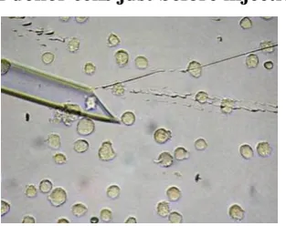
5.2.2. Donor cell preparation
Cell cycle of donor cells was synchronized in the G0/G1 stage by serum starvation starting 48h before nuclear transfer. On the day of nuclear transfer, donor cells were detached from culture dish and singularized by trypsinization. Figure 8: Appearance of donor cells just before injection
5.2.3. Donor cell injection
For cell insertion, round, small donor cells, with a smooth surface and a regular looking were selected. One single donor cell per enucleated oocyte was injected through the hole in the zona pelucida, originated by enucleation, into the perivitelline space.
Figure 9: Donor cell insertion
a) Introduction of the pipette
through the enucleation hole
b) Injection of one donor cell
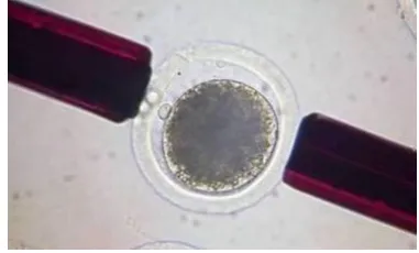
5.2.4. Fusion
Donor cell-oocyte complexes were placed in a droplet of 280 mM mannitol solution (pH 7.2) containing 0.15 mM MgSO4, 0.01% (w/v) PVA, and 0.5 mM Hepes and held between 2 electrode needles. Membrane fusion was induced using an Electro cell fusion LF101 (NEPA GENE Co. Ltd.) by applying a single direct current (DC) pulse (200 V/mm, 20 µs ×1) and a pre- and post-pulse alternating current (AC) field of 5 V, 1 MHz for 5 s, respectively. Reconstructed embryos were cultured in NCSU-23 for 0.5-1h, followed by electrical activation.
Figure 10: Reconstructed NT embryo in between two electrodes of the fusion aperture
5.2.5. Activation
Reconstructed embryos were washed twice in an activation solution consisting of 0.3 M mannitol, 50 µM CaCl2, 100 µM MgSO4 and 0.01% PVA (300 mOsm), then placed between 2 wire electrodes (1 mm apart) of a fusion chamber slide and overlaid with activation solution. A single DC pulse of 150 V/mm was applied for 100 µs. To suppress extrusion of the pseudo-second polar body, activated oocytes were subsequently cultured in a medium containing 5 µg/ml cytochalasin B (CB) for 3 h.
5.2.6. In vitro culture of reconstructed embryos
The reconstructed embryos were stored for one or two days, until embryo transfer took place, in 20-µl droplets of porcine zygote medium (PZM-5) under paraffin oil in a plastic Petri dish maintained under a humidified atmosphere of 5% CO2, 5% O2, 90% N2 at 38.5 °C.
5.3. Nuclear transfer in vitro experiment
enucleated oocytes were rejected. Cleavage and blastocyst formation of reconstructed embryos were monitored during culture for 7 days. In vitro culture of embryos was performed in 20-µl droplets of porcine zygote medium (PZM-5) under paraffin oil in a plastic Petri dish maintained under a humidified atmosphere of 5% CO2, 5% O2, 90% N2 at 38.5 °C.
5.4. Embryo transfer
Six to seven months old prepuberal gilts (hybrids of ―Schwäbisch-Hällisches Landschwein‖ and ―Deutsche Landrasse‖) were used as recipients.
5.4.1. Estrus synchronization
Estrus synchronization was conducted by oral administration of altrenogest (Regumate, Serumwerk Bernburg, Germany) over a 15 day period, followed by intramuscular injection of 750 IU eCG, (Intergonan, Intervet, Germany) which has a follicle stimulating effect, 24 hours after last gestagen administration. Ovulation was induced 3 days later by intramuscular injection of 750 IU hCG (Ovogest, Intervet, Germany).
5.4.2. Endoscopic embryo transfer
Endoscopic embryo transfer was performed two days after Ovogest treatment. Recipients were anesthesized with a combination of 1.2 ml per 10 kg ketamine hydrochloride (Ursotamin, Serumwerk Bernburg, Germany) and 0.5 ml per 10 kg xylazine (Xylazin 2%, WDT, Germany) intravenously.
Figure 11: Endoscopic embryo transfer
a) Prepared recipient b) operation field with the endoscopic optics (left), grasper (middle) and trocar (right)
Recipients were brought into 45° dorsal recumbency, and 53 to 147 embryos were transferred laparoscopically in to the right oviduct.
Figure 12: Introduction of the catheter into the oviduct
a) The right oviduct with inserted trocar b) expansion of the right oviduct through the introduced catheter
6.
Pregnancy control and birth
6.1. Pregnancy control
Pregnancy status was confirmed by ultrasound examination. These examinations were performed at least three times per recipient, the first check (TU1) always was scheduled for day 28, the second check (TU2) between day 50 and 70 and the third check (TU3) between day 90 and 110 after embryo transfer. Sows were classified as pregnant, when liquid filled chambered vesicles in the uterus were visible (TU1). At the later stages of pregnancy (TU2 + TU3) also fetuses were detected and evaluated.
6.2. Induction of labor
If spontaneous birth did not occur until day 117, labor was hormonally induced by intra muscular application of 0.7 ml Cloprostenol (Estrumate, Intervet, Germany).
7.
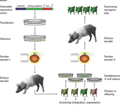
Overview: transgenic pig production via SCNT
Figure 13: Efficient production of transgenic pigs via SCNT (AIGNER et al., 2010)
8.
In vivo doxycycline stimulation
RANKL overexpression was in vivo stimulated by oral administration of Pulmodox® (Virbac Tierarzneimittel GmbH, Bad Oldesloe), with the active ingredient doxycyclinhyclat. Pigs were treated with doxycycline at a dosage of 40 mg/kg/day continuously over 6 days. Plasma blood samples were obtained on day 0 (before first doxycycline application), on day 3 and day 6 of doxycycline treatment. To distinguish the different RANKL plasma levels, ELISAs were performed by Tamara Radic at ExperiMed laboratory.
9.
Statistical analysis
IV.
R
ESULTS1.
Assessment of SCNT and ET
1.1. Overview of the years 2006 to 2009
Table 3: In vivo SCNT work of the years 2006 until 2009
Year 2006 2007 2008 2009 Total
Number of performed
embryo transfers 11 13 53 68 145
Total number of cultured
oocytes 1477 1933 13022 18006 34438
Average maturation rate in % 85 87 79 76 82
Average fusion rate in % 78 86 89 87 85
Average cleavage rate of NT
embryos in % 78 80 88 90 84
Total number of NT embryos
transferred 896 1121 4885 5978 12880
Average number of
transferred NT embryos per recipient
81 86 92 88 87
Pregnancy rate in % 64 31 60 54 52
Abortion rate in % 57 50 28 30 41
Number of birth 2 1 19 19 41
Number of pregnancy
stopped 1 1 4 7 13
Total number of piglets /
fetuses 13 10 100 112 235
Average number of piglets /
fetuses per recipient 4.33 5 5.26 4.31 4.73
Total number of stillborn or
degenerated piglets / fetuses 4 2 20 15 41
Average number of stillborn or degenerated piglets / fetuses per recipient
1.33 1 1.05 0.58 0.99
Cloning efficiency in % (offspring per transferred NT embryos)
1.2. Outcome of in vivo SCNT procedure
Of all 169 transgenic SCNT pigs born until June 2010, 37% were healthy and showed a normal development, the remaining 63% were lost. These losses can be divided in different groups, like still born, early neonatal death, killed by mother and lethal disease. The still born piglets died at different stages of pregnancy, but mainly they reached the last trimester. Some of the stillborn piglets showed abnormalities, but the majority appeared to have a normal development until shortly before birth.
Table 4: Upgrowth of SCNT piglets
Condition of transgenic SCNT
piglet
Overall number (%)
Cases and explanation
Healthy and normal development
63 (37) - Transgenic SCNT pigs up to three years of age until now
- Produced healthy and transgenic progeny
Still born 39 (23) - Full grown and fully developed - Mummification
- Abnormalities
Early neonatal death 52 (30) - Severe underweight (< 900g) - Weakness, low viability
- Abnormalities: Oversize tongue, Cleft palates, Atresia ani
Killed by mother 11 (7) - Prolonged and laborious birth - Nervous sow
Lethal disease 4 (2) - Malignant hyperthermia syndrome - Bacterial meningitis