of hematopoietic stem cell function and B-cell development
Thesis by Arnav Mehta
In Partial Fulfillment of the Requirements for the degree of
Doctor of Philosophy
CALIFORNIA INSTITUTE OF TECHNOLOGY Pasadena, California
© 2015
clever problem solver, and most importantly, a creative scientist. In addition, I thank
Jimmy Zhao for being a wonderful mentor and showing me the ropes on how to study HSC and microRNA biology. Thank you for always looking out for me, Jimmy, and for guiding me through the physician-scientist career path. I thank Devdoot Majumdar for many wonderful explorations into B cell biology and long noncoding RNAs, and for always being the first one to offer academic support when I’ve needed it. Finally, I thank the rest of the Baltimore lab for making my experience as wonderful as it has been!
Abstract
MicroRNAs are a class of small non-coding RNAs that negatively regulate gene
expression. Several microRNAs have been implicated in altering hematopoietic cell fate decisions. Importantly, deregulation of many microRNAs can lead to deleterious
Figure S4: Bone marrow and peripheral blood from MG-125b animals early post-reconstitution display hematopoietic
reconfiguration ... 173
Figure S5: Inhibiting miR-125b function decreases hematopoietic output ... 174
Figure S6: MiR-125b represses Lin28 expression in mouse and human hematopoietic cells ... 175
Figure S7: Lin28A and Lin28B expression in miR-125b over- expressing leukemic samples ... 176
Figure S8: Lin28 over-expression inhibits hematopoiesis ... 177
Chapter 5: Conclusions and Future Directions ... 178
A putative role of microRNAs in maintaining HSC function with age ... 179
Uncovering potential roles of miR-132 in normal and malignant B cell development ... 179
A proposal to better understand HSC aging with age at single-cell resolution ... 180
Significance ... 180
Innovation ... 181
Approach ... 182
C h a p t e r 1
Overview of thesis
In this thesis we explore the role of a class of noncoding RNAs, called microRNAs, in
fine-tuning hematopoietic cell fate decisions and function. MicroRNAs are key
post-transcriptional regulators of gene-expression and we seek to understand how these
microRNAs control the balance between normal and pathological hematopoiesis with age.
Aging of the hematopoietic system leads to an increased incidence of several hematopoietic
diseases, including cancer, autoimmune function, and a general failure to combat
infections. We uncover the role of two microRNAs that, when altered, severely affect stem
cell and B cell development, function, and survival in the immune system.
The first chapter will present background information on the normal function of
hematopoietic stem cells and B cells. We then delve into how aging may lead to
pathological hematopoiesis and the development of immune cell cancers. Next, we explore
microRNA biogenesis and the contribution of microRNAs to hematopoietic cell fate
decisions. In chapter 2, we describe the role of a previously unappreciated microRNA
cluster, miR-212/132, in hematopoietic stem cells. We find that miR-212/132 buffers the
expression of its target Foxo3 with age, and in doing so allows these stem cells to maintain
a critical balance between self-renewal, differentiation, and survival. When altered,
miR-212/132 can lead to poor stem cell function with age, and possibly to the onset of
pathological consequences such as anemia and cancer. In chapter 3, we continue the
exploration of miR-212/132 in the aging hematopoietic system by elucidating its role in B
mIR-212/132, the transcription factor Sox4, and demonstrate that when altered this microRNA
cluster severally inhibits B cell development and survival. We next take advantage of this
role of miR-212/132 in B cell development to alter the progression of B cell cancer, thus
revealing a potential therapeutic application of this microRNA. In chapter 4, we continue to
explore the role of microRNAs with potential therapeutic applications in blood cancers by
uncovering a mechanism by which miR-125b, another regulator of HSC function, causes
myeloid leukemia and inhibits B cell development by targeting the pluripotency factor
Lin28a. We consider the future directions and potential implications of this work in chapter
5, and discuss ongoing work to investigate the heterogeneous nature of young and aged
HSCs in order to understand the mechanisms underlying age-associated hematopoietic
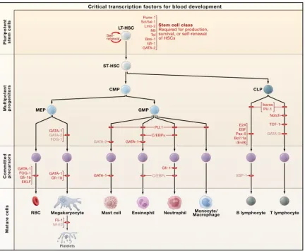
Overview of hematopoiesis
Normal functions of the hematopoietic system
We begin this introduction with an overview of normal hematopoietic function. The
hematopoietic system is remarkable in its ability to produce millions of cells every second
(Orkin and Zon, 2008). Each cell originates from a single hematopoietic stem cell (HSC)
that has the unique ability to self-renew (create identical copies of itself) and to
differentiate into more committed progenitors that lose their ability to self-renew but
eventually lead to all other cell types in the immune system (Figure 1) (Orkin and Zon,
2008). The goal of the hematopoietic system is to produce a balanced output of functional
red blood cells, myeloid cells (such as macrophages and granulocytes, which largely
govern innate immune function) and lymphoid cells such as B cells and T cells, which are
primarily responsible for adaptive immune function (Orkin and Zon, 2008). As such,
lineage commitment and cell fate in this system is governed by a complex set of gene
regulatory pathways, epigenetic changes, and post-transcriptional modifications
(Cabezas-Wallscheid et al., 2014; Laurenti et al., 2013; O'Connell et al., 2010b; Sun et al., 2014). The
developmental intermediates, or hematopoietic progenitors, that lead to each of these cell
types are being actively explored. While several lineage-committed progenitors such as the
common myeloid progenitor (CMP) and common lymphoid progenitor (CLP) have been
identified, it is becoming extremely clear that the hematopoietic tree is intricate and
complex, with several overlapping intermediates that maintain some potential to produce
more diverse cell types than originally anticipated (Orkin and Zon, 2008). We focus this
review on two particular cells in the hematopoietic stem cell relevant to the work discussed
later. These are the hematopoietic stem cell and the B cell.
Hematopoietic stem cells
The origin of all blood cells in the body is the hematopoietic stem cell (HSC) (Orkin and
Zon, 2008). HSCs must maintain an intricate balance between self-renewal and
differentiation throughout the course of our life (Morrison and Weissman, 1994; Rossi et
al., 2012). As such, many different genes, epigenetic modifications, and other intrinsic
factors regulate them (Cabezas-Wallscheid et al., 2014; Sun et al., 2014). Importantly,
HSCs reside in the bone marrow niche and are also strongly influenced by extrinsic signals,
increasingly apparent that HSCs might respond directly to foreign material, such as
pathogens, that enter the body (Nagai et al., 2006; Zhao et al., 2014).
At the peak of the cellular hierarchy is what is known as the long-term HSC (LT-HSCs).
These LT-HSCs are essential for maintaining a life-long supply of blood. Immediately
downstream of these cells are slightly more committed progenitors, known as short-term
HSCs (ST-HSCs) and multipotent progenitors (MPPs). ST-HSCs and MPPs have lost the
ability to self-renew; however, they maintain the ability to differentiate into all cell types.
Downstream of the MPPs are several lineage-restricted progenitors, which then give rise to
mature cells (Figure 1) (Orkin and Zon, 2008).
The majority of HSCs reside in the bone marrow in a quiescent state. It is believed that it is
these HSCs that have the best long-term reconstitution potential upon bone marrow
transplant (Morrison and Weissman, 1994; Orkin and Zon, 2008; Rossi et al., 2012). HSCs
that become activated cycle more rapidly, and this active proliferation is closely related to
differentiation (Morrison and Weissman, 1994; Rossi et al., 2012). Furthermore, many
HSCs periodically circulate in the peripheral blood and hone back to the bone marrow
eventually. Several genes regulate HSC cycling, including genes that control cell cycle
check-points such as p21, p27, and p57, as well as several other factors (Figure 2)
(Morrison and Weissman, 1994; Rossi et al., 2012). Other factors that alter HSC function
include those that regulate the ability of HSCs to stay in the bone marrow niche, such as
Mmp9, those that affect survival, such as Foxo3, and others that alter signaling in HSCs,
such as the Wnt pathway (Rossi et al., 2012).
B cells
B cells are the primary producers of immunoglobulin and play a critical role in adaptive
immunity (Mauri and Bosma, 2012). The maintenance of proper B cell output from early
hematopoietic progenitors, along with the production of an appropriate antibody repertoire,
is critical in maintaining the balance between normal immune function and diseases such as
many different transcription factors in a complex gene regulatory network that controls
lineage specification and commitment (Mandel and Grosschedl, 2010; Matthias and
Rolink, 2005; Nutt and Kee, 2007).
Antigen-independent B cell development begins with the differentiation of lymphoid
primed multipotent progenitors (LMPPs) to common lymphoid progenitors (CLPs), a
process driven by the expression of PU.1and Ikaros (Matthias and Rolink, 2005; Nutt and
Kee, 2007), both of which may play a role in regulating Flt3 and IL-7R expression
(DeKoter et al., 2002; Yoshida et al., 2006). These early progenitors also express Rag1 and
Rag2, and thus begin the process of rearrangement of the immunoglobulin heavy chain
(IgH) locus (Igarashi et al., 2002). Lineage specification to the next stages of B cell
development, the pre-pro-B cell and pro-B cell, involves the upregulation of several genes
controlled by E2A and Ebf1 (O'Riordan and Grosschedl, 1999), including Pax5 (Cobaleda
et al., 2007). Pax5 is essential for B cell lineage commitment, as it represses genes that are
inappropriate for B cell development (Souabni et al., 2002). The transition to pre-B cells,
the stage at which immunoglobulin light chain (IgL) rearrangement begins, and immature
B cells, involves many factors including Sox4 (Sun et al., 2013), which has also been
implicated in regulating the expression of the Rag genes (Mallampati et al., 2014).
The aging hematopoietic system
Aged HSCs are characterized by increased self-renewal potential, loss of long-term
reconstitution capability, myeloid-biased differentiation, and a change in niche localization.
HSCs with a poor ability to hone to the bone marrow niche (Geiger et al., 2013). These
aged HSCs also develop a requirement for basal autophagy for survival, because replication
stress and the accumulation of reactive oxygen species have harmful consequences on HSC
numbers and function with age (Flach et al., 2014; Tothova et al., 2007). The loss of critical
autophagic factors is often associated with altered cell cycling of HSCs, and leads to
apoptosis and a rapid loss of HSC numbers in aged mice (Miyamoto et al., 2007;
Rubinsztein et al., 2011; Warr et al., 2013). A critical balance between cell cycling and
differentiation, and survival of aged HSCs must therefore be established to maintain normal
hematopoietic output.
Hematopoietic malignancies
Deregulation of hematopoietic stem cell function or immune developmental processes can
have deleterious consequences, including the development of leukemias and lymphomas
(O'Connell et al., 2010b). MiR-125b is up-regulated in a range of human leukemias,
including acute myeloid leukemia (AML) (Bousquet et al., 2008; Enomoto et al., 2011),
chronic myeloid leukemia (CML) (Enomoto et al., 2011), acute megakaryocytic leukemia
(AMKL) (Klusmann et al., 2010), childhood acute lymphoblastic leukemia (ALL) with the
ETV6/Runx1 fusion protein (Gefen et al., 2010), and Philadelphia-chromosome positive
B-cell precursor ALL (Enomoto et al., 2011). Indeed, over-expression of miR-125b alone in
the bone marrow of mice is sufficient to induce leukemia (Bousquet et al., 2010; Enomoto
et al., 2011; O'Connell et al., 2010a). Recent in vitro work has also uncovered a role for miR-125b in the development of plasma cells (Gururajan et al., 2010) and effector T cells
addition to promoting leukemia.
Overview of microRNAs
MicroRNA biogenesis
miRs are ~18-22 nucleotide non-coding RNAs that negatively regulate gene expression
through translational inhibition and mRNA degradation (Friedman et al., 2009; Guo et al.,
2010a). They are transcribed by PolII and sequentially cleaved by the enzymes Drosha and
Dicer before being incorporated into the RNA-induced silencing complex (RISC) in their
mature form (He and Hannon, 2004). Current evidence suggests that miRs base-pair with
the 3’ untranslated region (UTR) of their mRNA targets, and this interaction is mediated by
a 6-8 nucleotide “seed sequence” at the 5’ end of the miR (Friedman et al., 2009). miRs
serve as “fine-tuners” of gene expression, and when deregulated they can drastically alter
the balance of dynamic biological processes, such as hematopoietic cell fate decisions
(O'Connell et al., 2010a; O'Connell et al., 2008; O'Connell et al., 2011).
MicroRNAs that regulate hematopoietic stem cell function
A number of microRNAs regulate the function of HSCs in a cell-intrinsic fashion. Our lab
has demonstrated that miR-125b potentiates the function of HSCs by increasing their
ability to hone to the bone marrow niche, engraft the bone marrow, and fully reconstitute a
mouse immune system (O'Connell et al., 2010a). It has also been demonstrated that
miR-125b may lead to an expansion of the number of HSCs in the bone marrow by targeting the
miR-125b family member, miR-125a, analogously leads to an accumulation of HSCs in the
bone marrow by targeting the apoptosis gene Bak1 (Guo et al., 2010b).
Other microRNAs regulate HSC function and longevity through different mechanisms.
Loss of miR-126 has been shown to lead to an accumulation of HSCs, and over-expression
to a loss of HSCs through alteration of cycling (Lechman et al., 2012). It is believed that
miR-126 does this by targeting several different mRNAs that are implicated in the
PI3-kinase/AKT axis (Lechman et al., 2012). Two other microRNAs, miR-146a, a tumor
suppressor, and miR-22, an oncomir, have also been shown to regulate cell cycling of
HSCs(Song et al., 2013; Zhao et al., 2013). The inhibition of miR-22 leads to decreased
HSC proliferation through upregulation of its target TET2 (Song et al., 2013). Conversely,
the loss of miR-146a leads to hyperporliferation and exhaustion of HSCs with age. This
hyperproliferation is linked to a defect in bone marrow reconstitution capability of HSCs
(Zhao et al., 2013).
MicroRNAs that regulate B cell development
Several microRNAs regulate key checkpoints in B cell development and the loss of a
microRNA processing protein, Dicer, results in a block in the pro-B to pre-B cell transition
(Koralov et al., 2008). In particular, both miR-150 and miR-34a regulate this transition by
targeting c-Myb and Foxp1, respectively (Rao et al., 2010; Xiao et al., 2007; Zhou et al.,
2007). Another example is miR-148a, which regulates plasma cell differentiation by
targeting Bach2 (Jordan et al., 2015). In addition, miR-181 and miR-155 play an important
hypermutation (de Yebenes et al., 2008; Teng et al., 2008; Thai et al., 2007).
Importantly, deregulation of the expression of many microRNAs important in B cell
development and function results in autoimmunity (Xiao et al., 2008) and the onset of B
cell cancers (Calin et al., 2008; Costinean et al., 2006; Eis et al., 2005; Puissegur et al.,
2012; Xiao et al., 2008).
The microRNA-212/132 cluster
MiR-132 is highly conserved among vertebrates and is expressed in a cluster with
miR-212, with which it shares an identical seed sequence (Ucar et al., 2010; Wanet et al., 2012).
In mice, miR-132 is transcribed from the first intron of a non-coding transcript on
chromosome 11; however, a recent report has demonstrated that it is also expressed on the
second exon of an alternatively spliced transcript variant, which is prevalent in immune
cells (Ucar et al., 2010). Since the discovery of miR-132, most studies have focused on its
role in neuronal development and in angiogenesis (Anand et al., 2010; Smith et al., 2011;
Wanet et al., 2012). Our lab first identified the potential importance of miR-132 in immune
function after observing that it was induced in response to toll-like receptor 4 (TLR4)
signaling in a human acute monocytic leukemia cell line (THP-1) (Taganov et al., 2006).
Recent reports have confirmed this finding, showing miR-132 is induced five-fold in
human macrophages in response to lipopolysaccharide (LPS) and CpG stimulation, and
approximately three-fold in the spleen and bone marrow of mice injected with LPS
(Shaked et al., 2009). Importantly, miR-132 negatively regulates acetylcholinesterase
expression in this context, and has thus been implicated in the inhibition of peripheral
MiR-132 has also been implicated in a broad range of other immunological processes. The
induction of miR-132 by immunoglobulin E (IgE) activation in mast cells leads to negative
regulation of heparin-binding epidermal growth factor (HB-EGF), which is important in
cell proliferation, migration, and wound healing (Molnar et al., 2012). Similarly, induction
of miR-132 by IL-12 in natural killer cells is responsible for tolerance to long-term IL-12
signaling through repression of STAT4 (Huang et al., 2011). A recent report also suggests
that miR-132 is induced in THP-1 cells in response to infection with herpes virus family
members, and that it plays a role in suppressing the host inflammatory response by
targeting the transcriptional co-activator p300 (Lagos et al., 2010). These results highlight
the importance of this miR as a breaking mechanism for uncontrolled activation of various
immune functions. To this end, miR-132 is also deregulated in human samples of acute
myeloid leukemia and B-cell chronic lymphocytic leukemia (Calin et al., 2004).
MicroRNA-125b and Lin28a
Our lab started studying miR-125b after noticing that it was enriched in hematopoietic stem
cells (HSCs) and conferred on them a competitive advantage for bone marrow engraftment
(O'Connell et al., 2010a). miR-125b is the mammalian homologue of the first discovered
miR, lin-4, found in C. elegans. Lin-4 has been shown to regulate the transition from the
early L1 stage of larval development to later stages, and does so by repressing its target
genes, the transcription factor Lin14 and the RNA-binding protein Lin28 (Feinbaum and
Ambros, 1999; Ha et al., 1996; Lee et al., 1993; Moss et al., 1997; Wightman et al., 1993).
loop of the let-7 precursor (pre-let-7) (Heo et al., 2008; Loughlin et al., 2012; Newman et
al., 2008; Viswanathan and Daley, 2010; Viswanathan et al., 2008). In mammals, Lin28
has two homologs, Lin28 and Lin28B. Lin28 recruits a terminal uridylyl transferase and
marks pre-let-7 for degradation, preventing Dicer from processing pre-let-7 into its mature
form, whereas Lin28B prevents processing of primary let-7 transcripts by Drosha (Heo et
al., 2008; Newman et al., 2008; Piskounova et al., 2011; Van Wynsberghe et al., 2011;
Viswanathan and Daley, 2010). Importantly, the lin-4:Lin28:let-7 axis is conserved in
Yoshida, T., Ng, S.Y., Zuniga-Pflucker, J.C., and Georgopoulos, K. (2006). Early hematopoietic lineage restrictions directed by Ikaros. Nature immunology 7, 382-391. Zhao, J.L., Ma, C., O'Connell, R.M., Mehta, A., DiLoreto, R., Heath, J.R., and Baltimore, D. (2014). Conversion of danger signals into cytokine signals by hematopoietic stem and progenitor cells for regulation of stress-induced hematopoiesis. Cell stem cell 14, 445-459.
Zhao, J.L., Rao, D.S., O'Connell, R.M., Garcia-Flores, Y., and Baltimore, D. (2013). MicroRNA-146a acts as a guardian of the quality and longevity of hematopoietic stem cells in mice. eLife 2, e00537.
C h a p t e r 2
Chapter 2: The microRNA-212/132 cluster buffers
hematopoietic stem cell function with age
Published as: A Mehta, JL Zhao, N Sinha, GK Marinov, M Mann, MS Kowalczyk, RP
Galimidi, X Du, E Erikci, A Regev, K Chowdhury, D Baltimore (2015). The
microRNA-212/132 cluster regulates hematopoietic stem cell maintenance and survival with age by
Abstract
MicroRNAs are critical post-transcriptional regulators of hematopoietic cell-fate decisions,
though little remains known about their role in aging hematopoietic stem cells (HSCs). The
microRNA-212/132 cluster (miR-212/132) is enriched in HSCs and is up-regulated during
hematopoietic aging. Both over-expression and deletion of microRNAs in this cluster leads
to inappropriate hematopoiesis with age. Enforced expression of miR-132 in the bone
marrow compartment of mice led to rapid HSC cycling followed by HSC depletion. A
genetic deletion of the miR-212/132 cluster in mice resulted in HSCs that had altered
cycling, function, and survival in response to growth factor starvation. We found that
miR-212/132 exerts its effect on aging HSCs by targeting the transcription factor FOXO3, a
known aging associated gene. Our data demonstrates that miR-212/132 plays a role in
maintaining balanced hematopoietic output by buffering FOXO3 expression. We have thus
Introduction
Hematopoietic stem cells (HSCs) are the source of most all the immune cells in our body
(Orkin and Zon, 2008). A complex gene regulatory network tightly regulates the function
and survival of HSCs to ensure balanced and appropriate hematopoietic output
(Novershtern et al., 2011). Alteration of the HSC niche and deregulation in cell-intrinsic
properties such as HSC self-renewal and cycling, metabolism, and survival can have drastic
consequences on hematopoietic output (Passegue et al., 2005; Suda et al., 2011; Takubo et
al., 2010). As an organism ages, the balance between HSC self-renewal, function, and
survival is drastically altered (Geiger et al., 2013), and this may lead to deleterious
consequences such as the inability to effectively combat infection, and the onset of
autoimmune disease or hematologic cancers (Frasca and Blomberg, 2011; Henry et al.,
2011).
Several genetic and epigenetic factors have been identified as important regulators of
hematopoietic stem cell aging (Geiger et al., 2013; Rossi et al., 2012; Sun et al., 2014). To
date, however, little is known about the role of noncoding RNAs in the regulation of
hematopoietic stem cells with age. MicroRNAs, a class of small-noncoding RNA
molecules, are important post-transcriptional regulators of hematopoietic cell-fate decisions
(Baltimore et al., 2008; Chen et al., 2004; Gangaraju and Lin, 2009). They alter cell fate by
negatively regulating gene expression through direct binding to the 3’untranslated regions
of target mRNAs (Filipowicz et al., 2008). Importantly, as post-transcriptional regulators
biological processes such as lineage commitment (Ebert and Sharp, 2012; Mukherji et
al., 2011; Strovas et al., 2014).
Several microRNAs have been found to regulate normal function of HSCs, including cell
cycling and engraftment potential (Guo et al., 2010; Lechman et al., 2012; Ooi et al., 2010;
Song et al., 2013; Zhao et al., 2013). However, it is unclear what role microRNAs might
play in regulating stem cell function in the aging hematopoietic system. In this work, we
study a previously unappreciated microRNA cluster, miR-212/132, that is enriched in
HSCs and up-regulated with age. These two microRNAs share a seed sequence and
therefore target many of the same genes. Several groups have demonstrated that the
miR-212/132 is an important regulator of immune function (Lagos et al., 2010; Nakahama et al.,
2013; Ni et al., 2014; Shaked et al., 2009). We now show that the miR-212/132 cluster
plays a critical role in maintaining the balance between function and survival of aged
HSCs. It does this by buffering the expression of its target FOXO3, one of only a few
known genes associated with human longevity (Willcox et al., 2008).
Results
Enforced expression of miR-132 leads to depletion of HSCs and extramedullary hematopoiesis
To understand the role of the microRNA-212/132 cluster (miR-212/132) in hematopoiesis,
we first examined the expression of both microRNAs during hematopoietic differentiation.
Sca1+ cKit+; LSK cells) and in long-term hematopoietic stem cells in particular (HSCs:
LSK CD150+ CD48-; Figure 1A). We initially focused on miR-132 since it was the more
enriched of the two microRNAs (Supplemental Figure 1A). To investigate the function of
miR-132 in these progenitors, we used a retroviral vector to ectopically express miR-132 in
hematopoietic stem and progenitor cells (HSPCs) and transferred these miR-132
over-expressing cells into lethally irradiated wild-type (WT) C57BL/6 recipient mice
(Supplemental Figure 1B-D). We then monitored mature cell output in the peripheral blood
of these mice using flow-cytometry to detect the cell-surface markers that identify each cell
type. Mice over-expressing miR-132 in the bone marrow compartment (herein referred to
as WTmiR-132) when compared to empty vector controls (WTMG) demonstrated a rapid
accumulation of CD45+ peripheral blood leukocytes at 2 months post-reconstitution,
followed by a progressive decline in the number of these cells by 4 months (Figure 1B). A
closer inspection of the bone marrow compartment at 2 months post-reconstitution revealed
that WTmiR-132 mice displayed an expansion in the total number of LSK cells and HSCs
(Figure 1C and Supplemental Figure 2A-C). These cells were additionally more
proliferative, as measured by the proportion of cells expressing the proliferation marker
Ki67, compared to LSK cells and HSCs from age-matched WTMG controls (Figure 1D and
Supplemental Figure 2D). WTmiR-132 HSPCs further demonstrated a down-regulation in
protein and RNA expression of several negative cell cycle regulators, including p27 and
p57, although no change in p21 transcript levels was observed (Figure 1E and
Supplemental Figure 2E). The mRNA expression of p27 remained down-regulated in
WTmiR-132 HSPCs compared to WTMG HSPCs at 4-months post-reconstitution
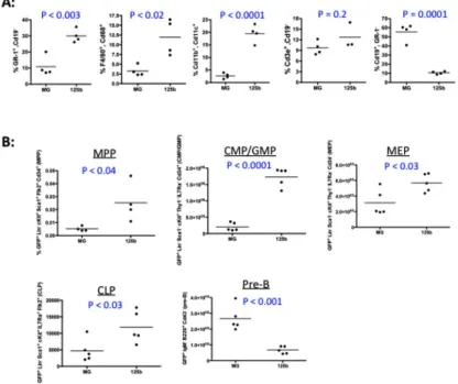
We next sought to characterize the cellular basis by which WTmiR-132 mice undergo
depletion in peripheral blood leukocytes at 4 months post-reconstitution. Almost two-thirds
(29/44) of the WTmiR-132 mice presented with gross pathology characteristic of
extramedullary hematopoiesis, including enlarged spleens and pale, fibrotic bone marrow
(Figure 1F and Supplemental Figure 2G). Strikingly, none of the age-matched WTMG mice
presented such a phenotype. Consistent with the onset of extramedullary hematopoiesis,
spleens from WTmiR-132 mice had a significant elevation of erythtroid cells (Ter11+) and a
slight elevation, albeit not statistically significant, of myeloid cells (CD11b+, Gr-1+) when
compared to WTMG mice (Supplemental Figure 2H). No elevation of myeloid cells was
found in the peripheral blood of WTmiR-132 mice compared to controls (Supplemental
Figure 2I). Examination of the bone marrow, however, revealed that WTmiR-132 mice had a
severe (approximately 3-fold) depletion in the frequency and total number of LSK cells and
HSCs compared to WTMG controls (Figure 1G and Supplementary Figure 2J,K). A similar,
though more dramatic, phenotype was observed at 9 months post-reconstitution in WT
miR-132 mice (Supplemental Figure 3A). This phenotype of rapid proliferation followed by
depletion of HSCs in the bone marrow compartment is an example of HSC exhaustion.
The depletion of HSCs in WTmiR-132 mice had the expected dramatic effect on the numbers
of more mature progenitor cells, including multi-potent progenitors (MPPs; LSK CD150-
CD48+), lymphoid-primed MPPs (LMPPs; LSK Flt3+), and megakaryocyte/erythroid
progenitors (MEPs; Lineage- Sca1- cKit+ CD34- FcRg-). However, no depletion in
granulocyte-myeloid progenitors (GMPs; Lineage- Sca1- cKit+ CD34+ FcRg+) was
observed (Supplemental Figure 3B-F). Importantly, the observed alteration in WTmiR-132
HSCs was intrinsic to the expression of the miR-132 over-expression vector, because no
depletion in the proportion of HSCs was evident among the GFP- cells of WTmiR-132 and
WTMG mice (Supplemental Figure 3H). Furthermore, we found that the observed
phenotype was specific to the expression of authentic miR-132 because over-expression of
a miR-132 mutant lacking the correct miR-132 seed sequence resulted in no observable
phenotype at 9 months post-reconstitution when compared to WTMG controls
(Supplemental Figure 3H).
We next sought to investigate the role of miR-212 in HSC maintenance. We found that
enforced expression of miR-212 in the bone marrow compartment of mice didn’t result in a
significant change in the total number of bone marrow CD45+ cells or LSK cells compared
to controls (Supplemental Figure 3I,J). However, we found that there was a significant
depletion of HSCs at 4-months post-reconstitution in these mice (Supplemental Figure 3K),
thus suggesting a less severe phenotype than enforced miR-132 expression, which is
consistent with the lower levels of enrichment of miR-212 in HSCs.
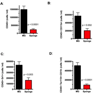
The microRNA-212/132 cluster has a physiological role of protecting the aging hematopoietic system
To determine if miR-132 has a physiological role in regulating hematopoietic stem cell
function, we obtained mice that had a genetic deletion in the entire miR-212/132 cluster
defect in the output of mature hematopoietic cells in the peripheral blood, spleen, and
bone marrow of 12-week old miR-212/132-/- mice when compared to age-matched
wild-type (WT) controls (Supplemental Figure 4). We noticed, however, an up-regulation of
miR-132 expression in the bone marrow and LSK compartment of aged (2-year old) WT
mice compared to young (12-week old) WT mice (Figure 2A), and posited a more
important role of miR-132 in maintaining the fidelity of aging HSCs. Consistent with this,
we found that unlike in 12-week old mice (Supplemental Figure 4D), aged (60-week old)
miR-212/132-/- mice had an elevation in the total number of HSCs (LSK CD150+ CD48-
and LSK EPCR+) in the bone marrow compartment compared to WT controls (Figure 2B).
Surprisingly, this was accompanied by a decrease in the total number of bone marrow LSK
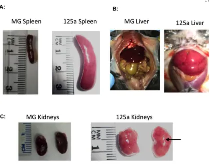
cells, which are mostly downstream products of HSCs (Figure 2B). Aged miR-212/132
-/-mice further presented with enlarged spleens (Supplemental Figure 5A) and a global
depletion of all major mature cell types in the bone marrow compartment (Supplemental
Figure 5B), indicative of a failure of HSCs to maintain normal hematopoietic output and
the onset of extramedullary hematopoiesis.
To investigate the molecular basis for the role of the miR-212/132 cluster in HSCs, we
performed gene expression analysis by bulk population RNA-sequencing on WT and
miR-212/132-/- long-term HSCs (LSK CD150+ ), short term HSCs (LSK CD150-
CD48-), and multipotent progenitors (LSK CD150- CD48+). Approximately 14,000 genes were
expressed in each sample (Supplemental Figure 6A), and clustering based on the number
differentially expressed genes revealed close similarity between WT short-term and
these groups differing significantly from the MPP populations (Figure 2C). Differentially
expressed genes between the WT and miR-212/132-/- HSC populations were enriched for
several functional annotations relevant to HSC biology, including regulation of cell-cycle,
cell differentiation, response to stress, and cell death (Figure 2D).
To investigate if the observed phenotype in miR-212/132-/- mice is intrinsic to the
hematopoietic system, we transferred bone marrow cells from 12-week old WT or
miR-2121/132-/- mice into irradiated WT recipients. After one year, we found that the
phenotypes in transplanted mice closely resembled that of aged WT and miR-212/132
-/-mice, respectively, consistent with a defect intrinsic to the hematopoietic system
(Supplemental Figure 5C). We next employed an inflammatory model for hematopoietic
aging (Esplin et al., 2011) to determine if this is sufficient to recreate the observed
alteration in hematopoiesis. We delivered LPS eight times over one month to 16-week old
miR-212/132-/- and WT mice. We found that, consistent with the altered hematopoietic
output we observed in aged mice, miR-212/132-/- mice presented with severely enlarged
spleens containing an enrichment of splenic HSCs compared to WT mice also injected with
LPS (Figure 3A and Supplemental Figure 5D). Similar to aged miR-212/132-/- mice, LPS
treated miR-212/132-/- mice also demonstrated an accumulation of HSCs and a decrease in
the frequency of LSK cells in the bone marrow compartment compared to LPS treated WT
controls (Figure 3B). This skewing of hematopoietic progenitor output in miR-212/132
-/-mice may be characterized by an increase in the total number of long-term HSCs and a
reduction in total number of short-term HSCs and MPPs in the bone marrow compartment
observed in miR-212/132-/- by exposing younger mice to chronic inflammatory stimuli
via repetitive LPS injections.
Loss of the miR-212/132 cluster reduces HSC cycling and improves engraftment potential
The cycling characteristics of HSCs are closely related to their ability to self-renew and
differentiate into committed progenitors(Pietras et al., 2011). Furthermore, increased HSC
quiescence and an increase in HSC number is characteristic of the aging hematopoietic
system(Geiger et al., 2013). We thus sought to determine if the accumulation of HSCs and
decrease in output of more committed progenitors in miR-212/132-/- mice might be a result
of altered cell cycling. We performed cell-cycling analysis using flow cytometry by
staining for the proliferation marker Ki67 and utilizing the dsDNA dye Hoescht33342.
Under steady-state conditions, we observed no major defect in cell cycling in 16-week old
miR-212/132-/- mice compared to age-matched WT mice (Supplemental Figure 5F).
However, under conditions of inflammatory stress such as low-grade LPS stimulation, we
found that miR-212/132-/- HSCs were far less proliferative, with an almost 50% increase in
the number of cells in the G0 phase of the cell cycle compared to WT HSCs (Figure 3C-E).
Importantly, we observed a substantial decrease in the number of HSCs in G1 and only a
small proportion of cells in S/M phases of the cell cycle in miR-212/132-/- mice (Figures
3C-E). No change in p27 mRNA expression was observed at steady state between
miR-212/132-/- and WT HSCs. However, in mice treated with either LPS or 5-fluorouracil,
which induce HSC proliferation, the expression of p27 in the bone marrow compartment
was up-regulated in miR-212/132-/- mice compared to WT mice (Supplemental Figure
quiescence and an accumulation of HSCs, with a concomitant decrease in the number of
more committed progenitors.
The majority of HSCs in the hematopoietic system remain in a dormant state, and
disruption of this quiescence can have serious consequences for HSC function (Rossi et al.,
2012). To investigate whether the alteration in cell cycling of aged miR-212/132-/- HSCs
might be related to altered HSC function, we performed competitive transplant assays.
Aged (60-weeks) CD45.2 miR-212/132-/- or WT-/- HSCs were transplanted with equal
numbers of CD45.1 WT HSCs into lethally irradiated CD45.2 recipient mice. The
peripheral blood of these mice was analyzed 4 months post-reconstitution for repopulation
of major mature cell types. The cells from aged miR-212/132-/- mice were more effective at
reconstituting most all immune cells than those from aged WT mice, as evidenced in total
blood leukocytes (CD45+), B-cells (CD19+), myeloid cells (CD11b+), and granulocytes
(Gr-1+) in the peripheral blood (Figure 4A). An insignificant difference in the relative
proportion of T-cells (CD3e+) was observed (Figure 4A). Competitive transplant of young
(12-weeks) miR-212/132-/- HSCs yielded no observable functional difference compared to
young WT HSCs except for defective repopulation of T-cells (Figure 4B); however,
secondary transplantation of young miR-212/132-/- HSCs yielded a similar phenotype to
that observed with primary transplantation of aged miR-212/132-/- HSCs (Supplemental
Figure 5I). Consistent with the increased quiescence of aged miR-212/132-/- HSCs
compared to WT HSCs, miR-212/132-/- cells performed better at long-term repopulation.
Additionally, HSCs obtained from WTmiR-132 mice, which ectopically over-express
compared to control HSCs obtained from WTMG mice (Figure 4C). It therefore appears
that the miR-212/132 cluster is important for tuning the interplay between quiescence and
functional output of the aging hematopoietic system.
FOXO3 is a target of miR-132 in bone marrow cells
To understand the molecular mechanism of miR-132 action, we characterized the
expression of the best computationally predicted targets of miR-132 from TargetScan under
conditions of miR-132 over-expression (Friedman et al., 2009). RNA was extracted from lineage-depleted bone marrow cells of WTmiR-132 and WTMG mice and was subjected to
quantitative polymerase chain reaction (qPCR) for target genes relevant to HSC function.
The most significantly down-regulated targets under conditions of ectopic expression of
miR-132 in WTmiR-132 bone marrow, relative to control WTMG bone marrow, were pursued
for further analysis. Messenger RNA expression of several genes relevant to hematopoiesis
was down regulated in WTmiR-132 bone marrow samples, including AchE, FOXO3, Lin28B,
MMP9, and SOX4 (Figure 5A). We sought to further investigate the role of FOXO3 in
mediating the effect of miR-132 on HSCs as it contains a perfect 8-mer binding site for
miR-132 (Figure 5B) and is the most significantly down regulated of these genes.
Importantly, we also found a global upregulation of miR-132 targets in our
RNA-sequencing analysis of miR-212/132-/- and WT HSCs, and this included an upregulation of
FOXO3 transcript expression as well as some of its downstream targets (Supplemental
Figure 6B-D). We validated that miR-132 binds directly to the FOXO3 3’-untranslated
expressed immediately downstream of luciferase. We found that in the presence of
miR-132 the expression of this reporter was significantly lower than from a vector lacking the
FOXO3 3‘UTR (Figure 5C). This binding was specific to miR-132 because mutating the
miR-132 binding site on the FOXO3 3’UTR normalized luciferase expression (Figure 5C).
We next quantified FOXO3 protein expression in bone marrow cells from WTmiR-132 and
WTMG mice. Consistent with FOXO3 being a target of miR-132, we found that protein
expression was significantly down regulated in WTmiR-132 mice compared to WTMG mice
(Figure 5D). Expression of FOXO1 and FOXO4, closely related family members of
FOXO3, remained unchanged in WTmiR-132 bone marrow cells (Supplemental Figure 7A).
Importantly, we also found that FOXO3 mRNA and protein expression levels were
elevated in lineage depleted bone marrow cells from miR-212/132-/- mice compared to WT
controls (Figure 5E,F). Expression of FOXO4 was also slightly elevated in miR-212/132-/-
cells and expression of FOXO1 was unchanged (Supplemental Figure 7B). We additionally
performed intracellular staining of FOXO3 and phospho-FOXO3 (p-FOXO3) protein. As
expected, we found elevated levels of FOXO3 in miR-212/132-/- HSCs compared to WT
controls. However, we saw only a marginal elevation in p-FOXO3 in miR-212/132-/- HSCs,
indicating that the majority of extra FOXO3 in these cells is likely in the nucleus in its
active, un-phosphorylated state (Supplemental Figure 7C,D). Together, the data indicate
that miR-132 is an important regulator of FOXO3 expression in bone marrow cells. As was
observed with miR-132 expression, FOXO3 mRNA expression was increased in bone
suggesting that miR-132 might serve to maintain FOXO3 protein expression within a
balanced range for normal hematopoietic function.
miR-132 regulates HSC cycling and function through FOXO3
To determine if FOXO3 is a key mediator of miR-132 function, we co-expressed FOXO3
with miR-132 in the bone marrow compartment of WT mice to see if it would rescue the
phenotype observed with miR-132 expression alone. FOXO3 cDNA lacking a miR-132
target site was cloned into the MSCV-IRES-eGFP (MIG) vector immediately downstream
of the MSCV promoter (Figure 6B). As previously described, miR-132 was cloned
immediately downstream of eGFP. Lethally irradiated mice were reconstituted with bone
marrow cells transduced with a control vector (WTMIG), or a vector expressing both
miR-132 and FOXO3 (WTFOXO3 + miR-132), miR-132 only (WTmiR-132) or FOXO3 only
(WTFOXO3). The expression of miR-132 and FOXO3 from these vectors was validated by
qPCR and Western Blot, respectively (Supplemental Figure 7E-F). We observed the mean
fluorescence intensity of eGFP in bone marrow cells expressing only FOXO3 to be lower
than that of the other vectors, suggesting that over-expression of FOXO3 above
endogenous levels may have a toxic effect on these cells (Supplemental Figure 7G). We
additionally observed that a larger fraction of bone marrow HSCs from WTFOXO3 mice
expressed AnnexinV compared to WTMIG controls (Supplemental Figure 7H). This effect
was not observed when FOXO3 was co-expressed with miR-132, presumably because
As expected, a reduction in peripheral blood leukocytes was observed in WTmiR-132 mice
compared to WTMIG mice at 4-months post-reconstitution (Figure 6C). Co-expression of
FOXO3 with miR-132, however, rescued this defect, as no significant change in peripheral
blood leukocytes was observed in WTFOXO3 + miR-132 mice compared to WTMIG controls
(Figure 6C). Examination of the bone marrow compartment of WTFOXO3 + miR-132 mice
revealed total numbers of HSCs and LSK cells comparable to WTMIG controls,
demonstrating a rescue of HSC depletion observed with the expression of miR-132 alone
(Figure 6D,E). In addition, expression of FOXO3 alone resulted in a moderate elevation in
the total number of bone marrow LSK cells compared to control mice (Figure 6E). HSCs
from WTFOXO3 + miR-132 mice also showed comparable proportions of Ki67 staining to HSCs
from WTMIG mice, indicating that these cells were not prone to cycling like WTmiR-132 cells
(Figure 5F). WTFOXO3 HSCs demonstrated a significant albeit moderate decrease in the
proportion of cycling HSCs compared to controls (Figure 6F). These experiments suggest
that co-expression of FOXO3 can rescue the phenotype observed with expression of
miR-132 alone. It seems likely that miR-miR-132 regulates hematopoiesis primarily by directly
modulating FOXO3 levels, although we cannot rule out that FOXO3 overexpression is able
to override the miR-132 effect while the true targets of miR-132 in the bone marrow are
other genes.
Loss of miR-212/132 affects HSC survival through protective autophagy
FOXO3 is critical for maintaining the hematopoietic stem cell pool by regulating HSC
cell-cycling and resistance to oxidative stress (Miyamoto et al., 2007; Tothova et al., 2007). It is
autophagy (Warr et al., 2013). To this end, we found that several autophagy-related
genes were up-regulated in miR-212/132-/- HSCs compared to WT controls upon inspection
of our RNA-sequencing dataset (Supplemental Figure 6E). Thus, to determine if miR-132
might play a role in altering survival of HSCs, we sorted HSCs from WT and miR-212/132
-/- mice and cultured them in the presence or absence of survival growth factors and
cytokines, including mSCF, mIL6, mIL3, TPO, and Flt3L. We used a luciferase-based
assay to monitor caspase 3 and caspase 7 activities after 12 hours in culture. In the presence
of survival factors, minimal caspase activity was observed in both WT and miR-212/132
-/-HSCs. However, under starvation conditions, which induce protective autophagy in aged
HSCs (Warr et al., 2013), miR-212/132-/- HSCs demonstrated a significant reduction in
induction of apoptosis compared to WT HSCs (Figure 7A). This is consistent with the
more rapid induction of a protective autophagy program due to higher levels of FOXO3 in
miR-212/132-/- HSCs. Importantly, when autophagy was inhibited by Bafilomycin A
(BafA), a known inhibitor of autophagosome fusion to lysosomes, miR-212/132-/- and WT
HSCs underwent comparable, higher levels of apoptosis (Figure 7A). As previously
reported, FOXO3 expression levels had no major effect on autophagy and apoptosis of
myeloid progenitors (Figure 7B).
To determine if miR-212/132-/- HSCs indeed induce the autophagy machinery more
potently than WT HSCs, we utilized a fluorescent reporter for autophagosome formation
that was detectable by flow cytometry. The efficacy of this assay in detecting
autophagosome formation was validated by comparing signal intensity in WT cells to
cultured under growth factor rich or starvation conditions as described above and were
stained for the presence of autophagosomes. Under starvation conditions, miR-212/132
-/-HSCs demonstrated higher levels of autophagosome formation compared to WT -/-HSCs
(Figure 7C). In the presence of LY2940002, a PI3-kinase inhibitor and early inhibitor of
autophagy, autophagosome formation was decreased to comparable levels in miR-212/132
-/- and WT HSCs (Figure 7C). We additionally sought to investigate whether the potent
induction of autophagy in miR-212/132-/- HSCs may improve survival by altering
reactive-oxygen species (ROS) accumulation. We utilized a fluorescent detection system for ROS
and found that miR-212/132-/- HSCs had lower levels of ROS accumulation compared to
WT HSCs under conditions of starvation (Figure 7D). The accumulation of ROS was
elevated to comparable levels in WT and miR-212/132-/- HSCs when autophagy was
inhibited with BafA (Figure 7D).
We employed an shRNA knockdown strategy for FOXO3 to determine if it is the key
mediator of autophagy in miR-212/132-/- HSCs. WT and miR-212/132-/- HSPCs were
transduced with either a control vector (MB) or a FOXO3 shRNA construct (shFOXO3),
and were subsequently used to reconstitute lethally irradiated WT mice. At 2 months
post-reconstitution, we sorted HSCs from these mice and subjected them to the aforementioned
assays for autophagy induction and caspase activation under conditions of growth factor
starvation. WT HSCs expressing shFOXO3 demonstrated lower autophagy activity and
higher levels of apoptosis compared to WT HSCs expressing MB (Figure 7E,F).
Importantly, knockdown of FOXO3 in miR-212/132-/- HSCs resulted in a significant
miR-212/132-/- HSCs expressing MB. However, this reduction did not reduce autophagy
activity completely to that of WT HSCs expressing MB. This may be due to incomplete
knockdown of FOXO3 in miR-212/132-/- cells or may suggest that factors other than
FOXO3 might be involved in mediating autophagy induction (Figure 7E,F).
Discussion
MicroRNAs are key regulators of lineage commitment and function in immune cells
(Baltimore et al., 2008; Gangaraju and Lin, 2009; O'Connell et al., 2010b). Several
microRNAs have been implicated in regulating diverse facets of normal HSC maintenance
and function, such as cell-cycling(Lechman et al., 2012; Song et al., 2013), apoptosis(Guo
et al., 2010), engraftment potential (O'Connell et al., 2010a; Ooi et al., 2010), and
resistance to inflammatory stress (Zhao et al., 2013). While much has been done to
characterize the functional differences between aged and young HSCs, little is known about
how microRNAs might contribute to maintaining balanced hematopoietic output as an
organism ages. Our findings suggest that the miR-212/132 cluster, particularly miR-132, is
critical in regulating the balance between HSC survival, and proliferation and
differentiation. We demonstrate that it does this primarily by buffering the expression of
FOXO3 in the aging hematopoietic system. Deregulation of this cluster, and in turn
FOXO3, can have negative consequences on the function of HSCs and the output of mature
Because the expression of miR-132 was higher in HSCs compared to total bone marrow
cells, we utilized both gain-of-function and loss-of-function approaches to investigate its
role in HSC function and survival. Ectopic expression of miR-132 resulted in
hyper-proliferation and depletion of HSCs within the bone marrow compartment. Enforced
expression of miR-212 produced a similar but less dramatic phenotype. This depletion of
HSCs with miR-132 over-expression coincided with the onset of extramedullary
hematopoiesis, including enlarged spleens and fibrotic bone marrow. We observed a drastic
decrease in protein expression of the miR-132 target FOXO3 within the bone marrow
compartment of miR-132 over-expressing mice. Consistent with our findings, a genetic
deletion of FOXO3 in hematopoietic cells leads to increased HSC proliferation and an
age-dependent depletion of the HSC pool with loss of HSC long-term reconstitution potential
(Miyamoto et al., 2007). This phenotype is exacerbated by the concomitant deletion of the
FOXO family members FOXO1 and FOXO4 (Tothova et al., 2007). We further found
several FOXO3 target genes, particularly the negative cell-cycle regulators p21, p57, and
p27, to be down regulated in miR-132 over-expressing bone marrow. Importantly,
replenishing levels of FOXO3 during miR-132 over-expression rescued the phenotype we
observed.
A genetic deletion of the miR-212/132 cluster led to higher basal expression of FOXO3 in
bone marrow cells. Over time, this led to a dramatic increase in the number of HSCs, a
decrease in production of more committed progenitors, and a defect in HSC cycling in
response to environmental stress, such as lipopolysaccharide treatment. Consistent with the
long-term reconstitution of the hematopoietic system than WT counterparts. FOXO3 is a
known regulator of apoptosis, and we further demonstrated that ectopic expression of
FOXO3 from a retroviral vector resulted in a selection for those cells expressing the lowest
amount of the vector, presumably because higher levels of FOXO3 expression were toxic.
We have therefore shown that the miR-212/132 cluster is important in regulating
expression of FOXO3, and that when this target is either up-regulated or down regulated,
there is a severe alteration in HSC function over time.
The expression of both miR-132 and FOXO3 transcripts is up-regulated with age in murine
bone marrow cells and early progenitors. The role of FOXO3 as a longevity-associated
gene remains unknown in the hematopoietic system. FOXO3 may be up-regulated in this
context due to its vital role in survival through autophagy and in cell cycling. MicroRNAs
play an important role in buffering perturbations in the expression of their targets in
response to environmental stress (Ebert and Sharp, 2012; Kim et al., 2013). Such stress
might include inflammation from repetitive exposure to environmental pathogens and
hematopoietic aging. Importantly, the abundance of microRNAs in any given cell plays an
important role in establishing a threshold for target expression (Mukherji et al., 2011); as
such, up-regulation of the microRNA may require higher target expression to maintain
important physiological functions. Given that both over-expression and deletion of
miR-132 in bone marrow cells led to inappropriate hematopoiesis, we believe that miR-miR-132
plays an important role in buffering FOXO3 expression levels within a defined range to
maintain normal HSC function as an organism ages. The concomitant increase in the
critical for maintaining an important balance between known FOXO3-regulated
processes, including cell-cycling and differentiation, and apoptosis of HSCs.
The aging hematopoietic system is characterized by an alteration of the balance between
self-renewal and differentiation, which leads to the accumulation of less-functional HSCs,
myeloid-biased differentiation, and a requirement for basal autophagy for survival (Geiger
et al., 2013; Rossi et al., 2005; Warr et al., 2013). Of note is the fact that the loss of critical
autophagy factors in the hematopoietic system leads to hyper-proliferation and poor
survival of HSCs (Mortensen et al., 2011). Recently, it has also been demonstrated that
FOXO3 plays a critical role in inducing protective autophagy of aging HSCs (Warr et al.,
2013), which is critical for their survival in response to oxidative stress(Eijkelenboom and
Burgering, 2013). The proposed mechanism of FOXO3 regulation of autophagy is through
the transcription of glutamine synthase(van der Vos et al., 2012). Consistent with this role
of FOXO3 in HSC survival, we found that miR-212/132-/- HSCs, when compared to WT
HSCs, demonstrated increased resistance to growth-factor starvation, as evidenced by the
decrease in presence of reactive oxygen species, lower levels of apoptosis induction, and an
increase in accumulation of autophagosomes. We observed an abrogation of this effect
when we knocked down FOXO3 in miR-212/132-/- HSCs, thus demonstrating that this
phenotype is mostly due to the up-regulation of FOXO3 in these cells. Importantly, this
improved survival of miR-212/132-/- cells in response to environmental stress may
contribute to the age-dependent accumulation of HSCs in miR-212/132-/- mice compared to
Our observations demonstrate that the miR-212/132-/- cluster is an important regulator of
HSC homeostasis by altering cell cycling, function, and survival. This is one of the first
clear examples of a microRNA playing a physiological role in maintaining the balance of
HSC functions during aging. The capacity of this microRNA to buffer expression of
FOXO3 is critical given the multiple roles FOXO3 plays in regulating HSC biology. Our
findings open the possibility of utilizing miR-132 mimics or antagonists to alter defects in
Experimental Procedures
DNA constructs
For in-vivo 132 over-expression and FOXO3 shRNA experiments, the mature miR-132 or FOXO3 shRNA sequence was placed in the microRNA-155 loop-and-arms format
(O'Connell et al., 2010a) and cloned into the MSCV-eGFP (MG) and MSCV-TagBFP
(MG) vectors, respectively. For FOXO3 rescue experiments, FOXO3 cDNA was cloned
into the MSCV-IRES-eGFP (MIG) vector. See supplemental procedures for more details
about these vectors. FOXO3 shRNA target sequences are given in Table S1.
For luciferase assays, the microRNA-132 expression cassette was sub-cloned into the
pCDNA3 vector. The 3’untranslated regions of relevant gene targets containing the
miR-132 binding region were cloned immediately downstream of luciferase in the pMiReport
vector as previously described (Chaudhuri et al., 2012).
Cell culture
Cells were cultured in a sterile incubator that was maintained at 370C and 5% CO2. 293T
cells were cultured in DMEM supplemented with 10% fetal bovine serum, 100 U/mL
penicillin, and 100 U/mL streptomycin. Primary cells were cultured in complete RPMI
supplemented with 10% FBS, 100 U/mL penicillin, 100 U/mL streptomycin, 50uM β
-mercaptoethanol, and appropriate growth cytokines as needed for the experiment (see
Cell sorting for RNA extraction
For miR-132 expression profiling, bone marrow cells from C57BL/6 mice were depleted of
RBCs and sorted for the respective cell populations at the Caltech Flow Cytometry Core
Facility. Detailed procedures are provided in the supplemental procedures. RNA was
harvested using the miRNAeasy RNA prep kit (Qiagen). For bone marrow samples from
MG and miR-132 mice, bone marrow was harvested from the respective mice, lysed of red
blood cells, and spun down. RNA was harvested as described above.
Expression profiling and qPCR
We performed real time qPCR (RT-qPCR) with a 7300 Real-Time PCR machine (Applied
Biosystems) as previously described (Chaudhuri et al., 2012). TaqMan qPCR was
performed for miR-132, miR-212 and snoRNA-202 (control) detection as per
manufacturers instructions using TaqMan MicroRNA Assays (Life Technologies). SYBR
Green-based RT-qPCR was performed for mRNA of mouse FOXO3, FOXO1, p27, p21,
p57, and relevant miR-132 targets following cDNA synthesis using qScript cDNA
SuperMix (Quanta) and detection with PerfeCTa qPCR Fastmix with ROX (Quanta) as per
manufacturers instructions. Gene-specific primers used for qPCR are listed in Table S2.
RNA-seq library construction and analysis are described in the Supplemental Information.
Target prediction and Luciferase reporter assays
Relevant targets for miR-132 were investigated using predictions from TargetScan Mouse
6.2 software (Friedman et al., 2009) and following sorting by probability of conserved
described (Chaudhuri et al., 2012). Briefly, 4 x 10⁵ cells were plated in 12-well plates for
24 hours and subsequently transfected with either pCDNA or pCDNA-miR-132, a
pMiReport vector, and a β-gal expression vector. 48 hours later, cells were lysed using
Reporter Lysis Buffer (Promega) and luciferase and β-gal expression was analyzed,
respectively, using a Dual Luciferase Kit (Promega) and a chemiluminescent β-gal reporter
kit (Roche).
Immunoblotting
Bone marrow samples were prepared as described for RNA preparation. Cell extracts were
collected using RIPA lysis buffer (Sigma), and were subjected to gel-electrophoresis and
transfer onto a PVDF membrane. Antibody staining was performed using antibodies for
FOXO3, p27, and actin. Detailed procedures are given in the supplemental information.
Animals
The California Institute of Technology Institutional Animal Care and Use Committee
approved all experiments. C57BL/6 WT and miR-212/132-/- mice were bred and housed in
the Caltech Office of Laboratory Animal Resources (OLAR) facility. Bone marrow
reconstitution experiments were performed as previously described (Chaudhuri et al., 2012)
with the aforementioned vectors and are explained in more detail in the Supplemental
Procedures. Recipient mice were monitored for health and peripheral blood was analyzed
for mature blood cell types each month up till the experimental end-point at either 16 or 36
weeks post-reconstitution. At each end-point, immune organs were harvested for further
figure legends. Each experiment was repeated at least twice, and in many cases three or
four times.
Competitive transplant experiments
Bone marrow cells from age and gender-matched WT CD45.2+ C57BL/6 mice,
miR-212/132-/- CD45.2+ C57BL/6 mice, and WT CD45.1+ C57BL/6 mice were harvested and
depleted of RBCs as described above. A 1:1 ratio of WT CD45.1+ HSCs with either WT or
miR-212/132-/- CD45.2+ HSCs were subsequently injected into lethally irradiated (1000
rads) WT CD45.2+ CD57BL/6 mice. Mice were monitored for up to 20 weeks
post-reconstitution and relevant tissues were harvested for further analysis by flow cytometry.
Flow cytometry
Relevant tissues were harvested and cells were homogenized and subsequently depleted of
red blood cells as described above. Flurophore-conjugated antibodies were used for the
indicated markers, and detected using a MACSQuant10 Flow Cytometry machine
(Miltenyi). Detailed procedures are given in the supplemental information.
Autophagy and reactive-oxygen species assays
HSCs were sorted as described above from either WT or miR-212/132-/- C57BL/6 mice, or
from reconstituted mice with donor WT or miR-212/132-/- bone marrow infected with
either MB or shFOXO3 retroviral constructs. Cells were then cultured with the appropriate
(Promega), the presence of ROS (Life Technologies), or autophagy activity (Enzo Life
Sciences). Detailed procedures are given in the supplemental information.
Statistical tests
All statistical analysis was done in Graphpad Prism software using an unpaired Student’s t
test. Data was reported as mean ± SEM. Significance measurements were marked as
follows: * p < 0.05, ** p < 0.01, ** p < 0.001, or ns for not significant.
Data access
The RNA-seq data used in this study can be accessed from the Gene Expression Omnibus
Figure legends
Figure 1. miR-132 is expressed in hematopoietic stem cells (HSCs) and over-expression alters hematopoiesis. (A) miR-132 expression in mature and progenitor hematopoietic
cells. Cell populations were sorted by FACS directly into RNA lysis buffer and miR-132
expression was detected using TaqMan RT-qPCR (n=3). (B) – (G) WT C57BL/6 mice
were lethally irradiated and reconstituted with donor bone marrow cells expressing either a
control (MG) or a miR-132 over-expressing (miR-132) retroviral vector (n=8-12 mice per
group). (B) Total numbers of mature leukocytes (CD45+) in the peripheral blood of MG
and miR-132 mice at the indicated time points post-reconstitution. (C) Total number of
HSCs (LSK CD150+ CD48-) in the bone marrow of MG and miR-132 mice at 8-weeks
post-reconstitution. (D) Percentage of Ki-67+ bone marrow HSCs in MG and miR-132
mice at 8-weeks post-reconstitution. (E) Protein and RNA expression of p27 in the bone
marrow of MG and miR-132 mice at 8-weeks post-reconstitution (n=3). (F) Representative
spleen and bone marrow gross pathology of MG and miR-132 mice at 16-weeks
post-reconstitution. (G) Total number of LSK cells and HSCs in the bone marrow of MG and
miR-132 mice at 16-weeks post-reconstitution. Data represents at least three independent
experiments and is represented as mean ± SEM. See also Figures S1-S3. * denotes p <
0.05, ** denotes p < 0.01 and *** denotes p < 0.001 using a Student’s t test.
Figure 2. Genetic deletion of the microRNA-212/132 cluster in mice alters hematopoietic output with age. (A) miR-132 expression in total bone marrow and LSK cells from 8-week