Regulatory T cells in the establishment and maintenance of
self-tolerance: role of the thymic epithelium
JOSSELYNE SALAÜN*, CATHERINE CORBEL
1and NICOLE M. LE DOUARIN
Laboratoire d’Embryologie Cellulaire et Moléculaire, Nogent-sur-Marne cedex, France and 1Institut Cochin, Département Hématologie, Paris, France
ABSTRACT The thymus constitutes the microenvironment for T lymphocyte differentiation and acquisition of self-tolerance. Aiming to specify the contributions of the two essential parts of the thymus, namely hemopoietic and epithelial, we have devised experimental models in birds and mice. Chimeric thymuses, xenogeneic in birds and allogeneic in mice, were constructed early in development. In both models we could demonstrate a critical role of the epithelial component of the thymic stroma in induction and maintenance of self-tolerance. These experiments showed that suppression mechanisms are also implicated in these events, strongly suggesting the existence of regulatory T cells in both models. Before these experiments the control of self-tolerance was usually attributed to suppressive cells. However, as the cell phenotypes were not identified, the role of these cells was disregarded. Numerous studies since our investigations argue in favour of regulatory mechanisms. The work we initiated several years ago represents a contribution to our understanding of the two linked and opposite aspects of immune-responded control, namely self-tolerance and autoimmunity.
KEY WORDS:
embryo, thymus, graft, self-recognition, regulatory T-cell
0214-6282/2005/$25.00 © UBC Press
Printed in Spain www.intjdevbiol.com
*Address correspondence to: Dr. Josselyne Salaün. Laboratoire d’Embryologie Cellulaire et Moléculaire, 49bis, av. de la Belle Gabrielle. 94736 Nogent-sur-Marne cedex, France. e-mail: josselyne.salaun@college-de-france.fr
Abbreviations used in this paper: MHC, major histocompatibility complex; Mls, minor lymphocyte stimulatory; TCR, T-cell receptor.
Introduction
T cell tolerance to self is acquired primarily within the thymus. As is well established, production of T cells in the thymus is controlled by a combination of positive and negative selection. Positive selection results from moderate affinity between T cells expressing the T Cell receptor (TCR) that recognize the Major Histocompatibility Complex (MHC) molecules and associated self-peptides. Negative selection leads to the elimination or apoptosis of T cells expressing TCR that bind the MHC-self peptide complex with too high affinity (von Boehmer, 1994 and for reviews, Sprent and Webb, 1995; Naji, 1996).
Clonal deletion of self-reactive T cells in the thymus was experimentally demonstrated by means of monoclonal antibodies (Mabs) directed against a defined variable region of the TCRβ chain (Kappler et al., 1987) and by means of appropriately
designed transgenic mice (Kisielow et al., 1988; Pircher et al.,
1989). At the same time analysis of the minor lymphocyte stimulatory (Mls) system provided insights into general aspects of T-cell recognition and repertoire generation (reviews in Abe and Hodes, 1989; Festenstein et al., 1989).
Transgenic mice also allowed the demonstration of “anergy”, a non-deletional mechanism, which inactivates post-thymic
autoreactive T cell clones (review in Fowlkes and Ramsdell, 1993).
On the other hand, the well established existence of autoreactive T cells in normal healthy individuals and the frequent occurrence of autoimmune diseases indicate that clonal deletion does not completely purge the repertoire of T cells able to respond to self-antigens. Besides these two mechanisms, designated as“recessive” resulting in the elimination and inactivation of autoreactive T cells, a “dominant” process involving regulatory T cells has been demonstrated in several experimental models (see for reviews: Le Douarin N. et al., 1996; Modigliani et al., 1996;
Sakaguchi, 2000; Annacker et al., 2001; Bach et al., 2003; Wood
and Sakaguchi, 2003).
In the present report we will review our studies based on the construction of chimeric thymus in which the thymic epithelial component was of one type and HC of another (Fig. 1 A, B).
Avian chimeras
The quail-chick model has yielded a precise picture of thymus development, in which three precisely-timed waves of colonization by HC during incubation was established precisely (Dieterlen-Lièvre and Le Douarin, 2004).
To investigate a possible role of the thymic epithelial component in inducing tolerance, pre-colonized quail thymic rudiments were implanted in situ into thymectomized chick hosts which also
received a quail limb bud from the same donor. The operations were performed at 4.5 days of incubation (E4.5), i.e. before the stage of thymic colonization by HC. Because the operation is tricky, some embryos turn out to be only partially thymectomized. In such chimeras either a definitive state of tolerance or a significant delay in the onset of wing rejection was observed in most animals kept alive between 2 and 16 months post hatching. In some cases, rejection was chronic and reversible whereas in non-thymus engrafted hosts rejection was acute (Okhi et al.,
1987).
Chimerism analysis of the thymus grafted in these chimeras was carried out with species-specific anti-class II Mabs. Epithelial cells in the cortex were of donor type whereas medullary dendritic cells were, like lymphoid cells, of recipient type thus of
hematopoietic origin. In order to induce tolerance, thymic epithelial tissue of donor origin in tolerant birds had to amount to more than one third of the total thymic tissue in the chimera (Okhi et al.,
1988). Tolerance to other somatic tissues than wing could also be induced (Belo et al., 1989). While chimeric bursas of Fabricius
where rejected after hatching (Belo et al., 1985; Corbel et al.,
1987), isotopic grafts of the thymus epitheliomesenchymal rudiment from the same quail donor as that of the bursal rudiment induced permanent acceptance of the bursa, which was invaded by chick hemopoietic progenitor cells (HPC) giving rise to lymphocytes and dendritic cells (Belo et al., 1989) (Fig. 1A).
Mouse thymic chimeras
Early embryonic allogeneic chimeras were performed in the mouse (Salaün et al., 1986). In this species the thymus arises
from the third pharyngeal pouches. (Metcalf and Moore, 1971; Dieterlen-Lièvre and Le Douarin, 2004). The first HPC enter the thymic anlagen at 11 days of gestation. Fontaine-Pérus et al.
(1981) showed further that the thymus in the third pharyngeal pouch from E10 embryos, cultured in vitro, remains entirely
epithelial.
In the mouse like in birds, it is possible to dissect the early uncolonized thymic rudiment (Jotereau et al., 1987). We grafted
E10 pouches into allogeneic nude neonates (Fig. 1B). Several months later, each of these mice was engrafted with tail skin from host, thymic donor and third party haplotypes. Third party skin was rejected whereas syngeneic and donor type skins were accepted. Thus, the fully allogeneic thymic epithelium has the capacity to induce tolerance to skin grafts belonging to its MHC haplotype. Ectopic heart grafts of donor type were also accepted. However, in a majority of the cases, cells from spleen and lymph node of these in vivo tolerant mice were reactive in vitro against
thymic donor type cells (Salaün et al., 1990; Thomas-Vaslin et al.,
1995).
These findings show that naked thymic epithelium is able to attract allogeneic precursors, restore T-cell function in nude mice and induce in vivo tolerance to tissue grafts of its haplotype:
however in vivo tolerance is not always correlated with in vitro
tolerance. The dichotomy between in vivo and in vitro responses
suggests that complete clonal deletion is not required to induce physiological tolerance to tissue grafts.
It is well established that the main mechanism imposing tolerance upon self-reactive T-cells occurs intrathymically. This mechanism involves deletion of cells bearing T-cells receptors with a high enough avidity for the antigens encountered within the thymus. The association between some Vβ families of T cell receptors and reactivities to endogenous superantigens (Mls) has contributed much of the experimental support for clonal deletion as well as positive selection operating on Mls-reactive Vβ TCR (Mac Donald et al., 1988; Bill and Palmer, 1989; Benoist and
Mathis, 1989). In order to elucidate the mechanisms involved in the induction of tissue tolerance, we constructed chimeras in which nude mice from non-deleting strains were reconstituted with thymic epithelium or colonized thymuses from embryos of deleting strains.
Restored mice were tolerant to recipient and donor skin grafts but rejected third party skin graft. The percentage of Mls-reactive Vβ T-cells in such mice was similar to this percentage of T-cells
Fig. 1. Construction of chimeric thymuses (A) in birds; chicken thymic anlagen were removed at E4.5, i.e. before the stage of HPC colonization and replaced by thymic anlagen from quail embryos. Quail wing or bursal buds grafted at the same time were definitively accepted. (B) In mice: athymic nude mice were grafted at birth with thymic rudiments removed from euthymic, allogeneic embryos at E10 (i.e. before the colonization by HPC). Tissular grafts from the haplotype of the donor were definitively accepted.
from nude mice engrafted with syngeneic thymic anlagen (Bandeira
et al., 1992; Salaün et al., 1992).
These results have shown that neither thymic epithelial cells nor the progeny of the first wave of HPC express or functionally present the studied superantigens and that tolerance to skin grafts and superantigens T-cell deletion are unrelated phenomena.
Peripheral tolerance
Since many antigens, such as tissue-specific antigens, are not represented in the thymus, tolerance must develop in the post-thymic environment. Peripheral tolerance induction has been demonstrated mainly by means of transgenic mice constructed so that a selected antigen is expressed in extrathymic tissues (Allison
et al., 1988; Morahan et al., 1989a-b, 1991; Hammerling et al.,
1991; Schonrich et al., 1991).
In these cases, anergy is responsible for tolerance induction. However, in the periphery, as well as inside the thymus, clonal deletion may occur (Webb et al., 1990).
Thus, different mechanisms seem to underlie peripheral tolerance induction; they might depend on the site where the antigen is expressed, the amount of antigen (Auphan et al., 1992)
and the cell type bearing the antigen.
Besides the transgenic model, exclusively locally expressed foreign antigens (i.e., not in the thymus), when introduced into the embryo, can induce tolerance to adult skin grafts of a same haplotype during the entire life span of the animal. Experiments designed according to this scheme had previously indicated the existence of peripheral tolerance inducing mechanisms in birds (Corbel et al., 1990; Martin et al., 1991). In the chick model,
allogeneic limb buds were transplanted from embryos (B4 or B12 MHC haplotype) into (B15xB21) F1 chick recipients. They were studied from birth onwards for periods ranging from 5 to 11 months posthatching. A lasting state of tolerance to the grafted wing was observed in most animals between 2 and 4 months. Moreover, adult skin grafts from a donor with the same MHC haplotype as the wing were also tolerated. Tolerance did not extend to the MLR (Mixed Lymphocyte Reaction) since proliferative responses of blood T cells from chimeras against wing donor MHC haplotype stimulator T cells were comparable to those elicited by T cells of control ungrafted chicks (Corbel et al., 1990).
In allogeneic bursal chimeras, a tolerant state was induced also, but it varied according to the haplotype combination and was short-lived compared with that induced by grafing in wings (Martin
et al., 1994).
Regulatory T cells
Our work both in birds and in mice demonstrates that thymic epithelium grafts induce tolerance, not only to the thymic epithelium itself, but also to peripheral tissues although effector cells, are present in uncompletely thymectomized chicken chimeras tolerate the grafted quail thymus and since some cells in grafted mice are reactive in vitro against donor type cells.
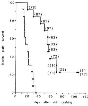
We have shown that peripheral spleen and lymph node T cells from nude mice restored with allogeneic thymic epithelium, when injected into naive nude recipients, are able to restore these mice and to induce tissue graft tolerance to donor type skin (Fig.2).
Transfer of tolerance however depends on the number of injected T cells: the fraction of tolerant recipients increases with increasing numbers of transferred cells, suggesting suppressive
Fig. 2. Transfer of tolerance. Nude mice reconstituted by allogeneic thymic epithelium are tolerant to skin graft of the donor haplotype. Different numbers of peripheral T cells from these mice were injected to naive nude mice. Tolerance to skin graft of the haplotype of the thymic epithelium depends of the number of the injected T cells.
mechanisms mediated by regulatory T cells. The phenotype of T cells able to transfer tolerance was shown to be CD4+ (Modigliani
et al., 1995).
In order to study whether T cells from chimeras are able to regulate activity of effectors from normal mice, we have devised a model in which naive nude C57BL/6 were reconstituted with a cell mixture of peripheral T cells from C57BL/6 Thy1.2 nude mice grafted with BALB/c thymic epithelium and from normal C57BL/6 Thy1.1 (Fig. 3).
These experiments have shown that the transfer of peripheral T cells from chimeras, together with syngeneic T cells from normal mice, induces a significant delay in the rejection of skin graft of the thymic haplotype (BALB/c). This delay depends on the ratio of the two types of injected cells.
Thus, regulatory T cells selected on an allogeneic thymic epithelium are able to control the effector activity of peripheral T cells derived from normal mice (Thomas-Vaslin et al., 1996). All
these results are in favour of “dominant” control mechanism where a population of T cells regulates the activation of self-reactive T cells.
Role of the thymic epithelium in autoimmunity
Evidence for the importance of the thymus in autoimmune disease development has been clearly demonstrated by the thymectomy experiments performed by Sakaguchi in mice. Thymectomy between days 3 and 5 after birth leads to the development of autoimmune diseases (Asano et al., 1996;
Sakaguchi et al., 1985). Transfer of CD4+ peripheral T cells from
normal animals to recipients with autoimmune manifestations can prevent these diseases (review in Seddon and Mason, 2000). In autoimmune diseases, the target organs are heavily infiltrated with T cells. This is particularly the case in insulinodependent diabetes, both in human and in Non Obese Diabetic mice (NOD).
In NOD mice, the disease is primarily mediated by CD4 and CD8 T cells (Bendelac et al., 1987; Wong et al., 1996). To
investigate the role of T cell repertoire selection in the immunopathenogenesis of the disease in the NOD strain, we have restored nude mice by grafting thymic rudiment from NOD embryos (Thomas-Vaslin et al., 1997). Autoimmune disease
development was monitored in these chimeras, after sacrifice. The pancreas and the salivary glands were heavily infiltrated with CD4+ and CD8+ infiltrating T cells.
These results demonstrate that non diabetic nude T cells selected in a chimeric thymus with a NOD embryo thymic epithelium are able to mediate insulitis and sialitis in nude mice. Thus, NOD thymic epithelium alone can select an autoimmune T cell repertoire, suggesting a defect of the thymic epithelium in selecting an appropriate repertoire of regulatory T cells. By using transgenic mice where major histocompatibility complex class II I-Ab expression is limited to thymic
cortical epithelium Bensinger and coll. (2001) have demonstrated that development of regulatory CD4+CD25+ T cells is dependent on
MHC class II-positive thymic cortical epithelium.
Regulatory T cells, “key controllers of immunologic self-tolerance” (Sakaguchi et al., 2000) are released from the thymus
as demonstrated by their functional defect in neonatally thymectomized mice (Sakaguchi, 1985).
Intrathymic transplantation of pancreatic islet allografts was initially investigated by Posselt et al. (1990) in diabetic rats. The
data in rats and subsequently in NOD mice (Mayo et al., 1994;
Gerling et al., 1992; Cetkovic-Cvrlje et al., 1997) were usually
interpreted as the results of negative selection of T cell precursors. We have investigated whether regulatory T cells could be selected by intrathymic islet graft. NOD mice were grafted with newborn NOD thymuses into which allogeneic pancreatic islets had been injected (Fig. 4): 70% of the controls developed the disease as compared with only 24% of the mice engrafted with NOD thymuses + allogeneic pancreatic islets (Salaün et al., 2002).
This protective effect of the pancreatic islet containing in the ectopic thymus is in favour of a role of regulatory T cells.
Conclusions
The first aim of our work, both in birds and in mice, was to determine the respective roles of the two thymic components, hemopoietic and epithelial cells, in T cell differentiation. Appropriate quail-chick chimeras had disclosed the contribution of the three embryonic germ layers, endoderm, mesoderm and ectoderm, to the ontogeny of the thymus. The timing of thymus colonization by hemopoietic cells was precisely defined in birds, then in mice (Jotereau et Le Douarin, 1982; Jotereau et al., 1987). Removing the
thymic rudiment before hemopoietic colonization has allowed the in vivo construction of chimeric thymuses in which epithelium is of one
species or, strain and hemopoietic cells of another. Such chimeric thymuses afforded the opportunity to reveal the role of thymic epithelium in self tolerance induction and to study mechanisms implicated in this event, namely deletion and suppression. In both avian and murine models we have observed tissular tolerance despite the persistence of autoreactive T cells. Our experiments of tolerance transfer by injection of peripheral T cells point out a crucial role of regulatory T lymphocytes in the maintenance of self tolerance (Modigliani et al., 1995a, b). Suppressive mechanisms in
transplantation tolerance were previously suggested by Kindred
Fig. 4. Protective effect of ectopic thymus injected with pancreatic islets in NOD mice.NOD female mice were grafted by an ectopic new-born NOD thymus (A) or by an ectopic new new-born NOD thymus containing allogeneic pancreatic islets (B). Frequence of the disease in A is similar as this frequence observed in non-operated NOD female mice. On the contrary, appearance of the disease is significantly decreased in (B).
(1971) and by Dorsch and Roser (1977). Since these results, the presence in the periphery of T cells, able to prevent autoaggressive immune reactions, has been established (see for review: Annacker
et al., 2001; Wood and Sakaguchi, 2003). Our experimental
reconstitution of nude mice with NOD thymic rudiments show further that abnormal thymic selection of T cells could induce autoimmune manifestations in non-autoimmune strains (Thomas-Vaslin et al.,
1997). Moreover, these results have indicated that a thymic epithelium graft is sufficient to induce autoimmunity in the host.
Thymic generation of T cells able to regulate the activation of autoreactive T lymphocytes is now well established. The thymic stroma has an important role in this process (our work and Jordan
et al., 2001). Failure of this mechanism can lead to the development
of autoimmunity (see for review Seddon and Mason, 2000). In NOD mice, improvement obtained by grafting supplementary thymuses injected with pancreatic islets suggests that the disease in this strain is due to an inefficient generation of regulatory T cells (Salaün et al., 2002).
Despite cumulative evidence for the crucial role of regulatory T cells in self tolerance, the ontogeny, phenotype and mode of action of these cells remain incompletely known. Different subsets of CD4+ regulatory T cells were found; CD4+ CD25+, CD4+ CD25+
CD45RBlow and CD4+ CD25+ CD62L+ (see for reviews Annacker
et al., 2001; Bach, 2003), showing the diversity of regulatory T
cells. Recently, genetic defects in Foxp3 which encodes a forkhead-winged-helix transcription factor were described in some inflammatory diseases both in man and in mouse (Bennett et al.,
2001). Hori and coll. (2003) have shown that Foxp3 is expressed at high levels in thymic and peripheric CD25+CD4+ regulatory T
cells. Foxp3 can be considered as a key gene for the development of regulatory T cells (Fontenot et al., 2005).
A better knowledge of how these cells are selected could help understanding the immune function both in normal development and in immunopathogenic processes.
Acknowledgements
The work summarized here has been supported by CNRS and grants from INSERM, ARC, Recherche et Partage and DRET. We are deeply grateful to Dr. Françoise Dieterlen-Lièvre for critical reading of the manuscript. We wish to thank Drs M. Belo, C. Martin, H. Okhi-Hamazaki and V. Thomas-Vaslin for their expert collaboration. We are also indebted to Pr. Antonio Coutinho (Gulbenkian Institute, Lisbon) and Dr. Antonio Bandeira (Institut Pasteur, Paris) for their close and fruitful collaboration that has importantly contributed to the immunological part of our study both through experiments and valuable and exciting discussions. We thank all the members of the Institute, past and present, for contributing the environment that allowed this work to evolve through the years. Josselyne Salaün and Catherine Corbel particularly wish to express their appreciation to Nicole Le Douarin for creating this environment.
References
ABE, R. and HODES, R.J. (1989). Properties of the Mls system: a revised formulation of Mls genetics and an analysis of T-cell recognition of Mls determinants. Immunol. Rev. 107: 5-28.
ALLISON, J., CAMPBELL, I.L., MORAHAN, G., MANDEL, T.E., HARRISON, L.C. and MILLER, J.F. (1988). Diabetes in transgenic mice resulting from over-expression of class I histocompatibility molecules in pancreatic beta cells. Nature 333: 529-533.
ANNACKER, O, PIMENTA-ARAUJO, R, BURLEN-DEFRANOUX, O and BANDEIRA, A. (2001). On the ontogeny and physiology of regulatory T cells. Immunol. Rev.
182: 5-17.
ASANO, M., TODA, M., SAKAGUCHI, N. and SAKAGUCHI, S. (1996). Autoimmune disease as a consequence of developmental abnormality of a T cell subpopulation.
J. Exp. Med. 184: 387-396.
AUPHAN, N., SCHONRICH, G., MALISSEN, M., BARAD, M., HAMMERLING, G., ARNOLD, B., MALISSEN, B. and SCHMITT-VERHULST, A.M. (1992). Influence of antigen density on degree of clonal deletion in T cell receptor transgenic mice.
Int. Immunol. 4: 541-547.
BACH, J.F. (2003). Regulatory T cells under scrutiny. Nat Rev. Immunol. 3 : 189-198.
BANDEIRA, A., COUTINHO, A., BURLEN-DEFRANOUX, O., KHAZAAL, I., COLTEY, M., JACQUEMART, F., LE DOUARIN, N.M. and SALAUN, J. (1992). Thymic epithelium induces neither clonal deletion nor anergy to Mls 1a antigens. Eur. J. Immunol. 22: 1397-1404.
BELO, M., MARTIN, C., CORBEL, C. and LE DOUARIN, N.M. (1985). A novel method to bursectomize avian embryos and obtain quail - chick bursal chimeras. I. Immunocytochemical analysis of such chimeras by using species-specific monoclonal antibodies. J. Immunol. 135: 3785-3794.
BELO, M., CORBEL, C., MARTIN, C. and LE DOUARIN, N.M. (1989). Thymic epithelium tolerizes chickens to embryonic graft of quail bursa of Fabricius. Int. Immunol. 1: 105-112.
BENDELAC, A., CARNAUD, C., BOITARD, C. and BACH, J.F. (1987). Syngeneic transfer of autoimmune diabetes from diabetic NOD mice to helathyneonates. Requirement for both L3T4+ and Lyt-2+ T cells. J. Exp. Med. 166: 823-832.
BENNETT, C.L., CHRISTIE, J., RAMSDELL, F., BRUNKOW, M.E., FERGUSON, P.J., WHITESELL, L., KELLY, T.E., SAULSBURY, F.T., CHANCE, P.F., OCHS, H.D. (2001). The immune dysregulation, polyendocrinopathy, enteropathy, X-linked syndrome (IPEX) is caused by mutations of FOXP3. Nat Genet. 1: 20-21.
BENOIST, C. and MATHIS, D. (1989). Positive selection of the T cell repertoire: where and when does it occur? Cell 58: 1027-1033.
BENSINGER, S.J., BANDEIRA, A., JORDAN, M.S., CATON, A.J. and LAUFER. T.M. (2001). Major histocompatibility complex class II-positive cortical epithelium mediates the selection of CD4+25+ immunoregulatory T cells. J. Exp. Med. 194: 427-438.
BILL, J. and PALMER, E. (1989). Positive selection of CD4+ T cells mediated by MHC class II-bearing stromal cell in the thymic cortex. Nature 341: 649-651.
CETKOVIC-CVRLJE, M., GERLING, I.C., MUIR, A., ATKINSON, M.A., ELLIOT, J.F. and LEITER, E.H. (1997). Retardation or acceleration of diabetes in NOD/Lt mice mediated by intrathymic administration of candidate beta-cell antigens. Diabetes
46: 1975-1982.
COLTEY, M., JOTEREAU, F.V. and LE DOUARIN, N.M. (1987). Evidence for a cyclic renewal of lymphocyte precursor cells in the embryonic chick thymus. Cell Diff. 22:
71-82.
CORBEL, C., BELO, M., MARTIN, C. and LE DOUARIN, N.M. (1987). A novel method to bursectomize avian embryos and obtain quail-chick bursal chimeras. II. Immune response of bursectomized chicks and chimeras and post-natal rejection of the grafted quail bursas. J. Immunol. 138: 2813-2821.
CORBEL, C., MARTIN, C., OHKI, H., COLTEY, M., HLOZANEK, I. and LE DOUARIN, N.M. (1990). Evidence for peripheral mechanisms inducing tissue tolerance during ontogeny. Int. Immunol. 2: 33-40.
DIETERLEN-LIÈVRE, F. and LE DOUARIN, N.M. (2004). From the hemangioblast to self tolerance: a series of innovations gained from studies on the avian embryo.
Mech. Dev. 121: 1117-1128
DORSCH, S. and ROSER, R. (1977). Recirculating, suppressor T cells in transplantation tolerance. J. Exp. Med. 145 : 1144-1157.
FESTENSTEIN, H., KUMURA, S. and BIASI, G. (1989). Mls and tolerance. Immunol. Rev. 107: 29-59.
FONTAINE-PERUS, J.C., CALMAN, F.M., KAPLAN, C. and LE DOUARIN, N.M. (1981). Seeding of the 10-day mouse embryo thymic rudiment by lymphocyte precursors in vitro. J. Immunol. 126: 2310-2316.
FONTENOT, J.D., RASMUSSEN, J.P., WILLIAMS, L.M., DOOLEY, J.L., FARR, A.G. and RUDENSKY, A.Y. (2005). Regulatory T cell lineage specification by the Forkhead Transcription Factor Foxp3. Immunity 22: 329-341.
FOWLKES, B.J. and RAMSDELL, F. (1993). T-cell tolerance. Curr. Opin. Immunol. 5:
873-879.
HAMMERLING, G.J., SCHONRICH, G., MOMBURG, F., AUPHAN, N., MALISSEN, M., MALISSEN, B., SCHMITT-VERHULST, A.M. and ARNOLD, B. (1991). Non-deletional mechanisms of peripheral and central tolerance: studies with transgenic mice with tissue-specific expression of a foreign MHC class I antigen. Immunol. Rev.
122: 47-67.
HORI, S., NOMURA, T. and SAKAGUCHI, S. (2003). Control of regulatory T cell development by the transcription factor Foxp3. Science 299: 1057-1061.
JORDAN, M.S., BOESTEANU, A., REED, A.J., PETRONE, A.L., HOLENBECK, A.E., LERMAN, M.A., NAJI, A. and CATON, A.J. (2001). Thymic selection of CD4+CD25+ regulatory T cells induced by an agonist self-peptide. Nat. Immunol. 2: 283-284.
JOTEREAU, F.V. and LE DOUARIN, N.M. (1982). Demonstration of a cyclic renewal of the lymphocyte precursor cells in the quail thymus during embryonic and perinatal life. J. Immunol. 129 : 1869-1877.
JOTEREAU, F., HEUZE, F., SALOMON-VIE, V. and GASCAN, H. (1987). Cell kinetics in the fetal mouse thymus: precursor cell input, proliferation and emigration. J. Immunol. 138: 1026-1030.
KAPPLER, J.W., WADE, T., WHITE, J., KUSHNIR, E., BLACKMAN, M., BILL, J., ROEHM, N. and MARRACK, P. (1987). A T cell receptor V beta segment that imparts reactivity to a class II major histocompatibility complex product. Cell 49:
263-271.
KINDRED, B. (1971). Antibody response in genetically thymus-less nude mice injected with normal thymus cells. J. Immunol. 107 : 1291-1295.
KISIELOW, P., TEH, H.S., BLUTHMANN, H. and VON BOEHMER, H. (1988). Positive selection of antigen-specific T cells in thymus by restricting MHC molecules. Nature
335: 730-733.
LE DOUARIN, N. and JOTEREAU, F. (1973). Embryologic origin of thymus lymphocytes in bird embryos. C. R. Acad. Sci., Série III, Paris 276 : 629-632.
LE DOUARIN, N.M. and JOTEREAU, F.V. (1975). Tracing of cells of the avian thymus through embryonic life in interspecific chimeras. J. Exp. Med. 142 : 17-40.
LE DOUARIN, N., CORBEL, C., BANDEIRA, A., THOMAS-VASLIN, V., MODIGLIANI, Y., COUTINHO, A. and SALAUN, J. (1996). Evidence for a thymus-dependent form of tolerance that is not based on elimination or anergy of reactive T cells. Immunol. Rev. 149: 35-53.
MACDONALD, H.R., LEES, R.K., SCHNEIDER, R., ZINKERNAGEL, R.M. and HENGARTNER, H. (1988). Positive selection of CD4+ thymocytes controlled by MHC class II gene products. Nature 336 : 471-473.
MARTIN, C., OHKI-HAMAZAKI, H., CORBEL, C., COLTEY, M. and LE DOUARIN, N.M. (1991). Successful xenogeneic transplantation in embryos: induction of tolerance by extrathymic chick tissue grafted into quail. Dev. Immunol. 1: 265-277.
MARTIN, C., BELO, M., LE DOUARIN, N.M. and CORBEL, C. (1994). A study of peripheral tolerance through embryonic grafts of the bursal epithelial rudiment between MHC-distinct chick embryos. Int. Immunol. 6 : 795-804.
MAYO, G.L., POSSELT, A.M., BARKER, C.F., ROSTAMI, S., MAYO, S.P., CAMPOS, L. and NAJI, A. (1994). Prolongation of survival of donor-strain islet xenografts (rat->mouse) by intrathymic inoculation of xenogeneic islet and bone marrow cells.
Transplantation 58 : 107-109.
METCALF, D. and MOORE, M.A.S. (1971). Haemopoietic cells. North Holland Publ., Amsterdam.
MODIGLIANI, Y., PEREIRA, P., THOMAS-VASLIN, V., SALAUN, J., BURLEN-DEFRANOUX, O., COUTINHO, A., LE DOUARIN, N. and BANDEIRA, A. (1995a). Regulatory T cells in thymic epithelium-induced tolerance.1. Suppression of mature peripheral non-tolerant T cells. Eur. J. Immunol. 25: 2563-2571.
MODIGLIANI, Y., THOMAS-VASLIN, V., BANDEIRA, A., COLTEY, M., LE DOUARIN, N.M., COUTINHO, A. and SALAUN, J. (1995b). Lymphocytes selected in allogeneic thymic epithelium mediate dominant tolerance toward tissue grafts of the thymic epithelium haplotype. Proc. Natl. Acad. Sci. USA 92: 7555-7559.
MODIGLIANI, Y., COUTINHO, A., PEREIRA, P., LE DOUARIN, N., THOMAS-VASLIN, V., BURLEN-DEFRANOUX, O., SALAUN, J. and BANDEIRA, A. (1996). Establishment of tissue-specific tolerance is driven by regulatory T cells selected by thymic epithelium. Eur. J. Immunol. 26 : 1807-1815.
MORAHAN, G., ALLISON, J. and MILLER, J.F. (1989). Tolerance of class I histocompatibility antigens expressed extrathymically. Nature 339: 622-624.
MORAHAN, G., BRENNAN, F.E., BHATHAL, P.S., ALLISON, J., COX, K.O. and MILLER, J.F. (1989). Expression in transgenic mice of class I histocompatibility
antigens controlled by the metallothionein promoter. Proc. Natl. Acad. Sci. USA 86:
3782-3786.
MORAHAN, G., HOFFMANN, M.W. and MILLER, J.F. (1991). A nondeletional mechanism of peripheral tolerance in T-cell receptor transgenic mice. Proc. Natl. Acad. Sci. USA 88: 11421-11425.
NAJI, A. (1996). Induction of tolerance by intrathymic inoculation of alloantigen. Curr. Opin. Immunol. 8: 704-709.
OHKI, H., MARTIN, C., CORBEL, C., COLTEY, M. and LE DOUARIN, N.M. (1987). Tolerance induced by thymic epithelial grafts in birds. Science 237: 1032-1035.
OHKI, H., MARTIN, C., COLTEY, M. and LE DOUARIN, N.M. (1988). Implants of quail thymic epithelium generate permanent tolerance in embryonically constructed quail/chick chimeras. Development 104 : 619-630.
PIRCHER, H., BURKI, K., LANG, R., HENGARTNER, H. and ZINKERNAGEL, R.M. (1989). Tolerance induction in double specific T-cell receptor transgenic mice varies with antigen. Nature 342: 559-561.
POSSELT, A.M., BARKER, C.F., TOMASZEWSKI, J.E., MARKMANN, J.F., CHOTI, M.A. and NAJI, A. (1990). Induction of donor-specific unresponsiveness by intrathymic islet transplantation. Science 249 : 1293-1295.
SAKAGUCHI, S., FUKUMA, K., KURIBAYASHI, K. and MASUDA, T. (1985). Organ-specific autoimmune diseases induced in mice by elimination of T cell subset. I. Evidence for the active participation of T cells in natural self-tolerance; deficit of a T cell subset as a possible cause of autoimmune disease. J. Exp. Med. 161: 72-87.
SAKAGUCHI, S. (2000). Regulatory T cells: key controllers of immunologic self-tolerance. Cell 101: 455-458.
SALAUN, J., BANDEIRA, A., KHAZAAL, I., CALMAN, F., COLTEY, M., COUTINHO, A. and LE DOUARIN, N.M. (1990). Thymic epithelium tolerizes for histocompatibility antigens. Science 247: 1471-1474.
SALAUN, J., CALMAN, F., COLTEY, M. and LE DOUARIN, N.M. (1986). Construction of chimeric thymuses in the mouse fetus by in utero surgery. Eur. J. Immunol. 16:
523-530.
SALAUN, J., BANDEIRA, A., KHAZAAL, I., BURLEN-DEFRANOUX, O., THOMAS-VASLIN, V., COLTEY, M., LE DOUARIN, N.M. and COUTINHO, A. (1992). Transplantation tolerance is unrelated to superantigen-dependent deletion and anergy. Proc. Natl. Acad. Sci. USA 89 : 10420-10424.
SALAUN, J., SIMMENAUER, N., BELO, P., COUTINHO, A. and LE DOUARIN, N.M. (2002). Grafts of supplementary thymuses injected with allogeneic pancreatic islets protect Nonobese diabetic mice against diabetes. Proc. Natl. Acad. Sci. USA 99 :
874-877.
SCHONRICH, G., KALINKE, U., MOMBURG, F., MALISSEN, M., SCHMITT-VERHULST, A.M., MALISSEN, B., HAMMERLING, G.J. and ARNOLD, B. (1991). Down-regulation of T cell receptors on self-reactive T cells as a novel mechanism for extrathymic tolerance induction. Cell 65: 293-304.
SEDDON, B and MASON, D. (2000). The third function of the thymus. Immunol. Today
21: 95-99
SPRENT, J. and WEBB, S.R. (1995). Intrathymic and extrathymic clonal deletion of T cells. Curr. Opin. Immunol. 7 : 196-205.
THOMAS-VASLIN, V., SALAUN, J., GAJDOS, B., LE DOUARIN, N., COUTINHO, A. and BANDEIRA, A. (1995). Thymic epithelium induces full tolerance to skin and heart but not to B lymphocyte grafts. Eur. J. Immunol. 25: 438-445.
THOMAS-VASLIN, V., COLTEY, M. and SALAUN, J. (1996). On the mechanisms of thymic epithelium induced tolerance. C. R. Acad. Sci., Paris 319: 401-404.
THOMAS-VASLIN, V., DAMOTTE, D., COLTEY, M., LE DOUARIN, N.M., COUTINHO, A. and SALAUN, J. (1997). Abnormal T cell selection on Nod thymic epithelium is sufficient to induce autoimmune manifestations in C57BL/6 athymic nude mice.
Proc. Natl. Acad. Sci. USA 94: 4598-4603.
VON BOEHMER, H. (1994). Positive selection of lymphocytes. Cell 76 : 219-228.
WEBB, S.R. and SPRENT, J. (1990). Tolerogenicity of thymic epithelium. Eur. J. Immunol. 20: 2525-2528.
WONG, F.S., VISINTIN, I., WEN, L., FLAVELL, R.A. and JANEWAY CA, J.R. (1996). CD8 T cell clones from young Nonobese diabetic (NOD) islets can transfer rapid onset of diabetes in NOD mice in the absence of CD4 cells. J. Exp. Med. 193 :
67-76.