Original Article
Expression of the mediators of dioxin toxicity, aryl hydrocarbon
receptor (AHR) and the AHR nuclear translocator (ARNT),
is developmentally regulated in mouse teeth
CARIN SAHLBERG*
,1, RAIMO POHJANVIRTA
3,4,5, YUGUANG GAO
1, SATU ALALUUSUA
1,7,
JOUKO TUOMISTO
5,6and PIRJO-LIISA LUKINMAA
2,81Department of Pedodontics and Orthodontics, and 2Department of Oral Pathology; Institute of Dentistry, University of Helsinki, Helsinki, 3Department of Food and Environmental Hygiene, Faculty of Veterinary Medicine, University of Helsinki, Helsinki, 4Kuopio Department, National Veterinary and Food Research Institute, Kuopio, 5Department of Environmental Health, National Public Health Institute, Kuopio,
6Department of Public Health and General Practice, University of Kuopio, Kuopio, 7Department of Oral and Maxillofacial Diseases and 8Department of Pathology, Helsinki University Central Hospital, Helsinki, Finland
ABSTRACT Dioxins are persistent and ubiquitous environmental poisons that become enriched in the food chain. Besides being acutely lethal, the most toxic dioxin congener, 2,3,7,8-tetrachlorodibenzo-p-dioxin (TCDD), is developmentally toxic to many animal species. We have previously found that developing teeth of children may be sensitive to environmental dioxins via their mother’s milk and that rat and mouse teeth are dioxin-sensitive throughout their develop-ment. The aryl hydrocarbon receptor (AHR) together with the AHR nuclear translocator (ARNT) protein is believed to mediate the toxic effects of dioxins. To study the potential involvement of the AHR-ARNT pathway in the dental toxicity of TCDD, we analysed the expression of AHR and ARNT by in situ hybridization and immunohistochemistry in developing mouse teeth. AHR mRNA first appeared in the epithelium of E12 first molar tooth buds and both proteins were weakly expressed in the bud. After cytodifferentiation the expression was upregulated and became intense in secretory odontoblasts and ameloblasts. The coexpression of AHR and ARNT during early tooth development as well as during the formation and mineralization of the dental matrices is suggestive of the AHR-ARNT pathway as a mediator of dental toxicity of TCDD.
KEY WORDS: Dioxin receptor, ARNT, odontogenesis, odontoblasts, ameloblasts
0214-6282/2002/$25.00
© UBC Press Printed in Spain
www.ijdb.ehu.es
*Address correspondence to: Dr. Carin Sahlberg. Institute of Dentistry, Biomedicum Helsinki P.O.Box 63, FIN–00014 University of Helsinki, Finland. Fax: +358–9–191-25371. e-mail: carin.sahlberg@helsinki.fi
Abbreviations used in this paper: AHR, aryl hydrocarbon receptor; ARNT, aryl
hydrocarbon receptor nuclear translocator; TCDD, 2,3,7,8-tetrachlorodibenzo-p-dioxin; XRE, xenobiotic response element; Hsp90, 90 kDa heat shock protein; bHLH/PAS, basic helix-loop-helix/Per-ARNT-Sim; EGFR, epidermal growth factor receptor.
Introduction
Dioxins and related halogenated aromatic hydrocarbons are highly toxic compounds that are formed as by-products in different industrial and combustion processes. These persistent and ubiq-uitous environmental poisons are bioaccumulated in tissue lipid and become enriched in the food chain. A well-established high-exposure effect in humans is chloracne (Assennato et al., 1989; Sweeney and Mocarelli, 2000) and currently, the most toxic dioxin congener, 2,3,7,8-tetrachlorodibenzo-p-dioxin (TCDD) is classi-fied as a human carcinogen (IARC, 1997). The consequences of dioxin poisoning of exposed animals vary from the eventually fatal wasting syndrome to chronic effects such as immunosuppression, thymic atrophy, teratogenesis and carcinogenesis (Peterson et al., 1993; Pohjanvirta and Tuomisto, 1994; Birnbaum, 1995). Dioxins also affect development and reproduction, alter hormone and receptor levels in the endocrine system and give rise to epithelial
hyperplasia and metaplasia (Birnbaum, 1995). Among the devel-opmental defects are cleft palate, hydronephrosis, accelerated eye opening and incisor eruption (Madhukar et al., 1984, 1988), and morphological changes in reproductive organs (Abbott et al., 1987; Abbott and Birnbaum, 1989; Roman et al., 1998; Peters et al., 1999; Hurst et al., 2000). Even very small amounts are still sufficient to produce adverse developmental effects (Hamm et al., 2000).
envi-ronmental dioxins via their mother’s milk (Alaluusua et al., 1996; 1999). As shown in different experimental conditions, sensitivity of rat and mouse teeth to TCDD extends from early morphogenetic stages to the completion of tooth development, with the conse-quences ranging from blockage of tooth development (Partanen et al., 1998; Kattainen et al., 2001; Lukinmaa et al., 2001) to defective dentinogenesis and amelogenesis (Alaluusua et al., 1993; Lukinmaa et al., 2001).
AHR and ARNT
The responses to the toxicity of polyhalogenated hydrocarbons are thought to mainly be mediated by the aryl hydrocarbon receptor (AHR, “dioxin receptor”), a ligand-activated transcription factor, which acts in concert with the AHR nuclear translocator (ARNT) protein. Both molecules are members of the basic helix-loop-helix/ Per-ARNT-Sim (bHLH/PAS) family of transcription factors (Okey et al., 1994; Schmidt and Bradfield, 1996).
Upon binding a ligand in the cytoplasm, AHR dissociates from its chaperone protein, the 90-kDa heat shock protein (Hsp90), and is translocated to the nucleus where it forms a heterodimer with ARNT. The heterodimer complex binds to DNA at a xenobiotic-responsive element (XRE) located upstream the target genes of many drug-metabolizing enzymes to activate their transcription (Sogawa and Fujii-Kuriyama, 1997). This activated gene battery is also thought to play an important role in both the cells’ defence against oxidative stress and in cell cycle control (Rowlands and Gustafsson, 1997; Gonzalez and Fernandez-Salguero, 1998; Nebert et al., 2000).
AHR apparently primarily dimerizes with ARNT, while ARNT can form dimers with other members of the PAS family (Sogawa et al., 1995). AHR and ARNT are widely and co-ordinately expressed in many cells and organs of the developing embryo and the adult. Among tissues and organs expressing AHR and ARNT are the neuroepithelium, heart, liver, ectoderm, muscle, bone, cartilage and adrenal glands. Generally, AHR and ARNT are found in the same or adjacent tissue areas during develop-ment, suggesting a coordinated regulation of expression (Abbott et al., 1995; Abbott and Probst, 1995). Jain et al. (1998) reported no AHR but wide ARNT expression in mice at E9.5, especially in neuroepithelium and the branchial arches. At later embryonic stages they reported AHR expression in the palatal shelf, nasal septal cartilage, olfactory epithelium and the dorsal surface of the tongue while ARNT expression continued at a high level in various tissues of mesodermal and endodermal origin, e.g., lung and tongue musculature.
Both AHR and ARNT null mutant mice have been generated, the phenotypes of which greatly differ. AHR knockout mice show no obvious phenotypic abnormalities at birth, survive into adulthood and are fertile (Gonzalez and Fernandez-Salguero, 1998; Peters et al., 1999; Shimizu et al., 2000). Foetal development therefore seems fairly unaffected by the absence of AHR, maybe due to gene redundancy. However, AHR null mice were resistant to the acute toxicity of TCDD (Mimura et al., 1997). ARNT null embryos, in contrast, are not viable past embryonic day 10.5, they showed defective angiogenesis of the yolk sac and branchial arches or failure of the embryonic component of the placenta to vascularize which eventually affected further growth and development (Kozak et al., 1997; Maltepe et al., 1997). The reason for this is thought to be the role that ARNT plays in the cellular response to hypoxia.
Tooth Development and its Relation to TCDD Exposure
Tooth development is regulated by inductive interactions be-tween cells of the ectodermal lining of the first branchial arch and the underlying mesenchyme. Development begins with a thicken-ing of the oral epithelium, which grows into the mesenchyme forming a bud and inducing the condensation of mesenchymal cells. Morphogenesis proceeds as the epithelial cells proliferate and form the enamel organ surrounding the mesenchymal dental papilla. The tooth crown shape is finally established during the bell stage by folding of the dental epithelium at the sites of future tooth cusps. At the late bell stage, the tooth-specific cells are formed as the dental papilla cells nearest to the epithelium differentiate into odontoblasts and cementoblasts and the inner dental epithelial cells into ameloblasts. Root formation starts only when the crown is fully developed and already for the most part is mineralized. Rodent incisor teeth differ from molars and from human teeth in that their eruption is continuous (Thesleff and Nieminen, 1996). To see if the AHR-ARNT pathway could be involved in the dental toxicity of TCDD, we analysed by in situ hybridization and immunohistochemistry the expression of AHR and ARNT during mouse molar tooth development from the early dental lamina to the onset of tooth eruption.
Results
In Situ Hybridization
The first signs of AHR mRNA expression in the dental tissues were seen in the forming epithelial bud of the first molar tooth germ at E12. The expression was most intense in some cells next to the mesenchyme (Fig. 1A).
After the differentiation of the tooth-specific cells by postnatal day 1 AHR mRNA expression had concentrated and was intense in both secretory odontoblasts and ameloblasts. Interestingly AHR transcripts were seen in scattered cells in the odontoblast cell layer. In the ameloblasts, intense expression was seen in the tips of the cusps and it corresponded to the areas of secretory ameloblasts (Fig. 1E). Scattered AHR expressing cells were present also in the stellate reticulum and developing bone.
Immunostaining
AHR. Our antibodies did not detect AHR protein at early stages of
tooth development in either the mesenchymal or epithelial com-partment (Fig. 1B).
However, at E14 the dental epithelium in incisors showed expression of AHR protein, as did osteogenic cells in the area of the future alveolar bone. Whisker follicles as well as some but not all cells in the basal cell layer of the oral epithelium also expressed AHR intensely (Fig. 1C). As development of the incisor continued expression was downregulated and only reappeared when matrix secretion started (Fig. ID).
In the dental mesenchyme of the molar tooth germ AHR protein was evident next to the inner dental epithelium at the bell stage (E17-newborn). After the terminal differentiation of odontoblasts the expression was upregulated in these cells and persisted throughout the deposition of the dentin matrix (E19–P12).
1 D,F). In both odontoblasts and ameloblasts the expression was first visible at the tips of the cusps from where it moved downwards as the cells differentiated. The expression was particularly strong at the beginning of matrix secretion and persisted throughout the deposition of the dentin matrix (Fig. 1 F,G). Immunostaining for AHR in odontoblasts and ameloblasts was intense as was the expression in the stratum intermedium and in osteogenic cells.
ARNT. ARNT protein was intensely expressed in the oral epithe-lium and in the area of bone formation at E13. The expression was particularly strong in the invaginating dental epithelium of the incisor and in the labial sulcus (Fig. 2A). The perichondrium of Meckel’s cartilage was negative (not shown). In molar tooth buds expression was detected in the dental lamina (Fig. 2B). Interest-ingly, whereas the dental mesenchyme was totally negative the surrounding osteogenic cells expressed ARNT intensely.
At E14 no ARNT expression was detected in either the epithe-lium or mesenchyme of the cap-staged tooth. In the area of the developing mandibular bone surrounding the tooth germ intense ARNT expression continued as well as in oral epithelium (Fig. 2C). However, after odontoblasts and ameloblasts had differen-tiated in the E17 incisor ARNT expression was upregulated in these tissues (Fig. 2D).
After the differentiation of odontoblasts and ameloblasts and the beginning of matrix secretion in neonatal molars staining for ARNT was mainly located in secretory ameloblasts. Presecretory odontoblasts stained faintly and some expression was also visible more centrally in the mesenchyme (Fig. 2E). In fully differentiated odontoblasts and ameloblasts ARNT protein was intensely ex-pressed (Fig. 2F).
The expression of AHR and ARNT in the incisors showed similar cellular and tissue distribution patterns to those in the molars (Figs. 1D and 2D).
Discussion
The present study shows that developing mouse teeth express both AHR and ARNT and that their expression patterns are related to the stage of tooth development. Expression of both AHR and ARNT was detected in the epithelium of early tooth germs, but not during morphogenesis from the bud to the bell stage. The proteins were intensely expressed in secretory odon-toblasts and ameloblasts, as well as in osteogenic cells and oral epithelium.
Two cellular pathways for the mediation of dioxin response in cells have been proposed. The effects of TCDD are thought primarily to be mediated by a heterodimeric complex formed by AHR and ARNT binding to a xenobiotic-responsive element on DNA and thereby activating gene expression. Transcriptional regulation of the target genes requires both AHR and ARNT, but both of these proteins can also dimerize with other members of the bHLH/PAS family of transcription factors (Schmidt and Bradfield, 1996). Especially ARNT is thought to be a partner in the regulation of several different developmental and physiological events (Crews and Fan, 1999). We have also shown that EGFR may act as a mediator of developmental toxicity of dioxin. Using EGFR null mutant mice (Miettinen et al., 1995) we showed that the effects of TCDD on cultured mouse embryonic teeth are dependent on the expression of EGFR (Partanen et al., 1998).
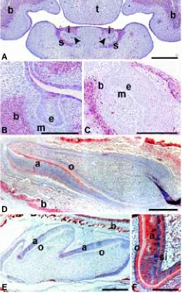
Fig. 1. In situ hybridization and immunohistochemical analysis of AHR expression in different stages of mouse tooth development.
(A-B) Frontal sections of E12 molars. (A) AHR is detected in the epithelium of the upper and lower molar tooth buds. (B) At this developmental stage no AHR protein is as yet localized in the dental area or in Meckel’s cartilage. (C) Sagittal section of E14 incisors. AHR protein is strongly expressed in the dental epithelium of the growing incisor as well as in the future alveolar bone and the oral epithelium. Some expression can also be seen in the dental mesenchyme while the expression in Meckel’s cartilage has almost completely disap-peared. Strong expression is localized to the whisker follicles (arrow-head). (D) Sagittal section of an E17 incisor. Faint expression can be seen in the epithelium of the apical loops (arrowheads). The undiffer-entiated dental mesenchyme and epithelium do not express AHR but as odontoblasts and ameloblasts differentiate expression is upregulated (arrows). Strong expression is present in bone. (E-F) Sagittal section of a 1 day postnatal molar. (E) The expression of AHR is upregulated in the secretory odontoblasts and ameloblasts. Some expression is also seen in the mesenchyme of the cusps and in the stellate reticulum. The dental follicle (arrow) and the alveolar bone are ex-pressing AHR as well. (F) AHR protein is localized to the same areas where the AHR mRNA can be seen. (G) Expression is intense in secretory ameloblasts and odontoblasts as well as in the stratum intermedium. a, ameloblasts; b, bone; e, dental epithelium; Mc, Meckel’s cartilage; o, odontoblasts; si, stratum intermedium; sr, stellate reticulum; t, tongue. A,C,E, in situ hybridization, B,D,F,G, immuno-histochemical staining. Scale bars A,B,G, 50 µm; C, 200 µm; D,E,F, 100 µm.
A
B
C
D
E
This study shows AHR and ARNT protein expression in the epithelium during budding of tooth germs. AHR mRNA as well as AHR and ARNT protein was seen in a few cells of the dental epithelial bud facing the condensed dental mesenchyme. During cap and early bell stages we detected only very little AHR mRNA and no AHR and ARNT protein in dental structures.
We have recently shown that rats exposed to TCDD through their dam’s milk show retardation or complete inhibition of the development of the third molar, which is at the bud stage in neonatal rats at the onset of lactation. The first and second molars, which already have undergone morphogenesis in neona-tal rats, developed normal crowns, but both showed arrest of root development (Lukinmaa et al., 2001). Rat lines A, B, and C, differing in their TCDD sensitivity and AHR structure, accounting for the resistance of rats to the acute lethality of TCDD, differ in responsiveness to the early dental toxicity of TCDD (Pohjanvirta et al., 1999). Development and eruption of the third molar are less affected in the more resistant rat lines carrying the Ahrhw or Bhw
alleles (lines A and B, respectively) than in a line lacking these genes (line C), thereby suggesting a role for AHR as a mediator (Kattainen et al., 2001).
At E14 the mouse first molar tooth germ is at an early cap stage, here found to lack AHR and ARNT expression. In many studies the cap stage has been shown to be a particularly vulnerable stage of tooth development (Thesleff and Åberg, 1999).
As a conclusion, epithelial expression of AHR and ARNT in the early tooth bud could imply mediation of TCDD toxicity via the AHR-ARNT pathway, resulting in the arrest of tooth development. During the developmental stages of dental morphogenesis and cellular differentiation when the expression of both AHR and especially ARNT were weak or absent, pathways other than the AHR-ARNT complex, for example, the tyrosine kinase pathway involving c-src and EGFR could potentially mediate the effect of dioxins (Partanen et al., 1998; Tiffee et al., 1999).
Later, AHR mRNA as well as the protein was strongly upregulated in the newly differentiated odontoblasts and amelo-blasts and the expression remained intense throughout the secre-tory stage. In the same cells ARNT showed a corresponding pattern of upregulation and protein distribution.
Dental hard tissue formation is a sequence of highly controlled events including the secretion of extracellular matrix, processing and degradation of the matrix proteins followed by mineral depo-sition and in the case of enamel, maturation. Disturbing any of these mechanisms could lead to defective matrix formation, degradation and mineralization.
Our previous study on rat pups exposed to TCDD during lactation showed that odontoblasts in the incisors were increas-ingly depolarized which resulted in disturbed dentinogenesis (Lukinmaa et al., 2001). Coexpression of AHR and ARNT in secretory odontoblasts and ameloblasts, shown here, implies that dental toxicity of TCDD involving matrix deposition and mineral-ization, could proceed via the AHR-ARNT pathway, although these findings still have to be verified in the rat.
The consistent spatial and temporal coexpression of AHR and ARNT in bone-forming cells may imply involvement of the AHR-ARNT pathway in the skeletal toxicity of TCDD, evident, e.g., as reduced dimensions and poor or retarded mineralization of the skull bones as well as reduced growth and torsional strength of tibial bone (Alaluusua et al., 1993; Jämsä et al., 2001; Lukinmaa et al., 2001). Fig. 2. Immunohistochemical staining for ARNT in different stages of
mouse tooth development.(A) Frontal section of an E13 mouse head. Intense expression is seen in the oral epithelium, in the invaginating incisor epithelium and in the labial sulcus. No expression is seen in the dental epithelium adjacent to the mesenchyme (arrowheads). ARNT protein is also localized to the area of the future maxillary bone. (B) Frontal section of an E13 lower first molar. ARNT protein is seen in the oral epithelium and in the future alveolar bone area but in the tooth germ neither the epithelium nor the mesenchyme expresses ARNT. (C) Sagit-tal section of an E14 incisor. ARNT expression is only seen in the oral epithelium and in the future bone area. ARNT is not seen in the dental tissues. (D) Sagittal section of an E17 incisor. The undifferentiated dental tissue does not express AHR but as odontoblasts and ameloblasts differentiate expression is upregulated. (E) Sagittal section of a 1 day postnatal molar. Faint staining is seen in secretory odontoblasts and a somewhat stronger expression in ameloblasts. Intense expression con-tinues in the bone area and in the oral epithelium. (F) Intense staining is seen in the secretory ameloblasts and odontoblasts and in the stratum intermedium. a, ameloblasts; b, bone; e, dental epithelium; i, incisor epithelium; m, dental mesenchyme; o, odontoblasts; s, labial sulcus; si, stratum intermedium; t, tongue. Scale bars A,C, 200 µm; B,D,E, 100 µm; F, 50 µm.
A
C
D
E
F
Thus, despite the fact that rat bone and molar teeth differ from the incisor teeth in terms of the dependence of TCDD sensitivity on the mutated AHR, mineralized tissues may share the AHR-ARNT pathway of mediation of TCDD toxicity.
Materials and Methods
Preparation of Tissue Sections
Mice of the NMRI strain were used for the experiments. The age of the embryos was set according to the day of the vaginal plug, which was designated day 0. The mice were anaesthetised with CO2 and killed by cervical dislocation. The use of animals has been approved by the Institutional Animal Care and Use Committee (IACUC) of the Faculty of Science of the University of Helsinki.
Whole heads or lower jaws from 12-d embryos to 12-d postnatal mice were fixed with 4% paraformaldehyde in PBS, washed in PBS, dehy-drated and embedded in paraffin. Postnatal tissues were demineralized with 2.5% PFA + 12.5% EDTA in PBS for up to 2 weeks before dehydration and embedding. For the immunostainings, sections of 7 µm thickness were prepared.
Probes
A mouse cDNA was reverse transcribed from mouse liver total RNA with random primers. The primers 5'-AAT-CCT-TCT-AAG-CGA-CAC-AGA-GAC-3' and 5'-AGT-GGA-GTA-GCT-ATT-GCA-AAC-AAA-G-3' were used to synthesize a 700 bp fragment by PCR corresponding to exons 2– 7of the mouse AHR gene. The fragment was cloned into the pCR® II-TOPO plasmid vector (Invitrogen BV, Groningen, The Netherlands) and subcloned into E.coli. Isolated plasmid was linearized with BamH1 and Not1 and digoxigenin-labelled riboprobes were transcribed using SP6 or T7 RNA polymerases, respectively. The labelled probes were precipi-tated with ethanol, dissolved in RNase free water and used at 0.5 µg/ml concentrations.
In Situ Hybridization
In situ hybridization was carried out according to Braissant and Wahli (1998). In short, the deparaffinized sections were pre-treated with pro-teinase K (Promega, Madison, WI 53711-5399, USA) and active DEPC, hybridized with the labelled probes in a humid chamber overnight at 58ºC, and washed under high-stringency conditions. Anti-DIG-antibodies coupled to alkaline phosphatase were applied. After washes, colour was devel-oped with NBT and BCIP over night, the staining was cleared in 95% ethanol, washed in H2O, dehydrated and mounted in Mountex.
Antibodies
The rabbit polyclonal antibody to rat AHR was from BioMol Feinchemikalien GmbH, Hamburg, Germany, and recognizes both mouse and rat AHR. It was a kind gift from Dr. Allan B. Okey.
The rabbit polyclonal antibody to ARNT/HIF-1β was from NOVUS Biologicals Inc., Littleton, CO 80160, USA.
Immunohistochemistry
The sections were deparaffinized and pre-treated with 10 mM citric acid for 3 x 5 min in a microwave oven, blocked with normal goat serum and BSA for 30 min at room temperature, washed in PBS and incubated with the rabbit primary antibodies to AHR or ARNT/HIF-1β for 30 min at 37°C. After washes in PBS, biotinylated secondary antibodies against rabbit IgG (Vector Laboratories, Burlingame, CA) were applied for 30 min at room temperature and washed. For the detection of the biotinylated antibodies the Vectastain ABC Elite Kit (Vector Laboratories, Burlingame Inc., CA, USA) was used and the colour reaction was generated with amino ethyl carbazole.
To confirm the specificity of the staining reactions the primary antibod-ies were replaced with non-specific rabbit IgG (Technical Grade, Sigma, St Louis, MO).
Acknowledgements
The skilful technical assistance of Ms Pirjo Jutila and Ms Marjatta Kivekäs is gratefully acknowledged. The work was supported by grants from the Finnish Academy and is part of an EU-funded project (QLK4-1999-01446)
References
ABBOTT, B.D. and BIRNBAUM, L.S. (1989). TCDD alters medial epithelial cell differentiation during palatogenesis. Toxicol. Appl. Pharmacol. 99: 276–278.
ABBOTT, B.D. and PROBST, M.R. (1995). Developmental expression of two mem-bers of a new class of transcription factors: II. Expression of aryl hydrocarbon receptor nuclear translocator in the C57BL/6N mouse embryo. Dev. Dyn. 204: 144–55.
ABBOTT B.D., BIRNBAUM L.S. and PRATT, R.M. (1987). TCDD-induced hyperpla-sia of the ureteral epithelium produces hydronephrosis in the murine fetuses. Teratology 35: 329–334.
ABBOTT, B.D., BIRNBAUM, L.S. and PERDEW, G.H. (1995). Developmental expres-sion of two members of a new class of transcription factors: I. Expresexpres-sion of aryl hydrocarbon receptor in the C57BL/6N mouse embryo. Dev. Dyn. 204: 133–43.
ALALUUSUA, S., LUKINMAA, P.-L., KOSKIMIES, M., PIRINEN, S., HÖLTTÄ, P., KALLIO, M., HOLTTINEN, T. and SALMENPERÄ, L. (1996). Developmental dental defects associated with long breast feeding. Eur. J. Oral. Sci. 104: 493–497.
ALALUUSUA, S., LUKINMAA, P.-L., POHJANVIRTA, R., UNKILA, M. and TUOMISTO, J. (1993). Exposure to 2,3,7,8-tetrachlorodibenzo-p-dioxin leads to defective dentin formation and pulpal perforation in rat incisor tooth. Toxicology 81: 1–13.
ALALUUSUA, S., LUKINMAA, P.-L., TORPPA, J., TUOMISTO, J. and VARTIAINEN, T. (1999). Developing teeth as biomarker of dioxin exposure. Lancet 353: 206.
ASSENNATO, G., CERVINO, D., EMMETT, E.A., LONGO, G. and MERLO, F. (1989). Follow-up of subjects who developed chloracne following TCDD exposure at Seveso. Am. J. Ind. Med. 16: 119–125.
BIRNBAUM, L.S. (1995). Developmental effects of dioxins and related endocrine disrupting chemicals. Toxicol. Lett. 82–83: 743–750.
BRAISSANT, O. and WAHLI, W. (1998). A simplified in situ hybridization protocol using non-radioactively labelled probes to detect abundant and rare mRNAs on tissue sections. Boehringer Mannheim Biochemica, No 1: 10–16.
CREWS, S.T. and FAN, C.M. (1999). Remembrance of things PAS: regulation of development by bHLH-PAS proteins. Curr. Opin. Genet. Dev. 9: 580–587.
GONZALEZ, F.J. and FERNANDEZ-SALGUERO, P. (1998). The aryl hydrocarbon receptor. Studies using the AHR-null mice. Drug. Metabol. Disposit. 26: 1194– 1198.
HAMM, J.T., SPARROW, B.R., WOLF, D. and BIRNBAUM, L.S. (2000). In utero and lactational exposure to 2,3,7,8-tetrachlorodibenzo-p-dioxin alters postnatal devel-opment of seminal vesicle epithelium. Toxicol. Sci. 54: 424–430.
HURST, C.H., DEVITO, M.J., SETZER, R.W. and BIRNBAUM, L.S. (2000). Acute administration of 2,3,7,8-tetrachlorodibenzo-p-dioxin (TCDD) in pregnant Long Evans rats: association of measured tissue concentrations with developmental effects. Toxicol. Sci. 53: 411–420.
IARC (1997). Monographs on the evaluation of carcinogenic risks to humans. Polychlorinated dibenzo-p-dioxins and polychlorinated dibenzofurans. Vol. 69. International Agency for Research on Cancer, Lyon.
JAIN, S., MALTEPE, E., LU, M.M, SIMON C. and BRADFIELD, C.A. (1998). Expression of ARNT, ARNT2, HIF1 alpha, HIF2 alpha and Ah receptor mRNAs in the developing mouse. Mech. Dev. 73: 117–123.
JÄMSÄ, T., VILUKSELA, M., TUOMISTO, J.T., TUOMISTO, J. and TUUKKANEN, J. (2001). Effects of 2,3,7,8-tetrachlorodibenzo-p-dioxin on bone in two rat strains with different aryl hydrocarbon receptor structures. J. Bone Miner. Res. 16: 1812– 1820.
KATTAINEN, H., TUUKKANEN, J., SIMANAINEN, U., TUOMISTO, J.T., KOVERO, O., LUKINMAA, P.-L., ALALUUSUA, S., TUOMISTO, J. and VILUKSELA, M. (2001). In utero/lactational 2,3,7,8-tetrachlorodibenzo-p-dioxin exposure impairs molar tooth development in rats. Toxicol. Appl. Pharmacol. 174: 216-224.
KOZAK, K.R., ABBOTT, B. and HANKINSON. O. (1997). ARNT-deficient mice and placental differentiation. Dev. Biol. 191: 297–305.
2,3,7,8-tetrachlorodibenzo-p-dioxin. Toxicol. Appl. Pharmacol. 173: 38–47.
MADHUKAR, B.V., BREWSTER, D.W. and MATSUMURA, F. (1984). Effects of in vivo-administered 2,3,7,8-tetrachlorodibenzo-p-dioxin on receptor-binding of epi-dermal growth factor in the hepatic plasma membrane of rat, guinea pig, mouse, and hamster. Proc. Natl Acad. Sci. USA 81: 7407–7411.
MADHUKAR, B.V., EBNER, K., MATSUMURA, F., BOMBICK, D.W., BREWSTER, D.W. and KAWAMOTO, T. (1988). 2:3,7,8-tetrachlorodibenzo-p-dioxin causes an increase in protein kinases associated with epidermal growth factor receptor in the hepatic plasma membrane. J. Biochem. Toxicol. 3: 261–277.
MALTEPE, E., SCHMIDT, J.V., BAUNOCH, D., BRADFIELD, C.A. and SIMON, M.C. (1997). Abnormal angiogenesis and responses to glucose and oxygen deprivation in mice lacking the protein ARNT. Nature 386: 403–407.
MIETTINEN, P.J., BERGER, J.E., MENESES, J., PHUNG, Y., PEDERSEN, R.A., WERB, Z. and DERYNCK, R. (1995). Epithelial immaturity and multiorgan failure in mice lacking epidermal growth factor receptor. Nature 376: 337–341.
MIMURA, J., YAMASHITA, K., NAKAMURA, K., MORITA, M., TAKAGI, T.N., NAKAO, K., EMA, M., SOGAWA, K., YASUDA, M., KATSUKI, M. and FUJII-KURIYAMA, Y. (1997). Loss of teratogenic response to 2,3,7,8-tetrachlorodibenzo-p-dioxin (TCDD) in mice lacking the Ah (dioxin) receptor. Genes Cells 2: 645–654.
NEBERT, D.W., ROE, A.L., DIETER, M.Z., SOLIS, W.A., YANG, Y. and DALTON, T.P. (2000). Role of the aromatic hydrocarbon receptor and (Ah) gene battery in the oxidative stress response, cell cycle control and apoptosis. Biochem. Pharmacol. 59: 65–85.
OKEY, A.B., RIDDICK, D.S. and HARPER, P.A. (1994). Molecular biology of the aromatic hydrocarbon (dioxin) receptor. Trends Pharmacol. Sci. 15: 226–232.
PARTANEN, A.-M., ALALUUSUA, S., MIETTINEN, P.J., THESLEFF, I., TUOMISTO, J., POHJANVIRTA, R. and LUKINMAA, P.-L. (1998). Epidermal growth factor receptor as a mediator of developmental toxicity of dioxin on mouse embryonic teeth. Lab. Invest. 78: 1473–1481.
PETERS, J.M., NAROTSKY, M.G., ELIZONDO, G., FERNANDEZ-SALGUERO, P.M., GONZALEZ, F.J. and ABBOTT, B.D. (1999). Amelioration of TCDD-induced teratogenesis in aryl hydrocarbon receptor (AHR)-null mice. Toxicol. Sci. 47: 86– 92.
PETERSON, R.E., THEOBALD, H.M. and KIMMEL, G.H. (1993). Developmental and reproductive toxicity of dioxins and related compounds: Cross-species compari-sons. Crit. Rev. Toxicol. 23: 283–335.
POHJANVIRTA, R. and TUOMISTO, J. (1994) Short-term toxicity of 2,3,7,8-tetrachlorodibenzo-p-dioxin in laboratory animals: effects, mechanisms, and animal models. Pharmacol. Rev. 46: 483-549.
POHJANVIRTA, R., VILUKSELA, M., TUOMISTO, J.T., UNKILA, M., KARASINSKA, J., FRANC, M.A., HOLOWENKO, M., GIANNONE, J.V., HARPER, P.A., TUOMISTO, J. and OKEY, A.B. (1999). Physicochemical differences in the AH receptors of the most TCDD-susceptible and the most TCDD-resistant rat strains. Toxicol. Appl. Pharmacol. 155: 82–95.
ROMAN, B.L., TIMMS, B.G., PRINS, G.S. and PETERSON, R.E. (1998). In utero and lactational exposure of the male rat to 2,3,7,8-tetrachlorodibenzo-p-dioxin impairs prostate development. 2. Effects on growth and cytodifferentiation. Toxicol. Appl. Pharmacol. 150: 254–70.
ROWLANDS, J.C. and GUSTAFSSON, J.A. (1997). Aryl hydrocarbon receptor-mediated signal transduction. Crit. Rev. Toxicol. 27: 109–134.
SCHMIDT, J.V. and BRADFIELD, C.A. (1996). AH receptor signalling pathways. Ann. Rev. Cell Dev. Biol. 12: 55–89.
SHIMIZU, Y., NAKATSURU, Y., ICHINOSE, M., TAKAHASHI, Y., KUME, H., MIMURA, J., FUJII-KURIYAMA, Y. and ISHIKAWA, T. (2000). Benzo[a]pyrene carcinoge-nicity is lost in mice lacking the aryl hydrocarbon receptor. Proc. Natl Acad. Sci. USA 97: 779–782.
SOGAWA, K. and FUJII-KURIYAMA, Y. (1997). Ah receptor, a novel ligand-activated transcription factor. J. Biochem. 122: 1075–1079.
SOGAWA, K., NAKANO, R., KOBAYASHI, A., KIKUCHI, Y., OHE, N., MATSUSHITA, N. and FUJII-KURIYAMA, Y. (1995). Possible function of Ah receptor nuclear translocator (Arnt) homodimer in transcriptional regulation. Proc. Natl Acad. Sci. USA 92: 1936–1940.
SWEENEY, M.H. and MOCARELLI, P. (2000). Human health effects after exposure to 2,3,7,8-TCDD. Food Addit. Contam. 17: 303–316.
THESLEFF, I. and ÅBERG, T. (1999). Molecular regulation of tooth development. Bone 25: 123–125.
THESLEFF, I. and NIEMINEN, P. (1996). Tooth induction. Encyclopedia of Life Sciences. Macmillan, http://www.els.net.
TIFFEE, J.C., XING, L., NILSSON, S. and BOYCE, B.F. (1999). Dental abnormalities associated with failure of tooth eruption in src knockout and op/op mice. Calcif. Tissue Int. 65: 53–58.