Cell death in the developing vertebrate retina
ELENA VECINO*, MARÍA HERNÁNDEZ and MÓNICA GARCÍA
Department of Cell Biology and Histology, University of the Basque Country, Leioa, Vizcaya, Spain
ABSTRACT Programmed cell death occurs naturally, as a physiological process, during the embryonic development of multicellular organisms. In the retina, which belongs to the central nervous system, at least two phases of cell death have been reported to occur during development. An early phase takes place concomitant with the processes of neurogenesis, cell migration and cell differentiation. A later phase affecting mainly neurons occurs when connections are established and synapses are formed, resulting in selective elimination of inappropriate connections. This pattern of cell death in the developing retina is common among different vertebrates. However, the timing and magnitude of retinal cell death varies among species. In addition, a precise regulation of apoptosis during retinal development has been described. Factors such as neurotrophins, among many others, and electrical activity influence the survival of retinal cells during the course of development. In this paper, we present a summary of these different aspects of programmed cell death during retinal development, and examine how these differ among different species.
KEY WORDS:
apoptosis, development, retina, glutamate, neurotrophin, insulin
0214-6282/2004/$25.00
© UBC Press Printed in Spain www.ijdb.ehu.es
*Address correspondence to: Dr. Elena Vecino. Department of Cell Biology and Histology, University of the Basque Country, E-48940 Leioa, Vizcaya, Spain. Fax: +34-94-601-3266. e-mail: [email protected]
Abbreviations used in this paper: AC, amacrine cell; ACD, displaced amacrine cell; BC, bipolar cell; C, cone; ER, endoplasmic reticulum; FasL, Fas ligand; GCL, ganglion cell layer; HC, horizontal cell; INL, inner nuclear layer; IPL, inner plexiform layer; MC, Müller cell; NBL, neuroblastic layer; ONL, outer nuclear layer; TNF, tumor necrosis factor.
Introduction
Apoptosis (derived from a Greek word meaning “falling off”, as leaves from a tree in Autumn) is the main morphological feature of the process of programmed cell death or “cell suicide”. It is a widespread, physiological phenomenon which occurs during the embryonic development of multicellular organisms (Glücksmann, 1965; Oppenheim, 1991) and represents the most common mechanism to regulate the size of cell populations during devel-opment, as well as in adult life. In the developing vertebrate nervous system, for example, around half or more of the nerve cells normally die soon after they are born. Moreover, deregulated cell death is increasingly recognized as a frequent component in the course of different diseases, and it has been considered the final common pathway resulting from a variety of primary defects. The concept of apoptosis was elaborated as a counterpoint to necrosis. The critical experiment which allowed the differentiation of these two forms of cell death was the production of ischemic damage to liver tissue (Kerr et al., 1972). Apoptosis is character-ized at the ultrastructural level by the condensation of nuclear chromatin, shrinkage of the cell soma, blebbing of the plasma membrane and fragmentation of the cell into pieces called apoptotic bodies. The cell surface is altered, displaying properties which cause the dying cell to be rapidly phagocytosed, either by neigh-boring cells or by macrophages, before any leakage of its contents occurs. Therefore, inflammatory reactions do not occur in apop-tosis, in which intracellular organelles such as mitochondria and
endoplasmic reticulum are relatively preserved. In contrast, ne-crosis is characterized by early swelling and rupture of intracellu-lar organelles and the plasma membrane, typically accompanied by inflammation in an extensive portion of tissue, either chal-lenged with harsh physical conditions or severely poisoned by chemicals.
During recent years, however, it has become evident that there is a continuum, rather than a clear-cut difference, between both cell death modalities. Some features which were initially regarded to be specific for one form of cell death have been recognized to be common to both. Furthermore, cells can switch from one mode of cell death to another in response to varying intensities of the same insult and depending on the availability of energy sub-strates (Pettmann and Henderson, 1998; Bähr, 2000).
according to instantaneous detection methods (Perry et al., 1983). Nevertheless, the precise magnitude and timing of the process of retinal cell death has not yet been completely charac-terized. To this end, it is very important to analyze retinal cell death in a quantitative and standardized manner. However, it is very difficult to estimate the magnitude of cell death in the retina, since cells cannot be specifically labeled before they have committed themselves to their definitive phenotype. For this reason, most of the data related to the quantification of cell death in the developing retina are based on studies of retinal ganglion cells (Bähr, 2000; Isenmann et al., 2003).
The purpose of the present article is to review the molecular bases and mechanisms of cell death which occurs during retinal development, as well as to summarize the timing of retinal cell death which occurs during the development of different species.
Intracellular pathways leading to cell apoptosis
A variety of stimuli can lead to apoptotic cell death. These proapoptotic stimuli include both extracellular and intracellular signals. The former include tumor necrosis factor (TNF) and Fas ligands (FasL) or the absence of trophic factors. In contrast, DNA damage and endoplasmic reticulum (ER) stress are examples of intracellular proapoptotic signals. These specific signals produce the activation of intracellular pathways which result in biochemical and morphological changes in the cell. Caspases, which are a family of proteases that have a cysteine at their active site and cleave their target proteins at specific aspartic acid residues, are typically activated during the early stages of apoptosis (Tornberry and Lazebnik, 1998). Caspases are synthesized in the cell as inactive precursors, or procaspases, which are usually activated
via proteolytic cleavage by another member of the caspase family, resulting in an amplifying proteolytic cascade. These proteases cleave key cellular substrates which are required for normal cellular function, including structural proteins in the cytoskeleton and nuclear proteins such as DNA repair enzymes. Caspases can also activate other degradative enzymes such as DNases, which cleave DNA in the nucleus. The activation of these powerful death effectors is a critical process. Mitochondria play an important role in the regulation of cell death through release of cytochrome C, which together with Apaf-1 and ATP form a complex with pro-caspase 9, leading to activation of pro-caspase 9 and the pro-caspase cascade (Fig. 1).
Methods to identify apoptotic cells
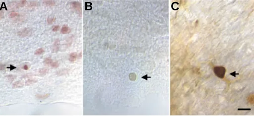
As we have mentioned above, it is difficult to estimate the number of apoptotic cells in the developing retina due to the lack of markers to identify the different cell types in an undifferentiated stage. Quantification of the apoptotic process is also difficult to perform, since the process is not restricted to a defined period, but rather occurs at different stages of development. Most of the studies reporting the quantification of retinal cell apoptosis during develop-ment are based on images taken at a particular time point of the process, but few studies have attempted to characterize the dynam-ics of retinal programmed cellular death. In one of these scarce studies, the progression of nuclear condensation was followed by time-lapse analysis in organotypic cultures of newborn rat retina, previously labeled with a nuclear dye (Cellerino et al., 2000). The process of apoptosis was found to last about 40 minutes and the duration of the whole process was found to be similar for the different cell types. However, the kinetics of RGC apoptosis cannot be observed in single neurons in vivo. Thus, the time course of the apoptotic process in these cells has been estimated from the number of neurons of the same population displaying morphological and/or biochemical signs of apoptosis during the period of neuronal death. The main methods used to identify apoptotic cells include histological methods such as Feulgen, TUNEL and immunocy-tochemical methods. The Feulgen staining procedure is a classical histochemical method based on the staining of DNA by a previous acid hydrolysis step, to remove purines from the deoxyribose backbone of the DNA molecule. Removal of purines leads to exposure of deoxyribose sugars with an aldehyde group. These groups can be identified by staining with a Schiff reagent (in this case, an aqueous solution of cresyl-violet and sulfurous acid used to test the presence of aldehydes). The amount of stain color developed is directly proportional to the amount of DNA present in the stained objects. Thus apoptotic cells can be identified as cells with a dark purple, compacted nucleus (Fig. 2A).
The TUNEL method (Terminal deoxynucleotidyl transferase-mediated dUTP nick-end labeling method) specifically stains apoptotic cells, making them easily distinguishable from normal cells. This technique consists of pre-treatment of cell or tissue samples with protease and then incorporation of a labeled oligo (dU) into the DNA breaks with terminal deoxy-transferase. Finally, labeled oligo (dU) is visualized either with peroxidase or fluores-cence. The TUNEL reaction is highly specific and only apoptotic nuclei are stained (Gavrieli et al., 1992) (Fig. 2B).
With respect to immunocytochemical methods, apoptotic ac-tivity can be identified by using antibodies to different proteins or
substrates which specifically participate in the apoptotic cascade. Proposed immunocytochemical markers for apoptosis include c-jun, proteases and other markers (Schemechel, 1999) (Fig. 2C).
Factors which regulate the apoptosis of retinal cells
during development
A common feature of apoptosis observed in all studied species is the existence of two phases of programmed cell death during retinal development. Neuronal cell death during the late phase of apoptosis seems to be associated with a lack of trophic factor. In contrast, survival of neural cells and developing neurons during the early phase of apoptosis is regulated by neurotrophic factors and/or by interactions of their afferents with neighboring glial cells, so that the number of afferent neurons matches available sites in the target-space (Clarke, 1985; Oppenheim, 1991; Lin-den, 1994). In addition, electrical activity can influence the early survival of both neurons and glial cells by activating intrinsic mechanisms (Meyer-Franke et al., 1998).
Evidence for the dependence of apoptosis on cell-cell interac-tions came from experimental manipulation of the developing nervous system. Moreover, the discovery and characterization of various factors derived from either neurons or glia have revealed some of the cues that are essential during programmed cell death. Pharmacological manipulation and the generation of trans-genic animals have allowed an elucidation, at least in part, of the mechanisms and timing of the apoptotic process in the develop-ing retina.
Neurotrophins and their receptors: NGF, BDNF, NT-3, NT-4, P75, Trks
Neurotrophins play an important role in the development and regeneration of the visual system in several species of verte-brates. Neurotrophins are polypeptide molecules belonging to a family which comprises six closely related factors: nerve growth factor (NGF), brain derived neurotrophic factor (BDNF), neurotrophin 3 (NT-3), neurotrophin 4 (NT-4), neurotrophin 6 (NT-6) and neurotrophin 7 (NT-7), a member of the neurotrophin family only found so far in zebrafish (Nilsson et al., 1998). Two subtypes of neurotrophin receptor have been characterized: the first consists of the tyrosine kinase (Trk) family of receptors known as TrkA, TrkB and TrkC, which specifically bind individual
Fig. 2. Three different methods to visualize apoptotic cells. (A) Feulgen staining, (B) the TUNEL method with peroxidase labeling and (C) immuno-histochemical detection of caspase activity. Arrows point to apoptotic cells visualized with the different methods. Scale bar, 10 µm. (Figure provided by Luis Martinez and María Inmaculada Guerricagoitia)
neurotrophins (Barbacid, 1994); the second type is the low-affinity NGF receptor, also known as low-affinity neurotrophic receptor (p75), which binds neurotrophins with varying affinities and does not possess intrinsic tyrosine kinase activity.
The localization and function of neurotrophins and their recep-tors in the developing retina has been analyzed in different species. It has been proposed that neurotrophins play a role in the topographic refinement of retinocollicular projections during the development of the retinocollicular system. This process is par-tially mediated by the selective elimination of retinal ganglion cells which establish inappropriate topographic projections (Herzog et al., 1994; Lein et al., 2000).
NGF controls the programmed cell death which affects early postmitotic neuroblasts during development. NGF, through bind-ing to the p75 receptor, acts as a killbind-ing factor, thereby controllbind-ing the provisional number of newly generated neurons (Frade et al., 1996). Moreover, it has been shown that the binding of NGF to p75 leads to the activation of intracellular pathways which involve ceramide production, in a way similar to that described following TNF and Fas receptor activation (Carter and Lewin, 1997; Dechant and Barde, 1997). Thus, anti-NGF or anti-p75 antibodies sub-stantially reduce the number of apoptotic cells in the E6 chick retina (Frade et al., 1996; 1999). Moreover p75 can bind to other neurotrophins and in that case its effect leads to a neuroprotective action (Butowt and von Bartheld, 2003).
More recently, a zinc finger protein designated neurotrophin receptor interacting factor (NRIF) has been reported to modulate p75 signaling. Thus, studies with NRIF knockout mice have dem-onstrated that animals which survive beyond E12 have reduced developmental cell death in the retina. Although p75 seems to be an important regulator of programmed cell death, its role in embry-onic and postnatal apoptosis has still not been fully characterized (Casademunt et al., 1999). However, it has been demonstrated that p75-mediated apoptosis is dependent on the expression of the signaling adaptor molecule named p75NTR-associated cell death executor (NADE). NADE has been shown to participate in p75-mediated apoptosis induced by NGF (Mukai et al., 2000).
BDNF, through TrkB activation, acts as a target-derived trophic factor controlling the survival of RGCs during the period of naturally occurring cell death. Recently it has been demonstrated that TrkB receptor signaling regulates developmental death dy-namics, but not final number of retinal ganglion cells (Pollok et al., 2003). In the chick retina, the application of BDNF to embryos in ovo prevented retinal cell death during the early period of devel-opment (E5-7), whereas exogenously applied NGF and NT-3 had no such effect. The addition of BDNF to embryos resulted in an approximately 70% increase in the number of RGCs (Frade et al., 1997). The influence of neurotrophins on retinal cell survival and differentiation depends on the stage of development of the retina and on the location to which the neurotrophins are applied. Thus, in Xenopus, BDNF is produced in the optic tectum soon after RGC axons have reached this target. This endogenous BDNF is necessary for the development of RGC axons and dendrites and increases the complexity of RGC dendritic and axonal arbors (Lom and Cohen-Cory, 1999). However, when BDNF is directly provided to cell bodies by intraocular injections, RGC axonal arbors are not influenced and dendrites undergo atrophy.
NT-3 is extensively expressed among most retinal cell types in many species during development. The conservation of this
pat-tern of expression of NT-3 during evolution may reflect the impor-tant functions which have been attributed to this neurotrophin as a factor required during normal development and cell differentiation (De la Rosa et al., 1994a; 1994b). Thus, it has been postulated that the lack of NT-3 results in a more massive early programmed cell death of newly generated RGCs (Bovolenta et al., 1996).
Immunohistochemical analysis of NT-3 in the developing retina demonstrated that this molecule predominates in the inner retinal layers (Das et al., 1997). Thus, NT-3 is located in the inner nuclear layer (Hallböök et al., 1996) and in the ganglion cell layer (Bennett et al., 1999) in the developing retina. NT-3 promotes the differen-tiation of neuroepithelial cells into neurons during chicken retina development (Frade et al., 1999) and facilitates the differentiation of retinal neurons in culture (De la Rosa et al., 1994a). On the other hand, NT-3 promotes the survival of differentiated ganglion cells (De la Rosa et al., 1994a). Responsiveness of differentiated RGCs to NT-3 is preferentially manifested within a narrow period of retinal development, which coincides with the time during which RGC axons reach their innervation field. This developmental period also coincides with the time of maximal arborization and synapse formation within the retina (De la Rosa et al., 1994a). NT-3 thus participates in early cell differentiation and in the later promotion of the survival of differentiated neurons. It has been suggested that these two roles are probably mediated via two distinct types of NT-3 receptors. The first type of receptor appears just coincidentally with the main onset of retinal differentiation, whereas the second type is expressed later, when most retinal cells have already differentiated (De la Rosa et al., 1994a). NT-3 thus plays a vital role in the development of the central nervous system.
NT-4 promotes the survival of developing and adult rat RGCs in vitro (Cohen et al., 1994; Ary-Pires et al., 1997) and rescues rat RGCs from developmental cell death in vivo (Cui and Harvey, 1994).
Insulin
Insulin-related growth factors positively modulate a number of processes during neural development, as well as in adult neu-ronal and glial physiology (Varela-Nieto et al., 2003). Extrapancreatic (pro)insulin and the insulin receptor seem to play an important role in the regulation of the proliferative stages of retinal neurogenesis. Thus, embryonic proinsulin could act as an endogenous protective factor (Díaz et al., 1999). Proinsulin is endogenously expressed in the retina (Hernández-Sánchez et al., 1995) and it has been reported that the physiological cell death occurring during the early stages of chicken retinal development is regulated by locally produced (pro)insulin through the activation of the Akt survival pathway (Díaz et al., 2000). Between E6 and E8 of chicken development, levels of proinsulin mRNA decrease in the retina. During the same stage, an increase in the expression of insulin-like growth factor I (IGF-I) mRNA, which is absent or very scarce at earlier stages, has been observed. In situ hybrid-ization studies have shown that IGF-I mRNA is present in the ganglion cell layer at advanced stages of retina development in the rat (Lee et al., 1992) and chick (De la Rosa et al., 1994b).
Glutamate
neu-rotransmitter, glutamate may also be important in retinal develop-ment (Bodnarenko et al., 1995). The three major classes of neurons which comprise the primary visual pathway in the retina are glutamatergic. These cells are generated during two separate developmental stages: one subclass of photoreceptor cells (cones) and ganglion cells are generated before birth; the other subclass of photoreceptor cells (rods) and bipolar cells are generated during the first week after birth. The total content of glutamate in the retina increases during the postnatal period in synchrony with the genera-tion and maturagenera-tion of glutamatergic cells.
High concentrations of extracellular glutamate in the neonatal retina may play an important role in a variety of developmental events, such as dendritic pruning and neurite sprouting (Haberecht and Redburn, 1996). Moreover, glutamate is involved in neuronal survival during the postnatal development of the retina (Nicoletti et al., 1996) and plays a key role in the formation of retinal synaptic circuitry (Massey, 1990; Davanger et al., 1991; Crookes and Kolb, 1992). This molecule is also involved in the establishment of connections between retinal cells and different cortical areas (Kleinschmidt et al., 1987; Bear et al., 1990).
Glutamate exerts its effects via two types of receptor: ionotropic glutamate receptors, which are ligand-gated ion channels com-prised of N-methyl-D-aspartate (NMDA), a-amino-3-hydroxy-5-methyl-4-isoxasole-propionate (AMPA) and kainate receptors, and metabotropic glutamate receptors, which act through second mes-senger systems (for reviews, see Hollmann and Heinemann, 1994; Mori and Mishina, 1995). Ionotropic glutamate receptors are made up of four subunits (Laube et al., 1998; Rosenmund et al., 1998) and it has been demonstrated that specific types of retinal cells selectively express glutamate receptors composed of different subunit combinations. As a consequence, different retinal cell types display different pharmacological and kinetic properties during retinal development. It has been demonstrated that NMDA receptors are expressed at high levels during the development of the rat retina (Gründer et al., 2000).
Blockade of NMDA receptors for only a few hours during late fetal or early neonatal life triggered widespread apoptotic neurodegeneration in the developing rat brain. This result sug-gests that the excitatory neurotransmitter glutamate, acting through NMDA receptors, controls neuronal survival (Ikonomidou et al., 1999). It has been shown that NMDA receptors participate in the regulation of apoptosis in the developing rat retina. However, systemic injections in rats with MK-801 (an NMDA channel blocker) during the first 14 days of life did not prevent retinal cell death (Bunch and Fawcett, 1993). Glutamate-mediated activity may be necessary for normal RGC development (Bodnarenko and Chalupa, 1993) and for the survival and differentiation of RGCs. This action may be mediated via both NMDA and non-NMDA receptors, since neonatal RGCs express both types of glutamate receptors (Roring and Grantyn, 1993). It has been shown that RGCs whose axons have grown into the wrong area of the colliculus have a greatly increased probability of dying during the period of naturally occur-ring cell death. This process is one way in which the postnatal refinement of the topography of the retino-collicular projection is achieved (Bunch and Fawcett, 1990) and is mediated by glutamate in lower vertebrates (Cline et al., 1987; Schmidt, 1990; Cline and Constantine-Paton, 1989; 1990).
In our laboratory, we have analyzed the effect of glutamate on retinal cell apoptosis during rat retinal development. Blockade of
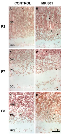
Fig. 3. The effect of MK-801 on cell apoptosis in the rat retina at different developmental stages. Sections were stained using the Feulgen method. (A,C,E) Control retinas from rats at post-natal stages P2, P7 and P8 respectively. (B,D,F) Retinas from P2, P7 and P8 rats treated with MK-801. Note the increase in the amount of apoptotic cells observed in the retina of rats treated with MK-801 during these developmental stages. Arrowheads point to apoptotic retinal cells. Scale bar, 100 µm. Abbreviations: GCL, ganglion cell layer; INL, inner nuclear layer; NBL, neuroblastic layer; ONL, outer nuclear layer.
A
B
C
D
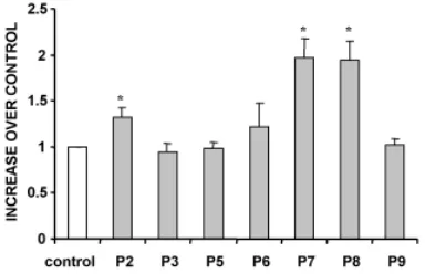
NMDA receptors was performed by administration of MK-801 at different developmental stages. We found that NMDA receptor antagonism during early post-natal developmental stages led to an increase in apoptosis in all retinal cell types. Moreover, our results indicate that the effect of glutamate on the developing retina varied according to the stage of development. Thus, the increase in apoptosis was particularly high when NMDA receptors were blocked during the postnatal stages P2, P7 and P8 in contrast with stages P4, P6 and P9, in which the increment was not significant (Figs. 3 and 4).
The effect of glutamate on apoptosis could be mediated by an increase in the levels of intracellular calcium leading the apoptosis, since it has been demonstrated that stimulation of neuronal cells by this excitatory amino acid often leads to a rise in the concentration of free cytosolic calcium (Wong, 1995).
Cell death in the retina of different species
During development, the processes of programmed cell death and cell differentiation coexist in the retina. The timing of both processes has been analyzed in different vertebrate species. The principal focus of the present article is on cell death in the developing retina. Nevertheless, we feel that it is instructive to compare the timing of both differentiation and apoptosis during the development of the retina in different vertebrate species (Tables 1 and 2).
Fish
The dynamics of cell death in the zebrafish embryo follow a similar time course to that described in other organisms (Jacobson et al., 1997; Hensey and Gautier, 1999). At 12 hours post-fertiliza-tion (hpf), one to five scattered apoptotic cells were observed primarily in the lateral margins of the optic primordium. The early onset and persistence of retina/optic vesicle apoptosis, between 12 and 30 hpf coincides with the period of evagination from the diencephalon (Schmitt and Dowling, 1994; Li et al., 2000).
Apoptotic processes in the zebrafish retina are detected in the GCL and INL at days 3-4 post-fertilization (dpf), followed by a second but clearly smaller wave at 6-7 dpf. Apoptosis in the ONL started at 5 dpf and peaked at 7 dpf (Biehlmaier et al., 2001). This peak of photoreceptor apoptosis is lower than that observed in other fish (Hoke and Fernald, 1998).
Amphibians
Pioneering studies of death in the developing frog retina showed that the physiological degeneration of retinal cells in these vertebrates takes place in three successive waves. In the first wave, mitotic cells confined to the center of the retina become pyknotic and degenerate prior to the onset of cell differentiation. Next, after the formation of the ganglion cell layer and the inner plexiform layer, many cells in the region of the incipient inner nuclear layer degenerate. Finally, a third wave of cell death is observed in the outer and inner nuclear layers in the periphery of the retina. This third wave occurs after the formation of the outer plexiform layer and the maturation of the central retina (Glucksmann, 1940).
Apoptosis of RGCs in the Xenopus retina occurs in two waves. It has been estimated that 40-70% of RGCs die by apoptosis from 1 dpf +16 hpf to 12 dpf. A second wave is observed after 26 dpf when binocular vision is developed and ipsilateral fibers begin to grow into the thalamic visual neuropil (Gaze and Grant, 1992).
Birds
Apoptosis has also been analyzed during the development of the chick retina (Cuadros and Ríos, 1988; Martín-Partido et al., 1988; Frade et al., 1996; 1997; Díaz et al., 1999, 2000; Mayor-domo et al., 2003). Two waves of programmed cellular death occur in the developing chicken retina. The first wave takes place between E4 and E7 (Fig. 5) and partially overlaps with the period of RGC neurogenesis, differentiation and migration (García-Porrero and Ojeda, 1979; Cuadros and Ríos, 1988). The later wave occurs between E10 and E14, when RGCs become depen-dent on trophic support from their targets (Hughes and La Velle, 1975; Rager and Rager, 1978; Hughes and McLoon, 1979).
In the INL, pyknotic cells appear during embryonic day 8. However, the highest levels of apoptosis are observed in this layer at E11 (Cook et al., 1998). Degenerating cells are initially located in the central retina. However, on subsequent days, they are also found in peripheral zones. It has been postulated that cell death occurs because of competition for adequate arborization space. If the number of retinal afferent fibers which arrive is higher than the number of available tectal termination sites, supernumerary fibers may degenerate (Rager and Rager, 1978).
Rodents
The normal RGC death period in the rat extends from around birth to about 6 days later and is especially high between postnatal days 1 to 4 (Galli-Resta and Ensini, 1996). The presence of degenerating bipolar cells, amacrine cells and rods has been described at postnatal day 4 in rat retinas. Subsequently, a slow decrease in the incidence of cell death occurs. However, amacrine cell death does not cease until day 26 and bipolar cell degeneration continues for 48 days. With respect to photoreceptor cell death, a second phase of apoptosis has been reported between days 12 and 72, peaking at P23 (Vogel and Möller, 1980).
Cell death in the mouse retina occurs primarily during the first 2 weeks after birth and is essentially completed by the end of the third week. The pattern of death of the different retinal cell types has been described in mice. RGC degeneration is prominent during the first 11 days, peaking on postnatal days 2 to 5 (Young, 1984). Amacrine cells die within the inner plexiform and inner nuclear layer, particularly between days 3 and 8, whereas bipolar and Müller cell degeneration reaches a peak at days 8-11.
Formation of the OPL at post-natal day 5 separates rods into two groups. Degeneration among the inner rods occurs rapidly from post-natal days 5 to 11. Nevertheless, the sporadic death of rods continues during the following two weeks (Young, 1984). Degeneration of the outer rods takes place over a longer period of time; it occurs at a much lower frequency and does not follow a center-periphery gradient. After day 18, cell death within the developing mouse retina is restricted to the sporadic, rare degen-eration of rods which ceases by postnatal day 30 (Young, 1984).
Humans
The number of retinal ganglion cells in retinas from the human fetus at 18 to 30 weeks of gestation (wg) has been estimated to be
around 2.2 - 2.5 million cells. After this stage, the number of RGCs declines rapidly to 1.5 - 1.7 million cells, through naturally occurring neuronal death (Provis et al., 1985). At early stages of fetal development, the distribution of cells in the ganglion cell layer of the human retina is almost uniform, but by the end of gestation, there is a central:peripheral gradient in cell density of about 10:1 respec-tively. Although the peripheral retina grows faster than the central retina during the first 23 weeks of gestation, this does not seem to be the cause of the centro-peripheral density gradient. Rather, the reason for this density gradient seems to be a non-uniform loss of cells in the entire retina. Between weeks 16 and 24, the relative frequency of pyknotic cells in the peripheral retina is higher than in the central retina (Provis et al., 1983; Provis and van Driel, 1985). Apoptosis in the INL is detected between 15 and 20 wg and the highest incidence of cell death in the ONL occurs at 23-24 wg (Georges et al., 1999).
Concluding comments
Many of the cellular and molecular mechanisms involved in cell death have been elucidated over the past 10 years. In particular, many of the molecular triggers which underlie cell suicide or programmed cell death have been discovered. Future research will undoubtedly focus on establishing the details of the molecular pathways activated by these triggering signals, and on establishing how particular types of cells are targeted for death or for protection from death. It will be important to establish for each cell type the precise point at which the cell is beyond salvation, i.e. the point beyond which the cell death programme is irreversible. Further studies using the retina will help to completely characterize the complex processes underlying cell death.
Acknowledgements
Financial Support: European Community (QLK6-CT-2001-00385), The Glaucoma Foundation Grant 2004, Spanish Ministry of Science and Technology (BFI 2003-07177), University of the Basque Country (E-4887/
TABLE 2
TIMING OF RETINAL CELL APOPTOSIS
Zebrafish Xenopus Chicken Rat Mouse Human Hatching/Birth 2-3 dpf 2dpf+2hpf 21 d 22 d 19 d 36 wg
ONL 5-7 dpf P12-P72 P5-P11 23-24 wg INL E8- E14 P3-P26 (AC) P3-P8 (AC) 15-20 wg
P4-P48 (BC) P8-P11 (BC,MC) GCL 3-4 dpf 1d 16h-12dpf E4-E7 P1-P6 P2-P11 14-30 wg
6-7 dpf 26 dpf E10-E14
Timing of retinal cell Apoptosis in the different layers of the retina in zebrafish (Biehlmaier et al., 2001), Xenopus (Gaze and Grant, 1992), chicken (García-Porrero and Ojeda, 1979; Cuadros and Ríos, 1988, Cook et al., 1998), rat (Vogel and Möller, 1980; Galli-Resta and Ensini, 1996), mouse (Young, 1984), and human (Provis and Van Driel, 1985; Georges et al., 1999) during retinal development. Abbreviations: AC, amacrine cells; d, days; dpf, days post-fertilization; E, embryonic day; GCL, ganglion cell layer; h, hours; hpf, hours post-fertilization; INL, inner nuclear layer; MC, Müller cells; ONL, outer nuclear layer; P, postnatal; wg, weeks of gestation.
TABLE 1
TIME OF RETINAL CELL DIFFERENTIATION
Zebrafish Xenopus Chicken Rat Mouse Human Hatching/Birth 2-3 dpf 2dpf+2hpf 21 d 22 d 19 d 36 wg
ONL 60 hpf 1d 20 1/2 h E4-E5 E16 E13-E14 (C) 13 wg (C)* E19 E13 (R) * 20-21 wg (C)**
P5 (R)**
INL 38 hpf 1d 16h-1d 20 1/2h E12 (MC) E13 –P5(AC) E17-P10 (MC) 16-17 wg (IPL) P1-P51 (MC, BC) 16-21 wg (HC) GCL 28 hpf 1 d 2 h 1/2h E2 E14-E20 E5-P0 11-12 wg
E16-E20 (ACD)
Time of retinal cell differentiation in hours, days or weeks in the retinal layers of: zebrafish (Branchek and Bremiller, 1984; Kljavin, 1987; Schmitt and Dowling, 1994), Xenopus (Stiemke and Hollyfield, 1995), chicken (Nakazawa et al., 1993; Bruhn and Cepko, 1996; Schlosshaver et al., 1997; Anezary et al., 2001; Rothermel and Layer, 2003), rat (Reese and Colello, 1992; Johanson et al., 2000), mouse (Young, 1985; Colello and Guillery, 1992), and human (Nag and Wadhwa, 1999), during retinal development. Abbreviations: AC, amacrine cells; ACD, displaced amacrine cells; BC, bipolar cells; C, cones; d, days; dpf, days post-fertilization; E, embryonic day; GCL, ganglion cell layer; h, hours; HC, horizontal cells; hpf, hours post-fertilization; INL, inner nuclear layer; IPL, inner plexiform layer; MC, Müller cells; ONL, outer nuclear layer; P, postnatal; R, rods; wg, weeks of gestation; *, first wave of differentiation; **, second wave of differentiation.
2002;15350/2003) to EV. MG holds a European Community postdoctoral fellowship. M.H. holds a pre-doctoral fellowship from the University of the Basque Country.
References
ANEZARY, L., MEDINA, J.I., SANCHEZ-NOGUEIRO, J., LOPEZ-GALLARDO, M. and PRADA, C. (2001). Shape diversity among chick retina Müller cells and their postnatal differentiation. J. Comp. Neurol. 438: 32-49.
ARY-PIRES, R., NAKATANI, M., REHEN, S.K. and LINDEN, R. (1997). Developmen-tally regulated release of intraretinal neurotrophic factors in vitro. Int. J. Dev. Neurosci. 15: 239-255.
BÄHR, M. (2000). Live or let die-retinal ganglion cell death and survival during development and in the lesioned adult CNS. Trends Neurosci. 23: 483-490.
BARBACID, M. (1994). The Trk family of neurotrophin receptors. J. Neurobiol. 25: 1386-1403.
BEAR, M.F., KLEINSCHMIDT, A., GU, Q. and SINGER, W. (1990). Disruption of experience dependent synaptic modifications in striate cortex by infusion of an NMDA receptor antagonist. J. Neurosci. 10: 909-925
BENNET, J.L., ZEILER, S.R. and JONES, K.R. (1999). Patterned expression of BDNF and NT-3 in the retina and anterior segment of the developing mammalian eye. Invest. Ophthalmol. Vis. Sci. 40: 2996-3005.
BIEHLMAIER, O., NEUHAUSS, S.C. and KOHLER, K. (2001). Onset and time course of apoptosis in the developing zebrafish retina. Cell. Tissue. Res. 306: 199-207.
BODNARENKO, S.R. and CHALUPA, L.M. (1993). Stratification of On and OFF ganglion cell dendritic depends on glutamate-mediated afferent activity in the developing retina. Nature. 364: 144-146
BODNARENKO, S.R., JEYARASASIMGAM, G. and CHALUPA, L.M. (1995). Devel-opment and regulation of dendritis stratification in retinal ganglion cells by glutamate-mediated afferent activity. J. Neurosci. 15: 7037-7045
BOVOLENTA, P., FRADE, J-M., MARTÍ, E., BARDE, Y-A. and RODRIGUEZ-TÉBAR, A. (1996). Neurotrophin 3 antibodies disrupt the normal development of the chick retina. J. Neurosci. 16: 4402-4410.
BRANCHEK, T. and BREMILLER, RJ. (1984). The development of photoreceptors in the zebrafish, Brachidanio rerio. I. Structure. J. Comp. Neurol. 224: 107-115.
BRUHN, S.L. and CEPKO, C.L. (1996). Development of the pattern of photoreceptors in the chick retina. J. Neurosci. 16: 1430-1439.
BUNCH, S.T. and FAWCEET, J. (1990). A comparison of the initial ganglion cell projection to the contralateral superior colliculus in albino and pigmented rats. Dev. Brain. Res. 52: 259-264.
BUNCH, S.T. and FAWCETT, J.W. (1993). NMDA receptors blockade alters the topography of naturally occurring ganglion cell death in the rat retina. Dev. Biol. 160-434-442.
BUTOWT, R. and VON BARTHELD, C.S. (2003). Connecting the dots: trafficking of neurotrophins, lectins and diverse pathogens by binding to the neurotrophin receptor p75 NTR. Eur. J. Neurosci.17: 673-680.
CARTER, B.D. and LEWIN, G.R. (1997). Neurotrophins live or let die: does p75NTR decide?. Neuron 18: 187-190.
CASADEMUNT, E., CARTER, B.D., BENZEL, I., FRADE, J.M., DECHANT, G. and BARDE, Y.A. (1999). The zinc finger protein NRIF interacts with the neurotrophin receptor p75NTR and participates in programmed cell death. EMBO J. 18: 6050-6061.
CELLERINO, A., BAHR, M. and ISENMANN, S. (2000). Apoptosis in the developing visual system. Cell. Tissue Res.301: 53-69
CLARKE, P.G.H. (1985). Neuronal death in the development of the vertebrate nervous system. Trends Neurosci. 8: 345-349.
CLINE, H.T. and CONSTANTINE-PATON, M. (1989). NMDA receptor antagonists disrupt the retinotectal topographic map. Neuron 3: 413-426.
CLINE, H.T. and CONSTANTINE-PATON, M. (1990). NMDA receptor agonist and antagonist alter retinal ganglion cells arbor structure in the developing frog retinotectal projection. J. Neurosci. 10: 1197-1216.
CLINE, H.T., DEBSKI, E.A. and CONSTANTINE-PATON, M. (1987). N-Methyl-D-aspartate receptor antagonist desegregates eye-specific stripes. Proc. Natl. Acad. Sci. USA 84: 4342-4345.
COHEN, A., BRAY, G.M. and AGUAYO, A.J. (1994). Neurotrophin 4 (NT-4/5) increases adult rat retinal ganglion cell survival and neurite outgrowth in vitro. J. Neurobiol. 25: 953-959.
COLELLO, R.J. and GUILLERY, R.W. (1992). Observations on the early development of the optic nerve and tract of the mouse. J. Comp. Neurol. 317: 357-378.
COOK, B., PORTERA-CAILLIAU, C. and ADLER, R. (1998). Developmental neuronal death is not a universal phenomenon among cell types in the chick embryo retina. J. Comp. Neurol. 396: 12-19.
CROOKES, J. and KOLB H. (1992). Localization of GABA, glycine, glutamate, and tyrosine hydroxylase in the human retina. J. Comp. Neurol. 315: 287-302.
CUADROS, M.A. and RIOS, A. (1988). Spatial and temporal correlation between early nerve fiber growth and neuroepithelial cell death in the chick embryo retina. Anat. Embryol. 178: 543-551.
CUI, Q. and HARVEY, A.R. (1994). NT-4/5 reduces naturally occurring retinal ganglion cell death in neonatal rats. NeuroReport. 5: 1882-1884.
DAS, I., HEMPSTEAD, B.L., MACLEISH, P.R. and SPARROW, J.R. (1997). Immu-nohistochemical analysis of the neurotrophins BDNF and NT-3 and their receptors TrkB, TrkC and p75 in the developing chick retina. Vis. Neurosci. 17: 835-842.
DAVANGER, S., OTTERSON, O.P. and MATISEN J.S. (1991). Glutamate, GABA and glycine in the human retina: an immunocytochemical investigation. J. Comp. Neurol. 311: 483-494.
DECHANT, G. and BARDE, Y.A. (1997). Signalling through the neurotrophin receptor p75NTR. Curr. Opin. Neurobiol. 7: 413-418.
DE LA ROSA, E. J., ARRIBAS, A., FRADE,.J. M. and RODRIGUEZ-TEBAR, A. (1994a). Role of neurotrophins in the control of neural development: neurotrophin-3 promotes both neuron differentiation and survival of cultured chick retinal cells. Neuroscience 58: 347-52.
DE LA ROSA, E.J., BONDY, C.A., HERNÁNDEZ-SÁNCHEZ, C., WU, X., ZHOU, J., LÓPEZ-CARRANZA, A., SCAVO, L.M. and DE PABLO, F. (1994b). Insulin and insulin-like growth factor system components gene expression in the chick from early neurogenesis until late development and the effect on neuroepithelial cells. Eur. J. Neurosci. 6: 1801-1810.
DE LA ROSA, E.J., and DE PABLO, F. (2000). Cell death in early neural development beyond the neurotrophic theory. Trends Neurosci. 23: 454-458.
DIAZ, B., PIMENTEL, B., DE PABLO, F. and DE LA ROSA, E.J. (1999). Apoptotic cell death of proliferating neuroepithelial cells in the embryonic retina is prevented by insulin. Eur. J. Neurosci. 11: 1624-1632.
DIAZ, B., SERNA, J., DE PABLO, F. and DE LA ROSA, E.J. (2000). In vivo regulation of cell death by embryonic (pro)insulin and the insulin receptor during early retinal neurogenesis. Development. 127: 1641-1649.
FRADE, J.M., BOVOLENTA, P., MARTINEZ-MORALES, J.R., ARRIBAS, A., BARBAS, J.A. and RODRÍGUEZ-TEBAR, A. (1997). Control of early cell death by BDNF in the chick retina. Development. 124: 3313-3320
FRADE, J.M., BOVOLENTA, P. and RODRÍGUEZ-TEBAR, A. (1999). Neurotrophins and other growth factors in the generation of retinal neurons. Microsc. Res. Tech. 45: 243-251
FRADE, J.M., RODRIGUEZ- TÉBAR, A., and BARDE, Y.A. (1996). Induction of cell death by endogenous nerve growth factor through its p75 receptor. Nature 383: 166-168
GALLI-RESTA, L. and ENSINI, M. (1996). An intrinsic limit between genesis and death of individual neurons in the developing retinal ganglion cell layer. J. Neurosci. 16: 2318-2324
GARCIA-PORRERO, J.A. and OJEDA, J.L. (1979). Cell death and phagocytosis in the neuroepithelium of the developing retina. A TEM and SEM study. Experientia. 35: 375 –376.
GAVRIELI, Y., SHERMAN, Y. and BEN-SASSON, S.A., (1992). Identification of programmed cell death in situ via specific labelling of nuclear DNA fragmentation. J. Cell Biol. 119: 493-501
GAZE, R.M. and GRANT, P. (1992). Spatio-temporal patterns of retinal ganglion cell death during Xenopus development. J. Comp. Neurol. 315: 264-274.
GEORGES, P., MADIGAN, M.C. and PROVIS, J.M. (1999). Apoptosis during devel-opmental of the human retina: relationship to foveal development and retinal synaptogenesis. J. Comp. Neurol. 413: 198-208
GLÜCKSMANN, A. (1965). Cell death in normal development. Arch Biol (Liege). 76: 419-37.
GRÜNDER, T., KOHLER, K., KALETTA, A. and GUENTHER, E. (2000). The distribution and developmental regulation of NMDA receptor subunit proteins in the outer and inner retina of the rat. J. Neurobiol. 44: 333-342.
HABERECHT, M.F. and REDBURN, D.A. (1996). High levels of extracellular glutamate are present in retina during neonatal development. Neurochem. Res. 21: 285-291
HALLBÖÖK, F., BÄCKSTRÖM, A., KULLANDER, K., EBENDAL, T. and CARRI, N.G. (1996). Expression of neurotrophins and trk receptors in the avian retina. J. Comp. Neurol. 364: 664-676.
HENSEY, C. and GAUTIER, J. (1999). Developmental regulation of induced and programmed cell death in Xenopus embryos. Ann. N.Y. Acad. Sci. 887: 105-119.
HERNÁNDEZ-SÁNCHEZ, C., LÓPEZ-CARRANZA, A., ALARCÓN, C., DE LA ROSA, E.J. and DE PABLO, F. (1995). Autocrine/paracrine role of insulin-related growth factors in neurogenesis: local expression and effects on cell proliferation and differentiation in retina. Proc. Natl. Acad. Sci. USA 92: 9834-9838
HERZOG, K.H., BAILEY, K. and BARDE, Y.A. (1994). Expression of the BDNF gene in the developing visual system of the chick. Development. 120: 1643-1649.
HOKE, K.L. and FERNALD, R.D. (1998). Cell death precedes rod neurogenesis in embryonic teleost retinal development. Dev. Brain Res. 11: 143-146.
HOLLMANN, M. and HEINEMANN, S. (1994). Cloned glutamate receptors. Ann. Rev. Neurosci. 17: 31-108
HUGHES, F. and LA VELLE, A. (1975). The effect of early tectal lesions on development in the retinal ganglion cell layer of chick embryos. J. Comp. Neurol. 163: 265-283
HUGHES,W.F. and MCLOON, S.C. (1979). Ganglion cell death during normal retinal development in the chick: comparisons with cell death induced by early target field destruction. Exp. Neurol. 66: 587 -601.
IKONOMIDOU, C., BOSCH, F., MIKSA, M., BITTIGAU, P., VÖCKLER, J., DIKRANIAN, K., TENKOVA, T.I., STEFOVSKA, V., TURSKI, L. and OLNEY, J.W. (1999). Blockade of NMDA receptors and apoptotic neurodegeneration in the developing brain. Science. 283: 70-74.
ISSENMANN, S., KRETZ, A. and CELLERINO, A. (2003). Molecular determinants of retinal ganglion cell development, survival, and regeneration. Prog. Ret. Eye Res. 22: 483-543.
JACOBSON, M.D., WEIL, M. and RAFF, M.C. (1997). Programmed cell death in animal development. Cell. 88: 347–354.
JOHANSSON, K., BRUUN, A., GRASBON, T. and EHINGER, B. (2000). Growth of postnatal rat retina in vitro. Development of neurotransmitter systems. J. Chem. Neuroanat. 19: 117–128.
KERR, J.F., WYLLIE, A.H. and CURRIE, A.R. (1972) Apoptosis: a basic biological phenomenon with wide- ranging implications in tissue kinetics. Br. J. Cancer. 26: 239-257.
KLEINSCHMIDT, A., BEAR, M.F. and SINGER, W. (1987). Blockade of NMDA receptors disrupts experience-dependent plasticity of kitten striate cortex. Sci-ence. 238: 355-358.
KLJAVIN, I.J. (1987). Early development of photoreceptors in the ventral retina of the zebrafish embryo. J. Comp. Neurol. 260: 461-471.
LAUBE, B., KUHSE, J. and BETZ, H. (1998). Evidence for a tetrameric structure of recombinant NMDA receptors. J. Neurosci. 18: 2954-2961
LEE, W.H., JAVEDAN, S. and BONDY, C.A. (1992). Coordinate expression of insulin-like growth factor system components by neurons and neuroglia during retinal and cerebellar development. J. Neurosci. 12: 4737-4744.
LEIN, E.S., HOHN, A. and SHATZ, C.J. (2000). Dynamic regulation of BDNF and NT-3 expression during visual system development. J. Comp. Neurol. 420: 1-18.
LI, Z., JOSEPH, N.M. and EASTER, S.S. (2000). The morphogenesis of the zebrafish eye, including a fate map of the optic vesicle. Dev. Dyn. 218: 175-188.
LINDEN, R. (1994). The survival of developing neurons-a review of afferent control. Neurosci. 85: 671-682
LOM, B. and COHEN-CORY, S. (1999). Brain-derived neurotrophic factor differen-tially regulates retinal ganglion cell dendritic and axonal arborisation in vivo. J. Neurosci. 22: 7639-7649.
MARTÍN-PARTIDO, G., RODRÍGUEZ-GALLARDO, L., ÁLVAREZ, I.S. and NAVASCUÉS, J. (1988). Cell death in the ventral region of the neural retina during early development of the chick embryo eye. Anat. Rec. 222: 272-281.
MASSEY, S.C. (1990). Cell types using glutamate as a neurotransmitter in the vertebrate retina. Prog. Retina Res. 9: 399-425.
MAYORDOMO, R., VALENCIANO, A. I., DE LA ROSA, E. J., HALLBOOK, F. (2003). Generation of retinal ganglion cells is modulated by caspase-dependent pro-grammed cell death. Eur. J. Neurosci. 18:1744-50.
MEYER-FRANKE, A., WILKINSON, G.A., KRUTTGEN, A., HU, M., MUNRO, E., HANSON, JR, M.G., REICHARDT, L.F. and BARRES, B.A. (1998). Depolariza-tion and camp elevaDepolariza-tion rapidly recruit TrkB to the plasma membrane of CNS neurons. Neuron 21: 681-693.
MORI, H. and MISHINA, M. (1995). Neurotransmitter receptors. Structure and function of NMDA receptor channel. Neuropharmacol. 34: 1219-1237
MUKAI, J., HACHIYA, T., SHOJI-HOSHINO, S., KIMURA, M.T., NADANO, D., SUVANTO, P., HANAOKA, T., LI, Y., IRIE, S., GREENE, L.A. and SATO, T.A. (2000). NADE, a p75NTR-associated cell death executor, is involved in signal transduction mediated by the common neurotrophin receptor p75NTR. J. Biol. Chem. 275: 17566-17570.
NAG, T. C. and WADHWA, S. (1999). Developmental expression of calretinin immunoreactivity in the human retina and a comparison with two other EF-hand calcium binding proteins. Neuroscience. 91: 41-50.
NAKAZAWA, T., TACHI, S., AIKAWA, E. and IHNUMA, M. (1993). Formation of the myelinated nerve fiber layer in the chicken retina. Glia. 8: 114-121.
NICOLETTI, F., BRUNO, V., COPANI, A., CASABONA, G. and KNOPFEL, T. (1996). Metabotropic glutamate receptors: a new target fro the therapy of neurodegenerative disorders? Trend. Neurosci. 19: 267-271.
NILSSON, A.S., FAINZILBER, M., FALCK, P. and IBAÑEZ, C.F. (1998). Neurotrophin-7: a novel member of the neurotrophin family from the zebrafish. FEBS Lett. 424: 285-290.
OPPENHEIM, R.W. (1991). Cell death during development of the nervous system. Annu. Rev. Neurosci. 14: 453-501.
PERRY, V.H., HENDERSON, Z. and LINDEN, R. (1983). Postnatal changes in retinal ganglion and optic axon populations in the pigmented rat. J. Comp. Neurol. 219: 356-368.
PETTMANN, B. and HENDERSON, C.E. (1998). Neuronal cell death. Neuron 20: 633-647.
POLLOCK, G., ROBICHON, R., BOYD, K.A., KERKEL, K.A. KRAMER, M., LYLES, J., AMBALAVANAR, R., KHAN, A., KAPLAN, D.R., WILLIAMS, R., and FROST, D. O. (2003) TrkB Receptor signaling regulates developmental death dynamics, but not final number, of retinal ganglion cells. J. Neurosci. 23: 10137-10145
PROVIS, J.M., BILLSON, F.A. and RUSSELL, P. (1983) Ganglion cell topography in human fetal retinae. Invest. Ophthalmol. Vis. Sci. 24: 1316-1320.
PROVIS, J.M. and VAN DRIEL, D. (1985). Retinal development in humans: the roles of differential growth rates, cell migration and naturally occurring cell death. Aust. N. Z. J. Ophthalmol. 13: 125-133.
PROVIS, J.M., VAN DRIEL, D., BILLSON, F.A. and RUSSELL, P. (1985). Develop-ment of the human retina: patterns of cell distribution and redistribution in the ganglion cell layer. J. Comp. Neurol. 22: 429-451.
RAFF, M.C., BARRES, B.A., BURNE, J.F., COLES, H.S., ISHIZAKI, Y. and JACOBSON, M.D. (1993). Programmed cell death and the control of cell survival: lesson from the nervous system. Science. 262: 695-700
RAGER, G. and RAGER, U. (1978). Systems-matching by degeneration. I. A quantitative electron microscopic study of the generation and degeneration of retinal ganglion cells in the chicken. Exp. Brain Res. 15: 33: 65-78.
REESE, B.E. and COLELLO, R.J. (1992). Neurogenesis in the retinal ganglion cell layer of the rat. Neuroscience. 46: 419-429.
RORING, B. and GRANTYN, R. (1993). Rat retinal ganglion cell express Ca2+-permeable-non-NMDA glutamate receptors during the period of histogenic cell death. Neurosci. Lett.153: 32-36
ROSENMUND, C., STERN-BACH, Y. and STEVENS, C.F. (1998). The tetrameric structure of a glutamate receptor channel. Science. 280: 1596-1599
ROTHERMEL, A. and LAYER, P.G. (2003). GDNF regulates chicken rod photorecep-tor development and survival in reaggregated histotypic retinal spheres. Invest. Ophthalmol. Vis. Sci. 44: 2221-2228.
SCHLOSSHAVER, B., BAUCH, H. and STIER, H. (1997). Photoreceptor differen-tiation analysed by the novel monoclonal antibody 1G1. Eur. J. Cell Biol. 73: 150-157.
SCHMIDT, J.T. (1990). Long-term potentiation and activity-dependent retinotopic sharpening in the regenerating retinotectal projection in goldfish: Common sensi-tive period and sensitivity to NMDA blockers. J. Neurosci. 10: 233-246.
SCHMITT, E.A., and DOWLING, J.E. (1994). Early eye morphogenesis in the zebrafish, Brachydanio derio. J. Comp. Neurol. 344: 532–542.
STIEMKE, M.M. and HOLLYFIELD, J.G. (1995). Cell birthdays in Xenopus laevis retina. Differentiation. 58: 183-193.
TORNBERRY, N.A. and LAZEBNIK, Y. (1998). Caspases: enemies within. Science. 281: 1312-1316
VARELA-NIETO, I., DE LA ROSA, E.J., VALENCIANO, A.I. and LEON, Y. (2003). Cell death in the nervous system: lessons from insulin and insulin-like growth factors. Mol. Neurobiol. 28: 23-50.
VOGEL, M. and MÖLLER, K. (1980). Cellular decay in the rat retina during normal post-natal development: A preliminary quantitative analysis of the basic endog-enous rhythm. Albrecht von Graefes Arch. Klin. Exp. Ophthalmol. 212: 243-260
WONG, R.O. (1995). Effects of glutamate and its analogs on intracellular calcium levels in the developing retina. Vis. Neurosci.12: 907-917.
YOUNG, R.W. (1984). Cell death during differentiation of the retina in the mouse. J. Comp. Neurol. 229: 362-373