Ridgeness Based Vessel Feature Detection for
Endoscopic Images
Khaleelu Rahman M1, A.Vidhyasekar2
P.G. Student, Department of Electronics and Communication Engineering, Akshaya College of Engineering and
Technology, Coimbatore, Tamil Nadu, India1
Assistant Professor, Department of Electronics and Communication Engineering, Akshaya College of Engineering and
Technology, Coimbatore, Tamil Nadu, India2
ABSTRACT: Distinctive features are crucial to many tasks in computer assisted minimally invasive surgeries (MIS). Most existing methods are difficult to extract distinctive features in MIS images. For better analysis of MIS images, resort to blood vessels that are abundant and distinctive on the tissue surfaces. In our paper, based on vascular branching points, propose a new type of vascular feature, branching segment. For special conditions in an MIS imaging environment, such as specular reflections and texture homogeneous areas, the feature points extracted by general feature point detectors are less distinctive and repeatable in MIS images. Abundant blood vessels are available on tissue surfaces and can be extracted as a new set of image features. Here two types of blood vessel features are proposed for endoscopic images: branching points and branching segments. Two novel methods, Ridgeness-Based Circle Test and Ridgeness-Based Branching Segment Detection are presented to extract branching points and branching segments, respectively.
KEYWORDS: MIS, Vascular feature, Branching segment, Branching points.
I. INTRODUCTION
Minimally Invasive Surgery (MIS) represents one of the major advances in modern healthcare. This approach has a number of well-known advantages for the patients including shorter hospitalization, reduced post-surgical trauma and morbidity. However, MIS procedures also have a number of limitations such as reduced instrument control, difficult hand-eye coordination and poor operating field localization.
Endoscopic surgery, also known as Minimally Invasive Surgery (MIS), is becoming widely deployed to replace classical surgery in order to reduce the recovery time and improve patient safety. However, there are still many challenges in improving the safety of doctors as well as patients, and reducing surgery time; namely, regular exposure to radiation, need for heavy cumbersome protective clothing covering only certain parts of the body, lack of real-time interactive 3D visualization, and automatic planning and tracking of a surgical end-effector.
Endoscopic procedures can be done to make a diagnosis, a biopsy, a surgery, etc. According to the area affected, endoscopy can also be called (non exhaustive list):
Laparoscopy (the abdominal cavity)
Thoracoscopy (the thorax area)
Arthroscopy (the joints surfaces, cartilage, and ligaments)
Rhinoscopy (the nose, nasal turbinates, frontal sinuses)
Otoscopy (the external ear)
Endovascular surgery (the blood vessels, the aorta)
Generally, the system developed is specific to the medical intervention; but, for a state-of-the-art about the actuators/sensors used, any procedures can be considered. In the literature, several solutions to enhance endoscope operability are available. Classical solutions are based on wire-driven robots. The main issue concerning wire-driven endoscope is the difficulty in identifying the friction phenomenon of the wires. Thus, the control may not be as accurate as desired. Usually, a wire-driven technology enables only the control of an endoscope tip. One solution is to assemble several independent units in a serial configuration, which results in greater design complexity. For example, Zhang et al. proposed a six degrees of freedom (dof) endoscope for fetal MIS. It is composed of 3 units of 2 dof each in order to obtain a 6 dof endoscope. Since the wire has to go through the different units, there may be interference between them. Blood vessel segmentation and visualization of blood vessels is important for clinical tasks such as diagnosis of vascular diseases, surgery planning and blood flow simulation.
A number of methods have been developed for vessel segmentation; however most of those techniques do not use a shape prior, or use a strong shape prior. With strong shape priors, such as shape templates, the segmentation is constrained to a particular shape space. Since diseased vessels can have abnormal shapes, a strict shape template may result in incorrect segmentation that misses important anatomical information. However, using image intensity alone for the task of segmentation often results in leakages perpendicular to the vessel walls at areas where the image information is ambiguous. Leakages cause the segmented model to expand in to areas that are not part of the vessel structure, and results in incorrect segmentation.
Paper is organized as follows. Section II describes the methods related to the approach of this work. The flow diagram represents the step of the algorithm. After detection vessels, how branching segment and branching point detection technique that is given in Section III. Section IV describes the steps to be followed in Image processing. Section V presents experimental results showing results of images tested. Finally, Section VI presents conclusion.
II. RELATED WORK
Different methods have been proposed to extract image features in computer vision. Depending on what information is used, these methods can be broadly classified into three categories: intensity-based detectors, first-derivative-based detectors, and second-derivative-based detectors.The methods are the first category directly relies on the comparison of pixel intensity. For example, in the Features from Accelerated Segment Test (FAST), Rosen and Drummond placed a circle at each pixel and determined that the pixel was a corner if there was a continuously bright or dark segment along the placed circle [9].
Methods in the second category are based on the first derivatives, namely Ix , Iy along x- and y- coordinates in a given raw image I. Since the first derivative is proportional to the intensity change, Ix and Iy are able to capture areas with large intensity change, such as edges and boundaries of objects. To overcome the difficulty of tissue deformation in MIS images[4], the Anisotropic Feature Detector (AFD) was introduced.
In the third category, the second derivatives of the raw image are analyzed and used for feature detection. The second derivatives have strong responses on blobs and ridges[9]. Many of these methods compute the Hessian matrix based on the second derivatives to detect interest points. Those pixels whose determinants of the Hessian matrices were local extrema in both image space and space were chosen as interest points in the Hessian-affine detector. As an approximation of Laplacian of Gaussian (the trace of the Hessian matrix), Difference of Gaussian (DoG) detected interest points as the local extreme points in both image space and scale space. Approximated the Gaussian filters with box filters in the calculation of the Hessian matrix, and theobtained Speeded Up Robust Features (SURF) detector was typically faster than the DoG detector[1].
taken in daily life. For example, MIS images contain abundant specular reflections, homogeneous areas, smokes, and so on. Much research has been presented to overcome those difficulties. Feature detectors and descriptors designed for MIS images to overcome tissue deformation. Puerto–Souza and Mariottini proposed the novel Hierarchical Multiaffine (HMA) and Adaptive Multiaffine (AMA) algorithms to improve the feature matching performance for endoscopic images.
Vesselness-Based Circle Test (VBCT) and Vesselness-Based Branching Segment Detection (VBSD) to extract the two types of vessel features based on Frangi vesselness[4]. The goal of this method is to develop a feature detector based on Frangi‟s vesselness that will be able to robustly and repeatedly produce feature points and curve segments
across different viewpoints and different lighting conditions in real time. Two types of blood vessel features are defined: branching points and branching segments[2]. Bifurcations and crossing points are defined as branching points. Branching segment is defined as a blood vessel segment which has both of its two endpoints as branching points[1]. Compared with feature points, branching segments are more powerful and distinctive visual cues whose locations, tangential directions and curvatures can all be exploited. The methods proposed in this paper to detect branching points and branching segments are named as Vesselness Based Circle Test and Vesselness Based Branching Segment Detection respectively.
III.PROPOSEDSYSTEM
Finding robust and repeatable features is a crucial problem in many tasks, such as in endoscope localization and surgical scene reconstruction. Different methods have been proposed in computer vision community to extract interest points in images based on intensity comparison, structure tensor and Hessian matrix. However, most of them are designed for general purposes and the datasets are mainly taken in the controlled environment. It is difficult for them to detect robust and repeatable features on endoscopic images because MIS images are taken inside of human body and contain homogeneously-textured organic surfaces, specular reflections and tissue-burning smokes.
Two types of blood vessel features are defined in this paper: branching points and branching segments. Bifurcations and crossing points are defined as branching points. We consider a blood vessel segment that has branching points at both ends as a branching segment as shown in the Fig 1.In the proposed system, First, image preprocessing, such as specular reflection removal, is applied on the input image. Then, Hessian matrix is calculated for each pixel, based on which Frangi vesselness and ridgeness are computed[2]. Next, circle tests are performed to detect branching points. Last, the vessel tracing technique is introduced to detect branching segments.
IV.IMAGE PROCESSING
The main type of scale space is the linear (Gaussian) scale space, which has wide applicability as well as the attractive property of being possible to derive from a small set of scale-space axioms[2]. The corresponding scale-space framework encompasses a theory for Gaussian derivative operators, which can be used as a basis for expressing a large class of visual operations for computerized systems that process visual information. This framework also allows visual operations to be made scale invariant, which is necessary for dealing with the size variations that may occur in image data, because real-world objects may be of different sizes and in addition the distance between the object and the camera may be unknown and may vary depending on the circumstances.
Frangi’s vessel detection is used to detect the vessel branches. It is based on eigenvalues of hessian matrix. Eigenvalues are important to find out tubular structures in images. In Frangi’s method it gives each pixel a 3D representation. It is done by adding the intensity value of each pixel as the Z –coordinate[2].
Hessian matrix is a square matrix of partial derivative of a function. The detector finds out hessian matrix of each pixel and calculates the eigenvalues corresponding to each pixel. This helps it to determine, whether the pixel is a part of a tubular structure or not. From hessian matrix two eigenvalues will get: -e1 and e2. For tubular structures such as vessels, one of the eigenvalue will be a large negative value (e1) and other will be a small positive or negative value (around zero) such that |e1| >> |e2|.
The Hessian detector searches for image locations that exhibit strong derivatives in two orthogonal directions. It is based on the matrix of second derivatives, the so-called Hessian: The detector computes the second derivatives Ixx, Ixy, and Iyy for each image pointand then searches for points where the determinant of the Hessian becomes maximal. The Hessian matrix describes the 2nd order local image intensity variations around the selected voxel. For the obtained
Hessian matrix its eigenvalues λi and eigenvectors are calculated[1].
Ridgeness Based on the well-known Frangi vesselness, a new blood vessel enhancement technique is introduced in this section, which is referred to as “ridgeness” in this paper. Different from the thick representation of vessels in the Frangi vesselness, we look for ridge pixels that achieve single-pixel width[4]. Here, the width of ridges is defined as the number of pixels in the direction of the eigenvector V2. For completeness, the definition of the Frangi vesselness is as
follows.
The ridge in a 2-D image is a good approximation of the vessel center line and has been extracted for vessel segmentation. Compared with the vessels in the vesselness image, the ridges are thinner and clearer. Ridges are defined
as pixels where the first derivative of the raw image intensity changes sign in the direction of the eigenvector V2
(across the vessel). Since a small amount of intensity change might flip the sign of the first derivative, the
aforementioned definition tends to detect massive “ridges” with many false positives, which include tiny vessels and
background noise. The width of the detected ridge is two pixels under this definition. Since the goal of our method is to robustly and repeatedly detect vessel features, the “false” ridges from the background need to be filtered out. Therefore,
instead of using the binary ridges directly, the pixels of the ridges are first weighted by their corresponding vesselness
Morphological image processing is a collection of non-linear operations related to the shape or morphology of features in an image. Morphological operations can also be applied to greyscale images such that their light transfer functions are unknown and therefore their absolute pixel values are of no or minor interest.
The detected candidates of branching points might contain blobs, specular reflections, branching points, and spurs. This section focuses on how to further distinguish branching points from the others. The major differences are their local structure patterns. One distinctive characteristic of branching points is that they have three or four connecting vessels. Many vessel segmentation methods have been proposed and the branching points can be identified after the vessels are
success fully segmented. Compare with those methods, the methods proposed in this paper have the advantage that they do not rely on any image segmentation techniques. Therefore, the proposed methods do not need to solve optimization problems required by many image segmentation methods. Inspired by FAST feature point detector , we propose to place a circle centered at each candidate point on the ridgeness image and examine the ridgeness value and intensity of each point along the circle to determine whether it is a branching point or not. For clarity, this process of using a circle is termed as Ridgness Based Circle Test (RBCT)[9].
The procedure of vessel tracing contained in RBSD. Since branching segment detection starts and ends at branching points, our algorithm starts from each branching point and initiates a vessel tracing process for each of its corresponding vessels. The vessel tracing process is the core of the branchin0g segment detection, and our algorithm is based on the binary mask of vessels, which is obtained by thresholding the ridgeness image and is referred to as “ridge mask.” The ridge mask has a single-pixel width in most areas, except the specular reflections[9].
V.EXPERIMENTAL RESULTS
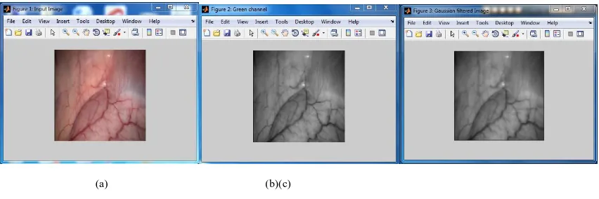
Endoscopic image is given to input as shown in Fig.2 (a) It is known that among all three channels in the RGB fundus image, the green channel provides the best contrast between vessels and the background green channel extraction has been done in image (b).
Fig. 2.(a) Original image (b) Image after applying Green Channel Extraction (c) Image after applying Gussian filtered image
Gussian filter is used for overall speckle reduction of the image, which eliminate the abundant vessels as shown in the image (c).
Fig. 3 (a) Enhanced Image (b) Eigen values of an image (c) Initial blood vessel detected image
The more structured vessels are enhanced as shown in Image (a) Negative eigenvalues indicate bright tubular structures as shown in (b) and positive eigenvalues represent dark tubular structures as shown in the (c).
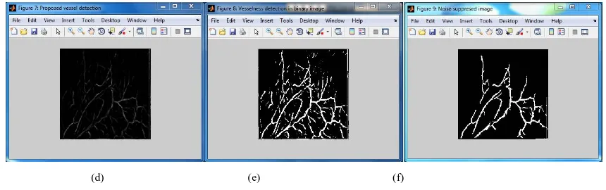
(d) Proposed vessel detection (e) Vesselness detection in binary image (f) Noise suppressed image
As we proposed the vessel detection Eigenvalues of Hessian matrix is shown in (d). To process the Circle test at a branching point we convert the image to Vesselness form as shown in the image (e). To enrich the image with robust boundary vessel noise suppression is done as shown in (f).
(g) Skeletons of vessels (h) Circle test (i) Branching Point and Branching Segment (a) (b) (c)
(d) (e) (f)
The ridgeness image as shown in the (g) is used to see the broken branching points and single pixel- width ridges.Candidate branching points after RBCT is as shown in the image (h). Those candidates are grouped into connected components. The image (i) illustrated those vessel features detected by RBSD: branching segments (green) and branching points (pink dots).
V. CONCLUSION
Blood vessel detection is one of the fundamental research topics in image guided surgeries and has many medical applications. The extraction of blood vessels provides a large number of new types of features for MIS image analysis. This paper proposes method to extract the branching segment features by using two novel methods, RBCT and RBSD, have been proposed to detect branching points and branching segments and the in vivo experimental results have validated that they are robust and distinctive features for MIS images.
REFERENCES
[1] Lin. B et al., “Vesselness based feature extraction for endoscopic image analysis”, inProc. Int. Symp. Biomed. Imag., pp. 1295–1298, 2014. [2] Baboiu D and Hamarneh G, “Vascular bifurcation detection in scale-space”, inProc. IEEEWorkshopMath. Meth. Biomed. Image Anal., pp.
41–46,2012.
[3] Lin. B et al, “Dense surface reconstruction with shadows in MIS”, IEEE Trans. Biomed. Eng., Vol. 60, No. 9, pp. 2411–2420, 2013.
[4] Lin. B, Sun.Y and X. Qian, “Thin plate spline feature point matching for organ surfaces in minimally invasive surgery imaging”, Proc. SPIE, Vol. 867112, pp. 867112-1–867112-7, 2013.
[5] Macedo M.M.G et al, “A center-line based estimator of vessel bifurcations in angiography images”, Proc. SPIE, pp. 86 703K- 1–86 703K-7, 2013.
[6] Maier-Hein L et al, “Optical techniques for 3D surface reconstruction in computer-assisted laparoscopic surgery”, Med. Image Anal., Vol. 17, No. 8, pp. 974–996, 2013.
[7] Peter Mountney et al, “A probabilistic framework for tracking deformable soft tissue in minimally invasive surgery”, in Proc.Int.Conf.Med. Image Comput. Comput-Assisted Intervention, pp. 34–41, 2013.
[8] Puerto-Souza G and Mariottini C.N, “A fast and accurate feature matching algorithm for minimally-invasive endoscopic images”, IEEE Trans. Med. Imag., Vol. 32, No. 7, pp. 1201–1214, 2013.
[9] Staal J et al, “Ridge-based vessel segmentation in color images of the retina”, IEEETrans. Med. Imag., Vol. 23, No. 4, pp. 501–509, 2014. [10] Stoyanov D et al, “Real-time stereo reconstruction in robotically assisted minimally invasive surgery”, in Proc. Int. Conf. Med. Image Comput.
Comput.-Assisted Intervention, pp. 275–282, 2013.