ABSTRACT
Stam, Christina Nicole. Prevalence and Persistence of Select Foodborne Pathogens in a mid-Atlantic Turkey Processing Facility. (Under the direction of Dr. Lee-Ann Jaykus). Listeria monocytogenes, Salmonella and Campylobacter combined are
responsible for the majority of foodborne disease hospitalizations and over 1200 deaths annually in the U.S. alone. Although raw poultry has been identified as a source of these pathogens, most microbiological studies have focused on broilers with little attention given to turkey processing. The purpose of this research was to investigate the
prevalence of select pathogens (L. monocytogenes, Salmonella spp., and Campylobacter
spp.) and microbiological indicators (Enterococcus spp.) in the turkey processing environment. Environmental samples were collected in one Southeastern processing facility using swab methods at two month intervals over a period of 14 months. Samples were taken from conveyors, drains, walls and various food contact surfaces. Isolation and identification of bacteria was done using the USDA-FSIS Microbiology Laboratory Guidebook protocols. The prevalence of contamination was 11.5%, 7.4%, and 0.4% for
L. monocytogenes, Salmonella, and Campylobacter, respectively. Enterococcus spp., an environmental indicator of fecal contamination, were isolated from over >75% of the samples screened. Salmonella isolates were typed using pulsed-field gel electrophoresis (PFGE) and Enterococcus isolates were speciated by PCR with antibiotic resistance profiles characterized using the SensiTitre system. A diverse set of relatively non-persistent Salmonella strains were obtained from the processing environment, as
susceptible to most antibiotics of human clinical relevance. Thirty-three L.
PREVALENCE AND PERSISTENCE OF SELECT FOODBORNE PATHOGENS IN A MID-ATLANTIC TURKEY PROCESSING FACILITY
By
CHRISTINA NICOLE STAM
A thesis submitted to the Graduate Faculty of North Carolina State University
In partial fulfillment of the Requirements for the Degree of
Masters of Science
DEPARTMENT OF FOOD SCIENCE Raleigh
2005
APPROVED BY
Dr. Brian Sheldon Dr. Sophia Kathariou
BIOGRAPHY
ACKNOWLEDGEMENTS
TABLE OF CONTENTS
LIST OF TABLES...v
LIST OF FIGURES...vi
CHAPTER 1 ...1
Public Health Implications of Foodborne Disease...1
HACCP and Performance Standards ...2
Role of Poultry in Foodborne Disease...4
Pathogens ...4
Prevalence of Contamination ...4
The Significance of Enterococcus spp...6
Biofilms ...8
Biofilms in Food Processing ...8
Formation of Biofilms...10
Sanitation and Control of Biofilms...14
Methods for Detecting and Studying Biofilms...16
Microbiological Strain Typing ...17
Pulsed field gel electrophoresis (PFGE) ...17
Instrumentation for PFGE...18
Advantages and Disadvantages to PFGE...20
References ...25
CHAPTER 2 ...33
Abstract...33
Introduction ...35
Materials and Methods...38
Sampling Procedure...38
Isolation of Listeria monocytogenes...38
Isolation of Salmonella...39
Isolation of Campylobacter...40
Isolation of Enterococcus...41
PCR of Enterococcus genus ...41
Enterococcus API Test Strips...43
Antiobiotic Resistance Testing of Enterococcus...43
CHROMagar Listeria...44
Biofilms...45
PFGE analysis ...46
Results ...47
Discussion...51
Conclusions ...60
LIST OF TABLES CHAPTER 1.
Table 1. Public Health implications of select foodborne pathogens ...21
Table 2. Methods of L. monocytogenes biofilms ...22
Table 3. Applications of PFGE to Salmonella typing ...23
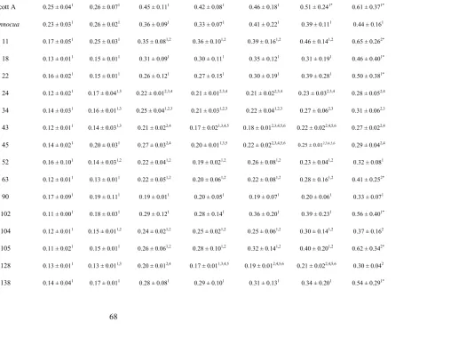
CHAPTER 2. Table 4. Pathogen prevalence of processing areas...62
Table 5. Enterococcus spp. environmental isolates ...64
Table 6. Antibiotic resistance profiles of E. faecium and E. faecalis...65
Table 7. Evaluation of BD ChromAgar...66
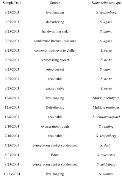
Table 8. Salmonella Serotypes...67
LIST OF FIGURES CHAPTER 1.
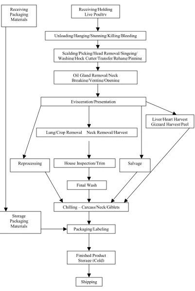
Figure 1. HACCP chart for Poultry processing facility ...24 CHAPTER 2.
CHAPTER 1 Literature Review Public Health Implications of Foodborne Disease
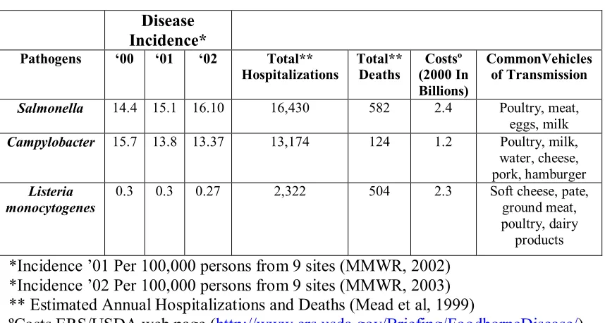
It is estimated that 76 million illnesses a year are caused by foodborne pathogens (Mead et al., 1999). Of those, 14 million illnesses are from known pathogens (Mead et al., 1999), with the bacterial pathogens of greatest concern being Listeria monocytogenes,
Campylobacter spp. and Salmonella spp.
(http://www.ers.usda.gov/Briefing/FoodborneDisease/foodandpathogens/). Listeria monocytogenes and Salmonella account for approximately 80% of all deaths from known foodborne disease agents (Mead et al., 1999). In 1999 alone, all three microorganisms combined were responsible for most of the foodborne disease hospitalizations and over 1200 deaths in the U.S. (Mead et al., 1999). The economic impact of illness associated with L. monocytogenes, Campylobacter and Salmonella has been estimated at 5.9 billion a year (http://www.ers.usda.gov/Briefing/FoodborneDisease/features.htm). This figure includes medical costs, productivity losses and costs associated with premature deaths (http://www.ers.usda.gov/Briefing/FoodborneDisease/features.htm). Interestingly, since the onset of the CDC FoodNet program, the incidence of listeriosis and
Campylobacteriosis appear to be steadily decreasing, while the incidence of Salmonella
HACCP and Performance Standards
HACCP
In 1996, the federal government passed the Pathogen Reduction: Hazard Analysis and Critical Control Point (HACCP) Systems final rule (USDA, 1996). The purpose of the HACCP rule was to provide a series of preventive controls based on 7 principles (USDA, 1996; Rose et al., 2002; McNarmara, 1997; Anonymous, 1999). These 7 principles include: (i) conducting a hazard analysis to determine where chemical,
biological and physical hazards occur in a process; (ii) establishing critical control points (CCP’s) that identify where a food safety hazard can best be controlled; (iii) setting critical limits to determine when a CCP is no longer in control and becomes a food safety hazard; (iv) monitoring CCP’s to ensure that they stay within the critical limit; (v)
establishing corrective actions when CCP’s breach the critical limit; (vi) keeping records to ensure compliance; and (vii) verification to ensure that the HACCP plan is working correctly (USDA, 1996; Anonymous, 1999) (Figure 1).
As of January 1998, all meat and poultry facilities with greater than 500
Performance Standards
In response to epidemiological data and concern over Salmonella contamination in poultry, pathogen performance standards were established for Salmonella because (i)
Salmonella was considered one of the two common causes of bacterial foodborne illness; (ii) Salmonella can colonize a variety of mammals and birds at high frequency; (iii)
Salmonella can be recovered from a variety of meat and poultry products; and (iv) reduction of fecal contamination should be effective at reducing both Salmonella and other foodborne pathogens of enteric origin (USDA, 1996). The initial baseline
performance standards for Salmonella in broilers was a prevalence of contamination of 20%; for ground chicken, prevalence was 44.6%; and for ground turkey, 49.9% (Rose et al., 2002). These percentages were based on a nationwide study and represent the maximum allowable Salmonella prevalence in a given sample set (Rose et al., 2002). The FSIS has determined that more than 80% of the facilities met the established
swine and 102 CFU/ml in cattle carcasses (USDA, 1996); also, 3 out of 13 samples can test positive for the presence of generic E.coli (USDA, 1996).
Role of Poultry in Foodborne Disease
Pathogens
Poultry and poultry products are a common vehicle of foodborne illness
(Uyttendaele et al., 1999). Ten to 18.5% of all reported foodborne disease outbreaks are associated with poultry, with turkey responsible for 56% and chickens responsible for 44% of these (Bryan and Doyle, 1995; Coleman et al., 2003). The pathogens most commonly associated with poultry include Salmonella spp., Campylobacter jejuni, Camplyobacter coli, and Listeria monocytogenes (Uyttendaele et al., 1999). Of the poultry associated foodborne disease outbreaks reported between 1992 and 1996, 67.3% were caused by Salmonella (Bryan and Doyle, 1995; Panisello et al., 2000). Per capita consumption of poultry products has been increasing (Brewer et al., 1995; Wallace et al., 1998), and the frequently cited factors contributing to poultry-associated foodborne disease continue to be improper handling and cooking of raw meat (Whyte et al., 2002; Stern et al., 2001).
Prevalence of Contamination
processing (Wallace et al., 1998). It is believed that technological changes associated with the mass production and processing of poultry have exacerbated these food safety issues (Fluckey et al., 2003).
Salmonella and Campylobacter are found in the intestinal tracts of birds, and appear to be acquired from feed and other environmental sources, with stress an
exacerbating factor (Fluckey et al., 2003; Bryan and Doyle, 1995). C. jejuni can colonize chicks, making it readily transmissible through the entire flock (Stern et al., 1988; Saleha et al., 1998). Only 3-4% of all flocks entering the processing plant are positive for
Salmonella,but 87.6% of the flocks are positive for Campylobacter (Stern et al., 2001; Sanchez et al., 2002). Campylobacter is usually found in higher amounts than
Salmonella on carcasses as well(Sanchez et al., 2002). During processing, there is no significant reduction in the numbers of Salmonella or Campylobacter, making these pathogens difficult to eliminate (Fluckey et al., 2003). S. enterica serovar Enteritidis is more prevalent on chicken carcasses and Campylobacter is more prevalent in turkeys (Wallace et al., 1998; Uyttendaele et al., 1998). For turkey carcasses, Campylobacter is usually present on 90% of carcasses and Salmonella on 2.6% (Zhao et al., 2001; Lam et al., 1992). Approximately 1.2% of turkeys and 2.2% of broiler flocks are contaminated with both C. jejuni/coli and Salmonella (Harns et al., 1986; Wedderkipp et al., 2001).
chickens, L. monocytogenes was isolated from 50-66% of all birds tested (Geornaras et al., 1995; Ojeniyi et al., 1996). Separated chicken pieces, including the skin of
drumsticks and wings, seem to have the highest level of contamination, presumably due to increased contact with processing equipment and manual handling (Geornaras and VonHoly, 2000; Franco et al., 1995). L. monocytogenes is usually detected on carcasses after evisceration, suggesting that environmental conditions during processing, rather than the bird itself, is the source of the organism (Geornaras et al., 1995; Ojeniyi et al., 1996).
Prevalence studies at the retail level show widely different contamination rates, depending upon commodity and study location. For example, 50-90% of broilers and turkeys were reportedly contaminated with Campylobacter at the retail level (Federighi et al., 1999; Sanchez et al., 2002; Lam et al., 1992). A survey done in Washington D.C. reported that 70.7% of chickens and 14% of turkeys were positive for Campylobacter
(Zhao et al., 2001). In Japan, 67.9% of chickens were positive for Campylobacter and 24.1% were positive for Salmonella (Tokumaro et al, 1990). However, in Belgian retail markets, L. monocytogenes was found more frequently on poultry, with contamination rates of 38.2%, while the prevalence of contamination with Salmonella and C. jejuni/coli
was 36.5% and 28.5%, respectively (Uyttendaele et al., 1999). The Significance of Enterococcus spp.
Enterococcus spp. have become a concern in the United States. In particular, multiple-drug-resistant E. faecalis and E. faecium are the third leading cause of all nosocomial infections in intensive care units (Johnston and Jaykus, 2004; Eaton and Gasson, 2002; Hayes et al., 2004). Historically, E. faecalis has been the dominant causative agent of these infections, but a recent increase in E. faecium infections is believed to be due to the emergence of vancomycin-resistance enterococci (VRE) in food animal production (Eaton and Gasson, 2002; Hayes et al., 2004). In the European Union, VRE have been found in broilers and pigs, and has been linked to the use of a human antimicrobial
glycopeptide, avoparcin, as a growth promoter in animal production (Manson et al., 2004; Hayes et al., 2004). Avoparcin use in animal production is thought to be responsible primarily for nonhospitalized increases in VRE infections in the European Union (Hayes et al., 2004).
production. To date, the actual importance of meat products, including poultry, to the evolution and transmission of VRE is unknown.
Biofilms
To maintain viability and growth, microorganisms seek surfaces that have been conditioned with nutrients that can promote their survival (Zottola, 1994; Kumar and Anand, 1998). Once the microorganisms multiply and colonize, they attach to the surface and form a biofilm (Donlan, 2002; Kumar and Anand, 1998; Zottola, 2001). A biofilm is a functional consortium of microbial cells that adhere to a wet surface and become immobilized in a protective polysaccharide matrix that can entrap nutrients and other microbes, allowing for subsequent microbial growth (Zottola, 2001; Donlan, 2002; Kumar and Anand, 1998; Zottola, 1994; Lindsay et al., 1996; Arnold and Silvers, 2000; Prigent-Combaret et al., 2000). The purpose of the biofilm is to provide the bacteria protection against adverse conditions in the environment such as toxicities, sanitizers, antibiotics, predators, desiccation and mechanical damage (Lindsay et al., 1996; Lee Wong, 1998; Watnick and Kolter, 1999).
Biofilms in Food Processing
1996; Kumar and Anand, 1998; Zottola, 2001). The areas of the equipment most prone to biofilm formation include dead ends, joints, valves and gaskets (LeeWong, 1998). Raw materials and ingredients have also been studied as the source of biofilm
development (Kumar and Anand, 1998). For instance, attached bacteria have been found in the collagen fibers of raw beef, pork and lamb, as well as on the skin surface of poultry (Zottola and Sasahara, 1994; Hood and Zottola, 1997; Arnold and Bailey, 2000).
Attached bacteria can enter the food supply by sloughing off from the biofilm during processing or sanitizing (Zottola and Sasahara, 1994). Contamination from biofilms is sporadic, as cells do not continuously slough, making control or determination of the source of the contaminant(s) more challenging (Zottola, 2001). If the cause of the contamination is spoilage bacteria, it can impact the processor by reducing product shelf life with associated economic losses (Kumar and Anand, 1998). Contamination from pathogenic microorganisms can have a major public health impact (Austin et al., 1998).
E. coli O157:H7, Salmonella enterica serovar. Enteritidis, L. monocytogenes, and C. jejuni have been all found to adhere to a variety of surfaces and have been resistant to sanitizers (LeeWong, 1998; Trachoo et al., 2002; Beresford et al., 2001; Joseph et al., 2001; Bonafonte et al., 2000).
Proper cleaning and sanitizing is the main element in controlling and removing biofilms (Zottola, 2001.). In one study, several biofilms consisting of various Listeria
spp. were found to be resistant to chlorine at the highest levels approved by the USDA for use in the food industry (Arnold and Silvers, 2000). In Canada, an outbreak of
Formation of Biofilms
Microorganisms form biofilms to protect themselves against adverse conditions (Lindsay et al., 1996; LeeWong, 1998). These biofilms can be comprised of single or mixed species (Kumar and Anand, 1998), although mixed species biofilms offer added protection because they are more stable and create a larger and thicker biofilm mass (Donlan, 2002). There are five stages associated with the formation and development of biofilms (Kumar and Anand, 1998; Zottola, 1994; Lindsay et al., 1996). These steps include the following: (i) transport of nutrients to a surface; (ii) surface conditioning of films; (iii) attachment of microorganisms; (iv) metabolism and growth; and finally (v) detachment and dispersal. These phases will be discussed in greater detail below.
Transport of Nutrients: Nutrients comprised of organic and inorganic molecules are transported to a surface that will promote and aid in the growth of bacteria (Zottola, 2001; Zottola, 1994). The nutrients become free-floating particles due to aerosolization during cleaning and ultimately come into contact with a solid surface and sediment, providing a substratum to allow biologically active organisms to adhere (Zottola and Sasahara, 1994).
conditioning film is not only to provide nutrients, which aid in microbial growth, but also to alter the physico-chemical properties of the surface so that the surface is conducive to microbial attachment (Kumar and Anand, 1998).
Attachment: For a bacterium to attach to a surface it first must overcome an electrostatic repulsion barrier (Donlan, 2002;). Once the cell is able to get less than 1nm from the surface, a strong attraction occurs between the surface and the bacterium, resulting in attachment (Watnick and Kolter, 1999). The initial bacterial adhesion to the surface is reversible and based on weak interactions of electrostatic attraction and Van der Waals forces (Zottola, 1994; Beresford et al., 2001; Hood and Zottola, 1997). Due to the attractive forces and Brownian motion exhibited by the bacteria, the cells can easily be removed from the surface (Kumar and Anand, 1998). At this point, product
contamination can occur more readily because the bacteria are not irreversibly attached to the surface (Arnold and Bailey, 2000).
particularly cracks and crevices that might be associated with worn stainless steel, may also promote bacterial attachment (Donlan, 2002; Zottola and Sasahara, 1994).
Metabolism and Growth: During metabolism and growth, the cells go from reversible to irreversible attachment, which is caused by the formation of extracellular polymeric substances (EPS) (Beresford et al., 2001). EPS is the most abundant material in biofilms and is comprised of polysaccharides and protein (Donlan, 2002; Hood and Zottola, 1997). EPS is produced by the microcolonies, creating a slime-like layer that binds cells to the surface, protecting and stabilizing them from the environment (Kumar and Anand, 1998; Ramesh et al., 2002; Zottola and Sasahara, 1994). The binding force created by the EPS is due to the ability of calcium and magnesium to crosslink in the polymer strands (Donlan, 2002).
In one study, L. monocytogenes was found to grow only in the presence of an EPS-producing microorganism such as P. fragi (Zottola and Sasahara, 1994). This was believed to be due in part to quorum sensing, where the bacteria behave collectively and form a biofilm to protect themselves and each other from the environment (DeKievit and Iglewski, 2000). Quorum sensing is a form of intracellular communication facilitated by molecular signals called autoinducers (DeKievit and Iglewski, 2000). When the signals reach a threshold concentration, they activate or repress the other organisms’ genes to allow for symbiotic behavior (Donlan, 2002;). The cell-to-cell signaling also allows for the bacteria to take on different behaviors (HaleBoothe et al., 1999). Quorum sensing is a fruitful area of further research and there is much to learn about its role in biofilm
formation.
Detachment and Dispersal: Detachment of biofilms is caused by physical forces of shearing, sloughing and abrasion (Donlan, 2002). Shearing is caused from flow effects that continuously remove parts of the biofilm (Donlan, 2002). Sloughing is sporadic and caused by cells at the edge of the biofilm that break off during routine cleaning (Zottola and Sasahara, 1994; Kumar and Anand, 1998). In a study by Austin et al (1998), serovar.
Enteritidis was easily sloughed from Teflon and stainless steel surfaces due to the combined effects of cell clumping and lack of fimbriae (Austin et al., 1998).
Sanitation and Control of Biofilms
Attached microorganisms are generally more resistant to sanitation chemicals than are their detached counterparts (LeeWong, 1998; Arnold and Silvers, 2000). This resistance is due to protection from organic materials and the EPS layer, which prevents chemicals from entering the biofilm or causes inactivation of the sanitizer (Donlan, 2002; Lindsay et al., 1996; Ramesh et al., 2002). Nutrient deficiency and the length of time the biofilm has been established will also affect resistance to sanitation (Kumar and Anand, 1998; Zottola, 1994). The surface may also influence the biofilm resistance because difficulties encountered when cleaning rough surfaces or non-food contact surfaces may allow for rapid biofilm reformation and/or continued growth (LeeWong, 1998; Bos et al., 1999; Zottola and Sasahara, 1994).
High temperatures, halogens, chlorine, iodine, quaternary ammonium compounds and acids are all used as sanitizers in food processing (Zottola and Sasahara, 1994). Those chemicals used for clean in place (CIP) procedures can still leave attached bacteria (LeeWong, 1998). In some instances, there is conflicting data regarding the efficacy of certain sanitizers in controlling biofilms. For example, cellulose associated with serovar.
Enteritidis biofilms correlated with chlorine resistance at approved concentrations for use (Solano et al., 2002). However, in another study, 500ppm of sodium hypochlorite and sodium chlorite with alkaline peroxide effectively eliminated Salmonella biofilms within 2 minutes (Ramesh et al., 2002).
the soil is composed of organic materials: fats, carbohydrates, proteins and minerals, all of which can inactivate the sanitizer (Zottola, 1994). After the soil is removed, a sanitizer can be used because the biofilm is no longer protected, so the sanitizer can disrupt the polysaccharide matrix (Kumar and Anand, 1998; Zottola and Sasahara, 1994; Zottola, 1994). For example, removal of necessary substrates that are rapidly metabolized is essential in controlling biofilm growth in poultry processing (HaleBoothe et al., 1999; HaleBoothe and Arnold, 2002).
There are several chemical mechanisms for controlling biofilm formation. Preventing the synthesis of EPS (Solano et al., 2002), which can be accomplished by the use of specific enzymes, is feasible but must be specific to the target biofilm microbiota (Kumar and Anand, 1998). In some instances, bacteriocins have been shown to inhibit bacterial attachment by exhibiting bactericidal properties (Kumar and Anand, 1998). For example, nisin effectively prevented surface growth of L. monocytogenes when the surface was adsorbed with the bacteriocin (Kumar and Anand, 1998). Electrochemical processes, such as electropolishing of stainless steel, results in the surface removal of metal ions, which reduces electrostatic or hydrophobic interactions, making surfaces less susceptible to bacterial attachment (Arnold and Bailey, 2000).
Various physical means to control biofilm formation are also being investigated. These include the use of electrical currents, magnetic fields and ultrasound treatments (Kumar and Anand, 1998). The use of electrical currents increases the transfer of lethal concentrations of antibiotics into the biofilm matrix by penetrating cell target sites more quickly (Kumar and Anand, 1998). Magnetic fields are more effective at higher
activity (Okuno et al., 1993). Ultrasound treatments have been used with antibiotics to speed up penetration of the antibiotics within the biofilm bacteria (Carmen et al., 2004).
Methods for Detecting and Studying Biofilms
Two methods are commonly used to screen bacterial strains for their potential as biofilm producers. As such, these are designated (i) indirect and (ii) direct methods (Djordjevic et al., 2002). For direct methods, the biofilm is actually produced on the surface of interest, and its integrity examined using microscopic methods. In the indirect approach, biofilms are produced on surfaces of interest, subsequently detached, and their formation evaluated indirectly by monitoring cell density. In this case, high cell density would suggest that a particular strain was also a good biofilm producer (Djordjevic et al., 2002). One common method involves inoculating a coupon or tube comprised of a specific material, such as stainless steel, plastic, rubber or some other material commonly used in the food-processing environment, and then using a direct method to assess growth (Beresford et al., 2001). The coupons and tubes are allowed to incubate for a select time with or without agitation (Hood and Zottola, 1997). After incubation, coupons are washed to remove any unattached cells (Beresford et al., 2001). Assessment of biofilm growth is directly measured through microscopy techniques, such as epifluorescence light and confocal scanning laser microscopy, to name a few (Kalmokoff et al., 2001; Chae and Schraft, 2000).
measurement of biofilm development, but rather uses cellular growth rates and final cell density as an indicator of biofilm formation capability (Djordjevic et al, 2002).
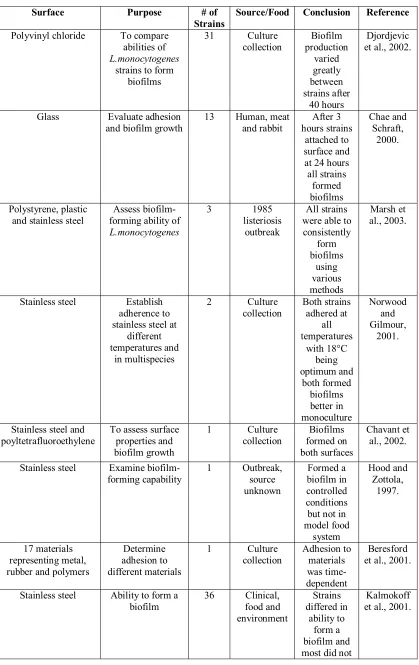
Representative studies of biofilm production among strains of Listeria monocytogenes are described in Table 2.
Microbiological Strain Typing
In microbiology, two general approaches are used to determine strain relatedness: phenotypic and genotypic methods (Farber, 1996). Phenotypic methods, including serotyping, biotyping, and bacteriophage typing, are used to determine if strains are related by virtue of phenotypic characteristics such as sugar utilization or surface antigens (Farber, 1996). The main advantage to genotyping is that it can discriminate between closely related strains (Farber, 1996). PCR typing, ribotyping, plasmid typing and pulsed-field gel electrophoresis (PFGE) are several methods commonly used to discriminate between organisms based on genetic relatedness (Farber, 1996).
Pulsed field gel electrophoresis (PFGE)
Instrumentation for PFGE
Contour-clamped homogeneous electric field (CHEF) is the most popular PFGE system (Wrestler, 1996). The CHEF system uses a hexagonal electrode array that can easily be programmed to the specific angle and pulse times (Maule, 1998). The pulses
switch through a range of 120° angles that will separate large DNA molecules with the small reorientation angles (Bustamante et al., 1993; Wrestler, 1996). The changing of angles of electric current and pulse times causes DNA to move in a zigzag motion through the gel (Wrestler, 1996; Farber, 1996). The slow migration speed of large molecules creates a longer aligning time when compared to smaller molecules (Wrestler, 1996). The size of the resolved fragments is determined by the pulse switch times; the smallest DNA fragment is controlled by the initial switch time and the largest fragment by the final switch time (Maule, 1998; Wrestler, 1996). A cooled recirculating buffer provides uniform pH of the gel, providing even discrimination of DNA banding patterns (Maule, 1998).
Sample Preparation for PFGE: Large DNA fragments are subject to shearing and need to be protected during electrophoresis (Farber, 1996). Therefore, DNA is embedded in low-melt temperature agarose plugs that protect the large DNA molecules by
different restriction enzymes because of variation in patterns. For PFGE applications to
Salmonella, the restriction enzymes XbaI and SpeI are the most commonly used (Fernandez et al., 2003).
Interpreting PFGE: Accurate comparison of PFGE patterns depends on having at least 10 fragments to interpret (Tenover et al., 1995). When determining pattern
relatedness, a scale of variations is used. In general, variation in 2-3 bands means that the strains are closely related; variations in 4-6 bands suggests that the strains are possibly related; and variations greater than 7 means the strains are different (Farber, 1996). In outbreak investigation, variation in a band or two is common and could be caused by insertions, deletions, or random point mutations (Laconcha et al., 1998).
Applications of PFGE: Early uses of PFGE for Salmonella strain typing focused almost exclusively on outbreak investigation. For instance, Salmonella banding patterns produced by the restriction enzyme BlnI were used to identify a European outbreak as the initial source of a Salmonella strain associated with a second outbreak in Chile
(Fernandez et al., 2003). Later applications focused on tracking strains through the environment. For example, PFGE patterns were used to study the source of Salmonella
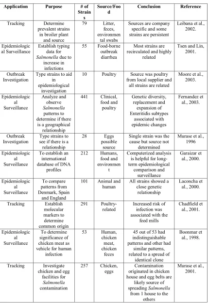
on a swine farm, where similar banding patterns suggested a connection between rodents, birds and the environment (Sandvang et al., 2000). Currently, similar yet unrelated patterns are being studied to evaluate relatedness between concurrent and/or epidemic strains (Tsen et al., 2000). Table 3 summarizes recent applications of PFGE to
Advantages and Disadvantages to PFGE
PFGE has become an invaluable tool because typing and discrimination of strains requires minimal band fragments, meaning that interpretation is relatively simple
(Garaizar et al., 2000). Once a strain is typed, its pattern will remain stable and can be duplicated for up to several years (Liesegang et al., 2002). In a study of 55 S. typhi
strains, PFGE typing using the XbaI enzyme produced 41 unique banding patterns, demonstrating that the technique can be highly discriminatory (Tsen et al., 2000). Furthermore, isolates indistinguishable by PFGE do not typically show different results when other methods are used (Sandvang et al., 2000).
However, PFGE is a time-consuming process and more rapid typing methods are available (Graves and Swaminathan, 2001; Laconcha et al., 2000). Lack of a
standardized protocol has created variability between results produced in separate labs, although this is less of a problem today (Kotetishvili et al., 2002). Standards for
Table 1. Public Health implications of select foodborne pathogens
*Incidence ’01 Per 100,000 persons from 9 sites (MMWR, 2002) *Incidence ’02 Per 100,000 persons from 9 sites (MMWR, 2003) ** Estimated Annual Hospitalizations and Deaths (Mead et al, 1999)
ºCosts ERS/USDA web page (http://www.ers.usda.gov/Briefing/FoodborneDisease/) Disease
Incidence*
Pathogens ‘00 ‘01 ‘02 Total**
Hospitalizations Total** Deaths (2000 In Costsº Billions)
CommonVehicles of Transmission
Salmonella 14.4 15.1 16.10 16,430 582 2.4 Poultry, meat, eggs, milk
Campylobacter 15.7 13.8 13.37 13,174 124 1.2 Poultry, milk, water, cheese, pork, hamburger
Listeria monocytogenes
0.3 0.3 0.27 2,322 504 2.3 Soft cheese, pate, ground meat, poultry, dairy
Table 2. Methods of L. monocytogenes biofilms
Surface Purpose # of
Strains
Source/Food Conclusion Reference
Polyvinyl chloride To compare abilities of
L.monocytogenes
strains to form biofilms
31 Culture
collection production Biofilm varied greatly between strains after 40 hours Djordjevic et al., 2002.
Glass Evaluate adhesion
and biofilm growth 13 Human, and rabbit meat hours strains After 3 attached to surface and at 24 hours all strains formed biofilms Chae and Schraft, 2000. Polystyrene, plastic and stainless steel
Assess biofilm-forming ability of
L.monocytogenes
3 1985 listeriosis
outbreak
All strains were able to consistently form biofilms using various methods Marsh et al., 2003.
Stainless steel Establish adherence to stainless steel at
different temperatures and in multispecies 2 Culture collection Both strains adhered at all temperatures with 18°C being optimum and both formed biofilms better in monoculture Norwood and Gilmour, 2001.
Stainless steel and
poyltetrafluoroethylene To assess surface properties and biofilm growth
1 Culture
collection formed on Biofilms both surfaces
Chavant et al., 2002.
Stainless steel Examine
biofilm-forming capability 1 Outbreak, source unknown
Formed a biofilm in controlled conditions but not in model food system Hood and Zottola, 1997. 17 materials representing metal, rubber and polymers
Determine adhesion to different materials
1 Culture
collection Adhesion to materials was time-dependent
Beresford et al., 2001.
Stainless steel Ability to form a
Table 3. Applications of PFGE to Salmonella typing
Application Purpose # of
Strain s Source/Foo d Conclusion Reference Tracking Determine prevalent strains
in broiler plant and source
79 Litter, feces, environmen
tal swabs
Sources are company specific and some strains are persistent
Leibana et al., 2002.
Epidemiologic al Surveillance
Establish typing data for
Salmonella due to increase in
infections
55 Food-borne outbreak
diarrhea
Most strains are recirculated and highly
related
Tsen and Lin, 2001.
Outbreak Investigation
Type strains to aid in epidemiological
investigation
10 Poultry Source was poultry from local supplier and
all strains are related
Moore et al., 2003. Epidemiologic al Surveillance Analyze and observe Salmonella patterns to determine if there
is a geographical relationship 441 Clinical, food and poultry Genetic diversity, replacement and expansion of Enteritidis subtypes associated with epidemic changes Fernandez et al., 2003. Outbreak
Investigation Type strains to see if there is a relationship
28 Eggs possible
source
Single strain was the cause but source not
determined
Murase et al., 1996
Epidemiologic al Surveillance
To establish an international database of DNA
profiles 212 Humans, food and environmen t Computerized analysis is helpful for long-term epidemiological comparison and surveillance Garaizar et al., 2000. Epidemiologic al Surveillance To compare patterns from Denmark, Spain and England
101 Animal and
human All strains showed a close genetic relationship Laconcha et al., 2000. Tracking Establish molecular markers to determine common origin 291
Poultry-related Increased risk of infection was associated with the
feed mills Chadfield et al., 2001. Epidemiologic al Surveillance To determine significance of chicken meat as vehicle for human
infection 53 Human, chicken meat, chicken feces
45 out of 53 had indistinguishable patterns and other had
similar patterns, related to a spread of
identical clone
Boonmar et al., 1998.
Tracking Investigate chicken and egg
facilities for
Salmonella
contamination
257 Chicken,
eggs originated in chicken Contamination house and egg belts are
likely source of spreading Salmonella
from 1 house to the
Figure 1. Processing flow chart for Poultry processing facility
Shipping Finished Product
Storage (Cold) Packaging/Labeling Chilling – Carcass/Neck/Giblets
Final Wash House Inspection/Trim
Lung/Crop Removal Neck Removal/Harvest Evisceration/Presentation
Oil Gland Removal/Neck Breaking/Venting/Opening
Scalding/Picking/Head Removal/Singeing/ Washing/Hock Cutter/Transfer/Rehang/Pinning
Receiving/Holding Live Poultry
Unloading/Hanging/Stunning/Killing/Bleeding
Liver/Heart Harvest Gizzard Harvest/Peel
Salvage Reprocessing
Storage Packaging
References
Anonymous. 2003. Preliminary FoodNet data on the incidence of Foodborne illnesses – Selected sites, United States, 2002. MMWR 52: 340-343.
Anonymous. 2002. Preliminary FoodNet data on the incidence of Foodborne illnesses – Selected sites, United States, 2001. MMWR 51: 325-329.
Anonymous. 2001. Preliminary FoodNet data on the incidence of Foodborne illnesses – Selected sites, United States, 2000. MMWR 50: 241-246.
Anonymous. 1999. Generic HACCP model for poultry slaughter. USDA FSIS. Arnold, J.W. and G.W. Bailey. 2000. Surface finishes on stainless steel reduced bacterial attachment and early biofilm formation: scanning electron and atomic force microscopy study. Poult. Sci. 79: 1839-1845.
Arnold, J.W. and S. Silvers. 2000. Comparison of poultry processing equipment
surfaces for susceptibility to bacterial attachment and biofilm formation. Poult. Sci. 79: 1215-1221.
Austin, J.W., G. Sanders, W. W. Kay, S. K. Collinson. 1998. Thin aggregative fimbriae enhance Salmonella enteritidis biofilm formation. FEMS Microbiol. Lett. 162: 295-301. Beresford, M.R., P.W. Andrew, G. Shama. 2001. Listeria monocytogenes adheres to many materials found in food-processing environments. J. Appl. Microbiol. 90: 1000-1005.
Bonafonte, M.A., C. Solano, B. Sesma, M. Alvarez, L. Montuenga, D. Garcia-Fos, C. Gamazo. 2000. The relationship between glycogen synthesis, biofilm formation, and virulence in Salmonella enteritidis. FEMS Microbiol. Lett. 191: 31-36.
Boonmar, S., A. Bangtrakulnonth, S. Pornrunangwong, J. Terajima, H. Watanabe, K.I. Kaneko, M. Ogawa. 1998. Epidemiological analysis of Salmonella enteritidis isolates from humans and broiler chickens in Thailand by phage typing and pulsed-field gel electrophoresis. J. Clin. Microbiol. 36: 971-974.
Bos, R., H.C. VanDermei, H.J. Busscher. 1999. Physico-chemistry of initial microbial adhesive interactions – its mechanisms and methods for study. FEMS Microbiol. Rev. 23: 179-230.
Bryan, F.L. and M.P. Doyle. 1995. Health risks and consequences of Salmonella and
Campylobacter jejuni in raw poultry. J. Food Prot. 58: 326-344.
Bustamante, C., S. Gurrieri, S. B. Smith. 1993. Towards a molecular description of pulsed-field gel electrophoresis. Trends Biotechnol. 11: 23-30.
Capita, R., C. Alonso-Callejo, B. Moreno, M.C. Garcia-Fernandez. 2001. Occurrence of Listeria species in retail poultry meat and comparison of a cultural/immunoassay for their detection. Int. J. Food Microbiol. 65: 75-82.
Carmen, J.C., J.L. Nelson, B.L. Beckstead, C.M Runyan, R.A. Robison, G.B Schaalje, W.G. Pitt. 2004. Ultrasonic-enhanced gentamicin transport through colony biofilms of
Pseudomonas aeruginosa and Escherichia coli. J. Infect. Chemother. 10: 193-199. Chadfield, M., M. Skov, J. Christensen, M. Madsen, M. Bisgaard. 2001. An
epidemiological study of Salmonella enterica serovar 4, 12:b:- in broiler chickens in Denmark. Vet. Microbiol. 825: 233-247.
Chae, M.S. and H. Schraft. 2000. Comparative evaluation of adhesion and biofilm formation of different Listeria monocytogenes strains. Int. J. Food Microbiol. 62: 103-111.
Chavant, P., B. Martinie, T. Meylheuc, M.N. Bellon-Fontaine, M. Hebraud. 2002.
Listeria monocytogenes LO28: surface physicochemical properties and ability to form biofilms at different temperatures and growth phases. Appl. Environ. Microbiol. 68: 728-737.
Coleman, M.E., S. Sandberg, S.A. Anderson. 2003. Impact of microbial ecology of meat and poultry products on predictions from exposure assessment scenarios for refrigerated storage. Risk Analysis 23: 215-228.
DeKievit, T.R. and B.H. Iglewski. 2000. Bacterial quorum sensing in pathogenic relationships. Infect. Immunol. 68: 4839-4849.
Djordjevic, D., M. Wiedmann, L.A. McLandsborough. 2002. Microtiter plate assay for assessment of Listeria monocytogenes biofilm formation. Appl. Environ. Microbiol. 68: 2950-2958.
Donlan, R.M. 2002. Biofilms: Microbial life on surfaces. EID. 8: 881-890. Eaton, T.J. and M.J. Gasson. 2002. A variant enterococcal surface protein Espfm in Enterococcus faecium; distribution among food, commensal, medical, and environmental isolates. FEMS Microbiol. Lett. 216: 269-275.
Federighi, M., C. Magras, M.F. Pilet, D. Woodward, W. Johnson, F. Juglau, J.L. Jouve. 1999. Incidence of thermotolerant Campylobacter in foods assessed by NF ISO 10272 standard: results of a two-year study. Food Microbiol. 16: 195-204.
Fernandez, J., A. Fica, G. Ebensperger, H. Calfullan, S. Prat, A. Fernandez, M. Alexandre, I. Heitmann. 2003. Analysis of molecular epidemiology of Chilean
Salmonella enterica Serotype Enteritidis isolates by pulsed-field gel electrophoresis and bacteriophage typing. J. Clin. Microbiol. 41: 1617-1622.
Franco, C.M., E.J. Quinto, C. Fente, J.L. Rodriguez-Otero, L. Dominguez, A. Cepeda. 1995. Determination of the principal sources of Listeria spp. contamination in poultry meat and a poultry processing plant. J. Food Prot. 58: 1320-1325.
Fluckey, W.M., M.X. Sanchez, S.R. McKee, D. Smith, E. Pendleton, M.M. Brashears. 2003. Establishment of a microbiological profile for an air-chilling poultry operation in the United States. J. Food Prot. 66: 272-279.
Garaizar, J., N. Lopez-Molina, I. Laconcha, D.L. Baggesen, A. Rementeria, A. Vivanco, A. Audicana, I. Perales. 2000. Suitability of PCR fingerprinting, infrequent-restriction-site PCR, and pulsed-field gel electrophoresis, combined with computerized gel analysis, in library typing of Salmonella enterica Serovar Enteritidis. Appl. Environ. Microbiol. 66: 5273-5281.
Geornaras, I., A. DeJesus, E. VanZyl, A. VonHoly. 1995. Microbiological survey of a South African poultry processing plant. J. Basic Microbiol. 35: 73-82.
Geornaras, I. and A. VonHoly. 2000. Bacterial counts associated with poultry processing at different sampling times. J. Basic Microbiol. 45: 343-349.
Graves, L.M. and B. Swaminathan. 2001. PulseNet standardized protocol for subtyping
Listeria monocytogenes by macrorestriction and pulsed-field gel electrophoresis. Int. J. Food Microbiol. 65: 55-62.
HaleBoothe, D.D. and J.W. Arnold. 2002. Nutrient substrates used by bacterial isolates from the poultry processing environment. Poult. Sci. 81: 1392-1405.
HaleBoothe, D.D., J.W. Arnold, V. Chew. 1999. Utilization of substrates by bacterial communities (biofilm) as they develop on stored chicken meat samples. Poult. Sci. 78: 1801-1809.
Harns, N.V., D. Thompson, D.C. Martin, C.M. Nolan. 1986. A survey of
Hayes, J.R., L.L. English, L.E. Carr, D.D. Wagner, S.W. Joseph. 2004. Multiple-antibiotic resistance of Enterococcus spp. isolated from commercial poultry production environments. Appl. Environ. Microbiol. 70: 6005-6011.
Hood, S.K. and E.A. Zottola. 1997. Adherence to stainless steel by foodborne
microorganisms during growth in model food systems. Int. J. Food Microbiol. 37: 145-153.
Johnston, L.M., L.-A. Jaykus. 2004. Antimicrobial resistance of Enterococcus species isolated from produce. Appl. Environ. Microbiol. 70: 3133-3137.
Joseph, B., S.K. Otta, I. Karunasagar, I. Karunasagar. 2001. Biofilm formation by
Salmonella spp. on food contact surfaces and their sensitivity to sanitizers. Int. J. Food Microbiol. 64: 367-372.
Kalmokoff, M.L., J.W. Austin, X.-D. Wan, G. Sanders, S. Banerjee, J.M. Farber. 2001. Adsorption, attachment and biofilm formation among isolates of Listeria monocytogenes
using model conditions. J. Appl. Microbiol. 91: 725-734.
Kotetishvili, M., O.C. Stine, A. Kreger, J.G. Morris, A. Sulakvelidze. 2002. Multilovus sequence typing for characterization of clinical and environmental Salmonella strains. J. Clin. Microbiol. 40: 1626-1635.
Kumar, C.G. and S.K. Anand. 1998. Significance of microbial biofilms in the food industry: a review. Int. J. Food Microbiol. 42: 9-27.
Laconcha, I., D.L. Baggesen, A. Rementeria, J. Garaizar. 2000. Genotypic
characterization by PFGE of Salmonella enterica serotype Enteritidis phage types 1, 4, 6, and 8 isolated from animal and human sources in three European countries. Vet.
Microbiol. 75: 155-165.
Laconcha, I., N. Lopez-Molina, A. Rementeria, A. Audicana, I. Perales, J. Garaizar. 1998. Phage typing combined with pulsed-field gel electrophoresis and random
amplified polymorphic DNA increases discrimination in the epidemiological analysis of
Salmonella enteritidis strains. Int. J. Food Microbiol. 40: 27-34.
Lam, K.M., A.J DaMassa, T.Y Morishita, H.L. Shivaprasad, A.A. Bickford. 1992. Pathogenicity of Campylobacter jejuni for turkey and chickens. Avian Dis. 36: 359-363. LeeWong, A.C. 1998. Biofilms in food processing environments. J. Dairy Sci. 81: 2765-2770.
Liesegang, A., D. Davos, J.C. Balzer, W. Rabsch, R. Prager, D. Lightfoot, A. Siitonen, H. Claus, H. Tschape. 2002. Phage typing and PFGE pattern analysis as tools for
epidemiological surveillance of Salmonella enterica serovar Bovismorbificans infections. Epidemiol. Infect. 128: 119-130.
Lindsay, D., I. Geornaras, A. VonHoly. 1996. Biofilms associated with poultry processing equipment. Microbios. 86: 105-116.
Manson, J.M., J.M.B. Smith, G.M. Cook. 2004. Persistence of vancomycin-resistant Enterococci in New Zealand broilers after discontinuation of avoparcin use. Appl. Environ. Microbiol. 70: 5764-5768.
Marsh, E.J., H. Luo, H. Wang. 2003. A three-tiered approach to differentiate Listeria monocytogenes biofilm-forming abilities. FEMS Microbiol. Lett. 228: 203-210. Maule, J. 1998. Pulsed-field gel electorphoresis. Molecular Biotechnol. 9: 107-126. McNamara, A.M. 1997. Generic HACCP application in broiler slaughter and
processing. J. Food Prot. 60: 579-604.
Mead, P.S., L. Slutsker, V. Dietz, L.F. McCaig, J.S. Bresee, C. Shapiro, P.M. Griffin, R.V. Tauxe. 1999. Food-related illness and death in the United States. EID. 5: 607-625. Moore, J.E., L. Murray, S. Fanning, M. Cormican, M. Daly, N. Delappe, B. Morgan, P.G. Murphy. 2003. Comparison of phenotypic and genotypic characteristics of Salmonella bredeney associated with a poultry-related outbreak of gastroenteritis in Northern Ireland. J. Infect. 47: 33-39.
Møretrø, T., L. Hermansen, A.L. Holck, M.S. Sidhu, K. Rudi, S. Langsrud. 2003. Biofilm formation and the presence of the intercellular adhesion locus ica among staphylococci from food and food processing environments. Appl. Environ. Microbiol. 69: 5648-5655.
Murase, T., A. Nakamura, A. Matsushima, S. Yamai. 1996. An epidemiological study of
Salmonella enteritidis by pulsed-field gel electrophoresis (PFGE): several PFGE patterns observed in isolates from a food poisoning outbreak. Microbiol. Immunol. 48: 873-875. Murase, T., T. Okitsu, R. Suzuki, H. Morozumi, A. Matsushima, A. Nakamura, S. Yamai. 1995. Evaluation of DNA fingerprinting by PFGE as an epidemiologic tool for
Salmonella infections. Microbiol. Immunol. 39: 673-676.
Nadeau, E., S. Messier, S. Quessy. 2003. Comparison of Campylobacter isolates from poultry and humans: association between in vitro virulence properties, biotypes, and pulsed-field gel electrophoresis clusters. Appl. Environ. Microbiol. 69: 6316-6320. Norwood, D.E. and A. Gilmour. 2001. The differential adherence capabilities of two
Listeria monocytogenes strains in monoculture and multispecies biofilms as a function of temperature. Lett. Appl. Microbiol. 33: 320-324.
Ojeniyi, B., A.C. Wegener, N.E. Jensen, M. Bisgaard. 1996. Listeria monocytogenes in poultry and poultry products: epidemiological investigations in seven Danish abattoirs. J. Appl. Bacteriol. 80: 395-401.
Okuno, K., K. Tuchiya, T. Ano, M. Shoda. 1993. Effect of super high magnetic field on the growth of Escherichia coli under various medium compositions and temperatures. J. Ferm. Bioeng. 75: 103-106.
Panisello P.J., R. Rooney, P.C. Quantick, R. Stanwell-Smith. 2000. Application of foodborne disease outbreak data in the development and maintenance of HACCP systems. Int. J. Food Microbiol. 59: 221-234.
Prigent-Combaret, C., G. Prensier, L. Thitt, O. Vidal, P. Lejeune, C. Doral. 2000.
Developmental pathway for biofilm formation in curli-producing Escherichia coli strains: role of flagella, curli and colanic acid. Environ. Microbiol. 2: 450-464.
Ramesh, N., S.W. Joseph, L.E. Carr, L.W. Douglass, F.W. Wheaton. 2002. Evaluation of chemical disinfectants for the elimination of Salmonella biofilms from poultry transport containers. Poult. Sci. 81: 904-910.
Rose, B.E., W.E. Hill, R. Umholtz, G.M. Ranson, W.O. James. 2002. Testing for
Salmonella in raw meat and poultry products collected at federally inspected establishments in the United States, 1998 through 2000. J. Food Prot. 65: 937-947. Saleha, A.A., G.C. Mead, A.L. Ibrahim. 1998. Campylobacter jejuni in poultry production and processing in relation to public health. Worlds Poult. Sci. J. 54: 49-58. Sanchez, M.X., W.M. Fluckey, M.M. Brashears, S.R. McKee. 2002. Microbial profile and antibiotic susceptibility of Campylobacter spp. and Salmonella spp. in broilers processed in air-chilled and immersion-chilled environments. J. Food Prot. 65: 948-956.
Sandvang, D., L.B. Jensen, D.L. Baggesen, S.B. Baloda. 2000. Persistence of a
Solano, C., B. Garcia, J. Valle, C. Berasain, J.M. Ghigo, C. Gamazo, I. Lasa. 2002. Genetic analysis of Salmonella enteritidis biofilm formation: critical role of cellulose. Mol. Microbiol. 43: 793-808.
Stern, N.J., J.S. Bailey, L.C. Blankenship, N.A. Cox, F. McHan. 1988. Colonization characteristics of Campylobacter jejeuni in chick ceca. Avain Dis. 32: 330-334. Stern, N.J., P. Fedorka-Cray, J.S. Bailey, N.A. Cox, S.E. Craven, K.L. Hiett, M.T. Musgrove, S. Ladely, D. Cosby, G.C. Mead. 2001. Distribution of Campylobacter spp. in selected U.S. poultry production and processing operations. J. Food Prot. 64: 1705-1710.
Tenover, F.C., R.D. Arbeit, R.V. Goering, P.A. Mickelsen, B.E. Murray, D.H. Persing, B. Swaminathan. 1995. Interpreting chromosomal DNA restriction patterns produced by pulsed-field gel electrophoresis: criteria for bacterial strain typing. J. Clin. Microbiol. 33: 2233-2239.
Tokumaro, M., H. Konuma, M. Umesako, S. Konno, K. Shinagawa. 1990. Rates of detection of Salmonella and Campylobacter in meats in response to the sample size and the infection level of each species. Int. J. Food Microbiol. 13: 41-46.
Trachoo, N., J.F. Frank, N.J. Stern. 2002. Survival of Campylobacter jejuni in biofilms isolated from chicken houses. J. Food Prot. 65: 1110-1116.
Tsen, H.Y., H.H. Hu, J.S. Lin, C.H. Huang, T.K. Wang. 2000. Analysis of the Salmonella typhimurium isolates from food-poisoning cases by molecular subtyping methods. Food Microbiol. 17: 143-152.
Tsen, H.Y. and J.S. Lin. 2001. Analysis of Salmonella enteritidis strains isolated from food-poisoning cases in Taiwan by pulsed field gel electrophoresis, plasmid profild and phage typing. J. Appl. Microbiol. 91: 72-79.
USDA. 1996. US Department of Agriculture. Pathogen reduction; hazard analysis and critical control point (HACCP) system; final rule. Fed. Regist. 61, 38805-38989. Uyttendaele, M.P., J.M. Debevere, R.M. Lips, K.D. Neyts. 1998. Prevalence of
Salmonella in poultry carcasses and their products in Belgium. Int. J. Food Microbiol. 40: 1-8.
Uyttendaele, M., P. DeTroy, J. Debevere. 1999. Incidence of Salmonella,
Uyttendaele, M., K.D. Neyts, R.N. Lips, J.M. Debevere. 1997. Incidence of Listeria monocytogenes in poultry and poultry products obtained from Belgian and French abattoirs. Food Microbiol. 14: 339-345.
Wallace, J.S., K.N. Stanley, K. Jones. 1998. The colonization of turkeys by thermophilic
Campylobacters. J. Appl. Microbiol. 85: 224-230.
Watnick, P.I. and R. Kolter. 1999. Steps in the development of a Vibrio cholerae El Tor biofilm. Mol. Microbiol. 34: 586-595.
Wedderkopp, A., K.O. Gradel, J.C. Jorgensen, M. Madsen. 2001. Pre-harvest
surveillance of Campylobacter and Salmonella in Danish broiler flocks: a 2-year study. Int. J. Food Microbiol. 68: 53-59.
Whyte, P., K. McGill, J.D. Collins, E. Gormley. 2002. The prevalence and PCR detection of Salmonella contamination in raw poultry. Vet Microbiol. 89: 53-60.
Wrestler, J.C., B.D. Lipes, B.W. Birren, E. Lai. 1996. Pulsed-field gel electorphoresis. Methods Enzymol. 270: 225-272.
Zhao, C., B. Ge, J. DeVillena, F. Sudler, E. Yeh, S. Zhao, D. White, D. Wagner, J. Meng. 2001. Prevalence of Campylobacter spp. Escherichia coli, and Salmonella serovars in retail chicken, turkey, pork, and beef from the greater Washington D.C., area. Appl. Environ. Microbiol. 67: 5431-5436.
Zottola, E.A. 1994. Microbial attachment and biofilm formation: a new problem for the food industry? Food Technol. 48: 107-114.
Zottola, E.A. 2001. Reflections on Salmonella and other “wee beasties” in foods. Food Technol. 55: 60-67.
Zottola, E.A. and K.C. Sasahara. 1994. Microbial biofilms in the food processing industry – should they be a concern? Int. J. Food Microbiol. 23: 125-148.
CHAPTER 2
Prevalence and Persistence of Select Foodborne Pathogens in a Mid-Atlantic Turkey Processing Facility
Abstract
Listeria monocytogenes, Salmonella and Campylobacter combined are
responsible for the majority of foodborne disease hospitalizations and over 1200 deaths annually in the U.S. alone. Although raw poultry has been identified as a source of these pathogens, most microbiological studies have focused on broilers with little attention given to turkey processing. The purpose of this research was to investigate the
prevalence of select pathogens (L. monocytogenes, Salmonella spp., and Campylobacter
spp.) and microbiological indicators (Enterococcus spp.) in the turkey processing plant environment. Environmental samples were collected in one Southeastern processing facility using swab methods at two month intervals over a period of 14 months. Samples were taken from conveyors, drains, walls and various food contact surfaces. Isolation and identification of bacteria was done using the USDA-FSIS Microbiology Laboratory Guidebook protocols. The prevalence of contamination was 11.5%, 7.4%, and 0.4% for
L. monocytogenes, Salmonella, and Campylobacter, respectively. Enterococcus spp., an environmental indicator of fecal contamination, were isolated from over >75% of the samples screened. Salmonella isolates were typed using pulsed-field gel electrophoresis (PFGE) and Enterococcus isolates were speciated by PCR with antibiotic resistance profiles characterized using the SensiTitre system. A diverse set of relatively non-persistent Salmonella strains were obtained from the processing environment, as
E. faecium and 55% were E. faecalis. Both E. faecalis as E. faecium strains were susceptible to most antibiotics of human clinical relevance. Thirty-three L.
Introduction
It is estimated that 76 million illnesses a year are caused by foodborne pathogens (Mead et al., 1999). Of those, 14 million illnesses occur from known pathogens (Mead et al., 1999), with the bacterial pathogens of greatest concern being Listeria monocytogenes,
Campylobacter spp. and Salmonella
(http://www.ers.usda.gov/Briefing/FoodborneDisease/foodandpathogens/). Listeria monocytogenes and Salmonella account for approximately 80% of all deaths from known foodborne disease agents (Mead et al., 1999). In 1999 alone, all three microorganisms combined were responsible for most of the foodborne disease hospitalizations and over 1200 deaths (Mead et al., 1999). The U.S. economic impact of illness associated with L. monocytogenes, Campylobacter and Salmonella has been estimated at 5.9 billion a year (http://www.ers.usda.gov/Briefing/FoodborneDisease/features/). Interestingly, since the onset of the CDC FoodNet program, the incidence of listeriosis and campylobacteriosis appear to be steadily decreasing, while the incidence of Salmonella infection has continued to increase over the 3 year period from 2000 to 2002 (Anonymous, 2003; Anonymous, 2002; Anonymous, 2001).
Wedderkipp et al., 2001). Unfortunately, most microbiological studies aimed at
estimating the incidence of foodborne pathogen contamination in poultry have focused on broilers, with relatively less attention given to turkey processing.
Enterococcus spp. have become a concern in the United States due to their ubiquitous nature and recent clinical disease associated with this genus. In particular, multiple-drug-resistant E. faecalis and E. faecium are the third leading cause of all nosocomial infections in intensive care units (Johnston and Jaykus, 2004; Eaton and Gasson, 2002; Hayes et al., 2004). Historically, E. faecalis has been the dominant causative agent of these infections, but a recent increase in E. faecium infections is believed to be due to the emergence of vancomycin-resistance enterococci (VRE) in food animal production (Eaton and Gasson, 2002; Hayes et al., 2004). In the European Union, VRE have been found in broilers and pigs, and has been linked to the use of a human antimicrobial glycopeptide, avoparcin, as a growth promoter in animal production
(Manson et al., 2004; Hayes et al., 2004). Avoparcin use in animal production is thought to be responsible primarily for nonhospitalized increases in VRE infections in the
European Union (Hayes et al., 2004).
Pathogenic and spoilage bacteria have been shown to attach to a wide variety of food-contact and non-contact surfaces (Lindsay et al., 1996; LeeWong, 1998). Such attachment of microorganisms to food contact surfaces, frequently referred to as biofilms, can impact the food industry by complicating sanitation and increasing the risk of cross contamination with spoilage and pathogenic microorganisms (Austin et al., 1998). E. coli
(LeeWong, 1998; Trachoo et al., 2002; Beresford et al., 2001; Joseph et al., 2001; Bonafonte et al., 2000). Furthermore, many different food contact surfaces are prone to biofilm production, including glass, rubber, plastics, aluminum, stainless steel, Teflon, Buna-N (dairy processing) and rubber “fingers” (poultry processing). Floor drains and crevices in floors, as well as dead ends, joints, valves and gaskets of processing
equipment, are also at risk for biofilm development (LeeWong, 1998).
The strains of pathogenic bacteria that colonize turkey processing plants and contaminate turkey products have not been characterized. There is a clear need to further investigate prevalence, strain subtypes and key strain attributes of these pathogens in this processing environment. This study was part of a broader project, the purpose of which was to investigate prevalence of select pathogens in the turkey processing industry by a systematic examination of environmental and product-associated contamination in processing facilities distributed in three geographical regions of the country (Eastern Seaboard, Midwest, West Coast). Accordingly, in this project, we report on the
prevalence of Listeria monocytogenes, Campylobacter, and Salmonella spp. in a single processing facility located in the mid-Atlantic States. We also had the opportunity to isolate Enterococcus spp. also naturally present in this environment, and characterize their antibiotic resistance profiles. Salmonella isolates were serotyped and genotyped, while L.monocytogenes strains were characterized for their ability to form biofilms. Finally, in cooperation with a multi-national media manufacturer, we had the opportunity to evaluate the performance of a new commercial Listeria selective medium as applied to
Materials and Methods
Sampling Procedure
All samples were collected from a mid-Atlantic turkey processing plant every other month for 14 months. Samples were collected only during the first shift while lines were operating and during sanitation breaks. All environmental samples were taken of a 3 x 3in area by wiping the surface with the sterile sponge in a back and forth motion according to the protocol established by International BioProducts Inc. [SpongeSicle, International BioProducts Inc., Bothell, WA]. Swabs were placed in a cooler for transport back to the laboratory. Samples were processed the same day for Salmonella, Campylobacter, Listeria monocytogenes and Enterococcus.Sponge swabs were
aseptically cut into 4 equal pieces using scissors. Unless otherwise specified all microbiological methods followed the protocol in the USDA-FSIS Microbiology Laboratory Guidebook (2002).
Isolation of Listeria monocytogenes
incubated for 30min at 37°C. Wells were emptied by quickly inverting the contents into a waste container. Residual liquid was removed by striking the holder several times face down onto absorbent paper towels. Using a nozzle squeeze bottle, the wells were washed with the supplied Tecra Wash Solution 3 times. 200µl of conjugate solution supplied in the kit was added to each well. The wells were covered with cling wrap and incubated for 30min at 37°C. After the 30min, the wells were washed 4 times with the supplied Tecra Wash Solution. 200µl of substrate was added to each well and incubated at room temperature for 10min. Test results were read using the supplied Tecra Color Card to verify proper color change. All presumptive positive samples was streaked onto modified oxford agar (Becton Dickinson and Co.) and incubated for 24 hrs at 37°C. Suspect colonies, approximately 5 were transferred to 5% sheep blood agar (Remel, Lenexa, KS) and incubated at 37°C for 24 hrs. β-lysin CAMP factor test was performed on suspect colonies by placing a β-lysin disc (Remel) in the center of a 5% sheep blood agar plate, and streaking 4-8 isolates in straight lines away from the disc. Plates were incubated at 37°C for 35hrs. Positive samples were determined by an arrowhead shaped
β-hemolysis zone around the disc. Presumptive Listeria Samples were verified using a MicroID test kit (Remel).
Isolation of Salmonella
(Becton Dickinson and Co.) and incubated at 37°C. After 24 hrs a Salmonella VIA (Tecra International Pty Ltd) kit was run according to the manufacturers’ instructions and as described above. Presumptive positive samples were streaked on double modified lysine iron agar (Oxoid Ltd), xylose lysine tergitol 4 and brilliant green sulfa agar (Becton Dickinson and Co.) and incubated at 37°C for 48 hrs. Select colonies, approximately 5 were inoculated on to triple sugar iron and lysine iron slants (Becton Dickinson and Co.) and incubated for 24 hrs at 37°C. All presumptive positive samples were sent to the Ames, IA USDA APHIS National Veterinary services laboratories for further serotyping.
Isolation of Campylobacter
The swab was enriched in 50mL Hunt Enrichment Broth (Oxoid; Becton Dickinson and Co.; Sigma, St. Louis, MO) in a quart-size ziplock freezer bag (S.C. Johnson and Son, Inc., Racine, WI), gased with a mixture of 5% O2, 10% CO2 and 85%
N2 and incubated at 37°C for 4 hrs. The enrichment was then re-gased and incubated for
an additional 20 hrs at 42°C. Samples were then streaked onto modified Campylobacter
(Becton Dickinson and Co.). A disc of 30µg nalidixic acid (Becton Dickinson and Co.) and a disc of 30µg cephalothin (Becton Dickinson and Co.) were aseptically placed on the Brucella-FBP agar using a single disc dispenser (Becton Dickinson and Co.). Plates were incubated in an anaerobic jar at 42° C for 1-3 days. Antibiotic resistance was determined by a clear zone around the discs.
Isolation of Enterococcus
The procedure for the isolation of Enterococcus was done according to U.S. Food and Drug Administration (Simjee et al., 2002; Hayes et al., 2003; D. D. Wagner [Food and Drug Administration], personal communication). Samples were first enriched in enterococcosel broth (Becton Dickinson and Co.) at 44°C for 48 hours and then streaked on enterococcosel agar (Becton Dickinson and Co.) and incubated at 37°C. After 24 hours, colonies showing esculin hydrolysis were streaked on Brain Heart Infusion agar (Becton Dickinson and Co.) and incubated at 37°C for 24 hours. The samples were then inoculated into BHI broth (Becton Dickinson and Co.) and incubated at 37°C. After 24 hours, samples were frozen in a 40% glycerol (Sigma) stock with brain heart infusion (Becton Dickinson and Co.) for further analysis by PCR.
PCR of Enterococcus genus
For PCR, DNA was extracted with the Ultra Clean microbial DNA isolation kit (Mo Bio Laboratories, Inc., Solana Beach, CA) in accordance with manufacturer recommendations. Primers were directed to the tuf gene (forward primer,
TACTGACAACCATTCATGATG; reverse primer,
DNA was added to a 45µl mixture containing 100nM Tris pH 8.3, 25mM MgCl2, 10mM
dNTP’s, 0.3µl concentration of each primer, 29.4µl of DNase free water and 1µl of
Ampli-Taq polymerase. The samples were subjected to an initial denaturation at 95°C for 3 min in the GeneAmp PCR System 9600 (Perkin Elmer, Wellesley, MA), followed by 35 cycles at 95°C for 30 sec, 55°C for 30 sec and 72°C for 70 sec, and a final cycle of 72°C for 7 min and a cool down to 4°C. Isolates producing an amplicon band of the appropriate size by agarose gel (1%) electrophoresis were considered presumptively positive for the genus Enterococcus and were further tested with API test strips for species-level identification.
PCR of Enterococcus species
For E. faecalis identification primers were directed to the ddl gene (forward primer, ATCAAGTACAGTTAGTCT; reverse primer, ACGATTCAAAGCTAACTG), yielding a 941-bp product. Five microliters of DNA was added to a 45µl mixture containing 10mM Tris pH 8.3, 2.5mM MgCl2, 10mM dNTP’s, 0.3µl concentration of
each primer, 29.4µl of DNase free water and 1µl of Ampli-Taq polymerase. The samples were subjected to an initial denaturation at 94°C for 2 min in the GeneAmp PCR System 9600 (Perkin Elmer, Wellesley, MA), followed by 30 cycles at 94°C for 1 min, 54°C for 1 min and 72°C for 1 min, and a final cycle of 72°C for 10 min and a cool down to 4°C. Isolates producing an amplicon band of the appropriate size by agarose gel (1%)
electrophoresis were considered presumptively positive for the E. faecalis species. For E. faecium identification the primers used were the forward primer,
containing 10mM Tris pH 8.3, 3.5mM MgCl2, 200µM dNTP’s, 1.0µM concentration of
each primer, 29.4µl of DNase free water and 1µl of Ampli-Taq polymerase. The samples were subjected to an initial denaturation at 94°C for 4 min in the GeneAmp PCR System 9600 (Perkin Elmer, Wellesley, MA), followed by 25 cycles at 94°C for 30 sec and 72°C for 1 min, and a final cycle of 72°C for 8 min and a cool down to 4°C. Isolates producing an amplicon band of the appropriate size by agarose gel (1%) electrophoresis were
considered presumptively positive for the E. faecium species.
Enterococcus API Test Strips
Species-level identification of Enterococcus isolates was done using the API 20 Strep test kits (BioMerieux, Hazelwood, MO). Enterococcus isolates were grown overnight in brain heart infusion (Becton Dickinson and Co.) at 37ºC. Samples were streaked on to Columbia Blood Agar (Remel) and incubated at 37ºC for 24 hrs. Colonies were harvested from the subculture plate and transferred into 2ml of distilled water, yielding a suspension turbidity of greater than 4 McFarland. Test strips were prepared and inoculated according to the manufacturers’ instructions and incubated at 37ºC for 24 hrs. After 24 hrs, reagents were added to the wells as specified in the manufacturers’ instructions and results read after 10 mins.
Antibiotic Resistance Testing of Enterococcus
concentration ranges (TREK Diagnostics), identical to that used in the National
Antimicrobial Resistance Monitoring System (NARMS 2001) program for gram-positive organisms was used in this study. The antibiotics and their concentration ranges were as follows: bacitracin, 8 to 128 IU/ml; chloramphenicol, 2 to 32 µg/ml; erythromycin, 0.5 to 8µg/ml; bambermycin (flavomycin), salinomycin, vancomycin, quinuprisitin-dalfopristin, and lincomycin, 1 to 32 µg/ml; penicillin, 0.5 to 16µg/ml; tetracycline, 4 to 32µg/ml; tylosin tartrate, 0.25 µg/ml; ciprofloxacin, 0.12 to 4 µg/ml; lizenzolid, 0.5 to 8µg/ml; nitrofurantoin, 2 to 128 µg/ml; kanamycin and gentamicin, 128 to 1,028µg/ml; and streptomycin, 512 to 2,048µg/ml. MICs were determined manually by assessing each antibiotic strain combination for growth. Isolates were categorized as susceptible, intermediate, or resistant, based on the NCCLS interpretive standards. The MICs, based on NCCLS breakpoints, were as follows: chloramphenicol and vancomycin, ≥32µg/ml; erythromycin and linezolid, ≥8µg/ml; penicillin and tetracycline, ≥16µg/ml; quinupristin-dalfopristin and ciprofloxacin, ≥4µg/ml; nitrofurantoin, ≥128µg/ml; gentamicin,
>500µg/ml; and streptomycin, >1,000µg/ml. Long Term Storage of Cultures
All positive cultures were grown at 37ºC overnight in brain heart infusion broth (Becton Dickinson and Co.). 500µl of the culture was pipetted into 1.0ml Nunc cryotube (Nalge Nunc International Corp., Rochester, NY) containing 500µl of a 40% glycerol (Sigma) stock in brain heart infusion broth. CryoTubes were stored at -20ºC indefinitely.
CHROMagar Listeria