UDOMKUSONSRI, PAREEYA. Pathogenesis of the Acute Ulceration Response (AUR) in Fish. (Under the direction of Dr. Edward J. Noga and Dr. Nancy A. Monteiro-Riviere.)
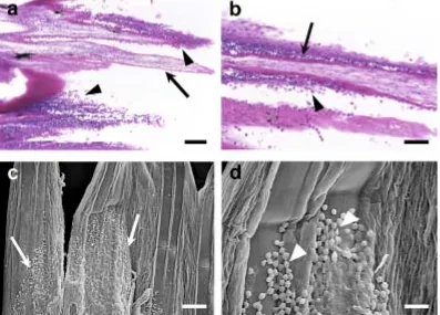
We demonstrated that rapidly confined hybrid striped bass (Morone saxatilis male x M. chrysops female) developed a syndrome characterized by the immediate and dramatic loss of their skin. We have named this phenomenon the Acute Ulceration Response (AUR). AUR is characterized by the rapid onset of severe epidermal erosion, ulceration and degeneration on the body skin and fins, as well as corneal ulceration, in stressed hybrid striped bass. Grossly, the distal edges of the fins became obviously ragged and blanched. The earliest microscopic change in the fins occurred within 15 min, with swelling of the outermost layers of the epidermis and epidermal erosion. After 30-min stress, the epidermis at the distal edges of the fins became ulcerated. Both apoptotic and necrotic epithelial cells were observed at 30 min confinement stress. The middle and basal epidermis developed severe spongiosis, and the dermis and hypodermis became edematous. Epidermal ulceration appeared on all fins of stressed fish and was significantly greater compared to fins from unstressed fish. A time-course study of the response to acute confinement stress showed a significant correlation between confinement period and severity of AUR.
DEDICATION
BIOGRAPHY
Pareeya Udomkusonsri grew up in Bangkok, Thailand. She was awarded the DVM
degree with the second honor in 1992 from the Kasetsart University, Bangkok. She served as a veterinarian for the Veterinary Teaching Hospital, Faculty of Veterinary Medicine,
Kasetsart University for 6 months. In 1992, she became a faculty member of the Department of Pharmacology and Toxicology, College of Veterinary Medicine, Kasetsart University. In 1997, the Royal Thai Government awarded her a fellowship to pursue doctoral studies at
North Carolina State University, Raleigh. During the course of her studies, together with her principal thesis advisor Dr. Edward J. Noga, she studied the pathogenesis of the Acute
Ulceration Response (AUR) in fish. She also awarded Dr. Monica Menard Award for Excellence in Veterinary Pathobiological Research by North Carolina Veterinary Medical Foundation, Inc, in 2002. In 2003, she achieved her Ph.D. degree in Comparative
ACKNOWLEDGEMENTS
The author would like to express her appreciation to Dr. Edward J. Noga, her principal advisor, for his efforts to provide her with invaluable knowledge. The author is
grateful to him for his wisdom, advice and support in many ways throughout her study. The author is grateful to Dr. Nancy A. Monteiro-Riviere, the co-chair of her advisory committee,
for her encouragement, advice and support throughout her program. The author would like to express her gratitude to Dr. Gregory A. Lewbart for his encouragement, facility, and kindness. The author would like to thank Dr. Mark G. Papich for being on the advisory
committee and for helpful comments on her work. The author thanks Dr. Umaporn Silphaduang for her support and friendship. The author extends her gratitude to Mr. Alfred
O. Inman and Mr. Stanley M. Dunston for their generous guidance and technical advice. The author is greatly indebted to the unconditional love and support of her family. Her parents always have supported her in many ways. Her husband, Dr. Suchin, has devoted
himself to her. He has encouraged and motivated her to fulfill her doctoral pursuit. She was also greatly inspired by her son, Napat, during the last year of her study.
The author gratefully acknowledge Mr. Lee Brothers (Carolina Fisheries), Mr. H. Maybury (Double MM Aquatics, Creedmoor, NC) and Dr. Jeff .M. Hinshaw (Department of Zoology, NCSU, Fletcher, NC) for providing fish for this research. Financial support for her
TABLE OF CONTENTS
Page
LIST OF TABLES . . . viii
LIST OF FIGURES . . . ix
I. LITERATURE REVIEW Fish skin: Normal structure and role in host defense . . . 2
Epidermis. . . 2
Basement membrane. . . 4
Dermis . . . . . . 5
Fin . . . 9
Pathological response of the skin to damage . . . . 10
Stress in fish . . . . . . . 11
Hormone basis of the stress response . . . . 13
Glucocorticoids. . . 13
Catecholamines. . . 16
Adrenergic receptors . . . 20
The effects of stress on skin integrity . . . 25
Toxin . . . 26
Stress hormones . . . .26
Temperature . . . 27
Psychology . . . 30
Crowding . . . . 31
The acute ulceration response (AUR): an extreme response to acute stress . 32 Probing the possible hormonal basis for AUR . . . 33
Stress and fish disease . . . 35
Water mold infection . . . . 37
Hypotheses and Research Objectives. . . . 43
II. PATHOGENESIS OF THE ACUTE ULCERATION RESPONSE (AUR) IN HYBRID STRIPED BASS
Abstract . . . 79
Introduction . . . 80
Materials and Methods . . . 81
Results . . . 85
Discussion . . . 91
References . . . 117
III. PHYSIOLOGICAL AND ENVIRONMENTAL EFFECTS ON THE DEVELOPMENT OF THE ACUTE ULCERATION RESPONSE (AUR) Abstract . . . . 123
Introduction . . . . 124
Materials and Methods . . . 127
Results . . . 133
Discussion . . . 138
References . . . 168
IV. THE ACUTE ULCERATION RESPONSE (AUR): A WIDESPREAD AND POTENTIALLY SERIOUS CAUSE OF SKIN INFECTION IN FISH HYBRID STRIPED BASS Abstract . . . 176
Introduction . . . 177
Materials and Methods . . . . 179
Results . . . 185
Discussion . . . 188
V. CONCLUSION, PERSPECTIVE OF AUR STUDIES AND TECHNOLOGY TRANSFER
LIST OF TABLES
Page Chapter I
Table 1.1 The effect of corticosteroid and catecholamine release
during stress in fish . . . 45
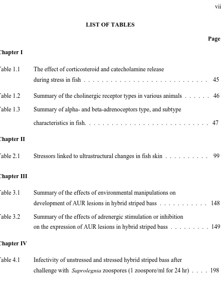
Table 1.2 Summary of the cholinergic receptor types in various animals . . . 46 Table 1.3 Summary of alpha- and beta-adrenoceptors type, and subtype
characteristics in fish. . . 47
Chapter II
Table 2.1 Stressors linked to ultrastructural changes in fish skin . . . 99
Chapter III
Table 3.1 Summary of the effects of environmental manipulations on
development of AUR lesions in hybrid striped bass . . . 148 Table 3.2 Summary of the effects of adrenergic stimulation or inhibition
on the expression of AUR lesions in hybrid striped bass . . . 149
Chapter IV
Table 4.1 Infectivity of unstressed and stressed hybrid striped bass after
LIST OF FIGURES
Page Chapter I
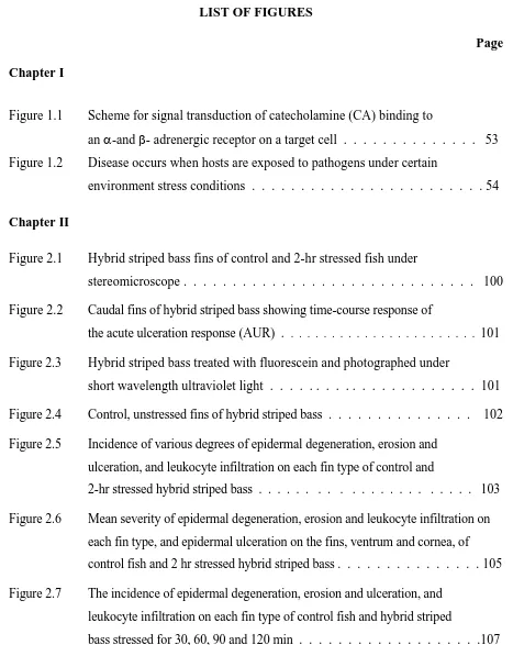
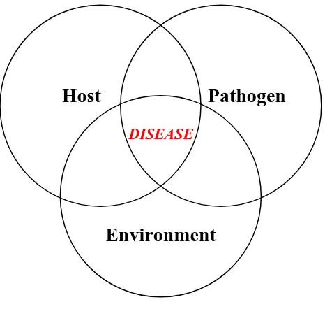
Figure 1.1 Scheme for signal transduction of catecholamine (CA) binding to an α-and β- adrenergic receptor on a target cell . . . 53 Figure 1.2 Disease occurs when hosts are exposed to pathogens under certain
environment stress conditions . . . . 54
Chapter II
Figure 2.1 Hybrid striped bass fins of control and 2-hr stressed fish under
stereomicroscope . . . 100
Figure 2.2 Caudal fins of hybrid striped bass showing time-course response of the acute ulceration response (AUR) . . . 101
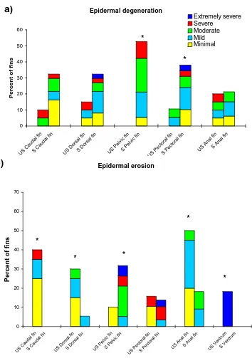
Figure 2.3 Hybrid striped bass treated with fluorescein and photographed under short wavelength ultraviolet light . . . . . . . . . . . . 101 Figure 2.4 Control, unstressed fins of hybrid striped bass . . . 102 Figure 2.5 Incidence of various degrees of epidermal degeneration, erosion and
ulceration, and leukocyte infiltration on each fin type of control and
2-hr stressed hybrid striped bass . . . . . . . . . . . . 103 Figure 2.6 Mean severity of epidermal degeneration, erosion and leukocyte infiltration on
each fin type, and epidermal ulceration on the fins, ventrum and cornea, of control fish and 2 hr stressed hybrid striped bass . . . . 105 Figure 2.7 The incidence of epidermal degeneration, erosion and ulceration, and
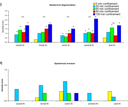
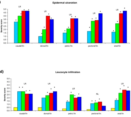
Figure 2.8 Mean severity of epidermal degeneration, erosion and ulceration, and leukocyte infiltration on each fin type of control fish and hybrid striped bass stressed
for 30, 60, 90 and 120 min . . . .109
Figure 2.9 Caudal fins of hybrid striped bass confined for 15 min . . . 111
Figure 2.10 Caudal fins of hybrid striped bass confined for 30 min . . . .112
Figure 2.11 Caudal fins of hybrid striped bass confined for 60 min . . . .113
Figure 2.12 TEM of caudal fin of hybrid striped bass confined for 90 min . . . .113
Figure 2.13 Fins of hybrid striped bass confined for 120 min . . . 114
Figure 2.14 Ventrum skin of hybrid striped bass . . . 115
Figure 2.15 Cornea of the eye in hybrid striped bass . . . 115
Chapter III Figure 3.1 Incidence of various degrees of severity of epidermal degeneration, erosion and ulceration, and lymphocyte infiltration on each fin type of unstressed and stressed fish acclimated to a small cage or to a large aquarium, respectively . . . 150
Figure 3.2 Comparison of mean severity score of pathological changes (epidermal degeneration, erosion, ulceration and leucocyte infiltration) between stressed fish acclimated in small cages versus the large aquarium . . . . . 153
Figure 3.3 Incidence of various degrees of severity of ulceration on each tissue of unstressed and stressed fish during stress at 13, 20, or 27°C . . . 155
Figure 3.4 The mean severity score of ulceration on each tissue of fish stressed at 13°, 20°, or 27°C . . . . 156 Figure 3.5 Incidence of various degrees of severity of ulceration on each fin type of
Figure 3.6 The mean severity score of ulceration on each tissue of stressed fish at 13°, 20°, or 30°C . . . .158 Figure 3.7 Incidence of severity of epidermal erosion and ulceration on each fin
type of hybrid striped bass administered epinephrine intraperitoneally at a dose of 1, 10, 100, 1,000 or 10,000 µg/kg body weight . . . . 159 Figure 3.8 The severity of epidermal erosion and ulceration on each fin type
of hybrid striped bass treated with either normal saline or epinephrine at 1, 10, 100, 1,000 or 10,000 µg/kg body weight . . . 160 Figure 3.9 Incidence of severity of epidermal erosion and ulceration on each fin
type of hybrid striped bass administered phentolamine (α-adrenergic antagonist) at 0, 10, 100 or 1000 µg/kg body weight prior to confinement stress at 27°C; control fish were treated with normal saline . . . 161 Figure 3.10 The severity of epidermal erosion and ulceration on each fin type of
hybrid striped bass treated with phentolamine at 0, 10, 100 or 1000 µg/kg body weight before acute confinement at 27°C . . . . 162
Figure 3.11 Incidence of severity of epidermal erosion and ulceration on each fin type of hybrid striped bass intraperitoneally administered normal
saline or propranolol (β-adrenergic antagonist) at 0, 10, 100 or 1000 µg/kg body weight before confinement stress at 27°C . . . .163
Figure 3.12 The severity score of epidermal erosion and ulceration on each fin type of hybrid striped bass treated with normal saline or propranolol at 0, 10, 100 or 1000 µg/kg body weight . . . . 164 Figure 3.13 Incidence of severity of epidermal erosion and ulceration on each fin
type of hybrid striped bass intraperitoneally administered saline or
Figure 3.14 The severity score of epidermal erosion and ulceration on each fin type of hybrid striped bass treated with normal saline or hexamethonium at 0, 10, 100 or 1000 µg/kg body weight . . . . . . 166
Chapter IV
Figure 4.1 Bacterial concentration (bacteria/g of tissue) isolated on cytophaga agar and BHI agar incubated at 25°C . . . 197
Figure 4.2 Skin lesions in hybrid striped bass with AUR that were challenged with Saprolegnia zoospores . . . 199 Figure 4.3 Caudal fins of control and 2-hr stressed guppies under stereomicroscopy . 200 Figure 4.4 Caudal, dorsal and anal fins of control and 2-hr stressed angelfish
under stereomicroscopy . . . .201 Figure 4.5 Caudal fins of control and 2-hr stressed channel catfish . . . . 202 Figure 4.6 Histological sections of caudal fins of control and 2-hr stressed
guppies, control and 2-hr stressed freshwater angelfish, and control and 2-hr stressed channel catfish . . . 203 Figure 4.7 Comparison of the severity of pathological changes (epidermal erosion,
I.
FISH SKIN: Normal Structure and Role in Host Defense
The skin of fish is a primary barrier against the external environment and preserves
the constancy of the internal milieu. The layers of fish skin, like those of all vertebrates,
consist of epidermis and dermis, which are separated by the basement membrane, or basal
lamina, which is visible under the electron microscope.
EPIDERMIS
Fish epidermis is a typical stratified, squamous, epithelium; however, unlike
mammalian skin, it is metabolically active, with mitotic capacity throughout all epidermal
layers (Bullock et al 1978). In all but a few fish (Mittal and Banerjee 1974), the outer surface
of the epidermis is not keratinized. The epidermis covers the body surface and is continuous
throughout the fins and cornea. Its thickness varies with body site, age, sex, condition, and
degree of maturation of fish (Ellis et al 1978; Whitear 1986b), and is frequently thicker in the
dorsal body areas in pelagic fish species and in the ventral surfaces of benthic fish. In
salmonids, the epidermis in non-scaled areas, such as the head and fins, is normally thicker
than in scaled areas (Harris and Hunt 1975). In addition, many factors, such as
environmental conditions, handling, nutritional status and other stressors, influence the
structural and cellular response of the epidermis (Blazer et al 1997; Ferguson 1989; Iger et al
1995; Iger et al 1994a, b; Pickering and Richards 1980; Quiniou et al 1998). For example,
the skin of cutthroat trout (Oncorhynchus claski henshawi) exhibited sunburn damage;
ultraviolet-B radiation (Blazer et al 1997). Fish epidermis also plays an important role in
wound healing and antigen uptake (Kiryu et al 2000; Ototake et al 1996).
The epidermal cell is known by many names, including malpighian cell, epithelial
cell, filament-containing cell, filamentous cell, polygonal cell, polyhedral cell, keratocyte,
keratinocyte, principal cell, and common cell. Epidermal cells in fish and mammals originate
from ectoderm. In mammals, the skin consists of five epidermal layers: stratum corneum,
stratum lucidum, stratum granulosum, stratum spinosum and stratum basale; however, fish
epidermis is generally separated into outer, middle and basal layers (stratum germinativum)
(Groman, 1982; Hawkes 1983). The epidermis consists of a non-vascularized stratified
squamous epithelium and has no stratum corneum which is normally found in the terrestrial
vertebrates. In primitive, jawless fish (Agnatha), the surface of the epithelium is pitted like a
sponge (Whitear 1986b). However, the exterior surface of the superficial epithelial cells of
teleosts is characterized by microridges or micropapillae, which often form regular,
fingerprint-like patterns. The function of microridges is unknown; however, they may
provide defense against trauma and might hold mucus secretions on the surface. They also
might increase gas exchange (Hawkes 1974) and might be involved in wound closure since
they can move by the contraction of basal actin microfilaments (Bereiter Hahn 1971).
Mucus cells are found in the epidermis, but the numbers vary with site, species and
sexual stage (Roberts and Bullock 1978). They usually originate from the middle layers of
epidermis, although they may be seen in the basal layer of a very thin epidermis. Immature
approach the surface. Mature mucus cells are filled with numerous mucosomes, with the
nucleus displaced to the periphery of the cell. When a mucus cell reaches the surface, the
plasma membrane ruptures at the apex, releasing its cell contents; then the cell dies. Mucus
secretions contain mucopolysaccharides (sialomucin, sulfomucin) that decrease friction with
water.
Club cells are large, oval or round cells and have one or two nuclei with prominent
nucleoli. They are found in the middle layers of epidermis in some fish species, and
normally are called alarm cells since they secrete potent “alarm” substances. Club cells
release fright pheromones into the surrounding water when the epidermis is damaged or the
cells are lysed (Smith 1992). Alarm substances also play a role in wound healing, and are
possibly protective agents against parasites, other pathogens, or irritants (Shiomi et al 1988;
Smith 1982).
BASEMENT MEMBRANE
The basement membrane in fish is similar to that of other vertebrates, acting as a
lining between the epidermis and dermis (Whitear 1986a). A lucent adepidermal space under
the basal epidermal layer contains fibrillar material (anchoring filaments) connecting
hemidesmosomes of the basal epidermal cells to the electron-dense basement membrane
(Ferri 1982; Whitear 1986a). The thickness of the basement membrane depends upon the
species and location on the body. It is generally less well-developed in scaled areas
compared to the head or fins (Roberts and Bullock 1978). The periodic acid Shiff reaction
Anchoring filaments connect the basement membrane to the collagenous tissues beneath.
Under the basement membrane, an electron-lucent layer is supplied with nerves and
capillaries.
The basement membrane acts as a barrier and controls the passage of cells and
chemicals. It also regulates morphogenesis and wound healing, serving as an attachment site
for epithelial cells or other cells.
DERMIS
The dermis consists of two major layers, stratum spongiosum (stratum laxum) and
stratum compactum. The upper dermis (stratum spongiosum) is a loose network of collagen
and reticulin fibers. The stratum spongiosum contains vascular and neural components, and
scales are anchored in the scale pockets in this layer. It also includes fibroblasts, pigment
cells, leucocytes and scale-synthesizing tissues (Whitear 1986a). The stratum compactum
consists of collagen bundles that form a dense matrix above the hypodermis. The collagen
fibers in these layers are high ordered at right angles to each other. The stratum compactum
may be reduced in fin tissue (Sharples and Evans 1996). Dermal fibroblasts are distributed
between collagen bundles. This layer is important in locomotor activity (Whitear 1986a) and
acts as a tendon in parallel with the muscles. The dermis in juvenile striped bass (Morone
saxatilis) has only a narrow layer of stratum compactum, composed of dense collagen fibers, elastic fibers, melanocytes and a small amount of connective tissue (Groman 1982). In adult
striped bass, the dermis is composed of 2 layers; a papillary layer (stratum compactum)
The fish mast cell, or eosinophilic granular cell (EGC), was first described in the skin
of plaice (Pleuronectus platessa L.) (Roberts et al 1971). Mast cells are common in the
dermis around blood vessels, and appear in many other tissues such as gill, oral epithelium,
swim bladder and alimentary tract (Powell et al 1993; Reite and Evensen 1994; Sire and
Vernier 1995; Zaccone 1980). Teleost mast cells are motile, mononuclear cells with
prominent granules and may play an important role in host defense (Silphaduang and Noga
2001). Teleost mast cells contain a number of mediators (e.g., histamine) and can be
stimulated by bacteria, parasites and chemical stimuli, resulting in their mobilization and
degranulation (Matsuyama and Lida 1999, 2000; Powell et al 1993; Sire and Vernier 1995).
Recently, Silphaduang and Noga (2001) discovered a novel family of peptide
antibiotics, named piscidins, from mast cells in hybrid striped bass (Morone saxatilis x M.
chrysops). Piscidins have potent, broad-spectrum activity against important bacterial pathogens of both fish and mammals, including multi-drug resistant bacteria. Hybrid striped
bass mast cells in gill, skin, stomach, intestine, and pyloric ceca are immunoreactive for
piscidins, including those lining blood vessels in the viscera. The mast cells of other fish are
also positive for piscidins, including white bass (M. chrysops), striped bass (M. saxatilis),
spot (Leistomus xanthurus) and croaker (Micropogonias undulatus). Those fish are all in the
Suborder Percoidei, Order Perciformes. This suggests that piscidins may be evolutionarily
conserved in this group.
In teleosts, chromatophores (pigment cells) are normally found in the stratum
membrane: in the upper portion of the stratum spongiosum in non-scaled areas, or under the
scales in scaled skin. Epidermal chromatophores are classified according to the color of their
pigment. Five different chromatophores are known, melanophores (black or brown pigment),
erythrophores (red or yellowish pigment), xanthophores (primarily yellow pigment),
leucophores and iridophores (colorless, reflecting pigment). The difference between
leucophores and iridophores is based upon the translocation of pigment-containing
organelles. Leucophores have fewer dendritic processes, but leucosomes can move
centripetally or centrifugally similar to other dendritic-chromatophores (Fujii 1993).
Leucophores reflect light in all directions. Iridophores are non-dendritic or occasionally
dendritic, and contain large crystalline platelets that form stacks in the cytoplasm with
uniform spacing between adjacent platelets within a stack (Fujii 1993).
Most vertebrate pigment cells originate from neural crest, and then migrate,
differentiating in the integument. Many fish do not develop a distinct neural crest; however,
fish chromatoblasts appear to migrate from a location close to the neural keel, which is
equivalent to the neural crest. Because chromatophores share an ontogenetic origin with
neurons, most of them are dentritic cells with multiple branched or unbranched processes
extending from the cell body. Chromatosomes, pigment-containing organelles, translocate
centripetally (aggregation) or centrifugally (dispersion) in response to various neuronal or
hormonal signals. The movement of chromatosomes involves two possible hypothesized
mechanisms (Fujii 1993). In the first model which has been widely accepted, pigment
granules are moved by interaction with microtubules that radiate from the center of the cell
microtubule-independent motility system, actin filaments provide the motive force for granule movement.
It is also possible that pigment granules may be enmeshed in a filamentous network which
passively drags the granules (Rodionov et al 1998).
The pigments in chromatophores are melanins, carotenoids, pteridines, and/or
purines. The black color of melanins is formed by polymerization of DOPA-quinone,
synthesized from tyrosine. In xanthophores, carotenoids (a yellowish color, highly
unsaturated hydrocarbon) are formed from four isoprene units and two ionone rings.
Pteridines are associated with red color and consist of a pyrimidine and a pyrazine ring.
Carotenoids and pteridines make yellowish to red color in erythrophores. Since animals
cannot synthesize carotenoids, pigmentation in xanthophores and erythrophores is influenced
by diet. The colorless pigments in leucophores and iridophores are purines. Guanine is the
main pigment, but other purines such as hypoxantine and uric acid are found in both
chromatophores.
Melanophores are the most common and usually the largest chromatophore in fish
(Fujii 1993). Melanophores usually respond to nervous and hormonal stimuli with rapid
aggregation or dispersion of melanosomes (Fujii 1993). Xanthophores and erythrophores are
generally smaller than melanophores, but the morphological features are similar to
melanophores. In contrast to the light-absorbing chromatophores (melanophore, xanthophore
and erythrophores), leucophores and iridophores are light-reflecting chromatophores that are
Chromatophore activity is regulated by humoral and neuronal controls. In the
humoral control, it requires stimulation of cells releasing hormones into the circulation.
Hormone is transported via blood and binds to receptors on the chromatophores.
Chromatophores are also directly innervated, which provide an instant response and specific
change. Neuronal control is dominant in higher teleosts allowing rapid body color change,
while humonal control is the rule in cyclostomes and elasmobranchs.
Color change in fish can occur via two different mechanisms. First, chromatophores
can redistribute the pigment-containing organelles within the cells. Color change often
appears immediately, through the activity of neuronal stimulation directly on the
chromatophores. Examples include adaptive skin color in response to background color or
lighting conditions and color change related to the physiological state of the fish. These
phenomena reflect the physiological control of the pigment cells, and therefore are called
physiological color changes. The other mechanism of color change, morphological color
change, results from changes in the total number and size of pigment cells, and or the amount
of pigments in the cells. This color change is gradual and much slower, taking several days
or weeks to be completed. However, both physiological and morphological color changes
occur simultaneously, with the latter providing the base coloration.
FIN
Fin consists of epidermal folds of skin which is supported by lepidotrichia, segmental
rays formed of giant fibers of collagen. Lepidotrichia first appear at the base of the fins, and
form an incomplete tube which is linked by connective tissue. Actinotrichia persist at the
distal end of each lepidotrichium to the margin of the fin (Beccera et al 1983). Each
lepidotrichal segment is composed of a pair of hemisegments, plate-like elements separated
by an intrasegmental region. The hemisegment or demiray consists of an acellular matrix or
has very few cells. The intrasegmental region contains nerve bundles, blood vessels and
connective tissues (Montes et al 1982). Lepidotrichia are divided lengthwise, and each
segment is connected with intersegmental joints. Fins are covered by a stratified squamous
epithelium that is continuous from the body epidermis. The dermis in fin has a reduced
stratum compactum and thicker hypodermis (Groman 1982; Whitear 1986a).
PATHOLOGICAL RESPONSES OF THE SKIN TO DAMAGE
The pathological responses to stress were well documented in which skin is capable
of expressing: degeneration, erosion, ulceration, leukocyte infiltration (reviewed in
Bodammer 2000; Iger et al 1992; Iger et al 1994a, b, c, d; Iger and Wendelaar Bonga 1994;
Noga 2000). Epidermal degeneration was defined as swollen epidermal cells (intracellular
edema) with pyknotic nuclei; epidermal erosion was the sloughing of epithelial layers, but
with the basement membrane still intact; ulceration was defined as complete loss of all
epithelial layers and the basement membrane. Leukocytes are normally found in the dermis
but in stressed skin, leukocytes may also infiltrate the basal epidermal layers.
Two modes of cell death can occur in skin cells, apoptosis and necrosis. They can be
distinguished based on differences in the morphological, chemical and molecular changes in
inflammatory response in damaged tissue, while apoptosis is a noninflammatory, programmed
cell death that is associated with embryogenesis, metamorphosis, and normal cell turnover
(Wyllie 1997; Wyllie et al 1980), but also with certain pathologies as well. Necrosis typically
occurs in response to toxins, hypoxia, or ischemia, and affects cells in groups rather than singly
(Anilkumar et al 1992; Wyllie et al 1980). Necrotic cells show destruction of organelles,
chromatin flocculation, mitochondrial swelling, rupture of the plasma membrane, and release
of cytoplasmic contents (Darzynkiewicz et al 1994; Lin et al 1996). Apoptosis is subjected to
control by genetic and normal physiological stimuli, such as endocrine changes (e.g., cortisol,
ACTH) (Iger et al 1995; Iger et al 1992; Wyllie 1997), as well as certain toxic agents such as
radiation and chemotherapeutic agents (Anilkumar et al 1992). Apoptosis is morphologically
characterized by cell shrinkage, nuclear condensation, membrane blebbing and
internucleosomal fragmentation of DNA into units of 180-200 base pairs (DNA laddering).
Apoptotic cells are usually phagocytosed and digested by resident cells (Anilkumar et
al 1992). Iger et al (1994a) reported that the autophagocytic vesicles, that appeared inside
the epidermal cells of both unstressed and stressed fish, were eliminated by the epidermal
cells. Thus, the autophagocytic vesicles in the epidermal cells may actually contain apoptotic
cells. Furthermore, apoptotic cells in unstressed fish are indicative of the normal turnover of
epidermal cells.
STRESS IN FISH
Stress refers to a condition in which organism equilibrium is disturbed due to a
areas. For example, physiologists describe stress as an increase of plasma hormones and
metabolites. Toxicologists describe stress as an induction of mixed-functional oxidase
enzymes, while fish culturists are concerned with growth and mortality of fish. In
aquaculture, a stimulus or stressor can place constraints on fish health. These constraints are
from changes in the physical environment (e.g., water pH, temperature, salinity), animal
interaction (e.g., competition for food, space and sexual partners), water pollution (e.g.,
organic chemicals and heavy metals) and aquaculture practices (e.g., handling, transport and
crowding) which cause stress to the fish. The acute stress response is an adaptive response
that promotes the best chance of survival in the threatening situation; thus, acute stress does
not necessarily harm an organism. However, if the stress is severe enough, or continuous and
prolonged, the response can result in severe damage to growth, reproductive capacities or
immune defense systems.
The stress response generally occurs in three stages. First is an initial alarm reaction
from the activation of stress-related hormones. This initiates energy production, helping to
maintain homeostasis and survival. Second is the resistance stage, which occurs when
physiological systems have successfully compensated and the organisms are acclimated.
There is an energy cost for compensation and thus growth is decreased. Third is the
exhaustion stage, when prolonged or severely stressful conditions exceed the ability of
acclimation to maintain homeostasis; this is maladaptive. In aquaculture, stress is often, if
not usually, a predisposing factor to an infectious disease (bacterial, fungal, parasitic, or viral
infection) outbreak (Pickering and Duston 1983). Thus, reducing stress is a major goal in
HORMONAL BASIS OF THE STRESS RESPONSE
Stress causes neuro-hormonal changes (primary response) and results in physiological
consequences of this neuro-hormonal stimulation (secondary response). The primary
response to stress in fish involves many hormones, e.g., catecholamines (CA),
glucocorticoids (GC), growth hormone and prolactin (Wendelaar Bonga, 1997); however,
CA and GC are generally recognized as the dominant stress hormones (Mazeaud and
Mazeaud 1981; Wendelaar Bonga 1997). In the secondary response to stress, both CA and
GC increase oxygen uptake, blood flow, cardiac output and energy consumption. It is
difficult to distinguish between the secondary effects of GC and CA (Mazeaud and Mazeaud
1981) since both CA and GC are usually activated in response to stress. When stress is
continuous or chronic, fish develop the tertiary response, with decreased growth rate,
modified behavior and increased susceptibility to diseases. In populations or ecosystems, a
higher order response (the quaternary response) is a result of alteration in species
composition which is caused by the disruption in energy flow through trophic levels
(Wedemeyer 1996).
GLUCOCORTICOIDS
Release of GC is caused by an activation of the hypothalamic-pituitary-interrenal
(HPI) axis (functionally similar to the hypothalamic-pituitary-adrenal axis in mammals).
Cortisol is the predominant glucocorticoid in most teleosts (Schreck et al 1991). In addition,
cortisone, a metabolite of cortisol, has been reported as a primary GC in unstressed coho
tissue, which originates from mesoderm and is located on the walls of the posterior cardinal
veins (PCV). Stress stimulates the release of adrenocorticotropic hormone (ACTH) from the
pituitary gland by the activation of corticotropin-releasing factor (CRF) from the
hypothalamus. The circulating ACTH stimulates the interrenal cells to produce cortisol.
Cortisol has broad activity and is important in regulating hydromineral balance and
metabolism in fish (Table 1.1). Other effects include reduction of growth and suppression of
immune function (Mazur and Iwama 1993; Schreck et al 1991; Wendelaar Bonga 1997). For
example, fish have increased susceptibility to water mold infection after prolonged oral
administration of cortisol, and implantation of the steroid increases fish mortality due to
furunculosis (Pickering and Duston 1983). Furthermore, prolonged elevated cortisol caused
brown trout (Salmo gairdneri) to die from a combination of water mold (Saprolegnia)
infection, severe bacterial fin-rot and furunculosis (Pickering and Pottinger 1989).
Elevated cortisol is one of the most widely used indicators of stress in fish
(Wendelaar Bonga 1997). After exposure to stress, the plasma cortisol concentration rises
rapidly in a few minutes. In fish, resting plasma cortisol levels are very low, 2-42 ng/ml
(reviewed in Gamperl et al 1994a). Acute and chronic stress causes an increase of plasma
cortisol which can vary from 20-740 ng/ml (Barton and Iwama 1991; Gamperl et al 1994a),
depending upon fish species, strain within a species, sexual maturation, season, temperature,
environment, and sampling time after stress (Barton and Iwama 1991; Pickering and
Pottinger 1989; Wendelaar Bonga 1997). For example, the plasma cortisol in catfish
2000). It was suggested that this catfish was very tolerant of stress.
Increased plasma cortisol was reported in response to many stressors, including
transport, increased water temperature, net confinement, or poor water quality (Davis and
Parker 1990; Fevolden et al 2003; Iger et al 1994b; Mazur and Iwama 1993). Cortisol levels
will return to normal if the stress is discontinued to allow the fish to recover. However, the
recovery period varies, depending on the type, intensity and duration of stress, as well as the
fish strain (Noga et al 1994; Pickering and Pottinger 1989; Pickering et al 1982). Acute
stress (1-hr confinement or 30-sec emersion), caused a significant increase of cortisol in
rainbow trout and brown trout; cortisol levels returned to control values within 24-48 hr
(Pickering and Pottinger 1989). The cortisol peak in rainbow trout was lower than that in
brown trout. In addition, chronic confinement caused a prolonged increase of cortisol in
rainbow trout that required 3 weeks for complete acclimation, while in brown trout it
required more than 7 weeks. Noga et al (1994) found that plasma cortisol concentration
increased significantly after exposure of striped bass and hybrid striped bass (Morone
saxatilis x M. chrysops and M. saxatilis x M. americana) to an acute net confinement for 45 minutes. Plasma cortisol in striped bass increased faster (from 3 ng/ml to 742 ng/ml) and
reached a higher level than in hybrid striped bass (from 212 ng/ml to 490 ng/ml). Plasma
cortisol concentration was still high in striped bass for at least 48 hours after the net
confinement. Conversely, plasma cortisol in hybrid striped bass decreased to normal or near
normal levels within this period. The mortality of stressed striped bass and its hybrids
increased markedly when fish were crowded at the high density. Domesticated sea trout
after exposure to stress (Lepage et al 2000). These studies suggested that strains of fish
respond differently to stress.
CATECHOLAMINES
The adrenergic function of fish is sensitive to stress and results in an increase of
plasma CA (Randall and Perry 1992; Thomas and Perry 1992). Epinephrine (E) and
norepinephrine (NE) are the predominant CAs in fish; however, epinephrine is usually the
predominant CA in teleosts while NE is dominant in elasmobranchs (Reid et al 1998;
Wendelaar Bonga 1997). The release of CA into the circulation is mediated by the
preganglionic-cholinergic innervation of the chromaffin tissues (Nilsson 1984; Thomas and
Perry 1992). The arrangement of chromaffin cells is quite diverse in fish. In the primitive
cyclostomes, chromaffin cells are associated with the systemic and portal hearts. In
elasmobranches, these cells are located near paravertebral autonomic ganglia. In teleosts, the
chromaffin tissues are equivalent to the adrenal medulla in mammals, which originates from
neuroectoderm. They are normally located within the wall of the posterior cardinal veins
(PCV) and in small clusters in the head kidney. In Perciformes and Salmoniformes,
chromaffin cells are also present in the posterior kidney in contact with the caudal veins
(Milano et al 1997). The chromaffin cells are closely associated with the interrenal cells
(steroidogenic cells) around the walls of the PCV.
Sympathetic stimulation by preganglionic nerve fibers causes the release of
acetylcholine, which stimulates cholinergic receptors, and ultimately initiates a series of
cholinergic receptors on the chromaffin cells of various animals is shown in Table 1.2. In
teleosts, the cholinergic receptors on teleost chromaffin cells are predominantly nicotinic
receptors (Abele et al 1998; Al-Kharrat et al 1997); thus, hexamethonium (a nicotinic
receptor antagonist) can prevent the secretion of CA in at least some fish (e.g., American eel,
Anguilla rostrata; hagfish, Myxine glutinosa; dogfish, Squalus acanthias) (Bernier and Perry 1996; Opdyke et al 1983a; Reid et al 1998; Reid and Perry 1995). Muscarinic receptors also
have an inhibitory effect, especially on norepinephrine-secreting cells (Al-Kharrat et al
1997). Muscarinic receptors on rainbow trout chromaffin cells are involved in CA secretion
(Montpetit and Perry 1999). Stimulation of muscarinic receptors increased the intracellular
Ca2+ concentration via activation of phospholipase C, but it was insufficient to trigger CA
secretion. However, it may be possible that the increase of intracellular Ca2+ concentration
enhanced the nicotinic receptor Ca2+ events and increased nicotinic-induced CA secretion.
CA are also synthesized and released from adrenergic nerve endings by adrenergic
stimulation (Nilsson 1984; Perry et al 1991).
In fish as in other vertebrates, the chromaffin cells contain a high CA content and are
stained dark by dichromate solution due to the oxidation of catecholamine stores to
adrenochromes. E and NE are stored in separate cells (Abelli et al 1996).
Norepinephrine-containing cells normally have spherical, strongly electron-dense secretory granules, and the
granules are evenly distributed. In epinephrine-containing cells, the granules are
electron-lucent, spherical or elongated, and distributed homogeneously. In all vertebrates, E and NE
are synthesized from tyrosine via the Holtz-Blaschko pathway (Randall and Perry 1992).
hydroxylase (TH) and subsequently decarboxylated to dopamine by dopamine decarboxylase
(DH). Dopamine is taken into the storage vesicles and is hydroxylated by dopamine-β
-hydroxylase (DβH) to NE. NE is released from the vesicles into the cytosol and is then
methylated by phenylethanolamine-N-methyl transferase (PNMT) to E. E is then moved to
storage vesicles. Normally, E and NE are produced in the different cells depending upon the
presence of PNMT and both CAs are released into the circulation by exocytosis. TH, DH
and PNMT are cytoplasmic enzymes while DβH is associated with the storage vesicles. The
rate-limiting step in the epinephrine biosynthesis pathway is normally the TH step in
mammals, but is the PNMT step in fish (Abrahamsson and Nilsson 1976; Senthilkumaran
and Joy 1995). Circulating CAs are catabolized by two enzymes, catechol-O-methyl
transferase (COMT) and mitochondrial monoaminoxidase (MOA) which are responsible for
ortho-methylation and deamination, respectively. COMT is mainly in the liver, which plays
a more important role in the catabolism of circulating CAs, while MOA is found mainly is
the kidney, as well as the brain (Randall and Perry 1992).
In teleosts, release of CA into the circulation (referred as a primary effect of stress) is
induced by a number of stimuli, such as preganglionic sympathetic nerve fibers, localized
changes in blood chemistry, activation of the renin-agiotensin system, serotonin, and
adrenocorticotropic hormone (Bernier and Perry 1996, 1997; Nilsson et al 1976; Opdyke et al
1981, 1983a; Perry et al 1991; Reid et al 1996). CA is released rapidly by the simulation of
various stresses, such as hypoxia (Perry et al 1991, 2000; Randall and Perry, 1992; Reid et al
1993; Thomas et al 1994), crowding and handling (FlØysand et al 1992; Gerwick et al 1999),
and McDonald 1995). The resting levels of plasma epinephrine range from 1 to 10 nM and
can be elevated up to 300-fold under acute stress (review in Gamperl et al 1994a; Reid et al
1998; Thomas and Perry 1992). The magnitude of CA release is related to the intensity and
type of stress; only severe stress (e.g., exhaustive exercise, severe hypoxia, air exposure)
causes an increase in circulating CA while mild or moderate stresses (e.g., sustained aerobic
exercise) usually do not change the CA levels (reviewed in Randall and Perry 1992; Perry
and Bernier 1999). In tilapia (Oreochromis aureus), an increase of CA levels to a 30-min
cold stress was detected earlier than an elevation in cortisol, but the duration of plasma CA
elevation was shorter than that of cortisol (Chen et al 2002).
While several factors are known to initiate CA secretion, neuronal activity of
preganglionic sympathetic nerve fibers is the predominant mechanism for CA secretion in
response to stress (Reid et al 1998). Furthermore, the pituitary-interrenal hormones can
modulate CA storage and release in rainbow trout (Reid et al 1996). It was demonstrated that
ACTH elicited CA release by stimulating the production of cAMP, then increasing CA
release via exocytosis. Prolonged plasma cortisol increased CA storage in the kidney and
tissue around the PCV. In mammals, cortisol increases transcription of the genes encoding
for PNMT; however, this effect did not occur in rainbow trout (Reid et al 1996).
Generally, CA are released much quicker than GC because chromaffin cells are
innervated directly with sympathetic ganglionic fibers and already store CA in their secretory
vesicles (Barton, 1988; Nilsson 1984). The secondary effects of CA are mainly on
(Table 1.1). CA stimulates gluconeogenesis and glycogenolysis, causing an increase of
blood glucose released from the liver (McKinley and Hazel 1993; Moon and Mommsen
1990). Thus, many studies have used blood glucose as an index for the stress response
(Lepage et al 2000; Reubush and Heath 1997). Increased CAs during stress causes
hyperventilation and increased stroke volume of the heart (Mazeaud and Mazeaud 1981;
Wedemeyer 1996). Epinephrine is linked to decreased blood PO2 and blood pH. CA
stimulates Na+/K+ exchange and inhibits CO2/HCO3- exchange across the plasma membrane
of red blood cells, which causes blood plasma acidification and cytoplasmic alkalinzation
and increases the affinity of hemoglobin for oxygen, suggesting that CA plays a role in the
optimization of oxygen transport (Perry et al 1991; Randall and Perry 1992; Witters et al
1991).
ADRENERGIC RECEPTORS (Adrenoceptors)
The activity of CA is a result of binding to specific receptors located on the
membrane of the target cell. In 1948, Ahlquist classified adrenergic receptors as α- and β
-adrenoceptors (AR). CA affects the circulatory system by interacting with α- and β
-adrenoceptors to increase oxygen uptake of the gill and increase oxygen transport capacity of
the blood. Increasing blood pressure and vasoconstriction is mediated by α-ARs to control
vascular resistance (Einstein et al 1994; Randall and Perry 1992; Wahlqvist and Nilsson
Adrenoceptors are classified based on the responses to various CAs, including
epinephrine, norepinephrine and isoproterenol (a synthetic CA). The α-and β-ARs are
defined in termed of agonist potencies as follows:
Receptor Order of agonist potency
α Norepinephrine = epinephrine >> isoproterenol
β Isoproterenol > norepinephrine > epinephrine
ARs are classified specifically into several types and subtypes according to their
pharmacology and molecular basis: α1 (A,B,D); α2 (A,B,C,D); β1, β2, β3 ARs (reviewed in
Hieble et al 1995). Most teleost ARs are classified based upon their phamacology (using
adrenergic agonists and antagonists) (Table 1.3). All ARs consist of a seven protein,
transmembrane-spanning domain of G-protein receptors. The hydrophobic domains are
connected by hydrophilic sequences forming loops that protrude out of the membrane. The
sequences of all G-proteins have high amino acid homologies; thus, ARs in animals are
highly conserved evolutionarily. All β-AR subtypes are in the same transduction pathways,
and are coupled with a G-stimulating protein named Gs, while α1-and α2-ARs are coupled
with Gqand Gi proteins, respectively (Figure 1.1).
Gs-protein means G-stimulating protein in which agonists (i.e., epinephrine,
norepinephrine) cause stimulation of the intracellular secondary messenger, cAMP. Gs
protein is αβγ heterotrimer in which the α-subunit has a GTP/GDP binding site and responds
form the βγ complex. The transduction pathway for fish β-AR is coupled with the
Gs-adenylyl cyclase system as is present in mammalian cells. When AR agonists or ligands bind
Gs, it causes the βγ complex to dissociate and the α subunit stimulates the AC enzyme which
is responsible for synthesizing the second messenger molecule, cAMP, from ATP. Fish AC
is also stimulated by forskolin which is a classical, direct AC activator in mammals (Moon et
al 1997). cAMP acts by activating protein kinases which catalyse the phosphorylation of
serine and threonine residues in different cellular proteins, using ATP as the source of the
phosphate groups. This mechanism acts to regulate cellular functions. The cellular functions
that cAMP can regulate include: cell division and cell differentiation, ion transport, ion
channel function which leads to changes in electrical excitability, the contractile proteins in
smooth muscle, and regulation of enzymes involved in energy metabolism. Increased cAMP
activates various effector systems (e.g., hepatic GPase and triacylglyceraol lipase, cardiac
Ca2+ channels and red blood cell Na+ /H+ exchange in many fish [Fabbri et al 1998; Fange
1994]).
α-adrenoceptors exist on peripheral sympathetic nerve terminals. α is found mostly
postsynaptically, while α2 is typically sited presynaptically and also occurs postsynaptically.
All α-adrenoceptors use G-proteins as their transduction mechanism (Figure 1.1). α1
-adrenoceptors are coupled through the Gq mechanism; Gq activates phospholipase C which
in turn phosphorylates phosphotidyl inositol 4,5, biphosphate (PIP2) to produce inositol 1,4,5
triphosphate (IP3), and diacylglycerol (DAG). The secondary messenger IP3 causes release
intracellular Ca2+; DAG stimulates protein kinase C (PKC) and activates calcium channels.
They both produce their effects by the release of calcium. The α2-adrenoceptor G-protein,
Gi, is negatively coupled to AC and so it reduces the formation of cAMP, which leads to a
decreased influx of calcium (the ion responsible for transmitter release) during the action
potential. Therefore, lower levels of calcium will correspondingly lead to a decrease in
transmitter release.
The effects of catecholamines on fish tissues
Liver
Epinephrine stimulates glucose release from the liver by binding to β-adrenoceptors,
typically the β2-AR subtype, in the hepatic plasma membrane (McKinley and Hazel 1993),
stimulating AC activity and producing cAMP. Cyclic AMP modulates the activity of protein
kinases and phosphatases, which affect the regulatory enzymes in the glycogenolysis
pathway by activating glycogen phosphorylase and inhibiting pyruvate kinase (Fabbri et al
1998; Wright et al 1989). In addition, the increase of blood glucose in goldfish (Carassius
auratus) hepatocytes, in vitro, was stimulated by α-ARs and was inhibited by phentolamine (an α-adrenergic antagonist) (Krumschnabel et al 2001). The stimulation of hepatic α-ARs
increased the intracellular [Ca2+], which activated the regulating enzymes in the
glycogenolysis pathway. The increase of blood glucose is normally used as a stress response
Cardiovascular system
Wood (1976) found a dominant α-adrenergic constrictor mechanism and β-adrenergic
dilatory mechanism in the systemic vasculature of rainbow trout. In fish heart, CA can
cause eitherpositive or negative chronotropic and inotropic effects (Temma et al 1989; Tirri
and Lehto 1984). The stimulation of β-ARs by CA causes positive chronotropic and
inotropic effects (Ask et al 1981; Gamperl et al 1998; Gamperl et al 1994b; Tirri and Lehto
1984), while the negative effect is a result of the coexistence of α-adrenoceptor stimulation
(Capra and Satchell 1977; Temma et al 1986; Temma et al 1989; Tirri and Ripatii 1982).
The subtype of β-adrenergic receptors in the heart is β2-AR (Ask 1983; Gamperl et al 1994b;
Temma et al 1986). Furthermore, the chronotropic effect depends on the concentration of
CA; a low concentration of NE caused positive chronotropy, while a high concentration
caused either positive or negative chronotropic effects (via the stimulation on β- and α-AR,
respectively) in carp (Cyprinus carpio) (Temma et al 1989). Tiiri and Lehto (1994) found
that CA concentration and temperature also had an influence on the inotropic effect in perch
(Perca fluviatilis). A biphasic inotropic effect was found when a ventricular strip was
incubated at a high temperature (24°C): a high concentration of NE caused positive inotropy,
while a low concentration caused a negative effect. Wood (1975) demonstrated that the
branchial vascular system in rainbow trout is controlled by an adrenergic mechanism.
Adrenergic stimulation caused vasodilatation, predominantly via β-adrenergic receptors, and a
Red blood cells
The presence of β-ARs on red blood cells has been reported in rainbow trout
(Gilmour et al 1994; Reid et al 1993). The activation of β-ARs probably helps to protect red
blood cells during hypoxia. The release of CAs during stress causes an increase of
intracellular cAMP in the red blood cells. The increase of cAMP stimulates a plasma
membrane Na+ /H+ exchange antiporter and then results in an increase of intracellular pH.
The intracellular acidosis in the erythrocyte increases oxygen’s affinity for hemoglobin. This
activation can be inhibited by propranolol, a β-AR antagonist (Nikinmaa 1992).
Melanophores
The control of pigment granules (melanosomes) in melanophores is primarily neural,
especially via sympathetic nerves. The pigment granule aggregation in melanophores,
causing blanching, is an effect of CA stimulation on the α1- and α2ARs (Burton and Vokey
2000; Grundstrom et al 1985; Svensson et al 1993), while the activation of β-ARs causes
melanosome dispersion (Mayo and Burton 1998a).
THE EFFECTS OF STRESS ON SKIN INTEGRITY
Stress responses can be expressed by the skin after exposure to many stressors,
including polluted water, acidified water, heavy metals, high water temperature, hormonal
imbalance and toxic algae. There are a number of other stressors associated with skin
damage (reviewed in Bodammer 2000; Iger et al 1992; Iger et al 1994a, b, c, d; Iger and
Toxins
Exposure to copper resulted in decreasing of the carp skin thickness which was
detectable within 24 hr (Iger et al 1994c). Necrotic, swollen, epidermal cells with disrupted
plasma membranes, electron lucent cytoplasm, and fragmentation of nuclear chromatin, were
observed during the entire experimental period (43 days). In addition, apoptotic cells
(shrunken cells showing condensation of cellular elements, increased electron density of
cytoplasm, and loss of junctional complexes) were common during the first 14 days. In carp
exposed to industrial pollutants, the skin was covered with a thick mucus, and mucus cells
differentiated near the epidermal edge (Iger et al 1992). In lead- or cadmium-exposed carp,
the epidermal cells had increased mitotic, necrotic and apoptotic rates. Lymphocytes
increased at the basal layers of the epidermis. Iger and Wendelaar Bonga (1994e) found
degenerative changes in carp skin after exposure to acidified water for 20 days. The
epidermis was initially thinned, but subsequently increased compared to the controls; there
was also an increase of mucus secretion during the early period of exposure. Apoptotic cells
increased during the first week.
Stress Hormones
After administration of a single meal containing cortisol to rainbow trout (causing
elevated plasma cortisol), degenerative cells appeared in the epidermis (Iger et al 1995). This
treatment also increased mitosis, secretory activity and apoptosis of the epidermal cells, as
well as increasing differentiation rate and apoptosis of normal mucus cells. Similar changes
temperature [Iger et al 1994a, b]). However, epidermal cell necrosis and leukocyte
migration, not observed after cortisol administration, occurred in fish acclimating to
environmental challenges (Abraham et al 1991; Iger et al 1992; Iger et al 1994b), and may
represent the direct effect of the stressors or locally controlled processes.
Temperature
Extreme temperature change is a hazard to fish health and causes stress (thermal
stress). For example, a difference between the hauling tank water and the receiving water
can cause stress to transported fish. The responses to thermal stress include disturbances in
growth, reproduction, behavior, metabolism, osmotic and ionic regulation, and ultimately
death (Elliott 1981).
Pathological changes in the skin can also occur after temperature stress. The
thickness of rainbow trout skin decreased after 3 hours exposure to an elevation of
temperature (from 15° to 20°C)(Iger et al 1994a). Many necrotic cells appeared in the
epidermal layer. The decreased epidermal thickness in stressed fish was associated with
degeneration and shedding of epidermal cells and enhanced mucus secretion. Necrotic cells
were prominent, possibly reflecting that the process was associated with accidental cell death
and perhaps was a direct effect of the stressor. The skin thickness was restored after 24 hr
and from day 4 the thickness was greater than the control. The increased thickness was
associated with enlargement of the intercellular space (spongiosis), leukocyte infiltration, and
increased mitoses. The higher mitotic rate, together with necrosis and apoptosis, indicated
The exact mechanism responsible for the effect of temperature stress on fish skin is
unknown, but temperature stress is known to cause serious changes in stress hormones.
Exposure of rainbow trout to chilled water (1°C) caused increased plasma cortisol by 4 hr
after exposure (Barton and Peter 1982). Plasma cortisol increased from 85.9 to 160.7 ng/ml
when Tilapia aurea were exposed to acute cold stress by reducing the temperature from 22°C
to 11°-12°C for 60 minutes (Kindle and Whitmore 1986). In addition, when tilapia were
chronically exposed to cold water for 5 weeks, cortisol levels were significantly higher
(119.6 ng/ml) compared to controls (50.3 ng/ml). Carmichael et al (1984) found that raising
the water temperature from 10° to 22°C in 20 minutes caused a rapid increase of plasma
corticosteroids in largemouth bass, while plasma corticosteroids only slightly increased if the
temperature was raised from 16° to 22°C in 18 minutes. Mazeaud et al (1977) found that
plasma epinephrine increased when sockeye salmon (Oncorhynchus nerka), acclimated to
11°C, were transferred into 21°C water. Plasma epinephrine levels increased during the first
10 minutes, and then remained at a steady level during a longer (3 hr) stress period.
Davis et al (1984) found that acclimation temperature affected the resting
concentration of cortisol in channel catfish (Ictalurus punctatus). The highest resting cortisol
concentrations were in fish acclimated to 5° or 10°C (25-29 ng/ml), intermediate in fish
maintained at 25°, 30° or 35°C (12-13 ng/ml) and lowest in fish held at 15°, 20° or 21°C (5-9
ng/ml). After confinement stress, cortisol levels increased most quickly in fish at the higher
temperatures, had a more delayed elevation at the intermediate temperatures, but were hardly
changed in fish held at 5° or 10°C. Basal concentrations of plasma cortisol (near 50 ng/ml)
30°C (near 25 ng/ml) (Strange 1980). However, fish at 20° and 30°C rapidly increased
plasma cortisol levels within 0.5 hr (average 75 ng/ml) of initial confinement stress, while
fish at 10°C had a slower increase in cortisol levels beginning after 1 hour of confinement
and reaching over 100 ng/ml at 6 hr.
Davis and Parker (1990) found that acclimation temperature (5°,10°,16°,21°,25° or
30°C) significantly affected plasma cortisol in yearling striped bass. A 12-minute
confinement stress increased cortisol levels in fish held at 16°C and higher. Cortisol levels
were highest in fish held at 30°C. Cortisol did not change significantly in fish held at 10°C
and a delayed increase at 6 and 24 hour of recovery occurred in fish held at 5°C. Cortisol
recovered rapidly in fish held at 10° and 16°C. They suggested that acclimating and moving
striped bass within this temperature range (10°-16°C) should decrease stress-related
responses. Furthermore, fish acclimated to different temperatures responded to acute and
chronic stress differently (Barton and Schreck 1987). It was demonstratedthat cortisol peaks
at 1 hour after a 30-second handling stress in juvenile chinook salmon (Oncorhynchus
tshawytscha) acclimated to low (7.5°C), medium (12.5°C) or high (21°C) temperature. Also, plasma cortisol response to continuous confinement peaked in 6 hours in the high
temperature group, as compared with 12 hours in the other 2 groups.
Besides increasing plasma epinephrine or cortisol, fish exposed to abrupt temperature
change might develop AUR. However, acclimation temperature may affect AUR
development after exposure to confinement stress in which fish acclimated in cold water
Psychology
The psychological aspects of stress appear important in determining the severity of a
stress (Schreck 1981). Some investigations have used an elimination of visual perception
and trained conditioning to elucidate the psychological aspects of stress in fish (Woodward
and Smith 1985; Young and Cech 1993). Exercise conditioning for 60 days significantly
improved physiological responses to and recovery from handling stress (capture, net
confinement and crowding) in cultured and wild young striped bass (Young and Cech 1993).
Plasma cortisol increased within 0.5 hr after acute handling in both unexercised and
exercise-conditioned fish. However, cortisol levels in exercise-exercise-conditioned striped bass returned to
prestress levels in 4 hr after handling, but not in the unexercised fish. Cortisol levels in
exercise-conditioned fish were significantly lower than those in the corresponding
unexercised fish at 4 hr after handling. In addition, faster clearance of plasma lactate
following handling stress was shown in exercise-conditioned fish, compared with
unexercised fish. These experiments suggest that fish can be conditioned to avoid or reduce
the stress response. Exercise conditioning may be useful in changing the perception of the
stressor (handling) and improve the psychological condition of the fish. Woodward and
Smith (1985) found that exercise-trained rainbow trout had lower resting levels of
epinephrine, norepinephrine and cortisol, compared with non-exercised control fish during
the last 3 weeks of a 6-week swimming training program. Initially, trained fish that were
swimming had higher epinephrine levels than resting, trained fish. After 2 weeks of exercise,
trained fish did not significantly elevate epinephrine levels during swimming. Presumably,
to the training routine. The decrease of plasma epinephrine might reflect an adaptation to the
stress.
Crowding
In intensive aquaculture, crowding can adversely affect health and physiological
condition (Schreck 1981; Wedemeyer 1996). Crowding is often used to describe a high
fish-loading density (weight of fish/unit of water). High fish density causes stress, reduced
growth, disease problems such as fin erosion, and mortality (Ruane et al 2002; Wendelaar
Bonga 1997). Crowding probably has a significant psychological component, but is a
complex event that also may comprise several other stressors. Increased fish density causes
increased biomass production, which can cause elevated ammonia and CO2. Failure to
remove dissolved CO2 results first in hypercapnia and acidosis, then tissue hypoxia and
eventually CO2 narcosis and death. Failure to allow sufficient areal space (kg fish/ m2
bottom surface area) caused fin erosion in Atlantic salmon (Salmo salar) and fin erosion
included partial or complete loss of the dorsal, pectoral and pelvic fins in 8°-10°C water
(Wedemeyer 1996). Crowding stress in fancy carp (Cyprinus carpio L.) increased plasma
cortisol and glucose, and decreased non-specific immunity, such as phagocytosis, serum
lysozyme and bactericidal complement activity (Yin et al 1995). However, there was no
difference in resistance to Aeromonas hydrophila between day 7 and day 30. They suggested
that the chronically stressed carp might have an adaptation to survive with a lower level of
non-specific immune defense mechanisms. After Atlantic salmon were exposed to crowding
and Iwama 1993). Besides the indirect effects of crowding described above, we suspect that
crowding may directly affect the development of AUR.
THE ACUTE ULCERATION RESPONSE (AUR): AN EXTREME RESPONSE TO ACUTE STRESS
Noga et al (1998) found that striped bass (Morone saxatilis) and hybrid striped bass
(Morone saxatilis female x Morone chrysops male) displayed epidermal ulceration on the
fins after exposure to an acute, 2 hour, confinement stress. Striped bass and hybrids striped
bass showed skin ulceration on all fins. The epidermal damage began to develop on the
distal edge of the fins, progressing towards the base of the fins. Because we discovered that
this ulceration response due to acute stress occurred not only on the fins, but also could affect
the ventrum and the cornea (see Chapter II, p. 77), we have named this syndrome the Acute
Ulceration Response (AUR).
The ulcerative lesions in striped bass appeared more severe than in hybrid striped
bass. AUR lesions expanded rapidly with various degrees of epidermal erosion and
ulceration. The epidermis had ballooned cells with nuclear debris, indicating epidermal
degeneration. Ulceration presented with dermal and hypodermal edema and necrosis of the
remaining stromal tissue and tips of bone in the fin rays. AUR-like lesions were induced by
high doses of exogenous epinephrine (>1 µg epinephrine/kg body weight). The blood
concentrations of epinephrine associated with acute stress in fish are within the range of
500-1500 nM (about 100-300 µg/kg)(Mazeaud and Mazeaud 1981). It may have been necessary
administered epinephrine may have decreased the epinephrine levels in blood and tissues.
Intraperitoneal epinephrine injection of unstressed fish induced lesions similar to AUR in
hybrid striped bass, suggesting that these lesions are a physiological adrenergic response of
the fish to an acute stress. It also suggests that epinephrine plays an important role in the
development of AUR. It is unknown why AUR lesions are specific for the fin epidermis. A
site-specific shutdown of peripheral vascular perfusion might lead to tissue hypoxia or the
release of cytotoxins from acute inflammation (Noga et al 1998).
PROBING THE POSSIBLE HORMONAL BASIS FOR AUR
As it has been shown that CA (i.e., epinephrine) could induce lesions similar to AUR
in hybrid striped bass (Noga et al 1998), stress hormones probably play an important role in
the development of AUR. Thus, inhibition or suppression of the stress hormone response
may prevent or reduce this damage. The experimental suppression of stress hormone release
in Atlantic cod (Gadus morhua) and eel has been accomplished using surgery or
pharmacological blockers (reviewed in Gamperl et al 1994a; Randall and Perry 1992;
Wendelaar Bonga 1997). Sectioning of sympathetic nerves was used to suppress or reduce
CAs (Bulter et al 1989). However, sectioning of the spinal nerves to the head kidney did not
completely abolish the increase of plasma CA in Atlantic cod during 10 min of air-exposure
(Wahlqvist and Nilsson 1980) or hypoxia (Perry et al 1991). They suggested that
mechanisms other than neural stimulation of head kidney chromaffin tissues were
contributing to the rise in plasma epinephrine, such as stimulation of adrenergic nerve
tissues in the head kidney and posterior cardinal veins suppressed cortisol release during
stress (Olivereau and Olivereau 1991). However, the diffuse nature of the interrenal tissues
makes complete surgical removal almost impossible (Pickering et al 1987). Furthermore,
interrenalectomy also causes osmotic stress. Surgical removal of the pituitary gland
suppresses interrenal activity, but also interferes with release of many other hormones (e.g.,
prolactin, growth hormone), making results often difficult to interpret.
A decrease of CA levels in the circulation can also be accomplished by inhibiting CA
synthesis at the adrenergic nerves and chromaffin cells, or inhibiting the response of
adrenoceptors by adrenergic antagonists. Diethyldithiocarbamate and amylxanthate reduced
head kidney epinephrine and norepinephrine in rainbow trout by inhibiting dopamine β
hydroxylase, one of the enzymes responsible for CA biosynthesis, but these drugs are
cytotoxic (Nilsson and Block 1991). Opdyke et al (1983) found that hexamethonium partly
inhibited CA secretion in exercised dogfish shark (Squalus acanthias). Hexamethonium, a
non-depolarizing, nicotinic ganglionic blocking agent, selectively competes with
acetylcholine, a neurotransmitter at sympathetic and parasympathetic nerve ganglia, for
nicotinic receptors on the postsynaptic membrane. In Opdyke et al’s (1983) investigation,
infused hexamethonium (0.83 mg/kg/min, 10 min) reduced the epinephrine and
norepinephrine peak, but failed to alter blood pressure response after exercise. As neural
control of catecholamine secretion from chromaffin tissues involves both nicotinic and
muscarinic cholinergic receptors in teleosts (Montpetit and Perry 1999), hexamethonium