Review
1
An Update on Antimicrobial Peptides (AMPs) and
2
Their Delivery Strategies for Wound Infections
3
Viorica Patrulea 1,2,*, Gerrit Borchard 1,2 and Olivier Jordan 1,2,*
4
1 University of Geneva, Institute of Pharmaceutical Sciences of Western Switzerland, 1 Rue Michel Servet,
5
1211 Geneva, Switzerland
6
2 University of Geneva, Section of Pharmaceutical Sciences, 1 Rue Michel Servet, 1211 Geneva, Switzerland
7
* Correspondence: viorica.patrulea@unige.ch; Tel.: 22379-3323 (V.P.); olivier.jordan@unige.ch; Tel.:
+41-8
22379-6586 (O.J.)
9
10
Abstract: Bacterial infections occur when wound healing fails to reach the final stage of healing,
11
usually hindered by the presence of different pathogens. Different topical antimicrobial agents are
12
used to inhibit bacterial growth due to antibiotic failure in reaching the infected site accompanied
13
very often by an increased drug resistance and other side effects. In this review, we focus on
14
antimicrobial peptides (AMPs), especially those with a high potential of efficacy against
multidrug-15
resistant and biofilm-forming bacteria and fungi present in wound infections. Currently, different
16
AMPs undergo preclinical and clinical phase to combat infection-related diseases. AMP dendrimers
17
(AMPDs) have been mentioned as potent microbial agents. Various AMP delivery strategies, such as
18
polymers, scaffolds, films and wound dressings, organic and inorganic nanoparticles, to combat
19
infection and modulate the healing rate have been discussed as well. New technologies such as
20
CRISPR-Cas are taken into consideration as potential future tools for AMP delivery in skin therapy.
21
Keywords: bacterial infection; non-healing wounds; antimicrobial resistance; multidrug resistance;
22
antimicrobial peptides (AMPs); AMP conjugates; AMP carriers and delivery systems
23
24
25
Table of content
26
1. Introduction ... 3
27
2. Antimicrobial peptides (AMPs) and AMP dendrimers (AMPDs) ... 3
28
2.1. AMPs: classification, mechanism of action ... 3
29
2.1.1. Wound healing promoting AMPs ... 5
30
2.1.2. The challenge of resistance development ... 11
31
2.1.3. AMP’s activity against biofilm formation ... 11
32
2.2. Antimicrobial dendrimer peptides ... 12
33
3. Peptide conjugates ... 14
34
3.1. Covalent coupling to polymers ... 14
35
3.2. Self-assembled AMPs ... 16
36
3.3. Combination of AMPs with antibiotics ... 17
37
4. Nanotechnological platforms and scaffolds for peptide delivery ... 17
38
4.1. Polymeric scaffolds and wound dressings ... 17
39
4.2. Organic nanoparticles ... 19
40
4.2.1. Polymeric nanoparticles, nanoemulsions and micelles ... 19
41
4.2.3. Liposomes ... 22
42
4.2. Inorganic NPs ... 22
43
4.2.1. Metallic NPs ... 22
44
4.2.2. Carbon nanotubes, graphene, fullerenes ... 23
45
4.3. Smart nanomaterials... 24
46
5. Discussion and further insights ... 26
47
References ... 28
48
1. Introduction
51
Skin wound healing is a complex and highly orchestrated process that consists of four
52
overlapping stages, namely inflammation, proliferation, migration and maturation of the new tissue
53
[1], [2]. Failure of any of these phases leads to chronic non-healing wounds, such as venous, diabetic
54
and pressure ulcers that compromise wound healing and subsequently maintains a steady-state of
55
“physiological inflammation” that allows microbial infections [3]. Nowadays, microbial infections
56
due to multidrug resistance (MDR) is an important threat, which needs to be seriously considered.
57
The most opportunistic and MDR pathogens, which have the ability to colonize the wound are
58
included in the ESKAPE bacterial collection (Enterococcus faecium, Staphylococcus aureus, Klebsiella
59
pneumonia, Acinetobacter baumannii, Pseudomonas aeruginosa and Enterobacter species) [4]. S. aureus and
60
P. aeruginosa have additionally the ability to develop a biofilm on the wound and medical devices,
61
which worsens the situation and makes biofilm bacteria extremely difficult or even impossible to
62
treat. This is attributed to the fact that bacteria within biofilms are 100- to 1000-fold more tolerant to
63
antimicrobial agents, thus delaying healing of infected wounds [5]. Additionally, bacteria within
64
biofilms are enclosed in a 3D network of self-produced matrix of extracellular polymeric substances
65
(EPS), such as exopolysaccharides, proteins, extracellular DNA, and teichoic and lipoteichoic acids,
66
that amplifies the resistance to most of the existing antimicrobial agents [6]. Infected wounds are an
67
alarming cause of death, especially in immunocompromised and diabetic patients, which poses a
68
significant clinical and economic burden for the patients and the healthcare system.
69
In this context, antimicrobial peptides (AMPs) are used as promising alternatives to counter
70
bacterial infections and controlling microbial spreading. As AMR is currently becoming a worldwide
71
threat, main focuses of research are: (i) to identify existing or new AMPs and to characterize their
72
efficacy against prevalent microorganisms, including their mechanism of action and toxicity towards
73
mammalian cells and/or blood cells; (ii) to assess the role of AMPs in modulating the
pro-74
inflammatory cytokines and in adaptive immune mechanisms [7]; and (iii) to identify new
75
approaches for AMPs delivery.
76
The main goal of this review is to identify and discuss potent AMPs able to eradicate MDR
77
bacteria in wound infections and AMPs acting as wound healing promotors. AMP dendrimers
78
(AMPDs), similar to AMPs are very potent against the ESKAPE collection and biofilm-related
79
bacteria, are discussed as well. We also review strategies to deliver AMPs topically, their covalent
80
coupling, self-assembly, coupling to antibiotics, embedding into 3D-scaffold or dressing, use of
81
nanotechnological platforms (i.e., organic or inorganic nanoparticles) and smart nanomaterials. As
82
future promising technologies to deliver AMPs efficiently to the site of infection, and CRISPR-Cas
83
are also discussed.
84
2. Antimicrobial peptides (AMPs) and AMP dendrimers (AMPDs)
85
AMPs are small molecules having a broad activity to treat microbial infections, especially those
86
causing antimicrobial resistance (AMR) [8]. They have several advantages over current antibiotic
87
treatment, which makes them good candidates especially for the treatment of topical and systemic
88
infections.
89
2.1. AMPs: classification, mechanism of action
90
Antimicrobial peptides (AMPs), also called host defense peptides (HDPs) are found in bacteria,
91
fungi, plants and animals. They typically consist of 10-50 amino acid residues (very rarely up to 100
92
amino acids) and generally possess cationic (net charge ranging from -4 to +20) and amphipathic
93
structures [9]. They were identified for the first time in 1922, almost a century ago, by Alexander
94
Fleming who discovered lysozyme [10], an antibacterial enzyme found in saliva, tears and human
95
urine. Soon after, in 1928, he discovered penicillin, the first antibiotic in human history, extracted
96
from the culture of green mold, Penicillium notatum [11]. Since then, more than 3171 AMPs have been
97
reported according to the Antimicrobial Peptide Database (APD) [12]. According to DRAMP (data
98
currently 20227 general entries related to AMPs, 5412 of which are general AMPs (including natural
100
and synthetic AMPs), 14739 entries of patented AMPs and 76 AMPs that are under development as
101
drugs (preclinical or clinical phase), most of them having antimicrobial activity [13]. The main focus
102
of the DRAMP database is to survey the haemolytic activity of AMPs, including detailed test
103
protocols essential to evaluate the efficacy/toxicity balance of the AMPs [14].
104
Based on their origin, data source, activity against microorganisms, taxonomy, conformational
105
structure and amino acid composition, AMPs can be divided into several categories, as depicted in
106
Figure 1. Very often, AMPs are classified based on their secondary conformation into four different
107
families: α-helix, β-sheet, loop and extended family. Most of the AMPs fall into the α-helix (e.g.,
108
cryptdin-4, human α-defensins (HD-5 and 6), magainin 1 and 2, melittin, moricin) or β-sheet families
109
(e.g., human β-defensins (hBD-1-6), lactoferricin B, protegrin-1, tachyplesin I) [15]. Very few AMPs
110
show a loop structure (e.g., thanatin), and extended structures with neither α-helical nor β-sheet
111
conformation (e.g., Indolicidin, Indolicidin analogue (CP10A), tritrpticin) [4].
112
Figure 1. AMP classification (partially inspired by the DRAMP database [13]).
113
The biological activity of AMPs against both Gram-positive (Gram (+)) and Gram-negative
114
(Gram (-)) bacteria, viruses and fungi has drawn special attention, particularly their ability to kill
115
MDR bacteria [16]. AMPs act mostly by disrupting the integrity of the cell membrane, which is
116
accompanied by leakage of vesicles and other cellular components within a very short time.
117
Typically, their interaction is based on the electrostatic attraction between the negatively charged
118
bacterial wall and positively charged AMPs [9]. Subsequently, once the interaction is established,
119
AMPs access the phospholipid bilayer membrane and start to aggregate, leading to the formation of
120
different complex structures through at least four distinct mechanisms, such as the formation of
121
“aggregate”, “toroidal pore”, “barrel-state” or “carpet” models, or via membrane permeabilization
122
that would lead finally to cell lysis. These mechanisms are described in detail by Rios et al. [8] and
123
Thapa et al. [3]. In addition to these bilayer membrane mechanisms, AMPs can diffuse via cytoplasmic
124
membrane and accumulate intracellularly, subsequently blocking DNA replication and disrupting
125
RNA and protein synthesis, which in turn leads to cell wall lysis. Buforin II, pleurocidin and
126
dermaseptin are examples of those [17]. A recent study showed that some AMPs such as cathelicidins
127
[18] and papiliocin [19], can act via the production of reactive oxygen species (ROS) and
128
2.1.1. Wound healing promoting AMPs
130
AMPs not only exhibit a broad spectrum of antibacterial activity, but many of them display
131
antibiofilm, anti-MRSA, anti-tuberculosis, anti-sepsis, anti-toxin, antiviral, anti-HIV, antifungal,
132
antiparasitic, anticancer, anti-diabetic, wound healing and anti-inflammatory activities, as well. Some
133
examples are listed in Table 1.
134
Table 1. AMPs with their potential activity selected from the APD database [12].
135
Activity
AMP Selected examples of AMPs Total no.
Antibacterial peptides
Abaecin; andropin; bombinin; 1bBD-1-13; cecropin A, B, C, D,P;
cryptdin; drosocin; esculentin-1-2; dermaseptin-B2-B5, B6, S1-S4;
2hBD-26,27; LL-37; magainin; melittin; nisin; protegrin 1;
pyrrhocoricin; temporin A,B,C, E, F,G, K, L; thanatin; tritrpticin
2678
Antibiofilm peptides
BMAP-27,28; citropin 1.1; colistin A; Dhvar4; gramicidin S; hBD-3; holothuroidin 1; indolicidin; LL-37; nisin A; polymyxin B; protegrin 1; SMAP-29 (Ovispirin); tachyplesin III; temporin B;
temporin-1CEb
57
Anti-MRSA peptides
Acipensin 1,2; BMAP-27,28; CAP18, citropin 1.1; clavanin A; cryptdin-4; 4Dhvar5; esculentin-1,2 ISa-ISb; hBD-3; hedistin; 3HNP-1; hominicin; imcroporin; indolicidin; LL-37; micasin-1;
omega76; SMAP-29; plectasin; pleurocidin; protegrin 1; ubiquicidin
165
Anti-tuberculosis
peptides
Griselimycin; hBD consensus; hBD10; human granulysin; lassomycin; laterosporulin10; LL-37; micrococcin P1; pantocin wh-1;
RNase 7; Teixobactin; VpAmp1.0, 2.0 13
Anti-sepsis peptides
Apidaecin IA; bactenecin 7; buforin II; cathelicidin-PY; cecropin 2, P1; drosocin; 5HD-5; HNP-1; lactoferricin B; LL-37; melittin; polymyxin
B; protegrin 1; pyrrhocoricin; SMAP-29; tachyplesin I; temporin L; thanatin
75
Anti-toxin
peptides hBD-1-4; HNP-1-5; retrocyclin-1-3 15
Antiviral peptides
Alloferon 1,2; antiviral protein Y3; aurein 1.2; BMAP-27,28; dermaseptin-S1, S4; hBD-1-3; HNP-1-6; indolicidin; lactoferricin B;
LL-37; magainin 2; melittin; mucroporin; protegrin 1-5; thanatin; temporin A,B
189
Anti-HIV peptides
Aurein 1.2; cecropin A; dermaseptin-S1,S4, S9; hBD-2,3; HNP-1-4;
indolicidin; lactoferricin B; LL-37; melittin; protegrin 1 109
Antifungal peptides
Androctonin; antifungal protein; aurein 1-3; cecropin 2, A, B; dermaseptin-S1-S5; HD-2-6; HNP-1-6; indolicidin; lactoferricin B; magainin 2; melittin; protegrin 1-5; ponericin G1-G4; G7, W1-W5;
thanatin; tritrpticin
1142
Antiparasitic
peptides Batroxicidin; cecropin A; dermaseptin-S1-S5; kalata B2, B5-B7; LL-37; magainin 2; melittin; temporin A, B, F, L; 116 Anticancer
peptides
Alloferon 1,2; aurein 1-3; buforin II; gomesin; indolicidin; lactoferricin B; LL-37; magainin 2; mastoparan B; melittin; nisin A,Z;
tritrpticin 237
Anti-diabetic
peptides Amolopin; brevinin-1E, 2EC; esculentin-1, 1B; magainin-AM2 15
Wound healing peptides
AH90; AG-30; AG-30/5C; bactenecin; coprisin; epinecidin-1; hBD-2, 3; HD-5; HNP-1; IDR-1018; indolicidin; LL-37; lucifensin; magainin 2;
Anti-inflammatory
peptides
Allomyrinasin; cathelicidin-PY; coprisin; defensin DEFB126; lucilin;
papiliocin 20
1 bBD: bovine beta defensin; 2 hBD: human beta-defensin; 3 HNP: human neutrophil peptide; 4Dhvar: human
136
Histatin; 5HD: human defensin
137
Many AMPs, beside their antimicrobial activity, additionally promote wound healing and
138
stimulate angiogenesis, key factors in the process of tissue regeneration. Several AMPs that have
139
shown promising results after topical application, both in vitro and in vivo, are mentioned in Table 1.
140
For instance, LL-37 (phase II in clinical trials) was shown to promote angiogenesis, migration and
141
proliferation of dermal cells, which is a crucial factor in wound restoration [20] and used for treatment
142
of diabetic foot ulcer (DFU). IDR-1018, an innate defense regulator peptide and less toxic than LL-37,
143
showed to significantly accelerate wound healing in non-infected and non-diabetic murine and S.
144
aureus-infected porcine models [21]. Human β-defensin (hBD-2) induces keratinocyte migration,
145
while hBD-3 has been additionally reported to accelerate wound closure with a ten-fold bacterial
146
reduction in a porcine model of S. aureus-infected diabetic wounds [22]. Furthermore, hBD-3 and
LL-147
37 were shown to stimulate not only dermal cells, but also corneal epithelial cells with an enhanced
148
ocular surface healing [23]. LL-37 induced migration, proliferation and wound closure of airway
149
epithelial cells [24]. Pexiganan, an analogue of magainin 2 and the first AMP evaluated for human
150
skin infections, was shown to promote dermal cell migration and antibacterial activity against Gram
151
(+) and Gram (-) cells (minimal inhibitory concentration (MIC) ≤16 µg/mL) in diabetic foot ulcer
152
models early in clinical trials. Consequently, Locilex®, a 0.8% pexiganan cream, was patented and
153
developed for DFU treatment. Locilex® failed later, during phase III of clinical trials due to lack of
154
efficacy compared to oral antibiotics [25].
155
Lack of stability of AMPs is an important issue and most of the AMPs could self-assemble, losing
156
their activity even in a saline solution. Furthermore, they may be sensitive to enzymatic degradation
157
[26]. For instance, Nishikawa et al. developed a helical AMP, AG-30, which has angiogenic properties
158
in a mouse ischaemic limb model. AG-30 exhibited high antimicrobial activity against P. aeruginosa
159
(MIC: 5 µg/mL), E. coli (MIC: 40 µg/mL) and S. aureus (MIC: 20 µg/mL) via a membrane disruption
160
mechanism [27], however, it was lacking stability in saline. An improved version of AG-30, in terms
161
of stability, is the cationic AG-30/5C, developed by replacing five residues of AG-30 with five cationic
162
amino acids. Noteworthy, AG-30/5C was shown to be stable in a saline solution for at least 12 months
163
at 5°C. In vitro, AG-30/5C showed a strong antimicrobial activity against P. aeruginosa (MIC: 5 µg/mL),
164
S. aureus (MIC: 50 µg/mL), MRSA (50 µg/mL), and Candida (MIC: 15.5 µg/mL). In vivo, AG-30/5C
165
accelerated re-epithelialization and angiogenesis in methicillin-resistant S. aureus (MRSA)-infected
166
diabetic mouse and porcine models [28]. Another cationic peptide, AH90, isolated from the skin of
167
frogs, promoted wound healing by stimulating transforming growth factor (TGF-β1) in a
full-168
thickness mouse model [29].
169
Tylotoin promotes both wound healing via induction of TGF-β1 and IL-6 secretion, responsible
170
factors for wound healing, and angiogenesis by inducing endothelial cell tube formation. Enhanced
171
cell motility and proliferation of both keratinocytes and fibroblast in vitro and formation of
172
granulation tissue at the wounded site in a murine model of full-thickness was shown as well [30]. A
173
small peptide, tiger17 (11-mer) promoted wound healing in vitro and in vivo. It enhanced migration
174
and proliferation of keratinocytes and significantly accelerated wound closure in a murine
full-175
thickness wound model. Tiger17 induced macrophage recruitment to the inflammation site and via
176
migration and proliferation of keratinocytes it induced re-epithelialization and granulation tissue
177
formation. Furthermore, tiger17 stimulated the release of TGF-β1 and IL-6, responsible for wound
178
regeneration [31]. A relatively small peptide, WRL3 (18-mer), exhibited high cell selectivity by its
179
ability to specifically eliminate MRSA cells (MIC: 2 µg/mL) grown in a co-culture model via
180
membrane lysis mechanism and efficiently eradicating biofilms of MRSA. In vivo, WRL3 successfully
181
removed bacteria, stimulated pro-inflammatory cytokine secretion, recruited macrophages and
182
WRL3 might be used not only for wound healing promotion, but for treating MRSA-related infections
184
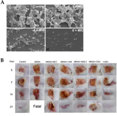
in skin burn wounds.
185
Figure 2. Antibacterial activity of WRL3 and antibiotics. (A) SEM images of the in vitro MRSA-biofilm
186
inhibition at indicated VWR3 concentrations compared to the control cells; (B) Photos of wound regions of
187
approximately 1 cm2 on the back of MRSA-infected mice and treated with MET (methicillin), VAN
188
(vancomycin), WRL3, CRO (ceftriaxone) or WRL3 and CRO at 3, 7, 14 and 21 days post-MRSA infection. “Fatal”
189
indicates no mice survival. From [32] with permission from American Chemical Society, Copyright © 2020.
190
Another potent AMP, Epinecidin-1 (Epi-1) induced keratinocytes proliferation and migration in
191
vitro. In vivo, Epi-1 induced complete healing in an MRSA-infected heat-burned pig skin over a period
192
of 25 days. The results showed that Epi-1 enhanced vascularization, increased epithelial activities and
193
stimulated collagen synthesis around the wound region [33].
194
Therefore, AMPs are considered to have a high potential in eradicating microorganisms, but as
195
well in promoting wound healing via stimulating re-epithelialization, angiogenesis, collagen and
196
granulation tissue formation.
197
The main hurdle of AMPs is their short elimination half-life due to their fast degradation in the
198
presence of proteases once the blood stream [34]. Therefore, most of the currently used AMPs are
199
directed for topical delivery, especially to wounds. Routinely used AMPs colistin, dalbavancin,
200
daptomycin, oritavancin and telavancin have a half-life of 5 h, 8 h, 8 h, 14 days and 195.4 h,
201
respectively, while the half-life of gramicidin has not been determined yet. On average, the median
202
half-life of FDA-approved peptides is 9 h [35]. As the main focus of this review is on topical
203
development stage, as well those routinely used FDA-approved AMPs for topical wound infections
205
(Table 2).
206
Table 2. AMPs for topical application under different phases of pre- and clinical trials, including
207
AMPs FDA-approved.
208
AMPs Mechanism of action Activity against Side effects
Application and administration
route
Ref.
Preclinical phase
Arenicin pore formation Membrane Gram (+), (-); MRSA infection
Significant toxicity to mammalian cells
Urinary tract infections;
hospital-acquired infections
[15, 36]
Avidocin and
purocin Membrane disruption Gram (+) and (-) Safety reported
Treatment of C. difficile infections (colitis); topical
[15]
Buforin II Inhibition of DNA/RNA synthesis
Gram (+), (-);
fungi Safety reported 37] [9,
Lactocin 160
(Bacteriocin) Membrane disruption G. vaginalis; P. bivia Safety reported
Urogenital tract infections;
Bacterial vaginosis
[38]
LTX-109 (Lutixar)
Membrane disruption and
cell lysis Gram (+) Not reported
Treatment of diabetic foot
ulcers; topical [15]
Mersacidin Inhibition of cell wall
Gram (+),
MRSA Safety reported
Treatment of hospital-acquired infections
[39]
Planosporicin
(Bacteriocin) Inhibition of cell wall
Planomonospora sp., MDR
strains
Not reported Hospital-acquired infections
[40]
Plectasin (NZ2114)
Inhibition of cell wall
synthesis Gram (+) Not reported
Pneumococcal peritonitis and pneumonia
infections
[41]
Clinical phase CZEN-002
(phase IIb) modulation Immuno- C. albicans Not reported
Vulvovaginal candidiasis;
topical [42] D2A21 (phase
III) Membrane disruption Gram (+), (-); fungi No side effects reported
Burn wound infections;
topical [43]
DPK-060 (phase II)
Membrane disruption and
immuno-modulation
Gram (+), (-);
EA-230 (phase IIb)
Immuno-modulation Gram (-) Safety reported
Sepsis and renal failure protection; IV
[44]
Histatin (phase
I) Membrane disruption Gram (-); C. albicans No side effects reported
Treatment of P. aeruginosa infections and oral candidiasis
[45]
hLF1-11 (phase
I/II) Membrane disruption Gram(+), (-); fungi
Little discomfort at the injection
site
LPS-related fungal
infections; IV [46]
IDR-1 (phase I) modulation
Immuno-MRSA;
vancomycin-resistant Enterococcus
No side effects reported Infection prevention in immuno-compromised patients [45] IMX942 (IDR-1 derivative; phase II)
Immuno-modulation Gram (+), (-) No side effects reported
Treatment of nosocomial infections, neutropenia [45] LL-37 (phase IIb) Barrel-stave mechanism of membrane disruption; inhibit LPS binding Bacteria, fungi and viral pathogens Cytotoxic Diabetic foot ulcers; chronic middle ear infection [47] LTX-109 (Lutixar, phase II) Membrane disruption and
cell lysis Gram (+)
Itching, pain and burning effects Treatment of nasal MRSA infections; nasal and topical [48] Mel4 (phase II/III) Membrane
disruption Gram (+), (-)
No cytotoxicity and no resistance
reported; no staining of human cornea
Contact lenses [49]
Melimine
(phase I/II) Membrane disruption Gram (+), (-)
No cytotoxicity and no resistance
reported; staining of human cornea
Contact lenses [49]
MX-226 (Omiganan®;
phase III) Cell disruption Gram (+), (-) Not reported
Prevention of device-related infections; topical [50] Novexatin (NP213; phase IIb) Membrane
disruption Fungi Not reported
Treatment of dermatophyte fungal infections [41] OP-145 (LL-37 derived; phase II) Membrane
disruption Gram (+)
infection; ear drops PAC113 (P113; histatin 5 analog; phase IIb) Membrane disruption and immuno-modulation Candida sp.; Gram (+), (-)
No cytotoxicity reported Oral candidiasis in HIV patients and prevention of bacterial periodontal disease; topical [15] p2TA (AB103; phase III)
Immuno-modulation Gram (-)
No adverse effects reported
Necrotizing soft tissue
infections; IV [52] Ramoplanin (NTI-851; phase II) Membrane disruption and cell wall synthesis
Gram (+); C. difficile
Low local tolerability when
injected IV
Treatment of C. difficile-associated infections; oral [53] SGX942 (Dusquetide; phase III) Modulates the innate immune
response Gram (+), (-)
Safe and well
tolerated Oral mucositis; oral rinse [54] FDA approved Anidulafungin (Eraxis™) in 2006 Inhibition of (1,3)-β-D-glucan synthase
Fungi Hypersensitivity; hepatic effects
Treatment of Candida infections, especially esophageal candidiasis; IV infusion [55] Caspofungin (Cancidas) in 2001
Inhibition of β (1,3)-D-glucan
production Fungi
Hypersensitivity; hepatic effects
Treatment of esophageal
candidiasis; IV [56]
Dalbavancin (Dalvance™) in
2014
Inhibition of bacterial cell
wall synthesis Gram (+)
May cause nausea, headache, and diarrhea Treatment of complicated skin and skin
structure infections (cSSSI); IV injection [57] Daptomycin (Cubicin®) in
2003
Membrane
lytic Gram (+)
Not approved for pediatric
patients
Treatment of cSSSI; IV
injection [58] Gramicidin
(Neosporin®) in
1955
Pore-forming; aggregation;
membrane disruption
Gram (+), (-) Hemolytic activity
Treatment of bacterial conjunctivitis; ointment [35] Polymyxins (Polymyxin E=colistin) in 1964 Membrane
disruption Gram (-)
Used only as “last-resort” due
to neuro- and nephrotoxic
effects and neuromuscular
blockage
against infections; cream, ear and
eye drops
Oritavancin (Orbactiv®) in
2014
Inhibition of bacterial cell wall synthesis and disruption
of bacterial membrane1
Gram (+) treatment is Long-term ambiguous
Treatment of
(cSSSI); IV [60]
Telavancin (Vibativ ™ & Vibativ®) in 2009
Inhibition of bacterial cell wall synthesis and disruption
of bacterial membrane1
Gram (+) acute kidney May induce injury
Treatment of
cSSSI; IV [60]
Vancomycin (Vancocin® HCl)
in 2016
Inhibition of bacterial cell wall synthesis
Gram (+) nephrotoxicity May cause
Treatment of severe MRSA infections; IV
and oral
[58]
1Oritavancin and telavancin may also act by membrane-pore or channel formation or lysis of the cell membrane;
209
Gram (+): Gram-positive bacteria; Gram (-): Gram-negative bacteria.
210
Currently, at least 10 AMPs failed in their clinical phases [3]. As examples, pexiganan (Locilex®),
211
which was used for treatment of infections in diabetic foot ulcers failed during clinical phase III.
212
Iseganan (IB-367, protegrin-I analogue), used for preventing polymicrobial oral infections such as
213
stomatitis failed in phase III, as well. Omiganan (indolicidin derived; MBI-226) failed during phase
214
III due to catheter-related infections [8]. XMP-629 used to treat impetigo and acne rosacea, and
215
Murepavadin (POL7080) [51], a protegrin analogue used to treat ventilator-associated bacterial
216
pneumonia, was halted recently in phase III. These failures were attributed to the lack of significant
217
efficacy compared to other antibacterial drugs, or to multiple side effects [15].
218
2.1.2. The challenge of resistance development
219
All types of pathogens tend to develop their own defense mechanism [45], adapting to their
220
specific environment. It is therefore unrealistic to expect no development of resistance to AMPs.
221
Generally, microbes create resistance via different modalities, such as modification of their structure
222
or electrical charge [61], modification in the cell wall and metabolism, LPS myristylation, acylation of
223
lipid A, etc. [62]. Additionally, microbes adapt to survive at high ionic concentration, a condition
224
known to hinder AMP potency [45].
225
However, it should be noted that bacteria will develop resistance to AMPs at a much slower rate
226
than to antibiotics [63]. This is attributed to the relatively low specificity of AMP mechanisms of
227
action, as well as to the variety of these mechanisms implied in bacterial killing. In general, AMPs are
228
quite resistant to the development of bacterial resistance [49].
229
2.1.3. AMP’s activity against biofilm formation
230
Though AMPs show high activity against a broad spectrum of bacteria, very little is known
231
about the resistance mechanisms developed by biofilms. Biofilm formation by bacteria is another
232
critical issue for infectious diseases. Most pathogens form microcolonies by adhering to each other
233
and then produce a biofilm as a protective environment. Biofilm formation is initiated by (i)
234
planktonic cell attachment to a surface; (ii) colony formation; (iii) biofilm formation, when bacterial
235
includes extracellular DNA, proteins, lipids and polysaccharides; and (iv) cell dispersion [64].
237
Further, the developed biofilm is dispersed and creates increased antimicrobial resistance.
238
Severe infections are associated with biomedical devices and implants due to their high
239
susceptibility to bacterial colonization [65]. Very often, implant surfaces become a reservoir of
240
bacteria that can spread very fast into the whole body, which leads to persistent chronic infections.
241
The only way to eradicate bacteria in this situation is implant removal, which implies surgery and
242
close follow-up of the patients for their complete recovery. In this context, it is worth noticing that
243
AMPs, such as AG-30 [27], AG-30/5C [28], WRL3 [32], melimine and Mel4 [49], 73c, D-73 [66] are very
244
potent against biofilm formation.
245
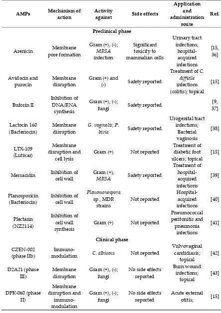
2.2. Antimicrobial dendrimer peptides
246
Antimicrobial peptide dendrimers (AMPDs) are highly branched 3D-structures with a central
247
core and high density of flexible surface groups for potential molecule attachment [67]. They can be
248
obtained by solid-phase peptide synthesis (SPPS) and purified by HPLC to ensure high
249
reproducibility [68]. Compared to polymeric dendrimers, they are built on covalently coupled amino
250
acid residues [64]. AMPDs can be built up to several generations based on the layers of branching
251
units from G0 to Gx, where x represents the number of branching cycles (Figure 3). They are a very
252
attractive class of molecules as they exhibit high antimicrobial activity against bacteria, fungi and
253
viruses [69]. Their high antimicrobial activity is mainly due to their positive charges of the amino acid
254
residues that interact with the negatively charged bacterial cell envelope and leads to bacterial death
255
[64, 70]. Moreover, AMPDs were shown to be more resistant to proteolysis and less toxic to both
256
mammalian and erythrocyte cells than AMPs [71].
257
In addition to their intrinsic activity and due to the fact that AMPDs have different functional
258
groups, it makes them very attractive delivery systems as drug carriers. Increasing the number of
259
ramification leads to an increase in functional groups, thus a higher opportunity to couple different
260
bioactive molecules.
261
Figure 3. Third generation AMPD structure, including the dendrimer core (peptidic or non-peptidic),
262
building blocks (amino acids) and functional groups (peptidic or non-peptidic) for coupling other bioactive
263
molecules. G1, G2 and G3 indicate the different generation levels.
264
Bacterial killing by AMPDs is governed by functional groups of the amino acid residues and
265
their ability to penetrate the cell membrane [64]. A recent example of effective third generation (G3)
266
AMPD, G3KL (incorporating repetitive lysine (K) and leucine (L) units), showed high activity against
267
4 out of 6 ESKAPE collection in 32 different strains of A. baumannii (MIC: 16 µg/mL), 35 strains of P.
268
mechanism of action is based on membrane disruption followed by vesicle leakage as shown by
270
transmission electron microscopy (TEM) analysis. G3KL AMPD showed as well to inhibit P.
271
aeruginosa biofilm formation [73]. Fluorescently labeled G3KL entirely diffused into P. aeruginosa
272
within 15 min as shown in a time-lapse assay, suggesting AMPD diffusion or translocation into the
273
cytoplasm through damaging both outer and inner bacterial membrane, as was confirmed by TEM
274
[74].
275
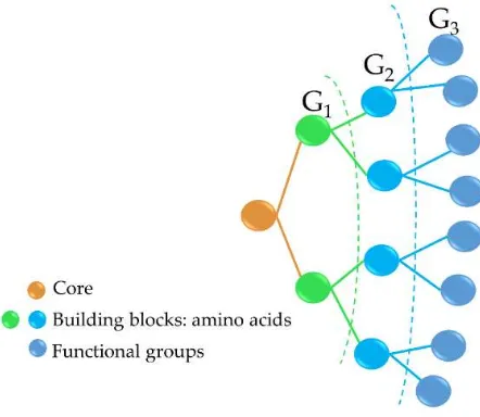
G3KL and another G3 (G3RL, consisting of arginine (R) and leucine (L) repeats), showed high
276
antibacterial activity against P. aeruginosa and neither toxicity, nor gene alteration towards progenitor
277
fibroblasts at an elevated concentration of 100 µg/mL. Furthermore, G3KL better promoted
278
angiogenesis than G3RL in a human umbilical vein endothelial cells (HUVEC) and chorioallantoic
279
membrane (CAM) model (Figure 4) [75], which is an important aspect for accelerated wound healing.
280
Figure 4. Images of the endothelial tubular networks in Matrigel after 5 hours treatment with G3KL and
281
G3RL at indicated concentrations versus PBS control. From [75] with the permission by Springer Nature,
282
Copyright © 2016.
283
A second generation (G2) AMPD, TNS18, showed the same activity against P. aeruginosa (MIC:
284
8-16 µg/mL), a much higher activity against S. aureus (MIC: 8-16 µg/mL) but less potency against K.
285
pneumoniae [68]. Combination of the peripheral branches of G2 (TNS18) with the core of a G3 (called
286
T7) resulted in a chimeric AMPD, named DC5. DC5 displayed significant activity against K.
287
pneumoniae (MIC: 16-32 µg/mL), MRSA (MIC: 32 µg/mL), E coli (MIC: 8 µg/mL), A. baumannii (MIC:
288
16 µg/mL) and P. aeruginosa (MIC: 4-8 µg/mL) strains [68]. In vivo on larvae, D-enantiomers of dG3KL
289
and dTNS18 showed high ability to kill P. aeruginosa biofilm (killing efficiency 90.2-100%) [76].
290
Another family of AMPDs based on R4 tetrapeptide (RLYR repetitive units) and R8 octapeptide
291
(RLYR-KVYG repeats) was tested against 10 different microbial strains (E. coli, P. aeruginosa, P.
292
vulgaris, K. oxytoca, S. aureus, M. luteus, E. faecalis, C. albicans, C. kefyr and C. tropicalis) with lower MICs
293
than 1 µM. Noteworthy, they proved as well to be more resistant to proteolysis and non-haemolytic
294
than the corresponding linearly repeating peptides [69]. A SB056 lipodimeric AMPD exhibited high
295
microbicidal activity against both Gram (+) and (-) bacteria with a MIC as low as 2-32 µg/mL against
296
A. baumannii, E. cloacae, E. coli, K. pneumoniae and P. aeruginosa, which is comparable activity to
297
polymyxin B. Importantly, SB056 strongly inhibited E. coli, S. aureus and S. epidermis biofilm
298
formation [77].
299
These AMPDs with preferred selectivity have proven to be highly active against the whole range
300
exhibits high antiviral activity against human papillomaviruses, human respiratory syncytial virus
302
(RSV) and human immunodeficiency virus type 1 (HIV-1), as described elsewhere [71]. Therefore,
303
further studies are warranted to bring good news in the fight against AMR in infection-related
304
diseases. So far, there are no further studies with regard to coupling of AMPDs with AMPs or other
305
chemistry involved to deliver AMPDs to the wound site.
306
3. Peptide conjugates
307
Needless to mention, most of recent studies report on new strategies to combat MDR and biofilm
308
formation. It is clear now that the antimicrobial activity could be considerably enhanced by several
309
strategies, such as chemical addition of different molecules via covalent grafting, incorporation into
310
different nanosystems, self-assembly, coupling with antibiotics, incorporation into polymer scaffolds,
311
while maintaining or improving AMP’s antibacterial activity with prolonged blood circulation and
312
reduced cytotoxicity. Other approaches for “tailoring” AMPs with enhanced stability, such as
313
modification of C- and N-terminus via acetylation and amidation, cyclisation, lactamisation,
314
lactonisation, macromolecular conjugation and PEGylation have been discussed elsewhere in detail
315
[45, 78, 79].
316
3.1. Covalent coupling to polymers
317
Polymers are composed of several repeating units of monomers. Many natural polymers, such
318
as proteins (i.e., collagen, elastin, keratin and silk fibroin) and polysaccharides (i.e., alginate, chitosan,
319
hyaluronic acid, cellulose), or synthetic polymers (i.e., poly(lactic acid) (PLA), poly(vinyl alcohol)
320
(PVAL), poly(caprolactone) (PCL), poly(lactic-co-glycolic acid) (PLGA) and poly(ethylene glycol
321
(PEG) have been widely used for medical applications [80]. Natural biopolymers (e.g., chitosan,
322
collagen and hyaluronic acid (HA)) have been shown to promote wound healing, for instance by
323
stimulating anti-inflammatory responses in chronic wounds [3]. Loss of antimicrobial activity or low
324
availability at the implant site when antimicrobial agents are covalently coupled to different surfaces
325
is a known problem related occurring with antimicrobial coatings [81]. To overcome, these issues,
326
AMPs are promising agents with high bioactivity and biocompatibility, which showed relatively low
327
bacterial resistance.
328
Among biopolymers, chitosan is widely used thanks to its amino and hydroxyl functional
329
groups [82, 83], which offer a variety of possibilities for covalent or non-covalent coupling. Chitosan
330
is a biocompatible, biodegradable polymer with antimicrobial activity and ability to promote wound
331
healing, dermal cell proliferation and migration [82, 83]. Several chitosan-AMP conjugates have been
332
designed, such as Dhvar-5 AMP immobilized to chitosan via azide-alkyne or “click” reaction which
333
showed promises as antibacterial coatings to prevent biomaterial-related infections. An ultrathin
334
layer was obtained by spin coating when dissolving AMP and chitosan in acetic acid. Higher
335
antibacterial (MIC: 4; 2; 16 and 4 µg/mL for S. aureus, S. epidermidis, E. coli and P. aeruginosa,
336
respectively) and antibiofilm activities were found when coupling Dhvar-5 peptide via C-terminus
337
than when coupling through the N-terminus, underlining the importance of the conformation of the
338
exposed peptide. Moreover, the AMP-chitosan conjugate did not show any cytotoxicity to fibroblasts
339
[84]. Similarly, hLF1-11 AMP was covalently immobilized by C-terminal unto chitosan thin films; the
340
presence of hLF1-11 inhibited the growth of MRSA in comparison to the control [46].
341
PEG has been extensively used to functionalise peptides, drugs and nanoparticles. This process
342
of PEGylation offers different advantages, such as increased resistance to degradation, increased
343
blood circulation time, reduced aggregation and toxicity [85]. Sometimes, peptide PEGylation may
344
lead to reduced proteolysis and toxicity at a cost of lower antibacterial effect. As such, Imura et al.
345
PEGylated tachyplesin I and found lower antimicrobial activity than the non-PEGylated peptide
346
against E. coli and significantly lower toxicity against CHO-K1 cells [86]. In another study, the same
347
authors loaded unilamellar vesicles with PEGylated magainin 2 and found, that PEG-magainin 2 had
348
weaker vesicle leakage, lower bactericidal activity and significantly lower toxicity to CHO-K1 cells
349
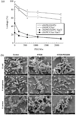
[87]. To investigate this decrease in antibacterial activity, Singh et al. PEGylated the KYE28 AMP
350
decreased against E. coli and P. aeruginosa and dramatically decreased towards S. aureus with the
352
increase of PEG length, irrespectively of the conjugation site. Cytotoxicity and hemolysis were
353
strongly reduced as well, while selectivity was improved (Figure 5) [88]. PEGylation is thus an
354
attractive technique to optimize the antibacterial efficacy of the peptide versus their toxicity. In
355
another study, peptide 73, a derivative of aurein 2.2Δ3, coupled to PEG exhibited a 2- to 8-fold
356
enhancement of antibacterial activity against S. aureus biofilms [66].
357
Figure 5. (a) Effect of PEG length on KYE28-PEG leakage induction of DOPE/DOPG (75/25 mol/mol) and
358
DOPC/cholesterol (60/40 mol/mol) liposomes at a peptide concentration of 1 µM. (b) Peptide-mediated
359
permeabilization of E. coli, P. aeruginosa, and S. aureus with the indicated peptides at 30 µM for 2 h, and analysed
360
by SEM. (DOPE (1,2-dioleoyl-sn-glycero-3-phosphoethanolamine), DOPC (1,2-dioleoyl-sn
-glycero-3-361
phosphocholine) and DOPG (1,2-dioleoyl-sn-glycero-3-phosphoglycerol, monosodium salt). Reprinted from [88]
362
with the permission of American Chemical Society, Copyright© 2014.
363
In general, bacterial colonization of medical devices favors biofilm development, which in turn
364
will lead to device failure due to peri-implantitis [89]. For instance, P. aeruginosa colonization of
365
catheters and other medical devices accounts for 10-20% of all related infections [49]. Therefore,
366
AMPs to surfaces are advantageous strategies to combat biofilm formation and reduce the rate of
368
infection. In this view, two cationic AMPs, melimine and Mel4 (melimine derivative), which are in
369
phase II/III clinical trials, were covalently attached to a model glass surface [49]. Surfaces coated with
370
both peptides lead to cytoplasmic bacteria leakage within 15 min. Moreover, the antimicrobial
371
activity of melimine was preserved upon covalent coupling to polymers or titanium, and
melimine-372
coated lenses did not show signs of conjunctival redness, though staining of human cornea was
373
observed in some cases [90]. On the contrary, Mel4-coated lenses showed no signs of corneal staining
374
or redness or ocular irritation [91]. Moreover, covalently coupled melimine and Mel4 to the glass
375
surface was shown to successfully eradicate P. aeruginosa within 15 min [49]. Overall AMP-polymer
376
conjugates can confer effective antibacterial properties to surfaces of medical devices at the preclinical
377
stage.
378
3.2. Self-assembled AMPs
379
Peptides can self-assemble into different nanostructures, such as micelles, nanotubes, vesicles
380
and fibrils. Their self-assembly mechanism is driven by spontaneous organization of peptides based
381
on their electrostatic or hydrophobic interactions, hydrogen bonding or π-π stacking [92]. An
382
important focus of self-assembled peptides research relates to the formation of amyloids, a key
383
process for the onset of neurodegenerative diseases, especially with regard to the link between
384
microbial infection and Alzheimer`s, Parkinson`s, Creutzfeldt–Jakob or related diseases [93-96]. For
385
example, Alzheimer`s β-amyloid diphenylalanine (FF) dipeptide was found to drive self-assembly of
386
FF into stiff nanotubes in Alzheimer`s disease [97]. A β-amyloid KLVFF peptide that has the same
387
mechanism as FF peptides could self-assemble into nanofibrils and then into a nanofibrillar gel in a
388
concentrated phosphate buffered saline solution [98]. Additionally, amyloid peptides, such as islet
389
amyloid polypeptide (IAPP) exhibited antimicrobial activity and sometimes their activity was higher
390
than that of LL-37 [99]. Therefore, AMPs could suggest the potential connection between amyloid
391
and AMPs in combating amyloid-related neurodegenerative diseases. AMP may be of therapeutic
392
interest in the treatment of neurodegenerative diseases, since persistent microbial infection is thought
393
to be one of the triggering factors of these diseases.
394
Melittin, a very potent AMP extracted from bee venom but severely cytotoxic, has been
co-395
assembled upon addition of a molecular block containing synthetic multidomain peptides. Upon
self-396
assembly of melittin into nanofibers, membrane selectivity and cytocompatibility was significantly
397
improved [100]. Yazici et al. combined a hydroxyapatite-binding peptide-1 (HABP1) with a tet127
398
AMP through a flexible linker as self-assembled antimicrobial agents against E. coli (MIC: 32 µg/mL)
399
and less efficient against Streptococcus mutant (MIC > 256 µg/mL). When using these self-assembled
400
HABP1-tet127 to functionalize calcium-phosphate coated on nanotubular titanium surfaces, a better
401
inhibition of E. coli (90%) than S. mutants (75%) was shown [101]. This type of self-assembled peptides
402
could be used for functionalizing surfaces of different medical implants for local therapy to prevent
403
bacterial infection. Bacitracin A modified with poly(D,L-lactic-co-glycolic acid) (PLGA) and
404
poly(ethylene glycol) (PEG) exhibited self-assembling properties with high antibacterial potency both
405
in vitro and in vivo. All PEGylated self-assemblies showed significantly stronger antibacterial activity
406
against Gram (+) and Gram (-) compared to the non-PEGylated system. The antibacterial activity for
407
non-PEGylated self-assemblies was severely compromised due to low water solubility. In contrast,
408
PEGylation of bacitracin-PLGA self-assemblies significantly improved water solubility of the system
409
and in turn, the antibacterial activity was improved against E. coli and S. aureus without significant
410
toxicity in a mouse thigh infection model. It was shown as well that PEGylation did not affect
411
antibacterial activity, on the contrary, PEGylated bacitracin-PLGA exhibited high accumulation in
412
inflammatory tissue and prolonged blood circulation [102]. This system could be efficiently used to
413
design novel antibiotic nano-assemblies in the treatment of invasive infections.
414
Self-assembly is a promising strategy towards the design of smart materials that could
415
incorporate antibiotics or other antimicrobial agents with stimuli-responsive properties. For example,
416
pH-sensitive materials could release antibiotics at a pre-defined pH, ionic strength or temperature,
417
biodegradability, and membrane selectivity, AMP self-assemblies hold great potential towards
419
eradicating MDR and biofilm-forming bacteria. Self-assembled peptides and factors influencing AMP
420
self-assembly have been described by Malekkhaiat Häffner and Malmsten in detail [96].
421
3.3. Combination of AMPs with antibiotics
422
AMPs associated with conventional antibiotics generally showed high synergism in terms of
423
their antimicrobial activity [45]. This is attributed to the fact that the combined
424
antibiotic/antimicrobial therapy causes bacterial cell membrane disruption, facilitating antibiotic
425
penetration and accumulation in the cytoplasm, ultimately leading to cell death. For example,
426
magainin 2 and cecropin A combined with rifampicin showed synergistic interactions by
427
significantly inhibiting MDR P. aeruginosa development in vitro and in vivo in rats [103]. This finding
428
suggested that the membrane-permeabilizing activity of peptides allows rifampicin to gain access to
429
its intracellular target. Stronger synergistic effects have been observed in vivo in a mouse model of
430
sepsis than in vitro when combining tachyplesin III with imipenem [104]. Synergistic effects against
431
MDR P. aeruginosa were seen when combining a peptide (18 mer) called “P5” with isepamicin.
432
Isepamicin could enter bacterial cells and inhibit protein synthesis assisted by P5, which lysed the
433
cell membrane [105]. Such synergy was not observed upon mixing P5 with cefpiramide. Thus,
434
combinations of P5 with isepamicin may be used for treatment of patients with cholithiasis, a
435
condition affected by antibiotic-resistant bacteria.
436
Similarly, B2088 AMP in combination with different conventional antibiotics, such as
437
chloramphenicol, tobramycin, gentamicin and imipenem exhibit synergistic antibacterial effects
438
against P. aeruginosa without cytotoxicity against mammalian cells [106]. For instance, WRL3 peptide
439
combined with ceftriaxone exhibited synergistic effects against MRSA-infected burn wounds in mice
440
[32]. This AMP-antibiotic mixture could be efficiently used in clinical applications for burn wound
441
infections, where MRSA and P. aeruginosa cause high rate of death.
442
Therefore, combined administration of AMPs and antibiotics appears to be a promising strategy
443
and is already applied in clinics for its synergistic antibacterial effects and ability to fight MDR
444
bacteria causing infectious diseases. Additionally, AMPs/antibiotics synergies against
difficult-to-445
eradicate biofilms have been reported [107-109].
446
4. Nanotechnological platforms and scaffolds for peptide delivery
447
As mentioned before, stability, short half-life and cytotoxicity are the key-factors limiting most
448
of the AMPs from further clinical application. During the last few decades, a plethora of studies have
449
been performed to find different strategies or systems to deliver AMPs. Nanotechnology is the field
450
of science that uses nanocarriers, such as nanoparticles (NPs) as drug delivery systems. NPs are
451
particles in the range of 0.1 to 100 nm. Conjugation of AMPs with NPs results in increased local
452
concentration at the delivery site with an enhanced AMP bioactivity, which might be attributed to a
453
synergistic effect [110]. Nanotechnology can add many advantages, such as improvement of
454
solubility, bioavailability, release and higher penetration within biofilms of AMPs [111, 112]. They
455
can provide protection to AMPs against degradation in different environments, such as enzymatic
456
degradation [26].
457
4.1. Polymeric scaffolds and wound dressings
458
Polymer scaffolds are the most used formulations to address wound healing and recovery due
459
to their ability to promote cell attachment and proliferation, extracellular matrix generation,
460
restoration of vessels via creating physical bonds between cells and 3D-network of the scaffolds [113].
461
Moreover, scaffolds can retain a significant amount of water or biological fluids within their
3D-462
network due to interaction of water with polymer hydrophilic groups (hydroxyl, amine, amide and
463
carboxyl groups), or hydrophobic interactions with specific biological fluid components, and/or
464
osmotic driving force effects [114]. Polymer-based scaffolds can be applied at the wound site and be
465
In this view, a new strategy for topical application was developed by tethering human
467
cathelicidin LL-37 on collagen scaffolds for the treatment of wound infections. LL-37 loaded scaffolds
468
exhibited no toxicity towards fibroblast at a peptide concentration 24-fold higher than the cytotoxic
469
threshold [116]. After 14 days, LL-37 loaded onto collagen domains (derived from collagenase or
470
fibronectin) was retained on the scaffolds and showed no inhibition of antimicrobial activity against
471
both Gram (+) and (-). This poor delivery may be due to too strong peptide-scaffold interactions.
472
Similarly, Cassin et al. evaluated incorporation of LL-37 into collagen and hyaluronic acid
473
polyelectrolyte multilayers (PEMs) via physisorption and covalent immobilization [117].
LL-37-474
loaded thin films effectively prevented E. coli adhesion, but cells treated with covalently immobilized
475
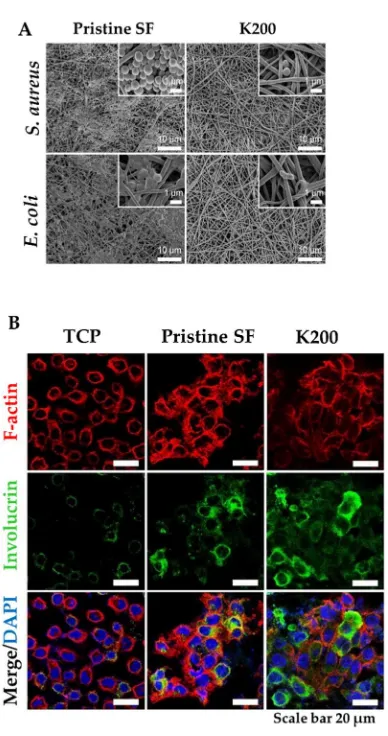
AMP presented morphological changes, suggesting cytotoxicity. In another study, Cys-KR12 AMP,
476
a LL-37 derivative, was covalently immobilized on an electrospun silk fibroin (SF) nanofiber
477
membranes via EDC/NHS and thiol-maleimide “click chemistry”. Chemical immobilization of the
478
AMP to SF membranes did not inhibit the biological activity of the peptide. On the contrary, the
479
conjugate was highly active against four different strains, such as S. aureus, S. epidermidis, E. coli, and
480
P. aeruginosa) with no biofilm formation (Figure 6) [118]. Cys-KR12-SF membranes promoted the
481
proliferation of keratinocytes and fibroblasts, and induced differentiation of keratinocytes with a
482
pronounced cell-adhesion, important key steps in wound healing. These membranes are promising
483
candidates for topical wound healing applications.
484
485
486
Figure 6. (A) FE-SEM images of S. aureus and E. coli cultured on Pristine SF and Cys-KR12 at 200 µg/mL
487
(K200). (B) Confocal immunofluorescence images of keratinocytes cultured on TCP (tissue culture treated),
488
pristine SF and K200 (red channel, F-actin; green, involucrin; blue, DAPI). Reprinted from [118] with permission
489
Cellulose and its derivatives loaded with AMPs have been tested in diabetic foot ulcer (DFU)
491
treatment. In a randomized phase III clinical trial [119], ACT1 AMP embedded in a
hydroxyethyl-492
cellulose (HEC) hydrogel (Granexin) was topically applied for DFU treatment. The results showed
493
that Granexin treatment was safe and effective for DFU treatment with a significant reduction of the
494
ulcer area at week 12 (72% reduction versus control with 57%). All patients treated with ACT1-based
495
hydrogels reached 100% ulcer re-epithelialization in a short time with no toxicity, no immunogenicity
496
or any side effects reported [120]. Such systems confirm their utility for the treatment of topical
497
wound infections, especially DFU patients.
498
All these studies support the potential for AMP-loaded into polymer scaffolds for sustained and
499
enhanced biological activity of the AMPs. Designing a polymer-based scaffold loaded with AMP that
500
could control the AMP release via binding to the matrix, while promoting wound healing via the
501
bioactivity of the scaffold itself would be beneficial not only for wound healing, but also for
502
preventing bacterial infection.
503
4.2. Organic nanoparticles
504
4.2.1. Polymeric nanoparticles, nanoemulsions and micelles
505
Polymer nanoparticles may be produced either in the form of gel-like particles, or solid polymer
506
particles. Nano- or microgel particles are prepared from swollen hydrophilic polymers, generally
507
obtained by ionotropic gelation, a one-step process in which a counter-ion is added to a
508
polyelectrolytic polymer thus producing micro- or nanoparticles though ionic interactions. In
509
contrast, solid polymer particles are based on organo-soluble, poorly hydrophilic polymers such as
510
PLGA or PLA, and may be produced by methods such as oil-in-water or water-in-oil-in-water
511
emulsion evaporation.
512
Carboxymethyl chitosan (CMC) NPs have been loaded with a very potent AMP OH30 extracted from
513
king cobra for potential wound healing. Surprisingly, positively charged CMC could interact with
514
negatively charged bacterial cell membranes and assist the internalization of the OH30 peptide. A
515
slow release of OH30 from the CMC-OH30 NPs was seen over 24 h and maintaining antimicrobial
516
activity as well. CMC-OH30 NPs could slightly enhance migration (95%) but not proliferation of
517
keratinocytes compared to OH30 (85%), CMC NPs (75%) or untreated cells (60%). In vivo, CMC-OH30
518
NPs significantly accelerated wound healing in a full-thickness excision mouse model. Moreover,
519
mice treated with CMC-OH30 NPs exhibited 70% wound closure at day 5 compared to CMC NPs or
520
OH30 alone, which maintained wound closure in the range of 36-58% [121]. A drawback of the
CMC-521
OH30 complex is its limited stability as shown by the variability in size and zeta potential over 28
522
days. Another 5-amino acid AMP, RBRBR, was encapsulated into chitosan NPs through the
523
ionotropic gelation process with a 51% encapsulation efficiency, slowly releasing the AMP over 14
524
days. AMP-loaded chitosan NPs showed at least 3-log increased antimicrobial activity against S.
525
aureus while decreasing the toxicity against both mammalian and human erythrocyte cells.
526
Importantly, positively charged (+33 mV) AMP-chitosan NPs (121 nm) significantly inhibited the
527
growth of biofilm (up to 98%) [122]. The straightforward approach of ionotropic gelation may be used
528
to deliver other potent AMP while reducing their cytotoxicity, in order to achieve efficient
529
antibacterial effects against MDR and biofilm-forming bacteria.
530
Nordström et al. investigated the ability of poly(ethyl acrylate-co-methacrylic acid) (MAA)
531
microgels to deliver two AMPs, LL-37 and DPK-060. AMP loading into the microgels decreased
532
toxicity towards erythrocytes and was suggested to protect peptides against protease degradation.
533
In turn, microgel-loaded peptides could kill MRSA, E. coli and P. aeruginosa via a membrane
534
microgel-loaded AMPs and cellular membranes, Nordström et al. included LL-37 into MAA
536
microgels. Insertion of the peptides released from LL-37-loaded microgels was shown to occur
537
without membrane defect formation at low concentrations, but with loss of lipid from the bilayer in
538
a concentration-dependent manner [124]. Similarly, LL-37 has been encapsulated into liquid
539
crystalline nanoparticles (LCNPs), such as cubosomes, for treatment of S. aureus skin infection. LL-37
540
loaded into cubosomes was protected against proteolytic degradation by P. aeruginosa elastase while
541
improving AMP bioavailability and efficiency. In an ex vivo wound infection model LL-37-loaded
542
cubosomes showed highest killing efficiency of S. aureus and absence of pig skin irritation [125].
543
Further molecular dynamic simulations confirmed these experimental results, revealing that charged
544
amine and guanidium groups from LL-37 are responsible for facilitating the interaction with the
545
bacterial membrane. Moreover, LL-37 stabilized cubosome through hydrophobic interactions, while
546
polar residues remained in solution [126].
547
Silva et al. loaded LLKKK18 (KEFKRIVKRIKKFLRKLV) into hyaluronic acid nanogels and
548
reported higher stability of the peptide and reduced toxicity to mammalian cells. Importantly, it was
549
found that nanogels were internalized by macrophages infected with Mycobacterium tuberculosis or
550
M. avium. Significant reduction of bacterial load in infected mice after tracheal administration of the
551
nanogels was seen [127]. Similarly, tet213 AMP was admixed to alginate, hyaluronic acid and
552
collagen for a potential wound dressing. Tet213-dressings showed antimicrobial activity against three
553
different bacterial strains (E. coli, MRSA and S. aureus) and promoted fibroblast proliferation in vitro.
554
Moreover, in vivo Tet213-dressings promoted re-epithelialization, enhanced collagen deposition,
555
angiogenesis and completion of wound healing in E. coli- and S. aureus-infected full-thickness
556
wounds in rats. The AMP-dressing had higher antibacterial activity than silver-containing
557
commercial Aquacel, alginate, hyaluronic acid and collagen dressing [128].
558
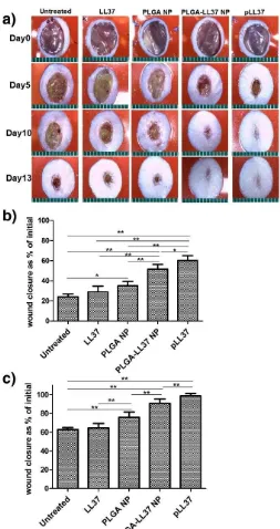
As for solid micro/nanoparticles, LL-37 loaded PLGA NPs were prepared via W/O/W
emulsion-559
solvent evaporation method and showed high antibacterial activity in vitro against E. coli. Topically
560
applied PLGA-LL-37 NPs enhanced wound closure in mouse full-thickness excisional wounds with
561
an average healing of 79 and 90 % at day 7 and 10, respectively (Figure 7). Moreover, advanced
562
granulation tissue formation, re-epithelialization and accelerated angiogenesis was observed in the
563
group of mice treated with PLGA-LL-37 NPs [129]. The only disadvantage of such system is the
564
limited residence time and bioadhesion in the wounds, which is not desired in case of hard-to-heal
565
wounds. Similarly, esculin1-a AMP loaded into PLGA NPs resulted in selective bioactivity with high
566
Figure 7. Accelerated wound healing in mice treated with PLGA-LL-37 NPs compared to controls. (a)
568
Images of wounds in mice of five tested groups: untreated, LL-37, PLGA-NP, PLGA-LL-37 NPs and pLL-37
569
(plasmid encoding hCAP18/LL-37). Representation of Wound area at (b) day 5 (n = 13) and (c) day 10 (n = 10)
570
(mean ± SD). Reprinted from [129] with permission of Elsevier, Copyright © 2014.
571
The performance of AMP incorporated into micelles was investigated by Wang et al. After
572
complexation process between MSI-78 (pexiganan) peptide and methoxy poly(ethylene
glycol)-b-573
poly(α-glutamic acid) (mPEG-b-PGlu) it was found that haemolytic toxicity was decreased through
574
polyelectrolyte complexation without hindering its antimicrobial activity against E. coli, B. subtilis
575
and S. aureus [131]. In another study, peptide 73c and D-73 encapsulated into PEGylated micelles
576
showed increased antibacterial activity against S. aureus biofilms by 510- and 9-fold, respectively,
577
compared to their parent peptide in a murine cutaneous abscess model. In vitro, peptides formulated
578
as PEGylated micelles exhibited decreased toxicity to mammalian cells [66].
579
LL-37 loaded nanostructured lipid carriers (NLC) were prepared by melt-emulsification method
580
and showed a preserved bioactivity against E. coli (killing efficiency 73%). Topically applied
![Figure 1. AMP classification (partially inspired by the DRAMP database [13]).](https://thumb-us.123doks.com/thumbv2/123dok_us/8068551.1345470/4.595.88.510.262.505/figure-amp-classification-partially-inspired-dramp-database.webp)
![Table 1. AMPs with their potential activity selected from the APD database [12].](https://thumb-us.123doks.com/thumbv2/123dok_us/8068551.1345470/5.595.69.510.163.792/table-amps-potential-activity-selected-apd-database.webp)