Open Access Journal of Contraception
Dove
press
R e v i e w
open access to scientific and medical research
Open Access Full Text Article
Folates for reduction of risk of neural tube
defects: using oral contraceptives as a source
of folate
Anita L Nelson
Obstetrics and Gynecology, David Geffen School of Medicine at UCLA, Harbor UCLA Medical Center, Torrance, CA, USA
Correspondence: Anita L Nelson 1457 3rd Street, Manhattan Beach, CA 90266, USA
email [email protected]
Abstract: The evidence that folates reduce the risk of neural tube defects (NTDs) is so compelling that supplementation has been recommended by every relevant authority. The Cochrane Database of Systematic Reviews has determined that folate supplementation should be rated as a Grade 1 recommendation. United States Preventive Health Services Task Force, the US Food and Drug Administration (FDA), and the Centers for Disease Control and Prevention (CDC) have all produced clear guidelines for such supplementation. Unfortunately, despite food fortification and targeted public health campaigns promoting folic acid supplementation, periconceptional utilization of folic acid supplements has decreased in the US in recent years. Worldwide, over 300,000 newborns are affected with NTDs every year. NTDs account for 10% of all neonatal mortality. This article will review the risk factors for NTDs and the evidence supporting folate supplementation. It will also describe the remaining problems and outline current ideas to solve them. Finally, new evidence of the effectiveness of adding metafolin to drospirenone-containing oral contraceptives in raising serum and red blood cell folate levels, the rationale for making such an addition, and an estimate of the magnitude of the contribution use of such pills might have on reducing NTDs will be discussed.
Keywords: neural tube defects, folate, metafolin, oral contraceptives
Introduction
Grains and cereals in the US have been fortified with folic acid for over a decade to reduce the risks of neural tube defects – so it is reasonable to ask: why do we need to provide any extra folate supplementation? It is also reasonable to ask: if women are using oral contraceptives to protect against pregnancy, why should we use those pills to reach women at risk for having pregnancies affected by neural tube defects?
In this article we will review the spectrum of neural tube defects (NTDs), the impact NTDs have on quality of life, what is known of their genetic causes, and the connection between NTDs and folates. We will also analyze the outcomes of the dif-ferent public health interventions, which have been adopted over time to increase folate for reproductive age women and we will describe the remaining challenges. Finally, we will describe the latest approach to providing folate supplementation to women at risk for pregnancy – the addition of tetrahydrofolate to drospirenone-containing oral contraceptives.
Definitions of neural tube defects
Neural tube defects are congenital malformations that result from failure of the neural tube to close during embryogenesis. The primordial central nervous system starts as
Open Access Journal of Contraception downloaded from https://www.dovepress.com/ by 118.70.13.36 on 26-Aug-2020
For personal use only.
Number of times this article has been viewed
This article was published in the following Dove Press journal: Open Access Journal of Contraception
Dovepress
Nelson
a plate of cells, which folds on itself to form a tube between days 21 and 28 postconception. Neural tube defects include spina bifida, anencephaly, encephalocele, craniorachischisis, and iniencephaly.1 The type of NTD that develops depends
upon the timing (and thus the location) of the defect. If the failure occurs at the cranial end, the resulting malformation is called anencephaly. Anencephalic fetuses lack variable amounts of brain and cranium above the base of the skull and the orbits. Defects at the cranial end of the neural tube, collectively called spina bifida, occur in the midline of the lumbosacral region. These defects involve dysplasia of the spinal cord, neural roots, and meninges. If the herni-ated sac contains neural elements, the defect is called a meningomyelocele; if only the sac protrudes, the defect is a meningocele. The defect may or may not be covered by skin. Encephalocele represents a postclosure NTD. It is a membrane-covered protrusion of the brain tissue through an abnormal opening in the skull, typically in the occipital area. Microcephaly and hydrocephalus are common findings associated with encephaloceles.2 This paper will focus on the
two main defects that have been linked with closure forma-tion defects – anencephaly and spina bifida.
Incidence of NTDs
Neural tube defects are the most common malformations of the central nervous system and collectively comprise the second most common congenital anomaly overall.3
Worldwide, spina bifida and anencephaly affect 300,000 newborns each year.1,4 These defects are responsible
annu-ally for 41,000 deaths and 2.3 million disability-adjusted life years.5 Neural tube defects alone account for 10% of total
neonatal mortality6 and comprise 10% of the burden of all
congenital conditions. The incidence of NTD varies between 1 and 10 per 10,000 live births each year.7 Prevalence differs
by geographic area, socioeconomic status, and ethnicity. The areas of highest prevalence in Western countries are Ireland and Scotland, where the baseline prevalence is 10/1000 live births.3 In the US, the greatest prevalence of NTDs is found
in the southern states.
The birth rate of infants affected by NTDs has dropped in the last 30–40 years because of better prenatal diagnosis utilizing widespread serum alpha fetoprotein testing and high resolution ultrasound, coupled with selective abor-tion practices. Prenatal detecabor-tion rates are reported to be in the range of 82%–98%.8,9 Once an NTD is detected, these
same studies report that 60%–83% of affected pregnancies are terminated. However, despite these antenatal screen-ing programs, food fortification, and extensive public
health campaigns recommending preconceptional folic acid supplementation, 2500 infants are born in the US each year with NTDs10 and another 1500 pregnancies affected by
NTDs are terminated.11,12 It is estimated that at this time about
30,000 Americans are living with spina bifida.3
NTDs represent a major health problem. The mortality rate for anencephaly is 100%; for infants affected by spina bifida, the mortality rate is about 30%. The survivors with spina bifida suffer significant morbidity from neurological defects that correspond to the level of their malformations. Most spina bifida results in paralysis in the lower extremities and loss of bladder and bowel function. Affected individuals usually require significant medical attention and experience long-term disabilities that affect their survival, functional abilities, and economic productivity. The development of intermittent bladder self-catheterization13 has been
recog-nized as providing major improvement in quality of life for those with spina bifida1 but latex allergy rates are among the
highest in this population and recurrent urinary tract infection can ultimately lead to renal failure.
NTD etiology
Neural tube defects have a heterogeneous and multifactorial etiology, including genetic and environmental factors as well as predisposing maternal factors.14 Genetic predisposition is
suspected because of the following: the prevalence of NTDs varies in different racial and ethnic groups,15,16 the excessive
numbers of affected females compared to males, the high rate of consanguinity seen in parents of NTD-affected infants, the association of NTDs with more than 80 genetic abnormalities (trisomy 13 and 18 and autosomal recessive syndromes, such as Meckel–Gruber)17, and the high rates of recurrence in
subsequent pregnancies.14,18 Environmental maternal health
influences are suspected because of wide geographic varia-tions in incidence as well as the identification of other risk factors, such as nutrition, maternal diabetes (relative risk for NTDs of 15.5),19 use of valproic acid,20 hyperthermia,21,22
obesity,23–25 and occupational exposure to agricultural26 and
cleaning products.27,28 In areas where a folate-rich diet is not
readily available, the risk of NTDs is highest among the most economically disadvantaged.29,30
Genetic etiology of NTDs
Knowledge about the genetic etiology of NTDs is limited because the techniques used to identify individual gene loci require genetic information from multiple affected family members, which is a rare occurrence with NTDs. There are 200 small animal models of NTDs, but most do not replicate
Open Access Journal of Contraception downloaded from https://www.dovepress.com/ by 118.70.13.36 on 26-Aug-2020
Dovepress Folates in oral contraceptives
the human disease phenotype. Reflecting known risk factors, over 100 candidate genes have been suggested for spina bifida, including those involved in folic acid metabolism, glucose metabolism, retinoid metabolism, and apoptosis, but fewer than 20% of the genes studied confer even minor risk association.7 NTDs have been found to be closely related to
genes that encode proteins that are directly or indirectly con-nected with folic acid and methionine metabolism. The most productive research has, therefore, focused on the variation in genes encoding for enzymes related to folate uptake and the metabolism of both folate and homocysteine.14
Genetic impacts on folate
metabolism
Natural dietary folates are polyglutamates that must be deconjugated into monoglutamates before they can be absorbed in the proximal small intestine. Deconjugation is accomplished by the enzyme folylpoly-gamma-glutamate carboxypeptidase. The folate most commonly found in the bloodstream is 5-methyltetrahydrofolate monoglutamate (THF). THF can enter cells directly by means of high affinity receptors that are found in the proximal tubules of the kidney, the choroid plexus and, importantly, in the placenta. There is also a high density of folate carrier in the embryonic neural tube, supporting the critical role for folate during neural tube closure. Other lower affinity receptors are located on numer-ous sites. In addition to receptor-mediated transport, THF can enter cells by a carrier-medicated transport via reduced folate carrier (RFC), which is ubiquitously expressed. Once THF enters the cell, another glutamyl group is added so THF cannot be transported out of the cell.
Delivery of folate to the embryo depends not only on maternal intake, but on maternal absorption and metabolism, as well as folate transport across the placenta and embryonal folate uptake.31 In either the mother or the embryo, mutations
in genes in the folate cycle, such as methylenetetrahydrofo-late reductase and fomethylenetetrahydrofo-late receptor genes, cause NTDs.32,33
The most significant genetic variations have been found in the genes encoding for enzymes of folate metabolism and the closely- related homocysteine metabolism.14 The most
important enzyme in the regulation of available folate is the enzyme methylenetetrahydrofolate reductase (MTHFR). A common mutation in the 5,10-methylenetetrahydrofolate reductase (MTHFR) gene has been identified as a risk fac-tor for NTDs.34,35 The MTHFR enzyme allows the cells to
regulate intracellular concentration of methionine and homo-cysteine. One specific thermolabile variant, MTHFR C677T SNP, in which there is an alanine-to-valine substitution, has
been associated with a 66%–100% increase in the risk of NTDs in their mothers,14,36 especially those with low folate
levels.37 This mutation has been reported to account for 26%
of Irish NTDs.38 This mutation causes the enzyme to lose
its activity in the face of an increase in temperature, results in increases in homocysteine, and requires higher levels of folate to stabilize the binding of the constituent parts of the MTHFR enzyme.59 This mutation is present in 4%–16% of
healthy adults, with the highest prevalence in Japanese men.39
A second SNP – the MTRR A66G SNP, which is involved in methionine synthetase reduction – has also been associated with a 48% increase in risk for NTDs.14
Because of altered folate-dependent metabolic activity, some segments of the population may require more folate than their local diet is capable of supplying.47 They may also
need higher amounts of supplementation to benefit from preconceptional supplementation. They would also benefit from using the most easily utilizable form of folate (L-5-methyltetrahydrofolate).
The connection between NTDs and
folate: an epidemiologic success story
There are only a few instances in which science has been able to identify preventable causes of birth defects. The most prominent of these includes recognition of rubella virus as a cause of congenital rubella syndrome, cytomegalovirus as a cause of birth defects and developmental disabilities, alcohol as a cause of a spectrum of defects, and folic acid as preventive for neural tube defects, especially the two defects we are discussing, spina bifida and anencephaly.40 A recent
Cochrane review concluded that folic acid alone or in combi-nation with vitamins and minerals prevents NTDs.41 Oakley
characterized the discovery that a simple cheap and effective public health intervention – giving a vitamin – could keep thousands of children alive each year and out of wheelchairs as a “humanistic miracle”.42 Indeed the folate story is quite
impressive. Folate pioneers had to overcome significant bar-riers on their path to discovery of that relationship and even more barriers to introduce the measures needed to prevent these birth defects.43–45
The relationship between NTDs (especially anencephaly and spina bifida) and folate deficiency was first suggested in 1964.46 It was not an easy association to recognize because
most women who deliver babies with NTDs have blood levels of folate that are within normal limits as defined for the gen-eral public.47 One early study indicated that levels of
abnor-mal formiminoglutamic acid excretion test were five-fold higher in women who delivered infants with NTDs than those
Open Access Journal of Contraception downloaded from https://www.dovepress.com/ by 118.70.13.36 on 26-Aug-2020
Dovepress
Nelson
who delivered normal infants, when the women were tested around the time of delivery. However, the authors concluded that that abnormality might indicate defective absorption or metabolism rather than a deficient intake of folate.48 On the
other hand, Smithells hypothesized that the cause of NTDs might be nutritional, because poor women had a higher risk than better fed, wealthier women.44 Unfortunately, three of
four hospitals in which Smithells sought to test his hypothesis refused to allow a randomized trial, so the power of his find-ings of a reduction in NTDs with folic acid supplementation in a nonrandomized trial were less persuasive than might have been expected for such an important breakthrough.42
The most convincing evidence of the association between NTDs and folic acid came from women who had experienced previous pregnancies affected by NTDs. The recurrence rate for NTDs is about 2.9% in North America and 4% in the British Isles, but it can be as high as 10% in other areas.49 The landmark
study by the Medical Research Council Vitamin Study Group was a randomized trial of folic acid (4 mg/day) vs placebo for the prevention of recurrent NTDs. When the 4 mg dose was started at least 1 month before conception and was continued through the first trimester, a 72% reduction in NTDs was seen in the intent-to-treat population and a 83% reduction was seen among the per-protocol women.43 A later meta-analysis
of four randomized trials of folic acid for the prevention of recurrent NTDs showed a 69% reduction in an intent-to-treat analysis and an 87% reduction in the risk of recurrence among women who started treatment prior to pregnancy.49 Studies
with lower doses of folic acid (0.35 mg) and less frequent doses failed to find benefit.50 Unfortunately, the percentage
of women with prior affected pregnancies who actually take
any folic acid supplements before subsequent pregnancies
has been reported to vary between 33% and 85%.49
Although studies of women with prior NTD-affected pregnancies persuasively demonstrated the protection folate provided, 90% of cases of NTDs occur in women who have never experienced a NTD-affected pregnancy. Since it is not clear that the defect mechanisms would be the same in those two different populations, it was important to also study interventions in the general population. Between 1988 and 1995, five case-controlled studies of women without prior pregnancies affected by NTDs (spina bifida or anencephaly) investigated the impact that preconceptional folate supple-ments with or without multivitamins and food folate would have on the incidence of NTDs.51–55
Only one study with all three interventions in the study arm failed to find any decrease in NTD incidence.53 The
other four studies reported reductions in the risk of NTDs
in the range of 35%–75%.51,52,54,55 A later large cohort study
in China also demonstrated the effectiveness of folic acid supplementation in preventing NTD in first pregnancies; the reductions seen in this study also showed geographic variation with protection ranging from 41% to 79%.56 One
prospective, but nonrandomized, cohort study with dietary folate and a folic-acid-containing multivitamin studied in 22,000 women found a mean reduction in NTDs of 71% among women who started the supplement at least 1 month prior to conception and continued to take it through the first trimester.57 The only randomized prospective trial of folic
acid compared folic acid (0.8 mg) with multivitamins to a supplement containing trace elements. In that study, no cases of NTDs occurred in the 2104 women in the folic acid arm, but six cases were seen in the 2052 women who were given trace element supplements.45
Today it is estimated that 50%–60% of NTDs could be prevented by adequate periconceptional folic acid.3 Some
experts have estimated that the lowest levels of NTDs to be expected with folate supplementation would be four to five cases per 10,000 births or seven to eight cases at birth or abortion per 10,000 births.58
Definition of folates
“Folate” is a generic term for a water soluble B-complex vitamin, which serves as an enzyme in transfer of single carbons during the synthesis of the purines and pyrimidines that are required for DNA synthesis and repair.3 The folate
cycle is also involved in another essential physiological process: the methylation associated with the methionine cycle, which is particularly important during the initial period of embryonic development.59 Folate helps produce
and maintain new cells, especially at a time of rapid cell division and growth. Low folate levels are also associated with megaloblastic anemia.
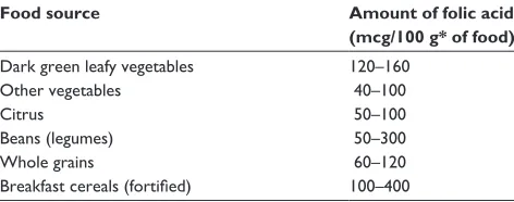
Folates occur in foods as polyglutamates. Foods that are good sources of dietary folate polyglutamates include spinach, lentils, chickpeas, asparagus, broccoli, peas, brus-sel sprouts, nuts, corn, and oranges as well as kidney beans (Table 1). Polyglutamates must be unconjugated to form monoglutamates before they can be absorbed. Folic acid is rarely found in food but is an oxidized monoglutamic acid (pteroylglutamic acid) that is the most active form of the vitamin. Polyglutamates from foods have only about half the absorbance of folic acid and have even less bioavail-ability on an empty stomach.3 Therefore, folic acid is the
form used both in vitamin supplementation and in food fortification.
Open Access Journal of Contraception downloaded from https://www.dovepress.com/ by 118.70.13.36 on 26-Aug-2020
Table 1 Food sources of folic acid145
Food source Amount of folic acid
(mcg/100 g* of food)
Dark green leafy vegetables 120–160
Other vegetables 40–100
Citrus 50–100
Beans (legumes) 50–300
whole grains 60–120
Breakfast cereals (fortified) 100–400 Note: *100 g = 3.5 oz.
Dovepress Folates in oral contraceptives
Folate requirements
Most fetal organogenesis is established within the first few months of pregnancy. A mother’s nutritional status precon-ceptionally can have lifelong consequences for the child. It is clear that it is not adequate for pregnant women to consume a diet that only avoids severe nutritional deficiencies; women need diets that provide sufficient nutrients of all types to optimize fetal anatomic and neurologic development.60 Both
excesses of certain nutrients (eg, glucose) and deficiencies in others (folate, protein, iron, vitamin B6, calcium, zinc) can cause deleterious changes in physiologic set points, organ size, and cell-signaling programming. Timing of these dietary changes is important; in particular, it is important that the intake of folates be increased prior to conception. Good nutritional assessment and counseling is a critical component of preconceptional care.60
The Institute of Medicine recommends consumption of folic acid 400 mcg/day for adult women (age 19 and over) and 800 mcg/day for women aged 14–18 years.61 The
aver-age amount of folic acid received through fortification from grains in the US is only 128 mcg/day, so supplementation is often needed.62 Even folic acid requires metabolism to
acti-vate it. Genetic influences in folate absorption and metabo-lism can occur at several levels and may limit the usefulness of supplementation for some individuals.
Historic approaches to enhancing
folate levels prior to pregnancy
At least 3 months of folic acid supplementation is needed to achieve adequate steady-state folate levels.60 Concentrations
of folate in red blood cells less than 906 nmol/L are associated with higher than baseline risk for NTDs.63 Folic acid
supple-mentation that is not initiated until the first prenatal visit is too late to prevent NTDs; the malformation may already be a reality. Classically, there have been three approaches tried in the US to ensure that women have adequate levels of folate prior to organogenesis.64 These approaches vary in their target
audiences as well as in their recommended interventions.
The CDC initially recommended that the target population for folic acid supplementation should be all reproductive-aged women.65 The Institute of Medicine (IOM) narrowed the
target population to “women capable of becoming pregnant”. In the assessment of the IOM, fewer than half of US women age 15–44 are at risk of pregnancy. This is because 22% of US women are permanently sterilized, 20% are using a “highly effective method”, 11% have never had coitus, and 5% are pregnant or recently delivered. The US Preventive Health Services Task Force recommends that all women planning or capable of pregnancy take a daily supplement containing 0.4–0.8 mg (400–800 mcg) of folic acid. They labeled the recommendation as Grade A.66 The two approaches that have
been tried over the years to increase periconceptional folate levels in women include:
• Recommend folic acid supplementation for the target population.
• Increase folic acid intake by fortifying food supplies so that all women will automatically be provided adequate levels.
These approaches may be complementary.
Folic acid supplements
Early efforts to target folic acid supplementation to women planning pregnancy were useful,64 but only had a modest
impact.67 In fact, the frequency of NTDs in the US did not
decline following extensive lay and professional educational campaigns about folic acid supplementation.68 Several
prob-lems with targeted supplementation have been identified. The first problem identified was that women were not aware of the need to consume extra folate. A survey in 1998 indicated that only 29% of US women said they were following the recom-mendation that they consume 400 mg of folic acid daily.69
In another survey, even among women who knew of the need for folic acid, knowledge did not translate into action. Following the campaigns, folic acid utilization rose overall from 14% to 23%.70 In 2007, the March of Dimes found in a
national survey in the US that 36% of women surveyed did not even know the meaning of the term “preconceptional care”.71 International studies have also reported infrequent
preconception use of folic acid supplement.72–74 A recent
study in Israel found that only 7.7% of Jewish women and 3.1% of Bedouin women used folic acid for 3 months before conception.75 Secondly, most pregnancies in the US
are not planned and prepared for, and we do not even have statistics estimating rates of planned pregnancies. All that is measured is “ unintended” pregnancy. Similarly, a recent special report from the European Surveillance on Congenital
Open Access Journal of Contraception downloaded from https://www.dovepress.com/ by 118.70.13.36 on 26-Aug-2020
Dovepress
Nelson
Anomalies group noted that in Europe, many pregnancies are unplanned.76 The other major problem with targeted
supplementation was that clinicians did not routinely screen women and educate them on the need for preconceptional folic acid supplementation.77,78
The bottom line was seen in a meta-analysis of eleven population-based registries and 15 surveillance systems from Europe, North America, and Australia, which found no significant changes in trends for NTDs in areas with supple-mentation recommendations alone.77 Another international
study of over 13 million births found that the incidence rates of NTDs were unchanged by the supplement campaigns.1
A Swiss study revealed recently that only 5% of women in that country correctly took periconceptional folic acid.79 In
fact, one newer study found that only 20% of Dutch women in lower socioeconomic status groups knew the correct period of use of folic acid. The study was conducted 8 years after a mass media campaign. The authors concluded the once-only campaign had only a short-term effect.80
Food fortification
Given the obvious failure of voluntary targeted folic acid supplementation, attention turned to food fortification. The topic was controversial because there were issues of costs and potential health risks from fortification.
Fortunately, fortification of specific frequently consumed food stuffs, such as cornmeal and wheat bread, does not affect their apparent taste or shelf life. In a benefit–cost analysis per-formed prior to the FDA mandate, the unit cost of folic acid was estimated to be US$0.0095/100 g of grain. The cost of testing to demonstrate appropriate levels in the final product was another $2.5 million. Labeling changes were expected to be the most costly item, totaling $20 million. On the benefits side, the cost per child with spina bifida was assumed to be nearly $350,000. Given these costs and benefits, the authors estimate a net annual benefit of $121 million and a benefit-to-cost ratio of 4.3 to 1.81
Beyond these economic costs, there was concern about adverse health effects that the addition of folic acid might have on vulnerable populations, such as the elderly and vegetarians. The most concerning impact would be that folic acid supplements would mask megaloblastic anemia due to vitamin B12 deficiency, ie, that folic acid would induce labora-tory normalization while there was progression of neurologi-cal manifestations of pernicious anemia, including cognitive impairment, subacute degeneration of the spinal cord or other neurological sequelae.82 Another possible adverse
effect of increasing folates in the diet would be experienced
by individuals using medications with intentional antifolate effects, such as methotrexate, pyrimethamine, trimethoprim, triamterene, and sulfasalazine. There was also concern that additional dietary folate might affect the absorption or metabolism of trace minerals, especially zinc, although decreased zinc excretion should balance that effect. The lack of data about long-term safety of continuous high intake of folate also troubled some people.83
Despite these concerns, the benefits expected from for-tification outweighed those risks. The US Food and Drug Administration mandated that by 1998 all enriched grain products would be fortified with 140 mcg folic acid per 100 g of grain.84 This level of enrichment was expected to raise
US consumption of folate by about 100 mcg a day. At folate levels achieved by the addition of 100 mcg of folic acid, it was expected that the percentage of US women who would consume the minimum recommended amount of folic acid a day (400 mg) would increase from 29% to 50%.43,85,86
Follow-up studies found that blood folate levels in the general adult population increased significantly following folic acid fortification of enriched grain products,87–91 and that
there was a measurable decline in neural tube defects after food fortification.92–96 These studies noted a 21%–34% reduction in
the occurrence of spina bifida and an 11%–20% reduction in anencephaly. Reductions were higher in Canada (53% reduction in spina bifida and 38% reduction in anencephaly)97 and
in Chile (51% lower rates of spina bifida and 45% lower rates of anencephaly).98 The gap identified between the results
seen with fortification experienced in the US and those seen in other countries raised many questions. It may have been that US women were already more folate-replete than other populations or there may have been other explanations.
The first answers to this question came from follow-up done by the National Health and Nutrition Examination Survey (NHANES III), which revealed that the proportion of women in the US aged 15–44 years who achieved thera-peutic levels following fortification increased by only 33%, not the expected 50%.85 In fact, the FDA’s goal to have 50%
of women of childbearing age consume 400 mcg folic acid was not reached by any racial or ethnic group in the US following food fortification. The percentage of women who consumed the desired amount after fortification varied. For Caucasian women, the percentage of women with adequate intake (400 mcg/day) rose from 30% to 39%; for African-American women, it rose from 20% to 26%; and for Hispanic women, the percentage change went from 17% to 28%. A lower goal of 200 mcg/day was met by about 90% of Caucasian and Hispanic women and by 70% of African-American
Open Access Journal of Contraception downloaded from https://www.dovepress.com/ by 118.70.13.36 on 26-Aug-2020
Dovepress Folates in oral contraceptives
women. Interestingly, NHANES data also found that after food fortification, the proportion of people taking folic acid supplements actually declined.85 Although with food
fortifi-cation, serum folate levels initially increased, some of those initial gains were lost over time. The CDC found that serum and red blood cell folate levels in nonpregnant reproductive age women decreased significantly from 2000 to 2004, by 16% and 8%, respectively.99 As a result of this shortfall in
increase in folate levels, NTD rates in the US decreased by only 20%–38% following fortification,12,96,100–102 not the
50%–70% reduction predicted.57
Several hypotheses have been advanced for this decline in folate levels, including a decline in the use of folic acid supplements, a decline in the consumption of fortified foods or foods rich in folates, and an increase in medical condi-tions, such as obesity, that affect blood folate levels.103 Folate
requirements may be higher in obese women due to abnor-malities in the release of that vitamin from body stores.23,104
A Gallup Poll in 2007 found that the groups least likely to use folic acid supplements were young women (aged 18–24), less educated, and less affluent women.71 Unfortunately, over
half of reproductive age women in that study reported that they had never been advised by a health care provider to take a multivitamin.
This shortfall in NTD reduction from the promise offered by food fortification is of particular concern for low income women, who are known to have 16%–24% lower serum folate levels than more affluent women.105 These same women are
also at higher risk for having suboptimal folate levels because of lower use of supplements.106 Over half (58%–63%) of low
income reproductive age women in California did not fol-low the recommendations for folic acid supplementation.107
Knowledge of the benefits of folic acid supplements is also low among low income women. Both serum and red blood cell folate levels for women of childbearing age are lowest among African-American women and highest among White women, with Hispanics in between. Birth certificates, which provided information on live births in 45 US states and in the District of Columbia, found that birth prevalence of NTDs decreased from 37.8 per 100,000 live births before food for-tification to 30.5 per 100,000 live births conceived after man-datory folic acid fortification. This represented only a 19% decline (prevalence ratio = 0.81; 95% confidence interval [CI]: 0.75–0.87). This same report found that during the same time period, NTD birth prevalence for women who received only third trimester or no prenatal care declined by almost the same percentage from 53.4 per 100,000 to 46.5 per 100,000 with a prevalence ratio of 0.87 (95% CI: 0.64–1.18).92 The
authors concluded that since the late prenatal care group had a decline of about the same magnitude as the overall group, both those improvements must be due to food fortification because the 13% decline observed in those with no or late prenatal heath care could not be due to significant increases in early diagnosis and pregnancy termination.
Possible solutions to the remaining
problem
Increased fortification
Some have suggested that to further reduce NTDs, we need to increase the levels of food fortification.108,109 Since the original
FDA mandate, many groups have requested that the supple-ment be raised from 140 mcg/100 g to 350 mcg/100 g.11 Some
experts have suggested that the threat to vulnerable popula-tions would not become clinically significant until fortifica-tion levels raised the daily intake of folic acid to 5 mg or more.81,110 One small study compared folate levels in women
randomized to two different folic acid doses: either 1.1 mg or 5 mg daily for 30 weeks. Red blood cell folate levels were 14.4 times higher with the 5 mg dose and therapeutic levels were achieved earlier.111 In another approach, investigators
in Honduras found that higher doses of folic acid (5 mg) given less frequently (weekly) did not increase blood folate levels more rapidly than a daily dose of 1 mg folic acid.112
Others have argued that the concern over pernicious anemia could be addressed directly by adding vitamin B12 as a sec-ond ingredient in the fortification (co-fortification) of the grains and cereals.113 In a 12-week study, men and women
aged 50–75 years were randomized to receive bread fortified with 130 mcg folic acid and 9.6 mcg vitamin B12 daily or unfortified bread. In those who received the vitamin supple-ments, both folate and vitamin B12 levels rose by 45%. All subjects in the active arm with borderline low vitamin B12 levels normalized their vitamin B12 levels.113
Recently a new concern has been raised about folic acid fortification – the potential for an increased risk of cancer.114 Theoretically, folic acid supplementation could
enhance DNA replication and increase the rate of growth of pre-existing carcinoma. Analysis of two recent randomized, double-blind, placebo-controlled clinical trials studied the impact of homocysteine-lowering drugs (folate and vitamin B12) on cardiovascular morbidity and mortality in patients with ischemic heart disease. In a post-hoc analysis, it was found that during the trial and the 3-year follow-up period, both the incidence and mortality of newly diagnosed “ cancer” was higher in the folic acid supplementation arms. The hazard ratio was 1.21 and 1.12 for each of these outcomes.
Open Access Journal of Contraception downloaded from https://www.dovepress.com/ by 118.70.13.36 on 26-Aug-2020
Dovepress
Nelson
The number of cases was too small to find a statistically significant risk for any of the very disparate cancers studied. Of note, the study was done in Norway, where there is no folate food fortification.115 The authors point out that other
investigators have not demonstrated any association between folate or folic acid and the risk of cancer of the lung116 or
colon or rectum.117,118 North American studies of folic acid
supplements also do not support these findings.119,120
The final argument against increasing the amount of folic acid fortification is that it will yield diminishing returns because an increasing proportion of the remaining cases of NTDs are not folate responsive. Not everyone agrees; one theoretical model predicted that folic acid 0.4 mg would reduce NTDs by 36% but 5 mg folic acid would reduce NTDs by 85%.121 However, this model has never been tested
clinically. To expose the entire population to higher folate lev-els to obtain smaller and smaller reductions in NTDs may not be good public policy. Other approaches may be needed for the remaining women at risk.
increase efforts at targeted
supplementation
Although significant progress has been made since 1995 in educating women about the importance of taking a vitamin with folic acid, only 12% of women in the 2007 survey knew that folic acid should be taken daily before pregnancy to reduce the risk of birth defects.71 Experts have suggested
that the failures of prior efforts at providing public educa-tion about the need for reproductive age women to use supplemental folic acid were due to the approaches used.103
Learner-centered approaches to adult nutritional education provide targeted messages using a wide variety of teaching styles to match the variety of learning styles seen among adults.122 However, the investigators in these low income
test populations found that their own interventions resulted in only a modest increase in folate intake.107
Unfortunately, shortly after food fortification was initiated in the US, surveys continued to demonstrate that even preg-nant women were unaware of the importance of preconcep-tional use of folic acid. In one survey in Arizona, only 7.9% of patients were aware of the specific association between folic acid and neural tube defects.123 This same study verified that,
despite many public health messages to many communities in the state, only 38% of Spanish speaking prenatal patients were taking a daily multivitamin.
In North Carolina, a public health initiative was launched to see if providing women with free bottles of multivitamins and education would enhance usage. Only 25% of women
reported vitamin use at baseline, but with the free vitamins, that percentage rose to 53%. Latinas experienced the larg-est increase in vitamin initiation, rising from 21% to 70%. Unfortunately, the program was later defunded by the State legislature.124
Given that clinicians have little time to counsel women about folic acid themselves, one unique trial tested a 15-minute computerized educational session combined with 200 free tablets to see if knowledge about and use of folic acid would increase. Follow-up interviews 6 months later found that women who got the education intervention were more likely to know that folate prevents birth defects than control women (46% vs 22%) and to know that its use is important in early pregnancy (36% vs 17%).125
At a minimum, clinicians should alert women who are using medications that are known to reduce folate levels of their increased risks for NTDs and of their need for pre-conceptional adjustments. It may be appropriate in at-risk women to assess their red blood cell folate levels to determine supplementation needs.63 Women with health problems such
as insulin-dependent diabetes, seizure disorders, obesity, family histories of NTDs, and high-risk ethnic groups may also need extra supplementation or longer times to prepare for pregnancy.110
why add folate to oral contraceptives?
Despite all of our current efforts, estimates are that only 10% of the preventable spina bifida and anencephaly are actually being prevented.126 Understanding that most women do not
get adequate amounts of folate in their diets and do not seek preconceptional care, it is obvious that newer approaches are needed to reach women at risk for pregnancy. Oral con-traceptive pill users may seem an unusual group to consider for targeting additional folate supplements, but there are several reasons to consider them. First, contraceptive users are clearly sexually active. Oral contraceptives are the most popular method of birth control, especially for younger reproductive age women (under the age of 35 years), the age group with the greatest number of pregnancies – both intended and unintended. For young women who stop tak-ing the pill to become pregnant, very few start folic acid supplements before they discontinue the pill. Estimates are that 21.6% of them will conceive the first month following pill cessation unless they adopt another effective method.127
Even if they started folic acid supplements as soon as they stopped their pills, 46.7% would be pregnant within the first 3 months, before they raised their serum folate levels to the protective range.127 Very importantly, folate supplements are
Open Access Journal of Contraception downloaded from https://www.dovepress.com/ by 118.70.13.36 on 26-Aug-2020
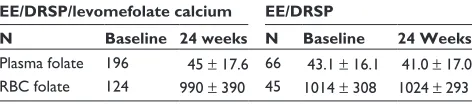
Table 2 Plasma folate and RBC folate baseline values vs value following 24-week therapy 20 mcg ee/DRSP vs levomefolate calcium142
EE/DRSP/levomefolate calcium EE/DRSP
N Baseline 24 weeks N Baseline 24 Weeks
Plasma folate 196 45 ± 17.6 66 43.1 ± 16.1 41.0 ± 17.0 RBC folate 124 990 ± 390 45 1014 ± 308 1024 ± 293 Abbreviations: DRSP, drospirenone; ee, ethinyl estradiol; RBC, red blood cell.
Dovepress Folates in oral contraceptives
also needed to prevent NTDs in the unintended pregnancies that occur while the woman is taking the pill and is certainly not prepared for pregnancy. Estimates are that each year, 1 million women who considered themselves to be “using the pill” become pregnant – these failures account for one out of every six pregnancies in the US.128
Very few of these pill failures occur to women who use the pill correctly and consistently. Clinical trials for virtually every pill demonstrate that the failure rate with correct and consistent pill use is less than 1%. However, when used in the real world where women experience barriers to access (most women are dispensed only one pack of pills at a time), have concerns about pill side effects or safety, and have strong ambivalence about pregnancy,129 consistent pill use
is relatively rare. In one study of over 1.7 million new start hormonal contraceptive users, less than one-third of women refilled their prescriptions “on time” (within 14 days of the next scheduled refill date) for 12 months.130 The rest either
completely discontinued or significantly interrupted their pill use. In two other studies, 30% of women completely dis-continued their pills by 3 months.131,132 International studies
show that of the women who stop their pills, only 35% switch to another modern, effective method within 3 months.133 In a
more recent US study in which women were provided effec-tive contraception for free, only 30% obtained a median of ten refills on time.134 These findings were substantiated by
a 12-month study in family planning clinics which reported that the 12-month continuation rate with pills was only 32.7 per 100 women years.135
Even when women do successfully gain possession of their pill packs, they often do not use them correctly and consistently. A retrospective study of 1311 women presenting for their first family planning visit in a public health depart-ment found that only 42% of users said they took their pills daily and only 20% claimed they took a pill within 2 hours of the same time each day.136 This bleak assessment has been
confirmed by a study in which computer chips embedded in the pill packs recorded the date and time pills were removed from the package. The researchers found that over 50% of established pill users missed three or more pills each cycle.137
Most women who discontinue pill use do not adopt another method immediately and if they do use a new method, it is generally one with a higher failure rate.138
In the face of these realities, it seems prudent to add a folate supplement to birth control pills. If a folate supplement were added to pills, it could raise plasma and red blood cell levels to reduce the risk of neural tube defects both during times of pill interruption and for several weeks after pill discontinuation. As a
safety net, it could provide needed elevated folate levels until such time as the woman adopts another effective contraceptive method or she initiates folic acid supplementation. This concept was unanimously endorsed at the FDA Health Drugs meeting in 2003.139 In a small study to test the FDA recommendation, it
was found that short-term use (8 weeks) of 500 mcg folic acid per day raised serum folate levels and that those levels did not return to baseline for 12 weeks following pill cessation.140
New products
There are two drospirenone-containing oral contraceptives that have added a very efficient form of folate in doses large enough to reliably raise plasma and red blood levels. The folate that was selected, metafolin, is levomefolate calcium, which is a synthetic calcium salt that is structurally identical to L-5-methyltetrahydrofolate (L-5-methyl-THF). As noted earlier, L-5-methyl-THF is the predominant folate transport seen in the blood with folic acid supplementation as well as with dietary folate. Tetrahydrofolate is directly absorbed from the intestine and requires no further processing before it can enter red blood cells. Genetic differences do not reduce the amount of folate that the body can utilize when tetrahydro-folate is used as the source of tetrahydro-folate.
The predictability of metafolin-enriched drospirenone-containing oral contraceptives was demonstrated in clinical trials used as the basis of FDA approval. In one trial, women who ate a folic acid-fortified diet (the US diet) were assigned to receive either the original 24/4 birth control pill with 20 mcg ethinyl estradiol and 3 mg drospirenone or the new for-mulation which added 0.45 1 mg metafolin to each pill.141,142
Folate levels were assessed in the plasma and red blood cells every 2 weeks for the 24-week study period (see Table 2). The women using the metafolin-supplemented oral contra-ceptives demonstrated significant increase (15.8 nmol/L) in serum folate levels. These levels were consistently maintained throughout the study period. In the conventional pill arm, plasma folate levels decreased by 2.2 nmol/L. Significant differences were also seen in the mean red blood cell folate levels. In the control group, red blood cell folate levels
Open Access Journal of Contraception downloaded from https://www.dovepress.com/ by 118.70.13.36 on 26-Aug-2020
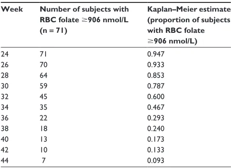
Table 3 Kaplan–Meier estimates and the proportion of subjects for the time to RBC folate falling below 906 nmol/L for the yasmin + metafolin treatment group in the folate elimination phase (week 26 to week 44)145
Week Number of subjects with RBC folate $906 nmol/L (n = 71)
Kaplan–Meier estimate (proportion of subjects with RBC folate $906 nmol/L)
24 71 0.947
26 70 0.933
28 64 0.853
30 59 0.787
32 45 0.600
34 35 0.467
36 22 0.293
38 18 0.240
40 13 0.173
42 10 0.133
44 7 0.093
Abbreviation: RBC, red blood cell.
Dovepress
Nelson
increased by 34.3 nmol/L, while the metafolin-supplemented pill users demonstrated a mean increase from baseline of 419.9 nmol/L (P , 0.001).141,143 While the P values in this
study were impressive, the FDA advises that they be should be used with caution because a substantial percentage of data was dropped from each arm due to blood sampling errors.
In the second trial, German women, who do not have folate food fortification, were randomized into two arms in a double-blind, double-dummy trial. Women in one arm were given the monophasic formulation with 30 mcg ethinyl estradiol and 3 mg drospirenone with 451 mcg levomefolate calcium added to each of the 21 active pills and to each of the seven placebo pills and a placebo pill as a second tablet to take daily. In the second arm, women were given the same 30 mcg ethinyl estradiol, 3 mg drospirenone pill without metafolin and a tablet with folic acid 0.4 mg to take each day. This was a 24-week trial of drug use followed by an open-label follow-up 20-week period in which women received only the unfortified pill. At each point in time, the metafolin-treated women had higher levels of folate in both serum and red blood cells than women who took the folic acid supplements. Folate levels returned to baseline at about 20 weeks in the plasma, but were still above baseline levels in the red blood cells at the end of the 20-week follow-up period. Table 3 demonstrates the percentage of subjects who remained at or above protective levels of folate in the red blood cells (906 nmol/L) by weeks following cessation of metafolin (trial weeks 24–44).139 Noninferiority was
estab-lished by these two trials and the persistence of the elevation in folate levels was quantified.
A recent report sought to estimate the impact that switching all oral contraceptive users in the US to metafolin-containing formulations might have on the incidence of anencephaly and spina bifida. The model predicted pregnancy outcomes (live births, stillbirth, and elective terminations) after 6 months of use of the fortified pills. The authors estimated accidental pregnancies among pill users would occur in about 5%–8% of users and they assumed that 23% of women on the pills would discontinue pill use. Both of these estimates are conservative compared to many of the studies cited above. Based on these assumptions and the patterns of folate measured in the clinical trials of these metafolin-containing pills, the authors concluded that the number of NTD cases would decline by 23.7% to 31.4%, depending on the baseline folate levels of women switching from traditional birth control pills.144
Conclusion
Neural tube defects affect 300,000 pregnancies worldwide each year. Estimates are that 50%–60% of these defects could be prevented by achieving and maintaining adequate folate level periconceptionally. Recent research suggests another benefit of maternal use of folic acid periconceptionally (from 4 weeks before to 8 weeks after conception) – a reduced rate of severe language delay in children at age 3 years.146 Food
fortification with folic acid has raised serum and red blood cell folate levels and reduced neural tube defects, but not as impressively as predicted. Targeted supplementation with dietary recommendations and vitamins has not reached the population at risk; in fact, supplementation seems to have declined in the US over the last decade. At the same time, risk factors for neural tube defects, especially those related to maternal conditions, such as obesity and diabetes, have increased. In a novel approach to reach at-risk women, first approved by an FDA committee in 2003, a more uniformly utilizable form of folic acid L-5-methyltetrahydrofolate (metafolin) has been added to two birth control pills con-taining drospirenone. Studies have shown that the amount of metafolin in the pills is sufficient to raise both serum and red blood cell folate significantly. Further studies have demonstrated that therapeutic levels (sufficient to prevent folate-dependent NTDs) persist in the red blood cells of half the users for about 10 weeks after pill cessation and that all women maintained folate levels above their baseline for 24 weeks.
This legacy effect provides protection during vulnerable times – when women temporarily interrupt pill use and when women discontinue pill use. Both of those events occur with
Open Access Journal of Contraception downloaded from https://www.dovepress.com/ by 118.70.13.36 on 26-Aug-2020
Dovepress Folates in oral contraceptives
surprising frequency, and help explain why 9% of pill users get pregnant in first year of pill use and why 1 million US “pill users” get pregnant each year. The addition of folate to pills offers the potential to reduce pregnancies affected by NTDs in these at-risk women.
Disclosure
The author serves on the advisory board for Bayer Health-care for both the LNG IUS and Bayer’s oral contraceptive products. Her clinic has received research grants from Bayer Healthcare and she has been active on Bayer’s Speaker’s Bureau.
References
1. Kondo A, Kamihira O, Ozawa H. Neural tube defects: prevalence, etiology and prevention. Int J Urol. 2009;16(1):49–57.
2. Padmanabhan R. Etiology, pathogenesis and prevention of neural tube defects. Congenit Anom (Kyoto). 2006;46(2):55–67.
3. Pitkin RM. Folate and neural tube defects. Am J Clin Nutr. 2007;85(1): 285S–288S.
4. Botto LD, Moore CA, Khoury MJ, Erickson JD. Neural-tube defects. N Engl J Med. 1999;341(20):1509–1519.
5. Blencowe H, Cousens S, Modell B, Lawn J. Folic acid to reduce neonatal mortality from neural tube disorders. Int J Epidemiol. 2010; 39 Suppl 1:i110–i121.
6. Finan A, Clarke TA, Matthews TG, et al. Strategies for reduction of neonatal mortality. Ir J Med Sci. 1999;168(4):265–267.
7. Au KS, Ashley-Koch A, Northrup H. Epidemiologic and genetic aspects of spina bifida and other neural tube defects. Dev Disabil Res Rev. 2010;16(1):6–15.
8. Van Allen MI, Boyle E, Thiessen P, et al. The impact of pre-natal diagnosis on neural tube defect (NTD) pregnancy versus birth incidence in British Columbia. J Appl Genet. 2006;47(2): 151–158.
9. Norem CT, Schoen EJ, Walton DL, et al. Routine ultrasonography compared with maternal serum alpha-fetoprotein for neural tube defect screening. Obstet Gynecol. 2005;106(4):747–752.
10. Centers for Disease Control and Prevention (CDC). Recommendations for use of folic acid to reduce number of spina bifida cases and other neural tube defects. JAMA. 1993;269(10):1233, 1236–8. Available at: http://www.cdc.gov/mmwr/preview/mmwrhtml/00019479.htm. Accessed on October 11, 2011.
11. Johnston RB Jr. Will increasing folic acid in fortified grain products further reduce neural tube defects without causing harm? consideration of the evidence. Pediatr Res. 2008;63(1):2–8.
12. Centers for Disease Control and Prevention (CDC). Spina bifida and anencephaly before and after folic acid mandate – United States, 1995–1996 and 1999–2000. MMWR Morb Mortal Wkly Rep. 2004;53(17):362–365.
13. Lapides J, Diokno AC, Silber SJ, Lowe BS. Clean, intermittent self-catheterization in the treatment of urinary tract disease. J Urol. 1972;107(3):458–461.
14. van der Linden IJ, Afman LA, Heil SG, Blom HJ. Genetic variation in genes of folate metabolism and neural-tube defect risk. Proc Nutr Soc. 2006;65(2):204–215.
15. Buccimazza SS, Molteno CD, Dunne TT, Viljoen DL. Prevalence of neural tube defects in Cape Town, South Africa. Teratology. 1994;50(3):194–199.
16. Chatkupt S, Skurnick JH, Jaggi M, Mitruka K, Koenigsberger MR, Johnson WG. Study of genetics, Epidemiology, and vitamin usage in familial spina bifida in the United States in the 1990s. Neurology. 1994;44(1):65–70.
17. Milunsky A, Alpert E, Neff RK, Frigoletto FD Jr. Prenatal diagnosis of neural tube defects. IV. Maternal serum alpha-fetoprotein screening. Obstet Gynecol. 1980;55(1):60–66.
18. Hall J, Solehdin F. Folic acid for the prevention of congenital anomalies. Eur J Pediatr. 1998;157(6):445–450.
19. Becerra JE, Khoury MJ, Cordero JF, Erickson JD. Diabetes mellitus during pregnancy and the risks for specific birth defects: a population-based case-control study. Pediatrics. 1990;85(1):1–9.
20. Lammer EJ, Sever LE, Oakley GP Jr. Teratogen update: valproic acid. Teratology. 1987;35(3):465–473.
21. Edwards MJ, Shiota K, Smith MS, Walsh DA. Hyperthermia and birth defects. Reprod Toxicol. 1995;9(5):411–425.
22. Graham JM Jr, Edwards MJ, Edwards MJ. Teratogen update: gestational effects of maternal hyperthermia due to febrile illnesses and resultant patterns of defects in humans. Teratology. 1998;58(5):209–221. 23. Shaw GM, Velie EM, Schaffer D. Risk of neural tube defect-affected
pregnancies among obese women. JAMA. 1996;275(14):1093–1096. 24. Watkins ML, Scanlon KS, Mulinare J, Khoury MJ. Is maternal
obesity a risk factor for anencephaly and spina bifida? Epidemiology. 1996;7(5):507–512.
25. Werler MM, Louik C, Shapiro S, Mitchell AA. Prepregnant weight in relation to risk of neural tube defects. JAMA. 1996;275(14): 1089–1092.
26. Brender JD, Felkner M, Suarez L, Canfield MA, Henry JP. Maternal pesticide exposure and neural tube defects in Mexican Americans. Ann Epidemiol. 2010;20(1):16–22.
27. Blatter BM, Roeleveld N, Zielhuis GA, Mullaart RA, Gabreëls FJ. Spina bifida and parental occupation. Epidemiology. 1996;7(2): 188–193.
28. Shaw GM, Nelson V, Olshan AF. Paternal occupational group and risk of offspring with neural tube defects. Paediatr Perinat Epidemiol. 2002;16(4):328–333.
29. Vrijheid M, Dolk H, Stone D, Abramsky L, Alberman E, Scott JE. Socioeconomic inequalities in risk of congenital anomaly. Arch Dis Child. 2000;82(5):349–352.
30. Wasserman CR, Shaw GM, Selvin S, Gould JB, Syme SL. Socioeconomic status, neighborhood social conditions, and neural tube defects. Am J Public Health. 1998;88(11):1674–1680.
31. Prasad PD, Ramamoorthy S, Moe AJ, Smith CH, Leibach FH, Ganapathy V. Selective expression of the high-affinity isoform of the folate receptor (FR-alpha) in the human placental syncytiotrophoblast and choriocarcinoma cells. Biochim Biophys Acta. 1994;1223(1): 71–75.
32. De Marco P, Moroni A, Merello E, et al. Folate pathway gene alterations in patients with neural tube defects. Am J Med Genet. 2000;95(3):216–223.
33. Boyles AL, Hammock P, Speer MC. Candidate gene analysis in human neural tube defects. Am J Med Genet C Semin Med Genet. 2005;135C(1):9–23.
34. Dalal A, Pradhan M, Tiwari D, et al. MTHFR 677C – .T and 1298A – .C polymorphisms: evaluation of maternal genotypic risk and association with level of neural tube defect. Gynecol Obstet Invest. 2007;63(3):146–150.
35. Muñoz JB, Lacasaña M, Cavazos RG, Borja-Aburto VH, Galavíz-Hernández C, Garduño CA. Methylenetetrahydrofolate reductase gene polymorphisms and the risk of anencephaly in Mexico. Mol Hum Reprod. 2007;13(6):419–424.
36. Botto LD, Yang Q. 5,10-Methylenetetrahydrofolate reductase gene variants and congenital anomalies: a HuGE review. Am J Epidemiol. 2000;151(9):862–877.
37. Harmon DL, Woodside JV, Yarnell JW, et al. The common ‘ thermolabile’ variant of methylene tetrahydrofolate reductase is a major determinant of mild hyperhomocysteinaemia. QJM. 1996;89(8): 571–577.
38. Kirke PN, Mills JL, Molloy AM, et al. Impact of the MTHFR C677T polymorphism on risk of neural tube defects: case-control study. BMJ. 2004;328(7455):1535–1536.
Open Access Journal of Contraception downloaded from https://www.dovepress.com/ by 118.70.13.36 on 26-Aug-2020
Dovepress
Nelson
39. Miyaki K, Murata M, Kikuchi H, et al. Assessment of tailor-made pre-vention of atherosclerosis with folic acid supplementation: randomized, double-blind, placebo-controlled trials in each MTHFR C677T geno-type. J Hum Genet. 2005;50(5):241–248.
40. Rasmussen SA, Erickson JD, Reef SE, Ross DS. Teratology: from sci-ence to birth defects prevention. Birth Defects Res A Clin Mol Teratol. 2009;85(1):82–92.
41. De-Regil LM, Fernández-Gaxiola AC, Dowswell T, Peña-Rosas JP. Effects and safety of periconceptional folate supplementation for preventing birth defects. Cochrane Database Syst Rev. 2010;10: CD007950.
42. Oakley GP Jr. The scientific basis for eliminating folic acid-preventable spina bifida: a modern miracle from epidemiology. Ann Epidemiol. 2009;19(4):226–230.
43. MRC Vitamin Study Research Group. Prevention of neural tube defects: results of the Medical Research Council Vitamin Study. Lancet. 1991;338(8760):131–137.
44. Smithells RW, Nevin NC, Seller MJ, et al. Further experience of vitamin supplementation for prevention of neural tube defect recurrences. Lancet. 1983;1(8332):1027–1031.
45. Czeizel AE, Dudás I. Prevention of the first occurrence of neural-tube defects by periconceptional vitamin supplementation. N Engl J Med. 1992;327(26):1832–1835.
46. Hibbard BM. The role of folic acid in pregnancy; with particular reference to anaemia, abruption and abortion. J Obstet Gynaecol Br Commonw. 1964;71:529–542.
47. Selhub J, Rosenberg IH. Public health significance of supplementa-tion or fortificasupplementa-tion of grain products with folic acid. Food Nutr Bull. 2008;29(2 Suppl):S173–S176.
48. Hibbard BM, Hibbard ED, Jeffcoate TN. Folic acid and reproduction. Acta Obstet Gynecol Scand. 1965;44(3):375–400.
49. Grosse SD, Collins JS. Folic acid supplementation and neural tube defect recurrence prevention. Birth Defects Res A Clin Mol Teratol. 2007;79(11):737–742.
50. Kirke PN, Daly LE, Elwood JH. A randomised trial of low dose folic acid to prevent neural tube defects. The Irish Vitamin Study Group. Arch Dis Child. 1992;67(12):1442–1446.
51. Mulinare J, Cordero JF, Erickson JD, Berry RJ. Periconceptional use of multivitamins and the occurrence of neural tube defects. JAMA. 1988;260(21):3141–3145.
52. Bower C, Stanley FJ. Dietary folate as a risk factor for neural-tube defects: evidence from a case-control study in Western Australia. Med J Aust. 1989;150(11):613–619.
53. Mills JL, Rhoads GG, Simpson JL, et al. The absence of a relation between the periconceptional use of vitamins and neural-tube defects. National Institute of Child Health and Human Development Neural Tube Defects Study Group. N Engl J Med. 1989;321(7): 430–435.
54. Werler MM, Shapiro S, Mitchell AA. Periconceptional folic acid exposure and risk of occurrent neural tube defects. JAMA. 1993;269(10):1257–1261.
55. Shaw GM, Schaffer D, Velie EM, Morland K, Harris JA. Periconceptional vitamin use, dietary folate, and the occurrence of neural tube defects. Epidemiology. 1995;6(3):219–226.
56. Berry RJ, Li Z, Erickson JD, et al. Prevention of neural-tube defects with folic acid in China. China US Collaborative Project for Neural Tube Defect Prevention. N Engl J Med. 1999;341(20):1485–1490. 57. Milunsky A, Jick H, Jick SS, et al. Multivitamin/folic acid
supplementa-tion in early pregnancy reduces the prevalence of neural tube defects. JAMA. 1989;262(20):2847–2852.
58. Heseker HB, Mason JB, Selhub J, Rosenberg IH, Jacques PF. Not all cases of neural-tube defect can be prevented by increasing the intake of folic acid. Br J Nutr. 2009;102(2):173–180.
59. Martínez-Frías ML. The biochemical structure and function of meth-ylenetetrahydrofolate reductase provide the rationale to interpret the epidemiological results on the risk for infants with Down syndrome. Am J Med Genet A. 2008;146A(11):1477–1482.
60. Carlson S, Aupperle P. Nutrient requirements and fetal development: recommendations for best outcomes. J Fam Pract. 2007;56(11 Suppl Womens):S1–S6; quiz S7–S8.
61. Institute of Medicine. Dietary reference intakes for thiamin, riboflavin, niacin, vitamin B6, folate, pantothenic acid, biotin and choline. Washington, DC: National Academy Press, 1998.
62. Simpson JL, Bailey LB, Pietrzik K, Shane B, Holzgreve W. Micronutrients and women of reproductive potential: required dietary intake and consequences of dietary deficiency or excess. Part I – Folate, Vitamin B12, Vitamin B6. J Matern Fetal Neonatal Med. 2010;23(12): 1323–1343.
63. Tam C, McKenna K, Goh YI, et al. Periconceptional folic acid supple-mentation: a new indication for therapeutic drug monitoring. Ther Drug Monit. 2009;31(3):319–326.
64. Butriss J. Strategies designed to increase awareness about folates and health and to increase folate intake: a review. Trends Food Sci Technol. 2005;16(6–7):246–252.
65. Centers for Disease Control. Recommendations for the use of folic acid to reduce the number of cases of spina bifida and other neural tube defects. MMWR Recomm Rep. 1992;41(RR-14):1–7.
66. US Preventive Services Task Force. Folic acid for the prevention of neural tube defects: US Preventive Services Task Force recommenda-tion statement. Ann Intern Med. 2009;150(9):626–631.
67. Botto LD, Lisi A, Bower C, et al. Trends of selected malforma-tions in relation to folic acid recommendamalforma-tions and fortification: an international assessment. Birth Defects Res A Clin Mol Teratol. 2006;76(10):693–705.
68. Olney RS, Mulinare J. Trends in neural tube defect prevalence, folic acid fortification, and vitamin supplement use. Semin Perinatol. 2002;26(4):277–285.
69. Centers for Disease Control and Prevention (CDC). Knowledge and use of folic acid by women of childbearing age – United States, 1995 and 1998. MMWR Morb Mortal Wkly Rep. 1999;48(16): 325–327.
70. Chivu CM, Tulchinsky TH, Soares-Weiser K, Braunstein R, Brezis M. A systematic review of interventions to increase awareness, knowledge, and folic acid consumption before and during pregnancy. Am J Health Promot. 2008;22(4):237–245.
71. March of Dimes. Improving Preconception Health: Women’s Knowledge and Use of Folic Acid, March of Dimes Foundation, White Plans, NY; 2007.
72. McDonnell R, Johnson Z, Doyle A, Sayers G. Determinants of folic acid knowledge and use among antenatal women. J Public Health Med. 1999;(2):145–149.
73. de la Vega A, et al. A nationwide program for the use of preconceptional folic acid to prevent the development of open neural tube defects. Who is really using folic acid? P R Health Sci J. 2002;21(1):7–9.
74. Morin VI, Mondor M, Wilson RD. Knowledge on periconceptional use of folic acid in women of British Columbia. Fetal Diagn Ther. 2001;16(2):111–115.
75. Auriel E, et al. Attitudes, and practice among women and doctors concerning the use of folic acid. ISRN Obstet Gynecol. 2011;2011: 946041.
76. European Surveillance of Congenital Anomalies. Special Report: Prevention of Neural Tube Defects by Periconceptional Folic Acid Supplementation in Europe. Updated December 2009, EUROCAT Central Registry, Newtownabbey, Northern Ireland, UK.
77. Botto LD, Lisi A, Robert-Gnansia E, et al. International retrospective cohort study of neural tube defects in relation to folic acid recommen-dations: are the recommendations working? BMJ. 2005;330(7491): 571.
78. Busby A, Abramsky L, Dolk H, et al. Preventing neural tube defects in Europe: a missed opportunity. Reprod Toxicol. 2005;20(3): 393–402. 79. Poretti A, Anheier T, Zimmermann R, Boltshauser E; Swiss Pediatric
Surveillance Unit (SPSU). Neural tube defects in Switzerland from 2001 to 2007: are periconceptual folic acid recommendations being followed? Swiss Med Wkly. 2008;138(41–42):608–613.
Open Access Journal of Contraception downloaded from https://www.dovepress.com/ by 118.70.13.36 on 26-Aug-2020