Journal of Inflammation Research
Dove
press
R e v i e w open access to scientific and medical research
Open Access Full Text Article
Journal of Inflammation Research 2016:9 13–20
submit your manuscript | www.dovepress.com 13
Familial Mediterranean fever: current perspectives
Hafize Emine Sönmez* Ezgi Deniz Batu* Seza Özen
Department of Pediatrics, Division of Rheumatology, Hacettepe University Faculty of Medicine, Ankara, Turkey
*These authors contributed equally to this work
Correspondence: Seza Özen Department of Pediatrics, Division of Rheumatology, Hacettepe University Faculty of Medicine, Sıhhiye, Ankara 06100, Turkey
Tel +90 312 305 1863
Fax +90 312 311 2398
Email sezaozen@hacettepe.edu.tr
Abstract: Familial Mediterranean fever (FMF) is the most frequent monogenic autoinflammatory
disease, and it is characterized by recurrent attacks of fever and polyserositis. The disease is associated with mutations in the MEFV gene encoding pyrin, which causes exaggerated inflammatory response through uncontrolled production of interleukin 1. The major long-term complication of FMF is amyloidosis. Colchicine remains the principle therapy, and the aim of treatment is to prevent acute attacks and the consequences of chronic inflammation. With the evolution in the concepts about the etiopathogenesis and genetics of the disease, we have understood that FMF is more complicated than an ordinary autosomal recessive monogenic disorder. Recently, recommendation sets have been generated for interpretation of genetic test-ing and genetic diagnosis of FMF. Here, we have reviewed the current perspectives in FMF in light of recent recommendations.
Keywords: familial Meditarranean fever, recommendation, child
Introduction
Familial Mediterranean fever (FMF) is the most common autoinflammatory disease (AID) mainly affecting the populations of eastern Mediterranean descent.1,2 It is
charac-terized by recurrent, self-limited flares of fever associated with polyserositis.3 The gene
mutated in patients with FMF is the MEFV gene, which encodes pyrin, which forms an element of the NLRP3 inflammasome complex.2 Mutations in the MEFV gene are
associated with increased interleukin-1β (IL-1β), which causes excess inflammation.4
The majority of FMF patients demonstrate a Mendelian autosomal recessive pattern of inheritance.5–7 However, since the MEFV gene was found to underlie FMF in 1997,5,6
the concepts about the etiopathogenesis and genetics of the disease have evolved. Progress in molecular biology suggested that this AID was more complicated than we had anticipated.
Colchicine still constitutes the mainstay of FMF treatment,8 and the aim of the
treatment should be preventing acute attacks and amyloidosis, decreasing the chronic inflammation, and providing an acceptable quality of life. Recent insights into the pathogenesis of FMF have made anti-IL 1 treatments important in colchicine-resistant or -intolerant patients. The most severe complication of FMF is secondary amyloidosis;9
however, it is less common in the colchicine and anti-IL 1 era.
In 2012, Shinar et al10 proposed recommendations for interpretation of genetic
test-ing in AIDs. Most recently, the SHARE (a pediatric initiative to develop better care and management for rheumatology patients; Single Hub and Access point for pediatric
Journal of Inflammation Research downloaded from https://www.dovepress.com/ by 118.70.13.36 on 24-Aug-2020
For personal use only.
Number of times this article has been viewed
This article was published in the following Dove Press journal:
Journal of Inflammation Research
Dovepress
Sönmez et al
Rheumatology in Europe) initiative has developed evidence-based recommendations for genetic diagnosis of FMF.11
In this paper, we have reviewed the current perspectives in FMF in the light of recent recommendations.
Etiopathogenesis
The MEFV gene encodes the protein pyrin, which is a part of the inflammasome complex, NLRP3, an intracellular organelle required for the production of IL-1β.12 Thus, FMF
may be classified as an inflammasomeopathy.2 Pyrin has been
suggested to interact with the inflammasome adaptor protein ASC, and this results in increased caspase-1 activation and IL-1β processing.13
In 2007, Yu et al14 have shown that activated pyrin
inter-acts with ASC and PSTPIP1 to form a trimolecular complex that directly activates caspase-1 and then leads to the secre-tion of active IL-1β. Booty et al15 demonstrated a significant
increase in pyrin expression in FMF patients in comparison with healthy controls. Supporting the hypothesis of a gain-of-function model, an elegant study by Chae et al16 revealed
that FMF-associated B30.2 mutations knock-in mice, but not pyrin deficient ones, showed severe spontaneous inflamma-tory phenotype.
On the other hand, Papin et al17 showed that pyrin
knock-down resulted in increased caspase-1 activation and IL-1β
secretion. In another study by Hesker et al,18 enhanced IL-1β
release by macrophages was demonstrated in response to a spectrum of inflammatory stimuli in a mouse line lacking the MEFV gene. The findings of these two studies suggest a loss-of-function model.
However, it is still controversial whether MEFV muta-tions cause loss of function or gain of function.
A recent study by Xu et al19 has elegantly shown that
pyrin is a specific immune sensor for bacterial modifications of Rho GTPases, and responds to Clostridium difficile, which is a frequent cause of nosocomial diarrhea. Pyrin does not directly recognize the microbial products but detects patho-gen virulence activity. This recent finding has shed some light on FMF pathogenesis.
Diagnosis
Since FMF usually requires lifelong treatment, it is crucial to establish a timely, correct diagnosis. The diagnosis of FMF relies mainly on clinical findings, and molecular analysis of the MEFV gene provides genetic confirmation.20
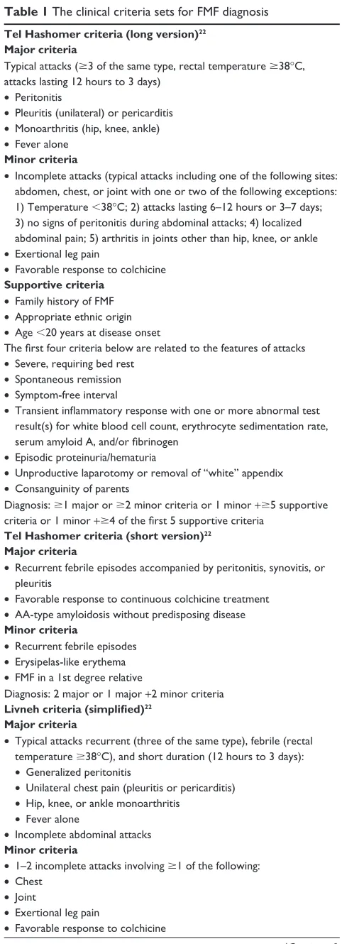
There are different sets of classification and diagnostic criteria for FMF. The first set of criteria was created for adults by the experts in Tel Hashomer Hospital (Table 1).21
Livneh et al22 validated the new criteria in 1997, excluding
some manifestations of the Tel Hashomer criteria such as amyloidosis (Table 1). However, there were certain dif-ferences between adult and pediatric FMF cases (such as shorter attacks in children, lack of unilateral characteristic of chest pain in some pediatric cases, more febrile attacks or even fever-only attacks in some children, and inability of some pediatric patients to express the severity and exact location of the pain), and some of the Tel Hashomer criteria were of less relevance to pediatric FMF patients. Thus, in 2009, our group attempted to define criteria for children as well (Table 1).23 Among Turkish children, the criteria
(two out of five criteria for diagnosis) reached a sensitiv-ity and specificsensitiv-ity of 88.8% and 92.2%, respectively.23 In
French children, the presence of three instead of two criteria yielded a better specificity of 95%.24 Validation of these
criteria in a larger and genetically more heterogeneous group is crucial.
It is important to keep in mind that FMF diagnosis is a clinical one, and if the phenotype is consistent with FMF, the physician should not exclude the diagnosis when there is no genetic confirmation.
Genetics
Variants to be checked in the
MEFV
gene
When the genetic association of FMF with the MEFV gene was first described in 1997,5,6 only a few mutations had been
reported. Since the number of variants recognized associ-ated with FMF has increased, concerns emerged about the adequacy of checking for four to six mutations only. Booty et al15 sequenced the MEFV gene in heterozygote FMF
patients and demonstrated that additional sequencing of the whole gene did not yield a second mutation in any of the screened patients. Currently, all reported MEFV variants and the associated phenotypes are recorded in the INFEVERS database (http://fmf.igh.cnrs.fr/ISSAID/infevers/), and there are approximately 300 known sequence variants of MEFV.11
The most common disease-associated pathogenic variants are M694V, V726A, M680I, and M694I, whereas E148Q is the most frequent variant among carriers.25,26 An agreed set
of best practice guidelines was proposed for genetic diag-nostic testing of hereditary recurrent fevers including FMF in 2012.10 The consensus was to test for a total of 14
vari-ants (the first nine are defined as clearly pathogenic, while the remaining five variants have unknown significance): M694V, M694I, M680I, V726A, R761H, A744S, E167D, T267I, I692del, K695R, E148Q, P369S, F479L, and I591T. We now suggest testing for these variants only.
Journal of Inflammation Research downloaded from https://www.dovepress.com/ by 118.70.13.36 on 24-Aug-2020
Dovepress Familial Mediterranean fever: current perspectives
Heterozygotes
FMF is an autosomal recessive disease. Thus, one would expect a heterozygote individual to be a carrier and lack the FMF phenotype. However, as FMF patients were genotyped, it has become evident that the mutation in the second allele could not be demonstrated in about one-quarter of patients with clinically confirmed FMF.10 As we previously mentioned,
resequencing of the entire MEFV genomic region did not provide additional benefit.15
In 2009, Marek-Yagel et al27 studied the clinical and
genetic characteristics of heterozygous FMF patients and performed haplotype studies. They concluded that although the heterozygotes tend to have a relatively mild disease, the disease could not be distinguished from that of homozygous patients and that FMF could be viewed as a dominant condi-tion with low penetrance in some cases.27
In addition, convincing data indicate that heterozygous individuals have an increased propensity to display signs of inflammation. In the eastern Mediterranean population, the carrier rate is higher among the patients with rheumatic diseases than in the general population.28,29 Kalyoncu et al30
revealed that rheumatoid arthritis, acute rheumatic fever, arthralgia, and febrile episodes of $4 times/year were signifi-cantly more common in asymptomatic heterozygous parents of children with FMF than healthy controls. Along the same lines, Lachmann et al31 showed greater basal and peak acute-phase
reactant concentrations in MEFV heterozygotes in comparison with wild-type controls which suggest a proinflammatory phenotype in carriers.
One explanation for heterozygote individuals displaying FMF phenotype may be the effect of other modifier genes associated with inflammation.15 One example is the serum
amyloid A (SAA) gene, encoding an acute-phase protein, SAA, which activates NLRP3 inflammasome causing increased secretion of active IL-1β.32
Table 1 The clinical criteria sets for FMF diagnosis
Tel Hashomer criteria (long version)22
Major criteria
Typical attacks ($3 of the same type, rectal temperature $38°C,
attacks lasting 12 hours to 3 days) • Peritonitis
• Pleuritis (unilateral) or pericarditis
• Monoarthritis (hip, knee, ankle)
• Fever alone
Minor criteria
• Incomplete attacks (typical attacks including one of the following sites:
abdomen, chest, or joint with one or two of the following exceptions:
1) Temperature 38°C; 2) attacks lasting 6–12 hours or 3–7 days;
3) no signs of peritonitis during abdominal attacks; 4) localized abdominal pain; 5) arthritis in joints other than hip, knee, or ankle
• exertional leg pain
• Favorable response to colchicine
Supportive criteria
• Family history of FMF
• Appropriate ethnic origin
• Age 20 years at disease onset
The first four criteria below are related to the features of attacks
• Severe, requiring bed rest
• Spontaneous remission
• Symptom-free interval
• Transient inflammatory response with one or more abnormal test
result(s) for white blood cell count, erythrocyte sedimentation rate, serum amyloid A, and/or fibrinogen
• Episodic proteinuria/hematuria
• Unproductive laparotomy or removal of “white” appendix
• Consanguinity of parents
Diagnosis: $1 major or $2 minor criteria or 1 minor +$5 supportive
criteria or 1 minor +$4 of the first 5 supportive criteria
Tel Hashomer criteria (short version)22
Major criteria
• Recurrent febrile episodes accompanied by peritonitis, synovitis, or
pleuritis
• Favorable response to continuous colchicine treatment
• AA-type amyloidosis without predisposing disease
Minor criteria
• Recurrent febrile episodes
• Erysipelas-like erythema
• FMF in a 1st degree relative
Diagnosis: 2 major or 1 major +2 minor criteria
Livneh criteria (simplified)22
Major criteria
• Typical attacks recurrent (three of the same type), febrile (rectal
temperature $38°C), and short duration (12 hours to 3 days):
• Generalized peritonitis
• Unilateral chest pain (pleuritis or pericarditis)
• Hip, knee, or ankle monoarthritis
• Fever alone
• Incomplete abdominal attacks
Minor criteria
• 1–2 incomplete attacks involving $1 of the following:
• Chest
• Joint
• exertional leg pain
• Favorable response to colchicine
(Continued)
Table 1 (Continued)
Diagnosis: 1 major or 2 minor criteria
Turkish FMF pediatric criteria23
• Fever (axillary, 38°C, $3 attacks of 6–72-hour duration)
• Abdominal pain ($3 attacks of 6–72-hour duration)
• Chest pain ($3 attacks of 6–72-hour duration)
• Arthritis (oligoarthritis, $3 attacks of 6–72-hour duration)
• Family history of FMF
Diagnosis: 2 out of 5 criteria
Notes: Adapted from: Livneh A, Langevitz P, Zemer D, et al. Criteria for the diagnosis of familial Mediterranean fever. Arthritis Rheum. 1997;40:1879–1885.22
Copyright © 1997 American College of Rheumatology Yalcinkaya F, Ozen S, Ozcakar ZB, et al. A new set of criteria for the diagnosis of familial Mediterranean fever in childhood. Rheumatology (Oxford). 2009;48:395–398.23
Abbreviation: FMF, familial Mediterranean fever.
Journal of Inflammation Research downloaded from https://www.dovepress.com/ by 118.70.13.36 on 24-Aug-2020
Dovepress
Sönmez et al
We also know that epigenetics and environment may affect the disease phenotype, and this will now be discussed later in the paper.
Genotype–phenotype correlation
Since the first description of the MEFV mutations, most experts agree that M694V was related with a severe disease phenotype.5,6,33 Ozturk et al34 demonstrated that both patients
homozygous and compound heterozygous for M694V were at increased risk for severe disease in comparison with patients with one mutant allele and patients not displaying M694V. Giaglis et al35 and Mattit et al36 confirmed these
findings in two separate studies. Giaglis et al35 also showed
that a more severe phenotype was evident in patients homozygous for M694V as well as mutations at position 680–694 on exon 10. In evidence-based recommendations, the experts concluded that FMF patients homozygous or compound heterozygous for M694V or with mutations at position 680–694 on exon 10 must be considered at risk of having a more severe and early-onset disease.11 In addition,
the individuals homozygous for M694V without disease manifestations should be evaluated and followed closely in order to decide about therapy.11
Besides issues about mutations at position 680–694 on exon 10, in a recent study, Federici et al37 demonstrated that
the frequency of FMF-like symptoms increased from patients carrying a single low penetrance mutation toward patients with two high penetrance mutations, suggesting a “dose effect” associated with the mutations.
The pathogenic role of E148Q on exon 2 (one of the most common alterations in the MEFV gene) still remains
controversial.11 It might be a polymorphism since it is
pres-ent in 1% of the healthy population. It has been reported to be present as high as 21% of healthy in some eastern Asian populations.38 A consensus conference led by Shinar
et al10 has defined E148Q as a variant of unknown significance.
According to the recent recommendations by SHARE, as the only MEFV variant, E148Q does not support the diagnosis of FMF.11 However, there are rare reports of patients with
homozy-gous E148Q variation having FMF phenotype.26 Furthermore,
patients carrying E148Q with a clearly pathogenic mutation on the other allele often display disease phenotype.2
The recent evidence-based recommendations for genetic diagnosis of FMF are as follows:11
• FMF is a clinical diagnosis, which can be supported but not excluded by genetic testing.
• Consider patients homozygous for M694V at risk of developing, with very high probability, a severe phenotype.
• FMF patients carrying two of the common mutated alleles (homozygotes or compound heterozygotes), especially for M694V mutation or mutations at position 680–694 on exon 10, must be considered at risk of having a more severe disease.
• The E148Q variant is common, of unknown pathogenic significance, and, as the only MEFV variant, does not support the diagnosis of FMF.
• Patients homozygous for M694V mutation are at risk of early-onset disease.
• Individuals homozygous for M694V who are not report-ing symptoms should be evaluated and followed closely to consider therapy.
Age:
Name: Month: Year:
Symptoms associated with autoinflammatory syndrome today
Day Overall
symptoms
Abdominal pain
Nausea/ vomiting
Head aches
Chest pain
Painful nodes
Arthralgia or myalgia
Swelling of the joints
Eye
manifestations Skin rash
Pain relief drug taken
0 or 1
1
2
3
…
31
Fever ≥38°C (100.4°F)
Diarrhea
0 or 1 0 or 1 0 or 1
0 or 1 0 or 1 0 or
1 0 or 1 0 or 1
0 or 1 0 or 1
0 or 1 Score
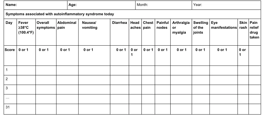
Figure 1 AID activity index diary.
Notes: Each line refers to a day in a month. Reproduced from Piram M, Kone-Paut I, Lachmann HJ, et al. Validation of the auto-inflammatory diseases activity index (AIDAI) for hereditary recurrent fever syndromes. Ann Rheum Dis. 2014;73:2168–2173.62
Abbreviation: AID, autoinflammatory disease.
Journal of Inflammation Research downloaded from https://www.dovepress.com/ by 118.70.13.36 on 24-Aug-2020
Dovepress Familial Mediterranean fever: current perspectives
• For individuals with two pathogenic mutations for FMF who do not report symptoms, if there are risk factors for AA amyloidosis (such as the country, family history, and persistently elevated inflammatory markers, particularly serum amyloid A protein), close follow-up should be started and treatment considered.
• Consultation with an AID specialist may be helpful to aid in the indication and interpretation of the genetic testing and diagnosis.
Epigenetics and environment
FMF patients with similar genotype may express different disease phenotypes. This difference may be due to other modifier genes, epigenetics, or the effect of the environment. The first clue suggesting the effect of environment on FMF was the observation of lack of secondary amyloidosis among Armenian FMF patients in the USA.39 We also know that the
eastern Mediterranean patients had a less severe disease if they migrated to Europe.9 Touitou et al40 demonstrated that
country of recruitment, rather than MEFV genotype, is the likely key risk factor for development of amyloidosis. We had previously shown that Turkish children with FMF living in Germany expressed a less severe disease phenotype in com-parison with the ones living in Turkey.41 These findings
empha-size the effect of environment on FMF disease severity. The epigenetic mechanisms such as histone modification, methylation, and microRNAs may play role in the pathogen-esis of FMF. Kirectepe et al42 demonstrated a slightly higher
methylation level of exon 2 of MEFV in FMF patients when compared to healthy controls.
MicroRNAs (miRNAs) are small, noncoding RNAs that regulate gene expression at a posttranscriptional level by degrading mRNA molecules or blocking their translation.43
Their circulating levels have already been described in inflammatory disorders such as rheumatoid arthritis, sys-temic lupus erythematosus, and tumor necrosis factor (TNF) receptor–associated periodic syndrome.44–47 Circulating
miR-NAs in FMF have not been investigated, but this might have a role in the disease pathogenesis of FMF as well.
Microorganisms may affect FMF since pyrin is a compo-nent of NLRP3, which is a pathogen recognition receptor.9
Pyrin has also been shown to detect virulent pathogenic activity.19 The cross-talk between the innate immune system
and commensal gut bacteria (microbiota) may affect (or may be affected by) the inflammatory status of the patient, as well. In a study by Khachatryan et al,48 they showed that the
composition and divergence of microbiota were different during attack and attack-free periods as well as between FMF patients and healthy controls.
Further studies on the effect of epigenetics, microbiota, and environment on FMF may enable us to answer more unsolved questions about the pathogenesis.
Treatment
Colchicine is still the main form of treatment for patients with FMF. Since 1972, colchicine has been used for treatment in FMF patients.8 It decreases attack frequency and increases
the quality of life.49 Furthermore, Livneh et al50 have shown
that colchicine can effectively prevent the complication of secondary amyloidosis in FMF patients.
In pediatric FMF patients, colchicine was shown to be a well-tolerated drug, even when given in infancy.51 The
com-monest side effects of colchicine are gastrointestinal, with vomiting, diarrhea, and transient elevation of transaminases. These mild side effects can occur in 5%–10% of patients even at recommended doses. It has a narrow therapeutic index; colchicine doses of 0.5–0.8 mg/kg are highly toxic, and doses of more than 0.8 mg/kg are typically lethal. In fact, the US Food and Drug Administration withdrew approval for intravenous colchicine to reduce the risk of irreversible bolus overdose.52,53
Colchicine is undoubtedly the main treatment for FMF, but approximately one-third of the patients treated with colchicine have a partial remission, and approximately 5% are non responders; another 2%–5% do not tolerate the drug mainly due to gastrointestinal symptoms.54 Lidar et al55 have
suggested that colchicine efficacy differs between patient groups depending on the MEFV genotype. M694V homozy-gotes showed a more severe disease and were treated with higher doses of colchicine compared to patients with the V726A genotype.
In recent years, biologic agents have been used in treat-ment of FMF patients unresponsive to colchicine therapy.56
Pyrin, the mutated protein in FMF, plays an important role in the regulation of interleukin-1 (IL-1) activation; anti-IL-1 treatment has proven beneficial in suppressing inflammation in colchicine-resistant FMF patients. Anakinra, a recom-binant, human IL-1 receptor antagonist that competitively inhibits binding of IL-1α and IL-1β to the IL-1 receptor; canakinumab, a human IgG1 monoclonal antibody directed against IL-1β; and rilonacept a fully human dimeric fusion protein that binds the extracellular domains of IL-1α and IL-1β were used in FMF patients.57 There are small series
or trials about anti-IL-1 treatment in colchicine-resistant FMF patients.56,58 The only randomized controlled study was
with rilonacept, in 14 colchicine-resistant FMF patients.59
Recently, a 6-month Phase II, open-label, single-arm pilot study showed the efficacy of canakinumab in pediatric
Journal of Inflammation Research downloaded from https://www.dovepress.com/ by 118.70.13.36 on 24-Aug-2020
Dovepress
Sönmez et al
patients with colchicine-resistant FMF.60 The major limitation
of this study was the small sample size. A larger controlled study is needed to explain the benefit, optimal dose, and side effect of canakinumab better in FMF patients.
Recently, French and Israeli experts have suggested evidence-based recommendations for the management of FMF.49 The treatment recommendations are about colchicine
dosage, maximum dosage of colchicine in children and adults, definition of colchicine resistance, and treatment alternatives in colchicine-resistant patients. Colchicine dose should be increased if patients have more than one FMF attack every 3 months and/or persistent elevation of inflammatory markers during the attack-free period. If a patient suffers from more than six typical FMF attacks per year, the patient should be accepted to be resistant to colchicine. The experts have suggested alternative treatment, such as IL-1 inhibitors, if there are persistent attacks despite the maximum doses of colchicine (2 mg in children; 3 mg in adults).49
TNF blockade with etanercept, infliximab, or adalimumab has been studied especially in FMF patients. Anti-TNF treat-ment can have beneficial effects for controlling FMF attacks in FMF patients with chronic arthritis and/or sacroiliitis.61
Monitoring disease activity and
response to treatment
The AutoInflammatory Disease Activity Index (AIDAI) is used to assess disease activity in FMF and the other common monogenic AIDs. AIDAI is a patient-based symptom diary that consists of 13 items as follows: fever, overall symptoms, abdominal pain, nausea/vomiting, diarrhea, headaches, chest pain, painful nodes, arthralgia or myalgia, swelling of the joints, eye manifestations, skin rash, and pain relief. It is scored daily as yes (1 point) or no (0 point) by patients or parents. All items but pain relief are used in the calculation of the activity score (Figure 1). Validation of AIDAI revealed a cutoff score of $9, discriminating active from inactive patients with a sensitivity of 89% and specificity of 92%.9,62
Turkish physicians have suggested the FMF50 score to assess the response to treatment, given the patient is com-pliant and is receiving the maximum tolerated dose of the drug. The items of this score are: percentage change in the frequency and duration of attacks with the treatment, patients/ parents’ and physicians’ global assessment of disease severity (10 cm visual analog scale), percentage change in arthritis attacks with the treatment, percentage change in C-reactive protein (CRP), erythrocyte sedimentation rate, or serum amyloid A level with the treatment. At least 50% improve-ment in five of six criteria, without worsening in any one, was
defined as response to treatment with very high sensitivity and specificity.63 However, this score needs further validation
before use in clinical trials.
Prognosis
The prognosis of a compliant FMF patient is now excellent thanks to the advances in management of FMF and the use of biologics, especially anti-IL-1 drugs. Secondary amyloidosis was the most serious complication. However, because of the advances in the management of AIDs, this complication is now very rare. Many studies have focused on the role of factors affecting the development of amyloidosis in patients with FMF, such as male sex, M694V homozygosity, and SAA1.1α/α genotype.64–66 Colchicine treatment prevents the
development of amyloidosis. However, some patients are refractory to colchicine treatment. Biologic treatments, including anti-TNF agents and IL-1 and IL-6 antagonists, have been suggested to be effective in the treatment of amyloidosis.67,68
There are limited data on long-term comorbidities and mortality among patients with FMF. Twig et al69 studied
long-term comorbidities and mortality among patients with FMF and found that FMF causes increased risk of mortality associated with amyloidosis, but does not have an increased incidence of cancer.
Rheumatic diseases may be risk factors for atherosclerosis. Studies have shown that inflammatory processes had played a significant role in atherosclerosis and have demonstrated an important association between high CRP levels and risk of atherosclerosis.70 Several studies have shown that FMF
might be a risk factor for early atherosclerosis because of its inflammatory nature.71–73 However, a recent study from Israel
showed that there were lower rates of metabolic syndrome compared to normal subjects, unlike other inflammatory diseases.74
Thus, FMF is a disease in which we can promise our compliant patients a normal life expectancy and remission with medication with a good quality of life.
Disclosure
The authors report no conflicts of interest in this work.
References
1. Aksentijevich I, Kastner DL. Genetics of monogenic autoinflamma-tory diseases: past successes, future challenges. Nat Rev Rheumatol. 2011;7:469–478.
2. Ozen S, Bilginer Y. A clinical guide to autoinflammatory diseases: familial Mediterranean fever and next-of-kin. Nat Rev Rheumatol. 2014;10:135–147.
Journal of Inflammation Research downloaded from https://www.dovepress.com/ by 118.70.13.36 on 24-Aug-2020
Dovepress Familial Mediterranean fever: current perspectives
3. Tunca M, Akar S, Onen F, et al. Familial Mediterranean fever (FMF) in Turkey: results of a nationwide multicenter study. Medicine (Baltimore). 2005;84:1–11.
4. Chae JJ, Wood G, Masters SL, et al. The B30.2 domain of pyrin, the familial Mediterranean fever protein, interacts directly with caspase-1 to modulate IL-1β production. Proc Natl Acad Sci U S A. 2006;103:9982–9987. 5. Ancient missense mutations in a new member of the RoRet gene family
are likely to cause familial Mediterranean fever. The International FMF Consortium. Cell. 1997;90:797–807.
6. French FMF Consortium. A candidate gene for familial Mediterranean fever. Nat Genet. 1997;17:25–31.
7. Stoffels M, Szperl A, Simon A, et al. MEFV mutations affecting pyrin amino acid 577 cause autosomal dominant autoinflammatory disease.
Ann Rheum Dis. 2014;73:455–461.
8. Goldfinger SE. Colchicine for familial Mediterranean fever. N Engl J
Med. 1972;287:1302.
9. Ozen S, Batu ED. The myths we believed in familial Mediterranean fever: what have we learned in the past years? Semin Immunopathol. 2015;37:363–369.
10. Shinar Y, Obici L, Aksentijevich I, et al. Guidelines for the genetic diagnosis of hereditary recurrent fevers. Ann Rheum Dis. 2012;71: 1599–1605.
11. Giancane G, Nienke ter H, Wulffraat N, Vastert B, et al. Evidence based recommendations for genetic diagnosis of Familial Mediterranean Fever. Pediatr Rheumatol. 2014;12(Suppl 1):241.
12. Chae JJ, Komarow HD, Cheng J, et al. Targeted disruption of pyrin, the FMF protein, causes heightened sensitivity to endotoxin and a defect in macrophage apoptosis. Mol Cell. 2003;11:591–604.
13. Park H, Bourla AB, Kastner DL, Colbert RA, Siegel RM. Lighting the fires within: the cell biology of autoinflammatory diseases. Nat Rev
Immunol. 2012;12:570–580.
14. Yu JW, Fernandes-Alnemri T, Datta P, et al. Pyrin activates the ASC pyroptosome in response to engagement by autoinflammatory PSTPIP1 mutants. Mol Cell. 2007;28:214–227.
15. Booty MG, Chae JJ, Masters SL, et al. Familial Mediterranean fever with a single MEFV mutation: where is the second hit? Arthritis Rheum. 2009;60:1851–1861.
16. Chae JJ, Cho YH, Lee GS, et al. Gain-of-function Pyrin mutations induce NLRP3 protein-independent interleukin-1β activation and severe autoinflammation in mice. Immunity. 2011;34:755–768.
17. Papin S, Cuenin S, Agostini L, et al. The SPRY domain of Pyrin, mutated in familial Mediterranean fever patients, interacts with inflammasome components and inhibits proIL-1β processing. Cell Death Differ. 2007;14:1457–1466.
18. Hesker PR, Nguyen M, Kovarova M, Ting JP, Koller BH. Genetic loss of murine pyrin, the Familial Mediterranean Fever protein, increases interleukin-1β levels. PloS One. 2012;7:e51105.
19. Xu H, Yang J, Gao W, et al. Innate immune sensing of bacterial modifications of Rho GTPases by the Pyrin inflammasome. Nature. 2014;513:237–241.
20. Soriano A, Manna R. Familial Mediterranean fever: new phenotypes.
Autoimmun Rev. 2012;12:31–37.
21. Sohar E, Gafni J, Pras M, Heller H. Familial Mediterranean fever. A survey of 470 cases and review of the literature. Am J Med. 1967;43: 227–253.
22. Livneh A, Langevitz P, Zemer D, et al. Criteria for the diagnosis of familial Mediterranean fever. Arthritis Rheum. 1997;40:1879–1885. 23. Yalcinkaya F, Ozen S, Ozcakar ZB, et al. A new set of criteria for the
diagnosis of familial Mediterranean fever in childhood. Rheumatology
(Oxford). 2009;48:395–398.
24. Kondi A, Hentgen V, Piram M, Letierce A, Guillaume-Czitrom S, Kone-Paut I. Validation of the new pediatric criteria for the diagnosis of familial Mediterranean fever: data from a mixed population of 100 children from the French reference centre for auto-inflammatory disorders. Rheumatology (Oxford). 2010;49:2200–2203.
25. Touitou I. The spectrum of familial Mediterranean fever (FMF) mutations. Eur J Hum Genet. 2001;9:473–483.
26. Yilmaz E, Ozen S, Balci B, et al. Mutation frequency of familial Mediterranean fever and evidence for a high carrier rate in the Turkish population. Eur J Hum Genet. 2001;9:553–555.
27. Marek-Yagel D, Berkun Y, Padeh S, et al. Clinical disease among patients heterozygous for familial Mediterranean fever. Arthritis Rheum. 2009;60:1862–1866.
28. Kogan A, Shinar Y, Lidar M, et al. Common MEFV mutations among Jewish ethnic groups in Israel: high frequency of carrier and phenotype III states and absence of a perceptible biological advantage for the carrier state. Am J Med Genet. 2001;102:272–276.
29. Ozen S, Bakkaloglu A, Yilmaz E, et al. Mutations in the gene for familial Mediterranean fever: do they predispose to inflammation? J Rheumatol. 2003;30:2014–2018.
30. Kalyoncu M, Acar BC, Cakar N, et al. Are carriers for MEFV mutations “healthy”? Clin Exp Rheumatol. 2006;24:S120–S122.
31. Lachmann HJ, Sengul B, Yavuzsen TU, et al. Clinical and subclinical inflammation in patients with familial Mediterranean fever and in heterozygous carriers of MEFV mutations. Rheumatology (Oxford). 2006;45:746–750.
32. Niemi K, Teirila L, Lappalainen J, et al. Serum amyloid A activates the NLRP3 inflammasome via P2X7 receptor and a cathepsin B-sensitive pathway. J Immunol. 2011;186:6119–6128.
33. Balow JE Jr, Shelton DA, Orsborn A, et al. A high-resolution genetic map of the familial Mediterranean fever candidate region allows identification of haplotype-sharing among ethnic groups. Genomics. 1997;44:280–291.
34. Ozturk C, Halicioglu O, Coker I, et al. Association of clinical and genetical features in FMF with focus on MEFV strip assay sensitivity in 452 children from western Anatolia, Turkey. Clin Rheumatol. 2012;31: 493–501. 35. Giaglis S, Papadopoulos V, Kambas K, et al. MEFV alterations and
population genetics analysis in a large cohort of Greek patients with familial Mediterranean fever. Clin Genet. 2007;71:458–467. 36. Mattit H, Joma M, Al-Cheikh S, et al. Familial Mediterranean fever in
the Syrian population: gene mutation frequencies, carrier rates and phe-notype-genotype correlation. Eur J Med Genet. 2006;49:481–486. 37. Federici S, Calcagno G, Finetti M, et al. Clinical impact of MEFV
muta-tions in children with periodic fever in a prevalent western European Caucasian population. Ann Rheum Dis. 2012;71:1961–1965. 38. Booth DR, Lachmann HJ, Gillmore JD, Booth SE, Hawkins PN.
Prevalence and significance of the familial Mediterranean fever gene mutation encoding pyrin Q148. QJM. 2001;94:527–531.
39. Schwabe AD, Peters RS. Familial Mediterranean fever in Armenians. Analysis of 100 cases. Medicine (Baltimore). 1974;53:453–462. 40. Touitou I, Sarkisian T, Medlej-Hashim M, et al. Country as the
pri-mary risk factor for renal amyloidosis in familial Mediterranean fever.
Arthritis Rheum. 2007;56:1706–1712.
41. Ozen S, Aktay N, Lainka E, Duzova A, Bakkaloglu A, Kallinich T. Disease severity in children and adolescents with familial Mediter-ranean fever: a comparative study to explore environmental effects on a monogenic disease. Ann Rheum Dis. 2009;68:246–248.
42. Kirectepe AK, Kasapcopur O, Arisoy N, et al. Analysis of MEFV exon methylation and expression patterns in familial Mediterranean fever.
BMC Med Genet. 2011;12:105.
43. Bartel DP. MicroRNAs: genomics, biogenesis, mechanism, and function.
Cell. 2004;116:281–297.
44. Ceribelli A, Yao B, Dominguez-Gutierrez PR, Nahid MA, Satoh M, Chan EK. MicroRNAs in systemic rheumatic diseases. Arthritis Res
Ther. 2011;13:229.
45. Dai Y, Sui W, Lan H, Yan Q, Huang H, Huang Y. Comprehensive analysis of microRNA expression patterns in renal biopsies of lupus nephritis patients. Rheumatol Int. 2009;29:749–754.
46. Lucherini OM, Obici L, Ferracin M, et al. First report of circulating microRNAs in tumour necrosis factor receptor-associated periodic syndrome (TRAPS). PloS One. 2013;8:e73443.
47. Wang GK, Zhu JQ, Zhang JT, et al. Circulating microRNA: a novel potential biomarker for early diagnosis of acute myocardial infarction in humans. Eur Heart J. 2010;31:659–666.
Journal of Inflammation Research downloaded from https://www.dovepress.com/ by 118.70.13.36 on 24-Aug-2020
Journal of Inflammation Research
Publish your work in this journal
Submit your manuscript here: http://www.dovepress.com/journal-of-inflammation-research-journal
The Journal of Inflammation Research is an international, peer-reviewed open-access journal that welcomes laboratory and clinical findings on the molecular basis, cell biology and pharmacology of inflammation including original research, reviews, symposium reports, hypothesis formation and commentaries on: acute/chronic inflammation; mediators of
inflamma-tion; cellular processes; molecular mechanisms; pharmacology and novel anti-inflammatory drugs; clinical conditions involving inflammation. The manuscript management system is completely online and includes a very quick and fair peer-review system. Visit http://www.dovepress.com/ testimonials.php to read real quotes from published authors.
Dovepress
Dove
press
Sönmez et al
48. Khachatryan ZA, Ktsoyan ZA, Manukyan GP, Kelly D, Ghazaryan KA, Aminov RI. Predominant role of host genetics in controlling the composition of gut microbiota. PloS One. 2008;3:e3064.
49. Hentgen V, Grateau G, Kone-Paut I, et al. Evidence-based recommen-dations for the practical management of Familial Mediterranean fever.
Semin Arthritis Rheum. 2013;43:387–391.
50. Livneh A, Zemer D, Langevitz P, Shemer J, Sohar E, Pras M. Colchicine in the treatment of AA and AL amyloidosis. Semin Arthritis Rheum. 1993;23:206–214.
51. Padeh S, Gerstein M, Berkun Y. Colchicine is a safe drug in children with familial Mediterranean fever. J Pediatr. 2012;161:1142–1146. 52. Terkeltaub RA. Colchicine update: 2008. Semin Arthritis Rheum.
2009;38:411–419.
53. Finkelstein Y, Aks SE, Hutson JR, et al. Colchicine poisoning: the dark side of an ancient drug. Clin Toxicol. 2010;48:407–414.
54. Kallinich T, Haffner D, Niehues T, et al. Colchicine use in children and adolescents with familial Mediterranean fever: literature review and consensus statement. Pediatrics. 2007;119:e474–e483.
55. Lidar M, Yonath H, Shechter N, et al. Incomplete response to colchicine in M694V homozygote FMF patients. Autoimmun Rev. 2012;12:72–76.
56. Meinzer U, Quartier P, Alexandra JF, Hentgen V, Retornaz F, Kone-Paut I. Interleukin-1 targeting drugs in familial Mediterranean fever: a case series and a review of the literature. Semin Arthritis Rheum. 2011;41: 265–271.
57. Grattagliano I, Bonfrate L, Ruggiero V, Scaccianoce G, Palasciano G, Portincasa P. Novel therapeutics for the treatment of familial Medi-terranean fever: from colchicine to biologics. Clin Pharmacol Ther. 2014;95:89–97.
58. Eroglu FK, Besbas N, Topaloglu R, Ozen S. Treatment of colchicine-resistant Familial Mediterranean fever in children and adolescents.
Rheumatol Int. 2015;35:1733–1737.
59. Hashkes PJ, Spalding SJ, Giannini EH, et al. Rilonacept for colchicine-resistant or -intolerant familial Mediterranean fever: a randomized trial.
Ann Intern Med. 2012;157:533–541.
60. Brik R, Butbul-Aviel Y, Lubin S, et al. Canakinumab for the treatment of children with colchicine-resistant familial Mediterranean fever: a 6-month open-label, single-arm pilot study. Arthritis Rheumatol. 2014;66:3241–3243.
61. Bilgen SA, Kilic L, Akdogan A, et al. Effects of anti-tumor necrosis factor agents for familial mediterranean fever patients with chronic arthritis and/or sacroiliitis who were resistant to colchicine treatment.
J Clin Rheumatol. 2011;17:358–362.
62. Piram M, Kone-Paut I, Lachmann HJ, et al. Validation of the auto-inflammatory diseases activity index (AIDAI) for hereditary recurrent fever syndromes. Ann Rheum Dis. 2014;73:2168–2173.
63. Ozen S, Demirkaya E, Duzova A, et al. FMF50: a score for assessing outcome in familial Mediterranean fever. Ann Rheum Dis. 2014;73: 897–901.
64. Saatci U, Ozen S, Ozdemir S, et al. Familial Mediterranean fever in children: report of a large series and discussion of the risk and prognostic factors of amyloidosis. Eur J Pediatr. 1997;156:619–623.
65. Shohat M, Magal N, Shohat T, et al. Phenotype-genotype correlation in familial Mediterranean fever: evidence for an association between Met694Val and amyloidosis. Eur J Hum Genet. 1999;7:287–292. 66. Gershoni-Baruch R, Brik R, Zacks N, Shinawi M, Lidar M, Livneh A.
The contribution of genotypes at the MEFV and SAA1 loci to amyloi-dosis and disease severity in patients with familial Mediterranean fever.
Arthritis Rheum. 2003;48:1149–1155.
67. Bilginer Y, Akpolat T, Ozen S. Renal amyloidosis in children. Pediatr
Nephrol. 2011;26:1215–1227.
68. Yilmaz S, Cinar M, Simsek I, Erdem H, Pay S. Tocilizumab in the treatment of patients with AA amyloidosis secondary to familial Medi-terranean fever. Rheumatology (Oxford). 2015;54:564–565.
69. Twig G, Livneh A, Vivante A, et al. Mortality risk factors associated with familial Mediterranean fever among a cohort of 1.25 million adolescents. Ann Rheum Dis. 2014;73:704–709.
70. Pearson TA, Mensah GA, Alexander RW, et al. Markers of inflammation and cardiovascular disease: application to clinical and public health practice: a statement for healthcare professionals from the Centers for Disease Control and Prevention and the American Heart Association.
Circulation. 2003;107:499–511.
71. Kucuk A, Uslu AU, Arslan S, et al. Ischemia-modified albumin and atherosclerosis in patients with familial Mediterranean fever. Angiology. Epub 2015 Aug 3.
72. Ugurlu S, Seyahi E, Cetinkaya F, Ozbakir F, Balci H, Ozdogan H. Intima-media thickening in patients with familial Mediterranean fever.
Rheumatology (Oxford). 2009;48:911–915.
73. Uluca U, Demir F, Ece A, et al. Assessment of epicardial adipose tis-sue thickness and the mean platelet volume in children with familial Mediterranean fever. Ital J Pediatr. 2015;41:15.
74. Twig G, Livneh A, Vivante A, et al. Cardiovascular and metabolic risk factors in inherited autoinflammation. J Clin Endocrinol Metab. 2014;99:E2123–E2128.
Journal of Inflammation Research downloaded from https://www.dovepress.com/ by 118.70.13.36 on 24-Aug-2020