O R I G I N A L R E S E A R C H
Diosmetin Inhibits Cell Proliferation, Induces Cell
Apoptosis and Cell Cycle Arrest in Liver Cancer
This article was published in the following Dove Press journal:
Cancer Management and Research
Aiqing Ma1
Rui Zhang 2
1Department of Operating Room, Linyi
Cancer Hospital, Linyi, Shandong, People’s Republic of China;2Department
of Thoracic Surgery, Linyi Cancer Hospital, Linyi, Shandong, People’s Republic of China
Objective:Diosmetin (DIOS) has been confirmed to possess anti-cancer effects in some types of tumors. However, it remains unclear whether DIOS exerts anti-cancer effects on liver cancer. Thus, our purpose was to observe the effect of DIOS on cell proliferation, cell apoptosis and cell cycle arrest in human liver cancer cells.
Materials and Methods:The cell viability of HepG2 and HCC-LM3 cells under different concentrations of DIOS was detected using MTT assay. The cell apoptosis and cell cycle
arrest were analyzed byflow cytometry. The expression levels of apoptosis/cell cycle-related
proteins including P53, Bcl-2, Bax, cleaved-caspase3, cleaved-caspase8, cleaved-PARP, Bak, cdc2, cyclinB1 and P21 were measured using Western blot. HepG2 cells were transfected by checkpoint kinase 1 (Chk1)-small interfering RNA (siRNA) and checkpoint kinase 2 (Chk2)-siRNA, respectively. After that, cell cycle was detected.
Results: DIOS significantly suppressed cell proliferation and induced cell apoptosis of HepG2 cells and HCC-LM3 cells. Moreover, DIOS promoted cell cycle arrest in G2/M
phase. Western blot results showed that DIOS significantly suppressed the expression levels
of Bcl-2, cdc2, cyclinB1, and promoted the expression levels of Bax, cleaved-caspase3, cleaved-caspase8, cleaved-PARP, Bak, P53, and P21. The G2/M phase arrest was observed in HepG2 cells transfected with Chk2-siRNA, while the G2/M phase arrest was not obvious in HepG2 cells transfected with Chk1-siRNA.
Conclusion: Ourfindings revealed that DIOS could inhibit cell proliferation and promote cell apoptosis and cell cycle arrest in liver cancer. Furthermore, DIOS could induce G2/M cell cycle arrest in HepG2 cell via targeting Chk2.
Keywords:diosmetin, cell apoptosis, cell cycle arrest, liver cancer, HepG2 cell
Introduction
Liver cancer is one of the most common malignant tumors worldwide.1–3 The
number of patients who die of liver cancer worldwide is as high as 16 million
per year.4 China is a high-risk area of primary liver cancer in the world.5 The
occurrence and development of liver cancer is closely related to hepatitis B virus
infection, long-term alcohol abuse, bad eating habits, and mildew food intake.6At
present, the number of liver cancer patients in China accounts for about half of the
total number of liver cancers in the world.7Liver cancer has become a malignant
disease that seriously threatens people’s health and life. The chemotherapy drugs
for postoperative liver cancer have improved the treatment of liver cancer patients
and improved their survival time.8 The significance of the expected treatment is
better when using these drugs for postoperative chemotherapy, but the main problem is that these chemotherapeutic drugs have a large cytotoxic effect, and
Correspondence: Rui Zhang
Department of Thoracic Surgery, Linyi Cancer Hospital, Linyi, Shandong 276001, People’s Republic of China
Email Zhangruidoctor123@163.com
Cancer Management and Research
Dove
press
open access to scientific and medical researchOpen Access Full Text Article
Cancer Management and Research downloaded from https://www.dovepress.com/ by 118.70.13.36 on 24-Aug-2020
the selectivity to tissue receptors is poor.9In recent years, targeted therapy of drugs has attracted attention due to
specific receptor proteins and small side effects on normal
cells.1,10,11Although the occurrence and development of
tumors is an extremely complicated process of genetic alteration, most tumors are accompanied by a process of
genetic mutations.12 Even for the same type of tumor,
malignant cell clusters are caused by changes in different genes leading to the formation of heterogeneous cells.
Moreover, along with the misconfiguration of tumors,
the mutated genes in tumor cells are constantly changing.
So it is extremely difficult to inhibit the proliferation of
tumor cells by targeting drugs targeting single gene.13
Therefore, through the use of the differences between the biological metabolism of tumor cells and normal
cells, further exploration of targeted specific anti-tumor
drugs is a new strategy for the development of anti-tumor drugs.
In thefield of drug development in the new era, natural
product can enhance the anti-tumor effect of natural
Chinese herbal medicine by improving the purity, effi
-ciency and targeting of tumor receptors. These natural products include some semi-synthetic taxane derivatives
andflavonoids.14,15Related studies have shown that most
flavonoids have anti-tumor characteristics against multiple
genes, multiple sites, and multiple pathways, and they also
have low toxicity.16,17 The pharmacological effects of
flavonoids in anti-tumor, cell cycle arrest and cell
apopto-sis have been widely recognized.18,19DIOS is aflavonoid
compound found mainly in the peels of oranges and lem-ons. It has anti-oxidant, anti-tumor and anti-mutagenic properties. Studies have found the anti-tumor effects of
DIOS on inhibiting tumor cell proliferation.20 However,
the mechanism of anti-tumor activity of DIOS in liver cancer is poorly understood. In this study, we aimed to investigate the effects of DIOS on the cell viability, apop-tosis and cell cycle arrest in human hepatoma HepG2 cells and HCC-LM3 cells.
Materials and Methods
Reagents
DIOS was purchased from Sigma (USA). AnnexinV-FITC/PI Apoptosis Assay Kit was purchased from BD (USA). Human
p53, Bcl-2, Bax, cleaved-caspase3, cleaved-caspase8,
cleaved-PARP, Bak, cdc2, cyclinB1 and p21 antibodies were purchased from CST (USA). Octamethylazozolium blue (MTT) and propidium iodide (PI) stains were purchased
from Sigma. Cell protein extraction kit was purchased from Biyuntian (China).
Cell Culture
The human hepatoma cell lines including HepG2 and HCC-LM3 and human normal liver cell line LO2 were
purchased from the Shanghai Cell Bank (China,
Shanghai). The cells were cultured in RPMI-1640 medium
(Gibco, Thermo Fisher Scientific, Inc., USA) containing
10% fetal bovine serum (FBS; Gibco, Thermo Fisher
Scientific, Inc., USA), 100 U/mL penicillin, 100 mg/L
streptomycin in 37°C incubator with 5% CO2. The cells
were passaged for 2 to 3 days, and the cells in the loga-rithmic growth phase were taken for further experiments.
MTT Assay
HepG2, HCC-LM3 and LO2 cells in the logarithmic growth phase were digested, counted, and mixed with
10% FBS to prepare 7.5×10³cells/well for inoculation in
96-well culture plates for 24 h. After the cells were attached, different concentrations of DIOS were added
(final drug concentrations were 0, 1, 2, 5, 10, 15, 20, 25,
30μg/mL). The cells were cultured for 6 h, 12 h, 24 h, 48
h, respectively. After that, the supernatant was discarded.
One hundredμL DMSO was added to each well. After the
crystals were sufficiently dissolved, the absorbance per
well (A) was measured at a wavelength of 490 nm. The cell proliferation inhibition rate was calculated as follows:
Inhibition rate = (control group A–test group A)/(control
group A–blank group A) × 100%.
AnnexinV-FITC/PI Apoptosis Assay
Flow cytometry was used to detect cell apoptosis. Briefly,
HepG2 and HCC-LM3 cells were treated with different concentrations of DIOS for 24 h, and the cell suspension was prepared. The cells were centrifuged for 5 min in pre-cooled phosphate buffer (PBS) to wash the cells. One
hundred μL of the cell suspension was incubated with 5
μL of AnnexinV-FITC and PI in the dark for 20 min.
Four hundred μL of buffer was added before operating
the machine to measure the apoptosis rate by flow
cytometry.
Cell Cycle Assay
HepG cells in logarithmic growth phase (concentration of
4.5×10³cells/mL) were treated with DISO (final
concen-trations of 0, 5, 10, 20 μg/mL). The cells were collected
after 24 h, centrifuged at 1000 r/min for 5 min, washed
Cancer Management and Research downloaded from https://www.dovepress.com/ by 118.70.13.36 on 24-Aug-2020
with pre-cooled PBS and then treated with 70% ethanol.
After overnight fixation, the ethanol was removed by
centrifugation, washed with pre-cooled PBS, stained with PI at 4°C for 30 min in the dark. The distribution of the
cell cycle phase was detected by flow cytometry.
The experiments were repeated at least three times independently.
Western Blot Assay
To detect the protein expression of cyclin p21, cdc2,
cyclinB1, Bcl-2, Bax, cleaved-caspase3,
cleaved-caspase8, cleaved-PARP and Bak protein, the HepG2 cells in logarithmic growth phase were collected and the total protein was extracted according to cellular protein extraction reagent. The protein content was determined by BCA method. The protein was denatured at 95°C for 10
min, and 20μg protein sample was loaded on
polyacryla-mide gel for electrophoresis. Then, the protein separated from the gel was transferred onto the PVDF membrane. After 5% skim milk was blocked for 2 h, the primary antibodies were incubated at 4°C for 24 h, and the
secondary antibodies were incubated at room temperature for 1 h. After washing with TBST solution, the expression of the target bands was detected by chemiluminescence kit (GE Healthcare Life Sciences, Chalfont, UK).
Cell Transfection
Chk1-siRNA, Chk2-siRNA, Bcl-2-siRNA,
cleaved-caspase8-siRNA, and their negative control (NC) were synthesized and purchased from Ribobio (Guangzhou, China). Transfection was performed using Lipofectamine
2000 (Invitrogen), according to the manufacturer’s
protocol.
Statistical Analysis
Statistical analyses were performed using SPSS 22.0 software (SPSS, Inc., Chicago, IL, USA) and Graphpad Prism 7.0 (San Diego, CA, USA). Data were expressed as mean ± standard deviation (SD). All experiments were repeated at least three
times. Comparisons were evaluated using student’sttest or
one-way analysis of variance. P<0.05 was considered statisti-cally significant.
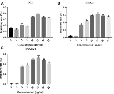
Figure 1DIOS inhibits the cell viability of liver cancer cells using MTT assay. (A) The normal hepatocyte LO2 cells and liver cancer HepG2 (B) and HCC-LM3 (C) cells were treated with different concentrations of DIOS, respectively. The MTT assay was used to detect the cell viability. *P<0.05, **P<0.01 and ***P<0.001.
Abbreviations:DIOS, diosmetin; MTT, 3-(4,5-dimethylthiazol-2-yl)-2,5-diphenyltetrazolium bromide.
Cancer Management and Research downloaded from https://www.dovepress.com/ by 118.70.13.36 on 24-Aug-2020
Results
DIOS Inhibits the Cell Viability of Liver
Cancer Cells
The normal hepatocyte cell line LO2 and liver cancer cell line HepG2 and HCC-LM3 cells were treated with differ-ent concdiffer-entrations of DIOS, respectively. MTT assay results showed that the cell viability of LO2 cells was
not significantly inhibited under different concentrations
of DIOS (Figure 1A). In contrast, we found that DIOS
significantly suppressed the cell viability of HepG2 and
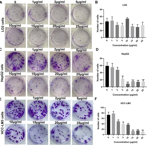
HCC-LM3 cells, with a concentration-dependent manner (Figure 1B and C). Similarly, the results of the clone formation experiments showed that different concentra-tions of DIOS could not affect the proliferation of LO2
cells (Figure 2A and B). However, we found that DIOS
significantly inhibited the proliferation of HepG2 and
HCC-LM3 cells, with a concentration-dependent manner (Figure 2C–F). HepG2 cells were treated with different
Figure 2Clone formation assay results showing the inhibitory effects of different concentrations of DIOS on the proliferation of LO2 cells (A, B), HepG2 (C, D) and HCC-LM3 cells (E, F). *P<0.05, **P<0.01 and ***P<0.001.
Abbreviation:DIOS, diosmetin.
Cancer Management and Research downloaded from https://www.dovepress.com/ by 118.70.13.36 on 24-Aug-2020
concentrations (0, 5, 10, 15 μg/mL) of DIOS for 24 h. Under the microscope, we found that the cells in the control group were slender, vigorously growing, reg-ular in morphology, clear in cell contour, and large in size (Figure 3A). However, as for the HepG2 and HCC-LM3 cells treated with DIOS, the cells were irregular in shape, the cell morphology became round, the cell gap increased,
some cells werefloating, and the cell debris increased with
the increase of concentrations (Figure 3A). Moreover,
DIOS significantly decreased the cells viability of HepG2
and HCC-LM3 cells with concentration-dependent and
time-dependent manners (Figure 3B).
DIOS Promotes Cell Cycle Arrest in G2/
M and Cell Apoptosis of HepG2 Cells
HepG2 cells were treated with different concentrations(0, 5, 10, 15 μg/mL) for 24 h, and flow cytometry was
employed to analyze the cell cycle change. As shown in
Figure 4A andC, the cells were blocked in G2/M phase. Furthermore, DIOS promoted the proportion of G2/M phase, with a concentration-dependent manner. We also examined the cells apoptosis of HepG2 cells under dif-ferent concentrations of DIOS. The results showed that
DIOS significantly promoted cell apoptosis of HepG2
cells, with a concentration-dependent manner (Figure 4B
and D). These results suggested that DIOS could induce
cell cycle arrest in G2/M and cell apoptosis of HepG2 cells.
DIOS Is Involved in Regulating the
Expression of Cell Cycle/
Apoptosis-Associated Proteins in HepG2
Cells
After HepG2 cells treated with different concentrations of
DIOS (0, 5, 10, 15μg/mL) for 24 h, we examined the
expres-sion levels of cell cycle/apoptosis-associated proteins in HepG2 cells using Western blot. The results showed that DIOS significantly inhibited the expression levels of cell cycle-related proteins including cyclin B1 and cdc2, apoptosis-related proteins including Bcl-2 and cleaved-caspase8 in HepG2 cells, with a concentration-dependent manner (Figure 5A). Furthermore, DIOS significantly promoted the expression levels of P53, P21, PARP, cleaved-caspase3, Bax, Bak, P-Chk1, P-cdc25c, P-Chk2 and P-cdk1 in HepG2 cells, with a concentration-dependent manner (Figure 5B).
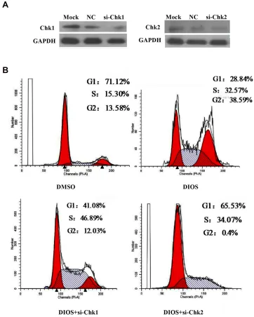
DIOS Might Promote G2/M Arrest via
Chk2 but Not Chk1 in HepG2 Cells
To further explore the mechanism by which DIOS might mediate G2/M cell cycle arrest, we knocked down the expression of Chk1 and Chk2 by the correspondingsiRNAs in HepG2 cells, respectively (Figure 6A). We
found that DIOS significantly promoted the proportion of
G2/M phase in HepG2 cells. After inhibiting Chk2, the
proportion of G2/M phase was significantly inhibited
(Figure 6B). However, after inhibiting Chk1, the proportion
Figure 3The cell morphology of HepG2 cells treated with DIOS. (A) HepG2 cells were treated with different concentrations (0, 5, 10, 15μg/mL) of DIOS for 24 h, and the cell morphology was observed under light microscopy. (B) MTT assay was used to detect the effect of different concentrations of DIOS on cell viability at different times (6, 12, 24, 48 h).
Abbreviations:DIOS, diosmetin; MTT, 3-(4,5-dimethylthiazol-2-yl)-2,5-diphenyltetrazolium bromide.
Cancer Management and Research downloaded from https://www.dovepress.com/ by 118.70.13.36 on 24-Aug-2020
of G2/M phase was not significantly inhibited (Figure 6B). DIOS might promote G2/M arrest via Chk2 but not Chk1 in HepG2 cells.
DIOS Might Promote HepG2 Cell
Apoptosis by Bcl-2 and Cleaved-Caspase8
We observed whether DIOS might promote HepG2 cell apoptosis by apoptosis-related proteins including Bcl-2 andcleaved-caspase8. As shown inFigure 7AandB, Bcl-2 and
cleaved-caspase8 were successfully silenced in HepG2 cells,
respectively. Flow cytometry results suggested that DIOS
significantly promoted HepG2 cell apoptosis. After
inhibit-ing Bcl-2 or cleaved-caspase8, the inhibitory effects induced
by DIOS were significantly reversed (Figure 7C). Thus,
DIOS might promote HepG2 cell apoptosis by Bcl-2 and cleaved-caspase8.
Discussion
Liver cancer is a highly malignant and highly invasive solid tumor.21,22At present, there is no effective treatment.
Figure 4DIOS promotes cell cycle arrest in G2/M and cell apoptosis of HepG2 cells. (A, C) Flow cytometry was used to detect the cell cycle of HepG2 cells treated with different concentrations of DIOS (0, 5, 10, 15μg/mL) for 24 h. (B, D) The apoptosis rate of HepG2 cells under different concentrations of DIOS (0, 5, 10, 15μg/mL) for 24 h was detected usingflow cytometry. *P<0.05, **P<0.01, ***P<0.001 and ****P<0.0001.
Abbreviations:PI-A, propidium iodide-area; G1, postsynthetic gap1 period; S, DNA synthesis phase; G2, postsynthetic gap2 period; M, mitotic phase; FITC-A,fluorescein isothiocyanate-area; DIOS, diosmetin.
Cancer Management and Research downloaded from https://www.dovepress.com/ by 118.70.13.36 on 24-Aug-2020
DIOS as aflavonoid anticancer drug provides a new idea
for the current anticancer treatment.23–25 In this study, we
explored the anti-tumor mechanism of DIOS in human hepatoma HepG2 cells and HCC-LM3 cells.
We found that DIOS promoted G2/M phase arrest in HepG2 cells, while normal human liver cell LO2 had no
significant changes after treatment with DIOS. It is well
known that inhibition of the hepatoma cell cycle can induce the repression of liver cancer cell proliferation. The combination of cyclin and cyclin-dependent kinase
(CDK) is closely related to cell cycle transition.26 Our
results showed that DIOS significantly inhibited the
pro-liferation of hepatoma cells and promoted G2/M cell cycle arrest, which was in close association with Chk signaling pathway. However, there have been few studies on signal pathways in which DIOS is involved, especially in the Chk signal transduction pathway. Therefore, we explored the relationship between Chk signaling pathway and cell cycle regulated by DIOS. The transition from G1 to S causes a cascade of cyclinD/CDK4 complexes to bind to CDK6, and the cyclinB/CDK1 complex will shift from G2 to
M during mitosis.27 When DNA is damaged, the G2
phase-detection site will cause the cells to enter the mitosis phase for automatic repair. When cyclinB and cyclinA form a complex with CDK1, it is extremely important to generate an M-phase transition of the cascade activation
pair.28In our experiments, it was found that DIOS
down-regulated the protein expression of cyclinB/CDK1. There
was no significant change in cyclinA expression after
DIOS treatment. This indicates that DIOS may induce G2/M phase arrest by down-regulating cyclinB/CDK1. In addition, with the accumulation of p53, dissociation from the MDM2 binding site, p53 gene promotes p21Cip1
accumulation, which is an inhibitor of Cdc2.29This results
in a decrease in the expression of cyclin B/cdc2; therefore, the DIOS-induced cell cycle arrest in G2/M phase may be through the induction of the cyclinB1/cdc2 pathway. Our study showed that the expression of p53 and p21 increased in DIOS-treated human hepatoma cell line HepG2, indi-cating that DIOS-induced cell cycle arrest may also be related to the p53 signaling pathway. Meanwhile, DIOS down-regulated Bcl-2 expression through P53 and mito-chondrial apoptosis pathway and up-regulated the expres-sion of Bax, Bak, cleaved-caspase3, cleaved-caspase8 and
Figure 5DIOS is involved in regulating the expression of cell cycle/apoptosis-associated proteins in HepG2 cells. HepG2 cells were treated with different concentrations of DIOS (0, 5, 10, 15μg/mL) for 24 h, and the expression levels of cell cycle/apoptosis-associated proteins were detected by Western blot (A, B).
Abbreviations:Cdc, cell division cycle 2, Bcl, B-cell lymphoma; PARP, poly ADP-ribose polymerase; Bax, BCL2-associated X; Bak, BCL2-Associated kinase; GAPDH, glyceraldehyde-3-phosphate dehydrogenase; Chk, cyclin-dependent kinase; DIOS, diosmetin.
Cancer Management and Research downloaded from https://www.dovepress.com/ by 118.70.13.36 on 24-Aug-2020
cleaved-PARP. Ultimately, DIOS promoted apoptosis and cell cycle arrest in G2/M phase through inducing the low expression of apoptosis-related proteins and cyclins.
DNA damage will result in the activation of ATM/ ATR, which induces Cdc25c phosphorylation to inhibit
the activation of Chk1 and Chk2.30,31 Chk1 promotes
phosphorylation by activating its ser317, ser345 and ser296 sites, and Chk2 activates its ser33/35, ser516,
ser296, and Thr68 sites.32,33 Our study found that DIOS
can induce high expression of p-Chk1 (ser317, ser345) and p-Chk2 (ser33/35). To elucidate the intrinsic mechanism of DIOS-induced cell cycle arrest, we detected the expression of Chk1 and Chk2 in HepG2 cells transfected with Chk1-siRNA and Chk2-Chk1-siRNA. We found that the change in G2 phase after Chk1-siRNA treatment was not obvious, but the proportion of G2 phase in HepG2 cells transfected
with Chk2-siRNA was significantly decreased. This result
demonstrated that the major signaling pathway regulating
Figure 6DIOS might promote G2/M arrest via Chk2 but not Chk1 in HepG2 cells. (A) Western blotting was used to detect the expression levels of cyclin Chk1 and Chk2 in HepG2 cells transfected with Chk1-siRNA and Chk2-siRNA. (B) Flow cytometry was used to detect the cell cycle changes in HepG2 cells transfected with Chk1-siRNA and Chk2-siRNA.
Abbreviations:PI-A, propidium iodide-area; DMSO, dimethyl sulfoxide; DIOS, diosmetin; Chk2, checkpoint kinase 2; siChk2, small interfering RNA-checkpoint kinase 2; NC, negative control; Chk1, checkpoint kinase 1; siChk1, small interfering RNA-checkpoint kinase 1; GAPDH, glyceraldehyde-3-phosphate dehydrogenase; G1, postsyn-thetic gap1 period; S, synpostsyn-thetic phase; G2, postsynpostsyn-thetic gap2 period; M, mitotic phase.
Cancer Management and Research downloaded from https://www.dovepress.com/ by 118.70.13.36 on 24-Aug-2020
G2/M cell arrest could be the Chk2 pathway. The cyclinB/ CDK and p53 signaling pathways are not fully involved in the regulation of G2/M cell arrest in HepG2 cells.
Taken together, our study showed that DIOS can cause proliferation inhibition, induce apoptosis and G2/M cell cycle arrest in human hepatoma HepG2 cells. Based on current research, DIOS may be a potential anti-tumor che-motherapeutic drug for liver cancer therapy. In future study, we will further clarify the role of DIOS in cell cycle arrest and signaling pathway in vitro and in vivo animal models, and provide new clinical treatment approaches for tumors.
Conclusion
In this study, ourfindings revealed that DIOS inhibited cell
proliferation and promoted cell apoptosis in liver cancer cells. Furthermore, DIOS treatment induced G2/M cell cycle arrest in HepG2 cell by down-regulation of cell cycle-related protein cdc2, cyclinB1 and up-regulation of P53 and P21. Moreover, we also found that DIOS might promote HepG2 cell apoptosis by Bcl-2 and cleaved-caspase8. Our research provides novel insights into the mechanism of DIOS against liver cancer.
Abbreviations
DIOS, diosmetin; Chk1, checkpoint kinase 1; siRNA, small-interfering RNA; Chk2, checkpoint kinase 2.
Data Sharing Statement
The datasets analyzed during the current study are avail-able from the corresponding author on reasonavail-able request.
Author Contributions
All authors made substantial contributions to conception and design, acquisition of data, analysis and interpretation of data, drafting the manuscript, revising the manuscript
critically, read and approve the final draft of the
manu-script for submission, gave final approval of the
manu-script version to be published and agreed to be accountable for every step of the work.
Disclosure
The authors declare no conflicts of interest.
References
1. Greten TF, Lai CW, Li G, Staveley-O’Carroll KF. Targeted and Immune-based therapies for hepatocellular carcinoma.Gastroenterology.
2019;156(2):510–524. doi:10.1053/j.gastro.2018.09.051
2. Villanueva A. Hepatocellular Carcinoma. N Engl J Med. 2019;380 (15):1450–1462. doi:10.1056/NEJMra1713263
3. Yang YM, Kim SY, Seki E. Inflammation and liver cancer: molecular mechanisms and therapeutic targets. Semin Liver Dis. 2019;39 (01):26–42. doi:10.1055/s-0038-1676806
4. Bray F, Ferlay J, Soerjomataram I, Siegel RL, Torre LA, Jemal A. Global cancer statistics 2018: GLOBOCAN estimates of incidence and mortality worldwide for 36 cancers in 185 countries. CA Cancer J Clin.2018;68(6):394–424. doi:10.3322/caac.21492
Figure 7DIOS might promote HepG2 cell apoptosis by Bcl-2 and cleaved-caspase8. (A, B) The transfection effects of si-Bcl-2 and si-cleaved-caspase8 in HepG2 cells were detected using Western blot. (C) Flow cytometry was used to detect HepG2 cell apoptosis.
Abbreviations:NC, negative control; si-Bcl-2, small interfering RNA-B-cell lymphoma-2; Bcl-2, B-cell lymphoma-2; GAPDH, glyceraldehyde-3-phosphate dehydrogenase; si, small interfering RNA; FITC-A,fluorescein isothiocyanate-area; DIOS, diosmetin.
Cancer Management and Research downloaded from https://www.dovepress.com/ by 118.70.13.36 on 24-Aug-2020
5. Zheng R, Qu C, Zhang S, et al. Liver cancer incidence and mortality in China: temporal trends and projections to 2030. Chin J Cancer Res.2018;30(6):571–579. doi:10.21147/j.issn.1000-9604.2018.06.01 6. Marengo A, Rosso C, Bugianesi E. Liver cancer: connections with obesity, fatty liver, and cirrhosis. Annu Rev Med. 2016;67 (1):103–117. doi:10.1146/annurev-med-090514-013832
7. Liu Z, Jiang Y, Yuan H, et al. The trends in incidence of primary liver cancer caused by specific etiologies: results from the global burden of disease study 2016 and implications for liver cancer prevention. J Hepatol.2019;70(4):674–683. doi:10.1016/j.jhep.2018.12.001 8. Llovet JM, Montal R, Sia D, Finn RS. Molecular therapies and
precision medicine for hepatocellular carcinoma. Nat Rev Clin Oncol.2018;15(10):599–616. doi:10.1038/s41571-018-0073-4 9. Neureiter D, Stintzing S, Kiesslich T, Ocker M. Hepatocellular
car-cinoma: therapeutic advances in signaling, epigenetic and immune targets.World J Gastroenterol.2019;25(25):3136–3150. doi:10.3748/ wjg.v25.i25.3136
10. Kudo M. Targeted and immune therapies for hepatocellular carci-noma: predictions for 2019 and beyond. World J Gastroenterol.
2019;25(7):789–807. doi:10.3748/wjg.v25.i7.789
11. Wang C, Jin H, Gao D, et al. Phospho-ERK is a biomarker of response to a synthetic lethal drug combination of sorafenib and MEK inhibition in liver cancer. J Hepatol.2018;69(5):1057–1065. doi:10.1016/j.jhep.2018.07.004
12. Tsilimigras DI, Ntanasis-Stathopoulos I, Bagante F, et al. Clinical significance and prognostic relevance of KRAS, BRAF, PI3K and TP53 genetic mutation analysis for resectable and unresectable color-ectal liver metastases: a systematic review of the current evidence. Surg Oncol.2018;27(2):280–288. doi:10.1016/j.suronc.2018.05.012 13. Li Z, Zhang Y, Wang R, Zou K, Zou L. Genetic alterations in
anaplastic thyroid carcinoma and targeted therapies.Exp Ther Med.
2019;18(4):2369–2377. doi:10.3892/etm.2019.7869
14. Cao X, Wang AH, Jiao RZ, et al. Surfactin induces apoptosis and G (2)/M arrest in human breast cancer MCF-7 cells through cell cycle factor regulation. Cell Biochem Biophys. 2009;55(3):163–171. doi:10.1007/s12013-009-9065-4
15. Cerella C, Scherer C, Cristofanon S, et al. Cell cycle arrest in early mitosis and induction of caspase-dependent apoptosis in U937 cells by diallyltetrasulfide (Al2S4). Apoptosis. 2009;14(5):641–654. doi:10.1007/s10495-009-0328-8
16. Dai QS, Liu W, Wang XB, et al. NCPMF-60 induces G2/M cell cycle arrest and apoptosis in human hepatocellular carcinoma HepG2 cells. Anticancer Drugs. 2011;22(1):46–57. doi:10.1097/CAD.0b013e3 283405801
17. Hu B, Shen KP, An HM, Wu Y, Du Q. Aqueous extract of curcuma aromatica induces apoptosis and G2/M arrest in human colon carci-noma LS-174-T cells independent of p53. Cancer Biother Radiopharm.2011;26(1):97–104. doi:10.1089/cbr.2010.0853 18. Suh H, Choi KW, Lee CH. CR389, a benzoimidazolyl pyridinone
analog, induces cell cycle arrest and apoptosis via p53 activation in human ovarian cancer PA-1 cells.J Microbiol Biotechnol.2015;25 (3):418–422. doi:10.4014/jmb.1412.12080
19. Wu W, Ye H, Wan L, et al. Millepachine, a novel chalcone, induces G2/M arrest by inhibiting CDK1 activity and causing apoptosis via ROS-mitochondrial apoptotic pathway in human hepatocarcinoma cells in vitro and in vivo.Carcinogenesis.2013;34(7):1636–1643. doi:10.1093/carcin/bgt087
20. Patel K, Gadewar M, Tahilyani V, Patel DK. A review on pharma-cological and analytical aspects of diosmetin: a concise report.Chin J Integr Med.2013;19(10):792–800. doi:10.1007/s11655-013-1595-3 21. Calderaro J, Couchy G, Imbeaud S, et al. Histological subtypes of hepatocellular carcinoma are related to gene mutations and molecular tumour classification.J Hepatol.2017;67(4):727–738. doi:10.1016/j. jhep.2017.05.014
22. Li M, Lai X, Zhao Y, et al. Loss of NDRG2 in liver microenviron-ment inhibits cancer liver metastasis by regulating tumor associate macrophages polarization. Cell Death Dis. 2018;9(2):248. doi:10.1038/s41419-018-0284-8
23. Chen X, Wu Q, Chen Y, et al. Diosmetin induces apoptosis and enhances the chemotherapeutic efficacy of paclitaxel in non-small cell lung cancer cells via Nrf2 inhibition.Br J Pharmacol.2019;176 (12):2079–2094. doi:10.1111/bph.14652
24. Choi J, Lee DH, Park SY, Seol JW. Diosmetin inhibits tumor devel-opment and block tumor angiogenesis in skin cancer. Biomed Pharmacother.2019;117:109091. doi:10.1016/j.biopha.2019.109091 25. Oak C, Khalifa AO, Isali I, Bhaskaran N, Walker E, Shukla S.
Diosmetin suppresses human prostate cancer cell proliferation through the induction of apoptosis and cell cycle arrest. Int J Oncol.2018;53(2):835–843. doi:10.3892/ijo.2018.4407 26. Palmer N, Talib SZA, Kaldis P. Diverse roles for CDK-associated
activity during spermatogenesis. FEBS Lett. 2019;593 (20):2925–2949.
27. Hu X, Eastman AE, Guo S. Cell cycle dynamics in the reprogram-ming of cellular identity.FEBS Lett.2019;593(20):2840–2852. 28. Hayward D, Alfonso-Perez T, Gruneberg U. Orchestration of the
spindle assembly checkpoint by CDK1-cyclin B1. FEBS Lett.
2019;593(20):2889–2907. doi:10.1002/1873-3468.13591
29. Cmielova J, Rezacova M. p21Cip1/Waf1 protein and its function based on a subcellular localization [corrected]. J Cell Biochem.
2011;112(12):3502–3506. doi:10.1002/jcb.23296
30. Nilsson I, Hoffmann I. Cell cycle regulation by the Cdc25 phospha-tase family.Prog Cell Cycle Res.2000;4:107–114.
31. Tao L, Cao Y, Wei Z, et al. Xanthatin triggers Chk1-mediated DNA damage response and destabilizes Cdc25C via lysosomal degradation in lung cancer cells. Toxicol Appl Pharmacol. 2017;337:85–94. doi:10.1016/j.taap.2017.10.015
32. Manic G, Obrist F, Sistigu A, Vitale I. Trial Watch: targeting ATM-CHK2 and ATR-CHK1 pathways for anticancer therapy.Mol Cell Oncol. 2015;2(4):e1012976. doi:10.1080/23723556.2015. 1012976
33. Ronco C, Martin AR, Demange L, Benhida R. ATM, ATR, CHK1, CHK2 and WEE1 inhibitors in cancer and cancer stem cells. Medchemcomm.2017;8(2):295–319. doi:10.1039/C6MD00439C
Cancer Management and Research
Dove
press
Publish your work in this journal
Cancer Management and Research is an international, peer-reviewed open access journal focusing on cancer research and the optimal use of preventative and integrated treatment interventions to achieve improved outcomes, enhanced survival and quality of life for the cancer patient.
The manuscript management system is completely online and includes a very quick and fair peer-review system, which is all easy to use. Visit http://www.dovepress.com/testimonials.php to read real quotes from published authors.
Submit your manuscript here:https://www.dovepress.com/cancer-management-and-research-journal
Cancer Management and Research downloaded from https://www.dovepress.com/ by 118.70.13.36 on 24-Aug-2020