R E S E A R C H
Open Access
The comparison of surgical patients with
primary hepatic squamous cell carcinoma or
adenosquamous carcinoma and surgical patients
with hepatocellular carcinoma
Long Yan, Feng Xie, Cheng Yang, Lihan Yu, Tao Zheng, Jun Fu and Jiamei Yang
*Abstract
Background:There are still several controversies and ambiguities in the aspects of primary hepatic squamous cell carcinoma and primary hepatic adenosquamous carcinoma. To further clarify the specific features of these two infrequent diseases and provide beneficial propose for clinical decision, we did this retrospective study.
Methods:We reviewed the clinical features and outcomes of three primary hepatic squamous cell carcinoma (SCC)
patients and six primary hepatic adenosquamous carcinoma (ASC) patients from January 1998 to December 2011 in Eastern Hepatobiliary Surgery Hospital in China. Meanwhile, 40 hepatocellular carcinoma (HCC) patients and 26 metastatic hepatic SCC or ASC patients who were hospitalized in the same period were also reviewed to make a comparison. In order to find out the definite survival information of SCC and ASC patients, 30 previous studies containing 37 primary hepatic SCC (16) and ASC (21) patients were reviewed, and their information of survival was analyzed together with the included patients in our hospital.
Results:Serum tumor markers showed significant differences between primary hepatic SCC/ASC and HCC patients,
especially for serum alpha fetal protein (AFP) level and carbohydrate antigen 19-9 (CA 19-9). On the pathologic performance, primary SCC/ASC tumor was rarely accompanied with tumor capsule. They presented peripheral hepatic lymph node metastasis more likely and showed low proportion of microvascular invasion. The median survival time of primary hepatic SCC/ASC patients after liver resection (LR) was 15 months. And the 1-, 3-, 5-year survival rates after LR were 60%, 24%, and 12%, respectively. Significant difference was not discovered when SCC and ASC patients were compared with HCC patients (P= 0.294). The median survival time after LR for primary SCC and ASC patients was respectively 23 months and 13 months.
Conclusions:The comprehensive application of some clinical characteristics, histopathologic features, and imaging findings may be useful for us in making definite diagnoses for primary hepatic SCC and ASC patients
preoperatively. And the treatment of liver resection was effective for those patients who met the selection criteria for liver resection.
Keywords:Hepatic squamous cell carcinoma (SCC), Adenosquamous carcinoma (ASC), Hepatocellar carcinoma (HCC)
* Correspondence:13386272885@189.cn
Department of Special Treatment and Liver Transplantation, Eastern Hepatobiliary Hospital, the Second Military Medical University, 255 Changhai Road, Shanghai 200438, China
Background
Due to the prevalence of hepatitis and some other reasons, the morbidity of primary liver tumor is high in Asia, espe-cially in China [1]. Though the majority of the primary tu-mors are hepatocellular carcinoma (HCC), some rare diseases also could not be ignored by the reason of their characteristics. Both primary hepatic squamous cell car-cinoma (SCC) and adenosquamous carcar-cinoma (ASC) are very rare. Few of them have been reported in the litera-tures. Primary hepatic SCC was considered to be associ-ated with hepatic cysts [2-5], hepatolithiasis [6,7] Caroli’s disease [8], and others. Primary hepatic ASC was defined as a liver tumor containing definite components of both adenocarcinoma and squamous cell carcinoma, and some-times it was considered as a variant of cholangiocarcinoma [9]. However, it was observed to have more aggressive clinical and pathological features than cholangiocarcinoma [10]. Both of SCC and ASC have squamous cell carcinoma components. These tumor components presented more malignant biological behavior, and that may be the com-mon reason for the poor prognosis of the both [11].
We retrospectively investigated some primary hepatic SCC or ASC patients in our hospital to research the characteristics of these two kinds of diseases. Meanwhile, some patients in previous studies were also reviewed.
Methods Patients
A total of 75 liver tumor patients, who had received liver resection (LR) during the period from January 1998 to December 2011 in Eastern Hepatobiliary Surgery Hospital, were picked out and included in this study.
There were just nine patients who suffered from histo-logically proven primary hepatic SCC (three patients) or ASC (six patients) had surgical treatment during the 14 years in our hospital, and all of them were included as the primary SCC/ASC group. Before their final diag-noses were made, each of these nine primary tumor pa-tients had undergone imageological examination and hematological and blood biochemical determinations to preclude other primary tumor diseases. The HCC group contained 40 patients who were selected randomly from our data base. Considering the significant impact of liver cirrhosis to the prognosis of liver cancer patients and half of patients in the primary SCC/ASC group had liver cirrhosis (four patients), the HCC group patients were picked randomly according to the factor that nearly half of patients suffered cirrhosis and half did not. Twenty-six pa-tients with metastatic hepatic SCC (16 papa-tients) or ASC (10 patients) in the hospital during the same time com-posed the metastatic SCC/ASC group. The primary car-cinomas of these patients contained lung cancer (12 patients, 46.2%), nasopharynx cancer (5 patients, 19.2%), gallbladder carcinoma (5 patients, 19.2%), esophagus
cancer (3 patients, 11.5%), and vocal cord carcinoma (1 patients, 3.8%).
We only compared the metastatic SCC/ASC patients with the primary SCC/ASC patients on histopathologic findings. That was because in the clinical practice, it is difficult to determine whether the tumor is primary or metastatic by the postoperative histopathologic examin-ation if other primary sites have not been discovered. So we intended to find some differences between them. All of the other comparisons, like demographic data, operative and laboratory findings, survival, were carried out between the primary SCC/ASC group and the HCC group.
Clinical and laboratory data
The clinical and laboratory data of the primary SCC/ ASC patients and HCC patients were reviewed retrospect-ively from the medical records and relevant databases of Eastern Hepatobiliary Surgery Hospital in China.
To analyze the clinical data, we reviewed the operation notes, like the number and size of tumors, TNM stage, and capsule of the tumors. The laboratory examinations were performed preoperatively, and the laboratory data contained biochemical tests, which reflected hepatic damage, such as prothrombin time (PT), total bilirubin (TBIL), serum alanine transaminase (ALT), and aspartate transaminase (AST). The data of serum alpha fetal pro-tein (AFP), serum carbohydrate antigen 19-9 (CA 19-9), and serum carcinoembryonic antigen (CEA) before LR was analyzed at the same time. We also summarized the features of patients’imagining and pathologic findings.
Survival data
We retrospectively referred to the record databases and completed the follow-up work to get the data associated with survival. Forty-nine patients in the primary SCC/ ASC group and the HCC group were followed up regu-larly until death or until the completion time of the data collecting work and writing the manuscript. The follow-up visit was conducted by phone.
ASC (21) patients were included in our study to provide the survival information [12-33].
Statistical analysis
The analyses were carried out by using SPSS 20. All data were presented as a median or the mean ± standard devi-ation (SD). The independent two-sample t-test was used to compare the numerical data. The Pearson chi-square test and Fisher’s exact test were performed for qualitative data. And the One-way analysis of variance (ANOVA), Kaplan-Meier, Cox regression analysis, was also used. AP value at <0.05 was regarded as statistically significant.
The study was approved by the Ethics Committee Eastern Hepatobiliary Surgery Hospital (approved num-ber: EHBHKY-005-7), informed consent which has been conducted according to the principles expressedin the Declaration of Helsinki was obtained from each patient. All participantsprovided their written informed consent to participate in this study
Results
Demographic data and baseline characteristics
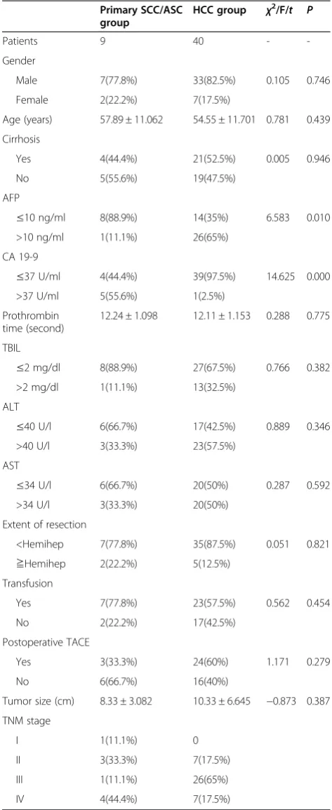
All related information of the primary SCC/ASC tients (9 patients) and primary HCC patients (40 pa-tients) were summarized in Table 1 for the comparison of the demographic data and clinical characteristics. These two groups of patients shared almost the same gender and age composition. Of these patients, 7 (77.8%) men and 2 (22.2%) women were in the primary SCC/ASC group, and 33 (82.5%) men and 7 (17.5%) women were in the HCC group (P= 0.746). The average age was respectively 57.89 ± 11.062 and 54.55 ± 11.701 (P= 0.439). To maintain consistent baselines for all pa-tients, the HCC group patients were selected according to the component ratio of patients with or without cir-rhosis in the primary SCC/ASC group. So there were also no significant differences between these two groups of patients on the baseline liver function indexes, like TBIL, ALT, AST, and PT.
Serum tumor markers showed significant differences for these two groups patients due to the different tumor tissues. As a classical marker of HCC, serum AFP level appeared to have higher frequency of exceeding normal value in HCC patients (P= 0.01) and there was only one primary ASC patient (no primary SCC patient) who was detected with a high level of serum AFP. However, the performance of CA 19-9 was just the opposite. When we defined 37 U/ml was the highest normal level of serum CA 19-9, we found only one (2.5%) HCC patient exceeded this standard value, and the ratio was much less than primary SCC/ASC patients (P= 0.000).
Three (33.3%) patients in primary SCC/ASC group and 24 (60%) patients in HCC group adopted postoperative transhepatic arterial chemotherapy and embolization.
Table 1 The demographic data and baseline characteristics of primary hepatic SCC/ASC patients and HCC patents
Primary SCC/ASC
Male 7(77.8%) 33(82.5%) 0.105 0.746
Female 2(22.2%) 7(17.5%)
Age (years) 57.89 ± 11.062 54.55 ± 11.701 0.781 0.439
Cirrhosis
Yes 4(44.4%) 21(52.5%) 0.005 0.946
No 5(55.6%) 19(47.5%)
AFP
≤10 ng/ml 8(88.9%) 14(35%) 6.583 0.010
>10 ng/ml 1(11.1%) 26(65%)
CA 19-9
≤37 U/ml 4(44.4%) 39(97.5%) 14.625 0.000
>37 U/ml 5(55.6%) 1(2.5%)
Prothrombin time (second)
12.24 ± 1.098 12.11 ± 1.153 0.288 0.775
TBIL
≤2 mg/dl 8(88.9%) 27(67.5%) 0.766 0.382
>2 mg/dl 1(11.1%) 13(32.5%)
ALT
≤40 U/l 6(66.7%) 17(42.5%) 0.889 0.346
>40 U/l 3(33.3%) 23(57.5%)
AST
≤34 U/l 6(66.7%) 20(50%) 0.287 0.592
>34 U/l 3(33.3%) 20(50%)
Extent of resection
<Hemihep 7(77.8%) 35(87.5%) 0.051 0.821
≧Hemihep 2(22.2%) 5(12.5%)
Transfusion
Yes 7(77.8%) 23(57.5%) 0.562 0.454
No 2(22.2%) 17(42.5%)
Postoperative TACE
Yes 3(33.3%) 24(60%) 1.171 0.279
When referring to the data of tumor sizes (the largest tumor size was chosen when several tumors were found in a patient), we found the average diameter of the tumors was generally large (>5 cm) for both two groups of pa-tients (P= 0.387). When the primary liver tumors were staged by the TNM stage system [12], the primary SCC/ ASC patients mainly distributed in stage II or IV, and the primary HCC patients mainly were stage IV. The extent of liver resection for most patients were less than hemihepa-tectomy (77.8% and 87.5%).
Histopathologic findings
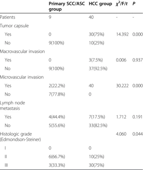
The comparison of general pathologic characteristics be-tween primary SCC/ASC patients and HCC patients was listed in Table 2. From the result of statistical analysis, we found that, because the main component was squa-mous carcinoma, the liver tumor in primary SCC/ASC patients usually did not accompany tumor capsule. This feature could make the primary hepatic SCC/ASC show more malignant biological behavior. Though the differ-ence was not significant (P= 0.191, >0.05), primary SCC/ ASC patients presented peripheral hepatic lymph node metastasis more likely (4 patients, 44.4%). This perform-ance might be related with these liver tumors’malignant behavior. The microvascular invasion of tumors was quite different when comparing the pathology of the two groups of patients. The microvascular invasion only emerged in a few part of primary SCC/ASC (2 patients,
22.2%) but almost in all of the HCC patients (40 pa-tients, 100%). The tumor tissues were staged in Edmondson-Steiner grading [13]. All these liver tumor patients were mainly distribute in stages II (moderately differentiated) and III (poorly differentiated). More than half of the primary SCC/ASC patients (6 patients, 66.7%) were stage II, and 75% of HCC patients (30 pa-tients) were stage III. It seemed that tumor tissues in primary hepatic SCC and ASC were better differentiated than that in HCC.
Microscopically, the postoperative tumor tissue slice of primary hepatic SCC presented that the cancer cells were polygonal and arranged as strips, and their nucleuses were round or orbicular-ovate, most of which even had patho-logic change, like karyomegaly and anachromasis. No cap-sule was found around the tumor and it appeared multifocal growth. Apart from the performance of squa-mous cancer cells, the tumor tissue of primary hepatic ASC arranged like glandular tube, adenocarcinoma cells were cubic or columnar and the tumor appeared infiltrative growth. We chose two typical HE tumor tissue slices from the nine primary carcinoma patients (Figure 1), of whom one was primary SCC (A), and the other was ASC (B).
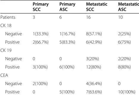
In order to find the immunohistochemical characteris-tics of hepatic tumor pathology for primary SCC and ASC, we compared some immunohistochemical markers of tumor tissues between primary hepatic SCC/ASC and metastatic hepatic SCC/ASC (Table 3). Unlike the meta-static tumor group (80% positive CK 19), all of the primary group patients expressed positive CK 19, and a half of the primary hepatic ASC patients (three patients) even had strong positive expression. Considering that CK 19 gener-ally appeared positive in the cholangiocellular carcinomas [34], the different performance of CK 19 between the four groups may provide some revelations for the origin of pri-mary hepatic SCC and ASC. We found the percentage of positive CK 18 for primary SCC patients was larger than that of metastatic SCC (valid percent, 66.7%:42.9%), and that was also adapted to the comparison between primary ASC and metastatic ASC (valid percent, 83.3%:75%).
Two patients in primary hepatic SCC group (1 did not have the test) were negative for CEA, and in contrast to that, all primary hepatic ASC patients (5 patients) were positive for CEA, excepted 1 person who did not conduct the test.
Survival
As mentioned above, because the survival of the meta-static hepatic SCC and ASC patients might be signifi-cantly affected by the primary tumor disease, it was insufficient to analyze the survival just basing on the metastatic hepatic tumor diseases. So the survival ana-lysis was only conducted in primary SCC/ASC and HCC patients (total 49 patients). The median time of follow-up was 20 months, arranged from 4 to 72 months. Till Table 2 The general pathologic characteristics of primary
SCC/ASC patients and HCC patents
Primary SCC/ASC group
HCC group χ2/F/t P
Patients 9 40 -
-Tumor capsule
Yes 0 30(75%) 14.392 0.000
No 9(100%) 10(25%)
Macrovascular invasion
Yes 0 3(7.5%) 0.006 0.937
No 9(100%) 37(92.5%)
Microvascular invasion
Yes 2(22.2%) 40 30.222 0.000
No 7(77.8%) 0
Lymph node metastasis
Yes 4(44.4%) 7(17.5%) 1.712 0.191
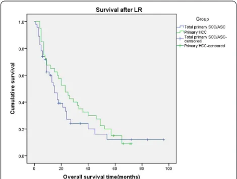
the end of follow-up period, there was only one patient alive in the primary SCC/ASC group. The median time of survival for these nine patients after LR was 15 months, and the mean of survival time was 19.778 ± 4.389 months. Primary SCC/ASC patients’1-, 3-, 5-year survival rates after LR were 67%, 20%, and 0%. For the 40 included HCC patients in this study, the median vival time after LR was 23 months and the mean of sur-vival time was 29.405 ± 3.614 months. We also found their 1-, 3-, 5-year survival rates after LR were 68%, 35%, and 14%. The short-term survival after LR of these two groups of patients was nearly the same, but the long-term survival of the HCC patients seemed better than that of the primary SCC/ASC patients. However, we found no significant difference of the overall survival
between two groups of patients (log rankχ2= 1.216,P= 0.27, Figure 2).
From the review of 30 previous studies, we extracted the survival information of 37 primary SCC (16, 43.2%) and ASC (21, 56.8%) patients, and their survival data was mixed together with our patients’ data to conduct the survival analysis. All of the 37 primary SCC/ASC patients from previous studies and our 9 primary SCC/ ASC patients were defined as total primary SCC/ASC group (46 patients), which would be compared with those 40 HCC patients. The median survival time after LR of the total primary SCC/ASC patients was 15 months, which was the same with our included 9 SCC/ASC patients in our hospital. And the mean sur-vival time after LR was 26.187 ± 4.876 months. The 1-, 3-, 5-year survival rates after LR for these primary SCC/ ASC patients were 60%, 24%, 12%, respectively. We also did not find any significant difference when compared with HCC patients (log rank χ2= 1.100, P= 0.294, Figure 3). At the same time, we compared all of primary SCC patients (19 patients) and primary ASC patients (27 patients) with HCC patients (40 patients), respect-ively. The median survival time after LR for primary SCC and ASC patients was 23 and 13 months. The 1-, 3-, 5-year survival rates after LR were 72%, 27%, and 18% and 51%, 21%, and 7% for SCC patients and ASC patients. Though the survival rates appeared to be different, from the analysis of comparison result, no significant difference was found when we compared the SCC and ASC patients with HCC patients respectively (log rank χ2= 4.330,P= 0.115, Figure 4). This result was similar with what we ob-tained from the comparison of patients in our hospital. So from the statistical analysis results, we found that the
Figure 1Two typical HE tumor tissue slices. (A)The squamous cancer cells were polygonal and arranged as strips, and their nucleuses were round or orbicular-ovate, most of which even had pathologic change, like karyomegaly and anachromasis (H & E stain, ×200).(B)Except the performance of squamous cell carcinoma components, adenocarcinoma cells were cubic or columnar and arranged like glandular tube (H & E stain, ×200).
Table 3 The immunohistochemical characteristics of primary SCC/ASC and metastatic SCC/ASC
Primary SCC
Primary ASC
Metastatic SCC
Metastatic ASC
Patients 3 6 16 10
CK 18
Negative 1(33.3%) 1(16.7%) 8(57.1%) 2(25%)
Positive 2(66.7%) 5(83.3%) 6(42.9%) 6(75%)
CK 19
Negative 0 0 3(20%) 2(20%)
Positive 3(100%) 6(100%) 12(80%) 8(80%)
CEA
Negative 2(100%) 0 4(36.4%) 0
survival after LR for primary hepatic SCC and ASC pa-tients seemed similar with HCC papa-tients.
Discussion
Though various hypotheses have been set forth previ-ously, the pathogenesis and precise steps leading up to the development of the carcinomas for both primary hepatic SCC and ASC are not exactly clear. Previous studies had reported that potentially pre-existent dis-eases, like hepatic cysts, containing congenital hepatic cyst or other benign non-parasitic cysts [2-5], calculus of intrahepatic duct [6], Caroli’s disease [7,8], may be the
aetiological agents for primary hepatic SCC. In our pri-mary SCC patients group, one patient had liver cirrhosis, and two had hepatic cyst, which was in accord with pre-vious reports and might explain the pathogenesis. Simi-larly, though some pathogenesis of primary hepatic ASC had been proposed previously, it was still undefined. It was suggested in some previous studies that primary hep-atic ASC might be caused by the squamous metaplasia of the intrahepatic bile duct [31]. The pathogenesis was also believed to be that the chronic inflammation con-tinuously irritated the bile ducts or various congenital cysts of the biliary tracts [32], while someone proposed squamous metaplasia of denocarcinoma cells might be the etiologic factors [35]. In our study, the six included primary ASC patients contained one liver cirrhosis pa-tients, one calculus of intrahepatic duct papa-tients, and three hepatic cyst patients, which was also complied with those previous opinions about the pathogenesis of primary hepatic ASC.
Since the absence of sufficient evidence and undeter-mined pathogenesis for both primary hepatic SCC and ASC, most patients’ preoperative diagnosis could not be confirmed timely. A previous study reported that the level of serum squamous cell carcinoma-related antigen (SCC-Ag) might be a useful marker for preoperative diagnosis for ASC [36]. They found patients suffering from primary hepatic ASC might have a high level of serum SCC-Ag and this marker could also indicate tumor recurrence. In this paper, none of our primary hepatic SCC or ASC pa-tients had definitive preoperative diagnosis.
We summarized and analyzed some kinds of preopera-tive clinical data and compared them between primary hepatic tumor (SCC and ASC) and HCC groups to
Figure 2Kaplan-Meier cumulative survival curves of the two groups of patients for overall survival (OS).
Figure 3Kaplan-Meier cumulative survival curves of the total primary SCC/ASC group (added 37 patients from previous studies) and HCC group of patients for overall survival (OS) after liver resection.
explore appropriate methods for the diagnosis. Patients with primary hepatic SCC and ASC shared the similar performance of common serum tumor biomarkers, like negative level of AFP (≤10 ng/ml), and a part of positive level of CA 19-9 (>37 U/ml) that may distinguish them from most hepatocellular carcinoma and part of meta-static liver tumor, but it was incapable of picking them out from other kinds of metastatic liver tumor and most of usual intrahepatic cholangiocarcinoma. The imaging performance of primary hepatic SCC and ASC in our study did not have much unique characteristic. The CT images could tell a low density mass with irregular rim enhancement for primary hepatic SCC and ASC, which was similar to the presentations of intrahepatic cholan-giocarcinoma, metastatic liver tumor, or even liver ab-scess. The imaging findings of primary SCC and ASC patients in our study were almost the same with previ-ous reports, and they also found most of the MRI of pri-mary hepatic SCC and ASC presented a low signal on T1WI image and high signal on T2WI image [33]. Someone detected that the ASC had a performance of high-signal T1WI image, which might relate to its solid-cystic structure and central necrosis [30,37].
Though a feasible and determinate preoperative diag-nostic approach has not been established yet for primary hepatic SCC and ASC patients, the comprehensive appli-cation of testing tumor biomarkers, like AFP, CA19-9, and especially SCC-Ag, could be useful. At present, the definitive diagnosis is made on the basis of preoperative needle biopsy, or even has to rely on postoperative histo-logical and immunohistochemical findings.
At present, both of the therapeutic regimes for pri-mary hepatic SCC and ASC are not certain, due to the low incidence and belated definitive diagnosis. Surgical procedures may be a more important treatment for both two [6-11,25-33]. Besides the resection, previously re-ported methods included transcatheter arterial infusion chemotherapy (TACE), which was considered to be use-ful for both primary hepatic SCC and ASC patients [22]. Systemic chemotherapy was also reported in the thera-peutic course of SCC and ASC [21,33]. Although these therapeutic methods were reported to have significant treatment effects, they usually were adopted just as adju-vant therapy [29]. In previous studies, whether TACE or systemic chemotherapy was always undertaken before or post resections.
With the high malignancy of tumor, most of the pri-mary SCC and ASC patients had dismal prognosis [38]. The squamous cell carcinoma components always present more malignant biological behavior, and it may be the common reason for the poor prognosis of these two dis-eases [39]. Takahashi found that, even in primary adenos-quamous carcinoma of liver, the sadenos-quamous cell carcinoma components had highly proliferative activity [9]. But if
surgery resection was undertaken, the situation might be improved. From the analysis of the survival data of the in-cluded patients in our hospital and the patients in the pre-vious studies, we even did not find any significant difference between the primary SCC/ASC patients and the HCC patients about the survival after the treatment of LR (log rank, P> 0.05). Of course, maybe we can not be so optimistic because we could not ignore some reasons which may play roles in achieving this improvement. Be-cause of the high malignancy of primary hepatic SCC and ASC, the tumor progresses rapidly, and it certainly leads to a small number of patients could get the treatment of LR according to the guideline. The prognoses of the rest were gloomy and we could not get sufficient information of this crowd who did not receive surgical treatment yet.
Conclusions
Though it was difficult to make a definite diagnosis for primary hepatic SCC and ASC patients preoperatively in the condition that biopsy has not been conducted, there still were some features for them in clinical characteris-tics, histopathologic features, and imaging findings. And the treatment of liver resection was effective for some of these patients. The low incidence of primary hepatic SCC and ASC resulted in difficulties to carry out large sample studies. Multicenter studies could be performed to further clarify the specific features and to confirm therapeutic methods.
Competing interests
The authors declare that they have no competing interests.
Authors’contributions
LY, J Y and FX carried out the studies. CY, LY, TZ, and JF drafted the manuscript. All authors read and approved the final manuscript.
Acknowledgements
This work was supported in part by grants from the China Postdoctoral Science Fund (20100470107), China Postdoctoral Science Special Fund (201104750), CSCO Fund (Y-B2011-006), Mengchao Fund for outstanding young scholar and the CFHPC Fund (CFHPC 20132193). The funders had no role in study design, data collection and analysis, decision to publish, or preparation of the manuscript.
Received: 26 June 2014 Accepted: 17 January 2015
References
1. Xavier Bosch F, Josepa R, Mireia D, Ramon C. Primary liver cancer: worldwide incidence and trends. Gastroenterology. 2004;127:S5–16. 2. Lynch MJ, McLeod MK, Weatherbee L, Gilsdorf JR, Guice KS, Eckhauser FE.
Squamous cell cancer of the liver arising from a solitary benign nonparasitic hepatic cyst. The Am J Gastroenterol. 1988;83(4):426–31.
3. Nieweg O, Slooff MJH, Grond J. A case of primary squamous cell carcinoma of the liver arising in a solitary cyst. HPB Surg. 1992;5:203–8.
4. Weimann A, Klempnauer J, Gebel M, Maschek H, Bartels M, Ringe B, et al. Squamous cell carcinoma of the liver originating from a solitary non-parasitic cyst: case report and review of the literature. HPB Surg. 1996;10:45–9.
6. Song E, Kew MC, Grieve T, Isaacson C, Myburgh JA. Primary squamous cell carcinoma of the liver occurring in association with hepatolithiasis. Cancer. 1984;53:542–6.
7. Zhu K, Li D, Jiang C. Primary squamous cell carcinoma of the liver associated with hepatolithiasis: A case report. World J Gastroenterol. 2012;18(40):5830–2.
8. Spaggiari M, Di Benedetto F, Ballarin R, Losi L, Cautero N, De Ruvo N, et al. Primary squamous cell carcinoma of the liver associated with Caroli’s disease: a case report. Onkologie. 2011;34:193–5.
9. Takahashi H, Hayakawa H, Tanaka M, Okamura K, Kosaka A, Mizumoto R, et al. Primary adenosquamous carcinoma of liver resected by right trisegmentectomy: report of a case and review of the literature. J Gastroenterol. 1997;32:843–7.
10. Shin Young P, Eun Jung C, Woo SM. Adenosquamous carcinoma of the liver. Clin Mole Hepatol. 2012;18:326–9.
11. Yeh C-N, Jan Y-Y, Chen M-F. Adenosquamous carcinoma of the liver: clinico-pathologic study of 10 surgically treated cases. World J Surg. 2003;27:168–72. 12. Fleming ID. AJCC/TNM cancer staging, present and future. J Surg Oncol.
2001;77:233–6.
13. Edmondson HA, Steiner PE. Primary carcinoma of the liver: a study of 100 cases among 48,900 necropsies. Cancer. 1954;7:462–503.
14. Bastiaan De Boer W. Basaloid squamous carcinoma in the liver. Pathology. 2000;32(2):147–51.
15. Furlanetto A, Tos APD. Squamous cell carcinoma arising in a ciliated hepatic foregut cyst. Virchows Arch. 2002;441:296–8.
16. Yagi H, Ueda M, Kawachi S, Tanabe M, Aiura K, Wakabayashi G, et al. Squamous cell carcinoma of the liver originating from nonparasitic cysts after a 15 year follow-up. Eur J Gastroenterol Hepatol. 2004;16(10):1051–6. 17. Boscolo G, Jirillo A, Da Pian P. Complete remission of poorly differentiated squamous liver carcinoma after systemic chemotherapy and surgery: a case report. Tumori. 2005;91:71–2.
18. Lee H-L, Liu Y-Y, Yeh C-N, Chiang K-C, Chen T-C, Jan Y-Y. Primary squamous cell carcinoma of the liver: a successful surgically treated case. World J Gastroenterol. 2006;12(33):5419–21.
19. Naik S, Waris W, Carmosino L, Mehrishi A, Muhammad WS. Primary squamous cell carcinoma of the liver. J Gastrointestin Liver Dis. 2009;18(4):487–9.
20. Zhang X, Wang Z, Dong Y. Squamous cell carcinoma arising in a ciliated hepatic foregut cyst: case report and literature review. Pathol Res Pract. 2009;205:498–501.
21. Iimuro Y, Asano Y, Suzumura K, Yada A, Hirano T, Iijima H, et al. Primary squamous cell carcinoma of the liver: an uncommon finding in contrast-enhanced ultrasonography imaging. Case Rep Gastroenterol. 2011;5:628–35. 22. L-w D. Sun H, Xiao R-x, Pan L-l, Li D-f. Primary squamous cell carcinoma of
the liver: a case report. Chin. J Hepatol. 2012;20(6):477–8.
23. Sasaki H, Hayashi M, Miyahara S, Shimomura M, Takahashi H, Kawarada Y. Adenosquamous carcinoma of the liver: case report. J Hep Bil Pancr Surg. 1994;2:179–83.
24. Maeda T, Takenaka K, Taguchi K, Kajiyama K, Shirabe K, Shimada M, et al. Adenosquamous carcinoma of the liver: clinicopathologic characteristics and cytokeratin profile. Cancer. 1997;80(3):364–71.
25. Isa T, Kusano T, Muto Y, Furukawa M, Kiyuna M, Toda T. Clinicopathologic features of resected primary adenosquamous carcinomas of the liver. J Clin Gastroenterol. 1997;25(4):623–7.
26. Zhang Z, Nonaka H, Nagayama T, Hatori T, Ihara F, Zhang L, et al. Double cancer consisting of adenosquamous and hepatocellular carcinomas of the liver. Pathol Int. 2001;51:961–4.
27. Kwon OS, Lee HS, Koh DW, Cho YJ, Park YH, Park DK, et al. A case of primary adenosquamous carcinoma of the liver presented with liver abscess. Korean J Intern Med. 2001;16(4):270–3.
28. Suzuki E, Hirai R, Ota T, Shimizu N. Primary adenosquamous carcinoma of the liver: case report. J Hepatobiliary Pancreat Surg. 2002;9:769–73. 29. Demir G, Yanmaz T, Aykut Ferhat C, Gülsen O, Suheyla S. Primary
adenosquamous carcinoma of the liver: case with the longest survival. J Clin Gastroenterol. 2005;39(10):924–5.
30. Yokota H, Matoba M, Tonami H, Hasegawa T, Saito H, Kurose N. Imaging findings in primary adenosquamous carcinoma of the liver: a case report. Clin Imaging. 2007;31(4):279–82.
31. Kobayashi M, Okabayashi T, Okamoto K, Namikawa T, Araki K. A clinicopathologic study of primary adenosquamous carcinoma of the liver. J Clin Gastroenterol. 2005;39(6):544–8.
32. Ochiai T, Yamamoto J, Kosuge T, Shimada K, Takayama T, Yamasaki S, et al. Adenosquamous carcinoma with different morphologic and histologic components arising from the intrahepatic bile duct: report of a case. Hepatogastroenterology. 1996;43(9):663–6.
33. Nakai T, Ono K, Terayama K, Yamagami T, Nishimura T. Case report: adenosquamous carcinoma of the liver successfully treated with repeated transcatheter arterial infusion chemotherapy (TACE) with degradable starch microspheres. Br J Radiol. 2004;77:516–8.
34. Jung Il L, Jin-Woo L, Joon Mee K, Ja Kyung K, Hyun Jung C, Young SK. Prognosis of hepatocellular carcinoma expressing cytokeratin 19: comparison with other liver cancers. World J Gastroenterol. 2012;18(34):4751–7. 35. Hamaya K, Nose S, Mimura T, Sasaki K. Solid adenosquamous carcinoma of
the liver: a case report and review of the literature. Acta Pathol Jpn. 1991;41:834.
36. Yamamoto K, Takenaka K, Kajiyama K, Maeda T, Shirabe K, Shimada M, et al. A primary adenosquamous carcinoma of the liver with an elevated level of serum squamous cell carcinoma related antigen. Hepatogastroenterology. 1996;43:658–62.
37. Tan Y, E-h X. Rare hepatic malignant tumors: dynamic CT, MRI, and clinicopathologic features: with analysis of 54 cases and review of the literature. Abdom Imaging. 2012;38(3):511–26.
38. Yuki N, Hijikata Y, Kato M, Kawahara K, Wakasa K. Squamous cell carcinoma as a rare entity of primary liver tumor with grave prognosis. Hepatol Res. 2006;36:322–7.
39. Sato M, Yajima Y, Takahashi N, Aoki C, Miyazaki A, Eda Y, et al. A case of adenosquamous carcinoma of the liver containing squamous components demonstrating a character of adenocarcinoma with immunohistochemical stains. Nippon Shokakibyo Gakkai Zasshi. 2001;98:964–9.
Submit your next manuscript to BioMed Central and take full advantage of:
• Convenient online submission • Thorough peer review
• No space constraints or color figure charges • Immediate publication on acceptance
• Inclusion in PubMed, CAS, Scopus and Google Scholar
• Research which is freely available for redistribution