Cellular and molecular function of Uhrf1 in
neural stem cells
“Dissertation der Graduate School of Systemic Neurosciences der
Ludwig-Maximilians-Universität München”
Prepared in the group of Professor Dr. Magdalena Götz at the Institute for
Stem Cell Research, Helmholtz Zentrum München
Vidya Ramesh
First supervisor: Professor Dr. Magdalena Götz, Institute for Stem Cell Research,
Helmholtz Zentrum München and Biomedical Center Munich, Germany
Second reviewer: Professor Dr. Peter Becker, Biomedical Center Munich, Germany
Third reviewer: Professor Dr. Wolfgang Enard, Biomedical Center Munich, Germany
External reviewer: Professor Dr. Stephan Beck, University College London, UK
i
Acknowledgements
This PhD would not have been possible without the support and friendship of many people in my
professional and personal life.
I would like to first thank my supervisor Dr. Magdalena Götz for her wonderful support over these
years of my PhD. Her guidance, knowledge and new unique ideas towards this work has been
extremely valuable. Her enthusiasm and motivation for science has been a great influence for
me. I am also glad that we had the opportunity to get to know each other and have a warm rapport.
I would like to thank Efil Bayam for all her help with this project and the opportunity to work on it.
I would also like to thank Dr. Gunnar Schotta and Dr. Filippo Cernilogar who were both excellent
collaborators. Their ideas and help towards this project has been invaluable. I am extremely glad
we had the chance to meet and have great discussions.
I would like to also particularly thank Dr. Jovica Ninkovic and Dr. Stefan Stricker for a lot of
interesting and useful scientific discussions about epigenetics. I would also like to thank Jovica
for his valuable suggestions during my thesis committee meetings. Thank you to my other thesis
committee members, Dr. Peter Becker and Dr. Francois Guillemot. I really value your great
suggestions and support during these years. To my graduate school – the Graduate School of
Systemic Neurosciences, I thank you for your excellent assistance during the PhD and helping
me attend many outstanding conferences. I would also like to thank the people involved in the
GSN for always being so kind and helpful.
I also thank Dr. Markus Riemenschneider and Dr. Markus Schulze for their great collaboration
and help towards this project.
A big thank you to all the fantastic people in the Götz lab. I have really loved working in this
environment and will always treasure all the emotional and scientific support from all of you! In
particular, I would like to acknowledge the embryo group, which taught me so many scientific
skills and a great foundation for neuroscience. I would also like to thank our wonderful technicians
in the lab – Andrea, Sarah, Angelika and Sabine for their help in running our lab smoothly. I thank
Andrea Steiner for so patiently and kindly helping me with cell culture experiments.
I would also like to thank my students – Isabel, Daniela and Linnea for their great help towards
ii
The time in my lab and these years in Munich were especially wonderful because of the great
people I have met and known.
I would like to thank Joana, Emily, Tessa and Francesca for their amazing support and affection
through all these years. Thank you to Elsa, Judith, Sven, Pia, Silvia, Ruth and Rossella for your
awesome friendship. I really enjoyed all the time we spent together and it has been a pleasure to
know you all.
I would also like to thank other people in the lab for always being so friendly, supportive and fun
to hang out with – Miriam, Tjasa, Chris, Maxi, Valentin, Filippo, Adam, Giacomo, Sergio, Ronny,
Gregor, Anita, Christina, Alessandro, Rosario, Kalina, Tamara and German.
Big thanks to my amazing ‘flowers’ –Noelia, Patricia and Roberta for their awesome friendship
and support through all my ‘neuroscience years’.
A very special thanks to Yamini and Katerina for always, always being there for me.
I am really very grateful to my parents, my brother and grandparents for always supporting me to
iii Table of Contents
1. Abstract ... 1
2. Introduction ... 3
2.1Epigenetic regulators of stem cells and NSCs ... 3
2.1.1 Histone marks and their role in neurogenesis ... 5
2.1.2 DNA modifications ... 6
2.1.3 Role of Dnmts and Tets in ESCs, early development and NSCs ... 10
2.1.4 Uhrf1 and Uhrf2 ... 11
2.2 Endogeneous retroviral elements ... 13
2.3 Embryonic neurogenesis ... 15
2.3.1 Development of the embryonic forebrain ... 15
2.3.1 Overview of embryonic neurogenesis ... 16
2.3.2 Cell types in the embryonic neocortex ... 17
2.3.3 Cortical layering and postnatal terminal neuron differentiation ... 19
2.4 Adult neurogenesis ... 22
2.4.1 Adult neurogenic niches and cell types ... 22
2.4.2 Gliogenesis ... 24
3. Aims of my thesis ... 26
4. Results ... 27
4.1Uhrf1 expression from embryonic to adult stages ... 27
4.2 Characterisation of the Uhrf1 cKO cellular phenotype ... 29
4.2.1 Comparable neuronal and glial cells at embryonic stages ... 29
4.2.2 Cortical layering appeared normal in the cKO ... 33
iv
4.3 Transcriptional profiling of Uhrf1 cKO ... 37
4.3.1 Activation of cellular stress genes at E16 and P5 Uhrf1 cKO ... 37
4.3.2 Specific endogeneous retroviral elements are activated in Uhrf1 cKO ... 47
4.4 Alterations in methylation and chromatin marks in Uhrf1 cKO ... 55
4.4.1 Loss of DNA methylation in Uhrf1 cKO ... 55
4.4.2 Loss of 5mC in specific genes and transposable elements ... 57
4.4.3 Gain of 5hmC in Uhrf1 cKO on IAP elements ... 61
4.4.4 Assessing changes in histone marks in the cKO ... 65
4.5 Manipulation of Tet enzymes to rescue IAP activation ... 66
4.5.1In vivo knockdown of Tet enzymes by in utero electroporation of the cKO cortex 66 4.5.2 Generating an in vitro system to investigate the relationship between Tet enzymes and IAP activation ... 71
4.6 Investigating changes in the proteasome of cKO cortices and controls ... 76
4.7 Generating neural stem cells from Uhrf1 KO ES cells ... 81
4.8 Conditional knockout of Uhrf1 in adult neurogenic niches ... 82
4.8.1Loss of Uhrf1 in the adult subependymal zone and dentate gyrus – further characterization of the cellular phenotype ... 82
4.8.2Differences in IAP activation in the adult cKO of the SEZ and dentate gyrus ... 87
5. Discussion 5.1Expression of Uhrf1 in the developing cerebral cortex ... 91
5.2Cellular function of Uhrf1 in the embryonic cerebral cortex ... 91
5.3Transcriptional control of Uhrf1 in the embryonic cortex ... 94
5.4Molecular mechanism of Uhrf1 in the developing neocortex ... 99
5.5Uhrf1 function in adult neurogenesis ... 103
v 6. Methods
6.1 Materials
6.1.1 Chemicals ... 107
6.1.2 Tissue culture reagents ... 109
6.1.3 Standard solutions and buffers ... 110
6.1.4 Cell culture media ... 112
6.1.5 Kits ... 113
6.1.6 Antibodies ... 114
6.1.6.1 Primary antibodies ... 114
6.1.6.2 Secondary antibodies ... 115
6.1.7 Primers ... 115
6.1.7.1 Genotyping primers ... 115
6.1.7.2 Real time PCR primers ... 116
6.1.7.3 Bisulfite sequencing primers ... 117
6.1.7.4 Bisulfite sequencing primers ... 117
6.1.8 Constructs ... 118
6.2 Methods 6.2.1 In vivo methods ... 118
6.2.1.1 Mouse lines ... 118
6.2.1.2 Perfusion and anesthesia ... 119
6.2.1.3 In utero electroporation ... 119
6.2.1.4 Tamoxifen induction ... 120
vi
6.2.2.1 Tissue dissection ... 120
6.2.2.2 Tissue sectioning ... 120
6.2.2.3 Immunostaining ... 121
6.2.2.4 Fluorescence activated cell sorting ... 121
6.2.3 Methods in molecular biology ... 122
6.2.3.1 Extraction of DNA from mouse tail ... 122
6.2.3.2 Plasmid DNA extraction ... 122
6.2.3.3 RNA extraction for cDNA synthesis and qRT-PCR ... 122
6.2.3.4 RNAseq at E16 - VZ ... 123
6.2.3.5 RNAseq at E16 full cortex and P5 cortex ... 123
6.2.3.6 Oxidative reduced representation bisulfite sequencing ... 123
6.2.3.7 Oxidative bisulfite sequencing ... 123
6.2.3.8 Hydroxy methylated DNA immunoprecipitation ... 124
6.2.3.9 Determining concentration and quality of nucleic acids ... 124
6.2.3.10 Genotyping PCR ... 125
6.2.3.11Bacterial transformation ... 125
6.2.3.12 Bacterial cultures ... 126
6.2.3.13 Gel electrophoresis ... 128
6.2.3.14 Western blotting and mass spectrometry ... 128
6.2.3.15 Methylation digests ... 129
6.2.4 Data analysis ... 129
6.2.4.1 Bioinformatic analysis ... 129
6.2.4.2 Analysis of immunostainings and qRT-PCR ... 131
vii
8. References ... 137
9. Curriculum vitae ... 155
10. List of publications ... 157
11. Affidavit ... 158
1 1. Abstract
Central Nervous System (CNS) injuries and neurodegenerative diseases can lead to a critical
loss of neurons and other nerve cells, which often in mammals cannot be regenerated by the
organism itself. To recover the neural damage, great efforts are being undertaken in the field of
neuronal reprogramming and other modes of repair. In order to execute neural repair, a thorough
understanding of the underlying molecular mechanisms of neurogenesis is required. Although
great strides have been made in uncovering key transcription factors and other modulators of
neurogenesis, the emerging field of neuroepigenetics is still in its infancy.
Many studies have individually highlighted the importance of novel and key players of embryonic
and adult neurogenesis. However, less is known about similarities and differences between the
molecular mechanisms underlying neurogenesis at these two stages. To shed light on these
processes, we decided to employ a candidate approach, focusing on a factor which is expressed
highly in both embryonic and adult neural stem cells and progenitors and downregulated in
differentiated cells. Our rationale was that such a factor could play a key role in instructing
neurogenesis and allow a comparison of embryonic and adult neurogenesis.
To identify new factors playing roles in neurogenesis, we mined data from transcriptional profiling
carried out by our lab previously. Our lab designed protocols to isolate embryonic progenitors
from the developing cerebral cortex (Pinto et al. 2008) and adult neural stem cells (NSCs) and
their progeny from the subependymal zone (SEZ) (Beckervordersandforth et al. 2010; Fischer et
al. 2011). From these data, we screened for epigenetic regulators of neurogenesis and identified
Uhrf1 (Ubiquitin-like with PHD and RING Finger Domain 1). Uhrf1 is a multi-domain protein,
described to play a role in DNA methylation, histone methylation and ubiquitination. Previous data
from our lab indicated that Uhrf1 is highly expressed in the embryonic cortex by progenitors and
adult neural stem cells in development and adulthood and played an important role in
neurogenesis (Bayam. 2014).
We observed that loss of Uhrf1 in the developing cortex primarily resulted in impaired terminal
neuronal differentiation, culminating in severe postnatal death. Surprisingly, we did not observe
significant changes in cell fate, progenitor proliferation or initial neuronal differentiation. Analysis
of transcriptional changes largely mirrored the cellular phenotypes observed. Additionally, we
could identify transcriptional programs related to cellular stress to be activated in the cortical
2
of specific retroviral elements (type of transposable element) known as IAPs in the cKO, which
are normally silenced in control animals. We determined a loss of DNA methylation of several
loci, including several groups of transposable elements. Interestingly, we could detect a selective
increase of 5-hydroxy methyl cytosine (5hmC, a mark associated with transcriptional activation)
on Intracisternal A-Particles (IAPs), as opposed to other elements. We could perform molecular
rescue experiments to elucidate interplay between Uhrf1 and Tet enzymes in the regulation of
IAP elements, in cortical neural stem cells.
To examine the role of Uhrf1 in adult neurogenesis we used the GlastcreERT2 mice (Mori et al.
2006) and CAG reporter mice (Nakamura et al. 2006) to inducibly delete Uhrf1 in adult neural
stem cells and all their progeny. As opposed to the role of Uhrf1 in embryonic neurogenesis, we
observed strong changes in progenitor proliferation in the adult cKO of Uhrf1 in the SEZ.
Moreover, in contrast to embryonic stages, we could not detect significantly altered cell death in
the adult cKO. Analysis of the molecular role of Uhrf1 in this niches, uncovered a surprising
difference in IAP regulation between the SEZ and dentate gyrus. Although we detected
upregulation of IAP structural proteins in the SEZ cKO, this was not the case in the dentate gyrus
cKO. However, the dentate gyrus adult neurogenesis was severely impaired, with much reduced
proliferation and neuroblasts. Thus, the cellular and molecular function of Uhrf1 is clearly different
in adult neurogenesis compared to development.
The data from my thesis allowed us to draw several key conclusions regarding the molecular role
of Uhrf1. Firstly, we observed specificity in regulation of retroviral elements by Uhrf1 in the
embryonic cortex and adult SEZ. Secondly, we uncovered a role for factors expressed in early
neural stem cells (in this case, Uhrf1) to exert long-term effects on their progeny. Altogether, our
data implicate an important unique function for Uhrf1 in neurogenesis and further research can
3 2. Introduction
2.1 Epigenetic regulators of neurogenesis
Epigenetics is the study of changes that occur in the DNA of a cell which affects gene expression
without modifying the original genetic code. The changes affect two classes of molecules – either
histones or DNA. The histones or DNA undergo certain modifications by methylation, acetylation
or other chemical modifications. Such modifications can either positively affect transcription, thus
termed ‘activating’, or negatively affect transcription, termed ‘repressive’. Epigenetic marks occur
on chromatin which is composed of nucleosomes (complex of histones and DNA) (Figure 2-1).
Chromatin conformation can vary from being tightly packaged (as observed in heterochromatin)
or as an open conformation (often occurring in euchromatin). Open chromatin is accessible for
transcription factors and is thus considered to be more transcriptionally active than the closed
conformation.
Epigenetics has been described to play important roles in cellular processes such as proliferation
and differentiation, in several cell types in tissues. One such region is the embryonic and adult
brain, wherein residing stem cells generate neurons, a process known as neurogenesis.
4
Fig. 2-1 Chromatin conformation showing epigenetic modifications
Adapted by permission from Macmillan Publishers Ltd: Nature (Marx 2012) copyright (2012).
License no. 4050260546065.
Chromosomes are packaged in cells in the form of chromatin which comprise of nucleosomes
containing histones and DNA. Chromatin can be both open and closed and modified by
epigenetic modifications such as DNA methylation and modified histone tails.
Most of the studies in neurogenesis have focused on the role of signaling pathways, transcription
factors and cytoplasmic/membrane related proteins. For example, the transcription factors Pax6,
Arx and Lhx2 have been shown to be control NSC proliferation and differentiation (Manuel et al.
2015). Moreover, the bHLH family of transcription factors such as Ngn2 and Ascl1 have been
described to act as important regulators of neurogenesis (Schuurmans and Guillemot 2002;
Wilkinson et al. 2013). Certain signaling pathways acting upstream of the transcription factors
have also been described to control stem cell renewal and proliferation, such as Notch, FGF and
Wnt signaling (Martynoga et al. 2012). However, there has been a recent burst of publications
illuminating the importance of chromatin and epigenetic factors in both embryonic and adult
neurogenesis.
Chromatin remodelers such as the SWI-SNF complex have been described to control neuronal
differentiation in the embryonic cerebral cortex, as well as cell fate in an adult neurogenic niche,
SEZ (Matsumoto et al. 2006; Ninkovic et al. 2013). Epigenetic regulators such as those controlling
histone methylation and DNA methylation can also regulate neurogenesis. Components of the
polycomb repressor complex, which are involved in setting the H3K27me3 mark, such as Ezh2
or Ring1b have been described to regulate cerebral cortical neurogenesis (Fasano et al. 2007;
Hirabayashi et al. 2009; Pereira et al. 2010). Moreover, ESET and Kap1, both involved in setting
the H3K9me3 mark, have also been implicated in cerebral cortical neuron differentiation (Tan et
al. 2012; Fasching et al. 2015). Besides histone methylation, components of the DNA methylation
machinery can impact neuronal survival. The DNA methyltransferases, Dnmt1, Dnmt3a and
Dnmt3b have been described to ensure appropriate cortical neuron numbers and neuronal
5
non-coding elements such as non-coding RNA and micro RNA in regulation of neurogenesis
(Iyengar et al. 2014).
2.1.1 Histone marks and their role in neurogenesis
The main histone marks which are ‘activating’ in terms of transcription are acetylation of H3K27
in enhancers (Creyghton et al. 2010) and H3K9 in promoters (Koch et al. 2007) and methylation
of H3K4 in promoters (Benevolenskaya 2007; Koch et al. 2007) (Figure 2-2). In contrast,
tri-methylation of H3K9 and H3K27 in genes (Barski et al. 2007) are considered largely repressive
marks. These modifications are carried out by specific enzymes which can acetylate histones (eg.
p300), deacetylate histones (eg. HDAC1) and methylate histones (eg. Setdb1).
Two of the repressive histone marks which have been widely studied in neurogenesis are
H3K9me3 and H3K27me3. H3K9me3 is set by a complex of ESET or Setdb1 which acts a histone
methyl transferase and Trim28 or Kap1 which acts a co-factor (Schultz et al. 2002). 2002). Both
Setdb1 and Kap1 are expressed by embryonic NSCs in the cerebral cortex (Tan et al. 2012;
Fasching et al. 2015). Conditional deletion of Setdb1 in the embryonic cortex resulted in defects
in neuronal differentiation and proliferation (Tan et al. 2012), suggesting an important role for
histone methylation in neurogenesis. Moreover, conditional deletion of both Setdb1 and Kap1
resulted in activation of transposable elements (Tan et al. 2012; Fasching et al. 2015).
H3K27me3 is regulated by the polycomb repressive complex PRC1 and PRC2 (Cao et al. 2002;
Cao and Zhang 2004; Leeb et al. 2010; Simon and Kingston 2013). Loss of certain components
of the polycomb machinery led to defects in neurogenesis. Conditional deletion of Bmi-1 in adult
NSCs, both in vitro and in vivo, affects their maintenance (Molofsky et al. 2003). However, acute
deletion of Bmi-1 in embryonic neural stem cells does not affect progenitor proliferation (Fasano
et al. 2007). These data highlight an importance difference between NSCs during development
and adult and between in vitro/in vivo experiments. Conditional deletion of Ezh2 and Ring1b in
cortical NSCs leads to a prolongation of the usual period of neurogenesis in this region
(Hirabayashi et al. 2009). However, early deletion of Ezh2 prior to neurogenesis disturbs the
6
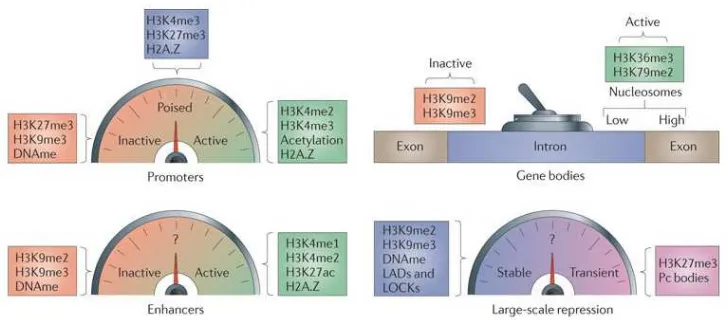
Fig. 2-2. Activated and repressed gene promoters and gene bodies by histone
modifications. Adapted by permission from Macmillan Publishers Ltd: Nature (Zhou et al. 2011)
copyright (2011). License no. 4050271026920
A. Active promoters often contain H3K4me3 and repressed promoters contain high levels of
H3K9me3 and H3K29me3.
B. Genes bodies vary in different histone marks depending on active and inactive regions. In
particular, exons of active genes often contain high levels of H3K36me3.
2.1.2 DNA modifications
The other key group of epigenetic modifications are DNA modifications. The main type of DNA
modification identified and studied till date is that of DNA methylation. DNA methylation involves
the addition of a methyl group to the fifth carbon atom of the cytosine residue which lies adjacent
to a guanine residue. This mode of methylation is an active process which is carried out by a
group of enzymes termed DNA methyl transferases (Dnmts). There are 5 known Dnmts – Dnmt1,
Dnmt3a, Dnmt3b, Dntm3c and Dnmt3L. Dnmt3a and Dnmt3b act as de novo methyltransferases
which initially adds the methyl group to unmethylated cytosine on both strands of DNA (Bird 1999).
Dnmt3L lacks the catalytic domain of other Dnmts but is important for establishing imprinting
control regions in gametes (Bourc'his et al. 2001; Bourc'his and Bestor 2004). Recently, a new
7
Dnmt3b (Barau et al. 2016). On the other hand, Dnmt1 is a maintenance methyl transferase
which adds the methyl group to the newly synthesized DNA strand wherein the other older strand
is already methylated (Bird 1999). Studies have indicated another molecule, Uhrf1 (Ubiquitin-like
with PHD and RING finger domains) to play a crucial role in DNA methylation. Uhrf1 binds
hemimethylated DNA (Arita et al. 2008) as well as Dnmt enzymes (Meilinger et al. 2009;
Berkyurek et al. 2014), thus maintaining DNA methylation. Another member of the Uhrf family,
Uhrf2, which has similar domains to Uhrf1 (Bronner et al. 2007) has also been implicated in DNA
methylation as it can bind hemimethylated DNA (Pichler et al. 2011).
DNA can be either actively or passively demethylated. Until recently, the only form of
demethylation was considered passive wherein lack of maintenance of DNA methylation in
dividing cells causes the daughter cells to loss the 5mC mark. However, recent evidence has
shown the presence of enzymes capable of converting the 5mC mark to 5-hydroxy methyl
cytosine (5hmC) (Tahiliani et al. 2009; Ito et al. 2010) (Figure 2-3). These enzymes are termed
as Ten-Eleven-Translocation (Tet) enzymes. The Tet family of enzymes has 3 members till date
– Tet1, Tet2 and Tet3. The Tet enzymes function by oxidizing 5mC to 5hmC as an initial
intermediate (Ito et al. 2011). The 5hmC mark can be further oxidized to 5-carboxyl cytosine
(5CaC) and 5-formyl cytosine (5fC) (Ito et al. 2011) (Figure 2-3). The 5fC and 5caC mark can be
reverted to an unmodified cytosine by Base Exchange Repair (BER) (He et al. 2011; Maiti and
Drohat 2011). Interestingly, a recent study has elucidated that Uhrf1 can bind the 5hmC mark with
8
Fig. 2-3. Epigenetic modifications of cytosine residue in DNA. Reprinted by permission from
Macmillan Publishers Ltd: Nature biotechnology (Song et al. 2012) copyright (2012). License no.
4056700245047
Cytosine can be modified from C to 5mC by methyltransferases such as Dnmt enzymes. 5mC
can be converted to 5hmC, 5fC and 5caC by the Tet enzymes. The oxidation products of 5fC and
5caC can be removed by TDG to generate an abasic site which is converted to a cytosine by BER
(Base Exchange Repair). 5hmC can be converted to 5hmU by AID/APOBEC and 5caC can be
converted to C by decorboxylation. The solid arrows indicate pathways which have been validated
whereas the dotted arrows need yet to be confirmed.
DNA methylation is a largely repressive mark wherein stretches of CpGs, identified as CpG
islands, are methylated and subsequently the corresponding genes are transcriptionally silent
(Deaton and Bird 2011). Methylated CpGs can be recognized by Methylated DNA binding domain
proteins (MBDs) such as MecP2 (Fraga et al. 2003). Of the 28 million CpGs in the human genome,
about 60-80% are methylated (Smith and Meissner 2013), including CpG islands and other
regions. DNA methylation is extremely widespread and can occur both in promoters and gene
9
the genome are static across development and into adulthood and across somatic tissues. From
the 60-80% of CpG methylation (among all CpGs) only about 10% occurs at CpG islands which
are mainly at the transcription start sites of either housekeeping genes or developmentally
regulated genes (Deaton and Bird 2011). Mechanisms by which DNA methylation prevents
transcriptional activation have been studied widely and are largely attributed to the activity of CpG
binding proteins and histone deacytelases and histone methylases. There are some hypotheses
that DNA methylation in promoters can prevent access for transcription factors due to the binding
of MBDs (Bird 1992). This additional layer of epigenetic complexity allows certain genes to be
expressed in specific tissues and not others.
Compared to 5mC, the other DNA marks are much lower in abundance in the genome. Whole
genome mapping of the mouse genome revealed the 5hmC is present in about 0.1% of all
cytosine in mouse embryonic stem cells (ESC) (Yu et al. 2012). However, the highest abundance
of 5hmC has been described to be in the mouse brain, about 0.4-0.7% of all cytosines (Szulwach
et al. 2011; Song et al. 2011). In comparison, 5caC and 5fC are extremely rare with only about
20ppm (parts per million) of all cytosines and 3 ppm of all cytosines respectively in mouse ESCs
(Ito et al. 2011). Studies are still early in understanding the stability of these new DNA marks and
their functions. However, there is indication that the 5hmC mark can correlate with transcriptional
activation (Wu et al. 2011).
There are two instances during development wherein the global static nature of DNA methylation
is drastically erased. These are during early pre-implantation development and in the germ line.
During early pre-implantation development, the hypermethylated sperm undergoes rapid loss of
5mC (Santos et al. 2002). Interestingly, although majority of the genome is demethylated, certain
transposable elements called Intracisternal A-Particles (IAP) are kept methylated (Lane et al.
2003)
The other instance wherein DNA methylation is massively erased is in primordial germ cells at
about embryonic day E10-11. Genome-wide studies indicate that also in these cells, although
methylation is almost completely erased, on IAPs and some other retroviral elements, methylation
10
2.1.3 Role of Dnmts and Tets in ESCs, early development and NSCs
To understand the importance of epigenetic regulators in stem cells and neurogenesis, most
studies have employed loss-of-function studies. Both Dnmt1 and Uhrf1 are highly expressed by
mouse ESCs (Bostick et al. 2007; Sharif et al. 2007). Dnmt3a is predominantly expressed in
oocytes and early preimplantation embryos where it establishes DNA methylation at imprinting
control regions in gametes (Kaneda et al. 2004; Kato et al. 2007). Dnmt3b is strongly expressed
at the blastocyst stage (Watanabe et al. 2002).
Complete knockout of Dnmt1 enzyme is embryonically lethal due to a massive loss of methylation
(Li et al. 1992). Interestingly, loss of Uhrf1 causes a similar phenotype (Sharif et al. 2007). Deletion
of Dnmt3b also causes embryonic lethality (Okano et al 1999). However, knockout of Dnmt3a is
partially viable (Okano et al. 1999). Dnmt3L knockout mice are also partially viable (Bourc'his et
al. 2001; Bourc'his and Bestor 2004)
In contrast to the Dnmts and Uhrf1, the Tet enzymes are more dispensable in terms of embryonic
lethality. Tet1 single knockout and Tet1/Tet2 double knockouts are viable with a slight
developmental delay (Dawlaty et al. 2011; Dawlaty et al. 2013). Triple knockout of Tet1/Tet2/Tet3
led to a significant loss of 5hmC and defects in ESC differentiation (Dawlaty et al. 2014).
Conditional knockout of Dnmt enzymes have been generated and studied in neurogenic niches.
Dnmt1 is expressed by cortical radial glia, adult neurons and adult neural stem cells (Hutnick et
al. 2009; Feng et al. 2010; Noguchi et al. 2015). Deletion of Dnmt1 in the dorsal telencephalon at
early embryonic stages resulted in severe neuronal death with an activation of the retroviral
elements, IAPs (Fan et al. 2001; Hutnick et al. 2009). Interestingly, deletion of Dnmt1 in
hippocampal neurons did not affect their numbers but synaptic plasticity was affected (Feng et al.
2010). Loss of Dnmt1 in aNSCs of the hippocampus affected the production of new neurons
(Noguchi et al. 2015).
Dnmt3a is expressed by NSCs derived from ESCs and loss of function studies indicated defects
in both proliferation and differentiation (Wu et al. 2010). Dnmt3b function in NSCs was studied by
using Dnmt3b knockout ESCs and differentiating them into early neuroepithelial stages. In this
study, Dnmt3b was found to be important for early neuroepithelial specification (Martins-Taylor et
al. 2012). A comprehensive study of Dnmt3a expression and function was performed in postnatal
neural stem cells (Wu et al. 2010). Dnmt3a was found to be highly expressed by postnatal NSCs
11
Dnmt3a was found to occupy and methylate intergenic regions and gene bodies of several
neurogenic genes (Wu et al. 2010).
Besides Dnmts, many studies have investigated the role of proteins binding methylated DNA
(MBDs) in the neurogenic niches. MBD1 knockout mice showed impaired neurogenesis in the
adult (Zhao et al. 2003; Li et al. 2008). Another heavily studied protein is MeCP2 which is
implicated in Rett’s syndrome. MeCP2 is most expressed in neurons and MeCP2 knockout mice
exhibit defects in neuronal maturation but not in their numbers (Smrt et al. 2007).
As 5hmC is most abundant in the brain, many studies have investigated the role of 5hmC and Tet
proteins in neurogenesis. In the embryonic cortex, 5hmC is abundant in neurons and accumulates
in gene bodies of neuronal genes (Hahn et al. 2013). Tet1 has very low expression in the
embryonic cortex, with Tet2 and Tet3 being highly expressed by progenitors and neurons (Hahn
et al. 2013). Acute knockdown of Tet2 and Tet3 in cortical radial glia resulted in accumulation of
cell clusters in the cortex, suggesting aberrant neuronal migration (Hahn et al. 2013). Tet1
knockout mice exhibited impaired hippocampal neurogenesis with some genes being
hypermethylated
In summary, loss of function studies of Dnmt and Tet protein suggest that these proteins play key
cell-type and niche specific roles in regulating transcription and cell fate.
2.1.4 Uhrf1 and Uhrf2
Besides Dnmts and Tet enzymes, another protein has been described to play a key role in
maintaining DNA methylation, Uhrf1. Uhrf1 is a multi-functional protein with several domains. As
mentioned previously, Uhrf1 can bind hemi-methylated DNA with its SRA domain. Uhrf1
possesses a RING finger domain, linked to histone and protein ubiquitination and turnover
(Citterio et al. 2004; Liu et al. 2013; Nishiyama et al. 2013; Qin et al. 2015). Furthermore, Uhrf1
has also been described to establish and reorganize heterochromatin (Papait et al. 2008; De Vos
et al. 2014). Uhrf1 also possesses a tandem tudor domain (TTD) which can bind the H3K9me3
mark (Karagianni et al. 2008). Besides Dnmts, Uhrf1 can also interact with histone modifiying
enzymes such as HDAC1, Tip60 and G9a (Unoki et al. 2004; Achour et al. 2009; Kim et al. 2009)
(Figure 2-4). These data suggest Uhrf1 to be an important link between DNA and histone
methylation. Indeed, deletion of the RING finger domain which binds methylated histones, can
12
Fig.2-4 The different domains of Uhrf1 and its interaction partners. Modified from (Alhosin et
al. 2011) (Open Access article distributed under the terms of the Creative Commons Attribution
License)
Schematic of the domains of Uhrf1 – SRA interacting with DNA and Dnmt1, RING, TTD and PHD
domain which binds histones. Uhrf1 can interact with the proteins HDAC1, G9a and Tip60.
Besides ESCs, Uhrf1 is expressed in skin stem cells during proliferation and downregulated upon
differentiation (Sharif et al. 2007; Sen et al. 2010). It has also been described to play a crucial role
in regulatory T cell differentiation, whereby loss of Uhrf1 in these cells led to defects in proliferation
and differentiation (Obata et al. 2014). Many studies have implicated a role for Uhrf1 in cancer
formation and progression (Hervouet et al. 2010; Tien et al. 2011; Babbio et al. 2012; Pacaud et
al. 2014). Moreover, Uhrf1 has been shown to negatively regulate the Pml protein in human
endothelial cells (Guan et al. 2013). Uhrf1 has been recently been shown to be important in
maintaining the balance between self-renewal and differentiation of hematopoietic stem cells by
regulating DNA methylation of key stem cell genes (Zhao et al. 2016).
Uhrf2 is also expressed by mouse ESCs (Pichler et al. 2011). However, in contrast to Uhrf1, Uhrf2
13
to exhibit contrasting modes of expression. Genetic knockouts of Uhrf2 has not been studied yet
and thus its exact function is largely unknown.
Although many studies have examined the role of Dnmts and Tet enzymes in neurogenesis, the
function of Uhrf1 has not been examined in NSCs.
2.2 Transposable elements
One group of genomic loci wherein DNA methylation has been described to play a key role in
regulating their epigenomic landscape and transcription, is that of ERVs. ERVs form a class of
transposable elements (TEs) which are sequences of DNA that can jump around the genome.
However, ERVs form a part of the retrotransposon family which can multiply prior to transposition
(Sharif et al. 2013). Nearly half of the genome is comprised of TEs and ERVs are about 5% of the
mouse and human genome (Kassiotis 2014) (Figure 2-5). Other types of retrotransposons are
more abundant such as LINE-1 elements (19% of genome) and SINE elements (8% of genome)
(Kassiotis 2014). ERVs have arisen from germline integration of retroviruses which have infected
mammals through evolution (Dewannieux and Heidmann 2013). These ERVs largely resemble
retroviruses, comprising of gag, pro, pol and env genes flanked by long-terminal repeats or LTRs.
The LTR region often act as promoters. There are many different types of ERVs in both the mouse
and human genome. The mouse genome has a specific ERV known as IAP (Figure 2-5) which is
present as about 1000 copies in the genome (Barbot et al. 2002). The IAP elements themselves
comprise of several subtypes, largely based on their structure, as some of them are heavily
14
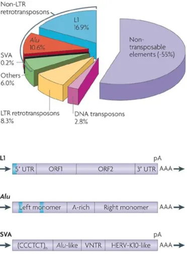
Fig. 2-5 Transposable elements in the genome. Reprinted by permission from Macmillan
Publishers Ltd: Nature Reviews Genetics (Cordaux and Batzer 2009) copyright (2009). License
no. 4056710870507
Schematic showing the percentage of different types of transposable elements in the genome.
The sequence of LINE-1 elements with its open reading frames (orf) and the sequences of ERVs
is depicted. Note that IAP elements form a part of ERVs which contain gag, pro, pol and env
genes with flanked LTRs.
During development, in preimplantation blastocysts and in primordial germ cells, there is a
genome- wide erasure of 5mC which occurs (Messerschmidt et al. 2014). During this time, many
regions in the genome have been shown to resist the loss of 5mC, such as the IAP subclass of
ERVs (Guibert et al. 2012; Hackett and Surani 2013). However, interestingly, loss of 5mC in
15
depending on the cell-type and developmental time, IAP transcription can be under the control of
levels of DNA methylation.
The impact of retrotransposons on mammalian cells has been described to be both beneficial and
harmful. Studies have suggested that LINE-1 elements can contribute to the evolution of complex
brains and the generation of neuronal diversity (Muotri et al. 2005; Singer et al. 2010) . However,
there are also multiple reports which have linked overexpression of retrotransposons to cell death,
genome instability, stress response and cancer (Morrish et al. 2002; Belgnaoui et al. 2006;
Sinibaldi-Vallebona et al. 2006; Pasquarella et al. 2016).
Regulation of retrotransposons in neural development is still in its infancy and data from the
LINE-1 studies suggest that it may be of importance in this niche.
2.3 Embryonic forebrain
2.3.1 Development of the embryonic forebrain
Neurogenesis is a dynamic process which requires tight transcriptional control in a spatial and
temporal manner. The main events occurring during neurogenesis are neural stem and progenitor
proliferation and neuronal differentiation and maturation. Each of these steps are regulated by
several key coding genes and non-coding elements in the genome.
Neurogenesis is widespread during embryonic development, occurring in several regions of the
brain. In the embryonic cerebral cortex, neurogenesis commences at about E10. Prior to this
stages, the developing cortex is derived from the neural tube. The neural tube is initially divided
into the prosencephalon (which forms the telencephalon or anterior embryonic forebrain and
diencephalon or posterior forebrain), the mesencephalon (midbrain), the rombhencephalon or
hindbrain (develops into pons, cerebellum and medulla oblongata) and the spinal cord.
At about E8.5, the telencephalic primordium is specified by the induction of the transcription factor
Foxg1 (Tao and Lai 1992; Shimamura and Rubenstein 1997). Following this, the telencephalon
is subdivided into two specific domains - the ventral and dorsal telencephalon. The specific
domains of the telencephalon are established by signaling gradients which control the expression
of specific transcription factors in each domain. The ventral telencephalon is further subdivided
16
develops in the hippocampus, cortical hem and the choroid plexus. The ventral telencephalon is
also divided into the medial region termed the medial ganglionic eminence (MGE) and the
posterior lateral regions are termed lateral ganglionic eminence (LGE) and caudal ganglionic
eminence (CGE).
The telencephalon is composed of the lateral ventricles which are lined by germinal zones. The
ventricle itself is composed of cerebral-spinal fluid produced by the choroid plexus (a network of
ependymal cells).
2.3.2 Overview of embryonic neurogenesis
At about E9-E10, the process of neurogenesis commences in the telencephalon. Neurogenesis
is the process by which neurons are generated from neural stem and progenitor cells. The main
difference between the dorsal and ventral region of the telencephalon lies in the type of neurons
they produce. The dorsal telencephalon primarily produces glutamatergic neurons whereas the
ventral telencephalon generates mainly GABAergic neurons. Neurons generated in the neocortex
migrate radially and remain in the neocortical domain. However, neurons generated in the
ganglionic eminences can migrate tangentially to different regions (Figure 2-6A). The LGE derived
neurons migrate primarily to the olfactory bulb and amygdala. The MGE and CGE derived
neurons migrate tangentially to the neocortex and integrate into the existing neuronal network
(Marin and Rubenstein 2001) (Figure 2-6A). The different regions of the telencephalon and their
molecular markers are depicted in Figure 2-6B. These molecular markers are specific
transcription factors which instruct certain cell fates in a spatial and temporal manner. The sharp
border between the dorsal and ventral telencephalon is primarily setup by the transcription factors
Pax6 and Gsx2 (Figure 2-6B). In fact, in Pax6 knockout embryos, the ventral part of the neocortex
become specified as the dorsal LGE and in the Gsh2 knockout embryos, the dorsal LGE is
specified into the ventral cortex (Corbin et al. 2000; Stoykova et al. 2000; Toresson et al. 2000;
Yun et al. 2001). In the mouse telencephalon, neurogenesis occurs until about E18 with the
process peaking at E14. Following neurogenesis, the generation of glia, a process known as
gliogenesis, occurs from the end of development and into postnatal stages.
Much of our understanding of neurogenesis in the telencephalon is derived from studies in the
neocortex, with the ganglionic eminence exhibiting some differing and some common features
17
Fig.2-6 Regions of the telencephalon
Adapted by permission from Macmillan Publishers Ltd: Neuron (Ross et al. 2003) copyright
(2003). License no. 4050741125950. (A) and (Nat et al. 2013) (open access under CC BY 3.0 license) (B)
A. Coronal section of the telencephalon depicting the dorsal and ventral regions and their
respective types of neurons.
B. Coronal section of the telencephalon depicting the specific transcription factors in each domain.
Emx1/2 and Pax6 are dorsally expressed. Nkx2.1 is specific to the MGE and Gsh2 is in the LGE
and MGE.
2.3.3 Cell types in the embryonic neocortex
The neuroepithelium of the telencephalon at the onset of neurogenesis is composed of
neuroepithelial cells termed radial glia. These cells act as neural stem cells - capable of
self-renewal and multipotency, generating both neurons and glia. Radial glia are situated in the
ventricular zone (VZ) and maintain contact to the inner ventricular surface and the outer pial
surface via apical and basal process. However, their cell body is stationed in the ventricular zone,
18
E11, the radial glia mainly undergo symmetric self-renewal cell divisions in order to expand their
pool (Gotz and Huttner 2005). Following this time, they start generating a population of
transit-amplifying progenitors termed basal or intermediate progenitors via asymmetric divisions
(Haubensak et al. 2004; Noctor et al. 2004; Noctor et al. 2007). These basal progenitors do not
exhibit apical-basal polarity and are multipolar in nature. In addition to apically located radial glia,
another type of cell termed as subapical progenitors has been recently reported (Pilz et al. 2013).
Moreover, another type of progenitor has been identified termed as basal radial glia (Reillo et al.
2011; Wang et al. 2011; Florio and Huttner 2014) which are cells harboring radial glia features
but with only a basal process in contact with the pial surface (Figure 2-7). Moreover, they are
situated more basally outside the ventricular zone.
Generation of basal progenitors leads to the appearance of an additional zone in the neocortex
(Figure 2-7) termed as the subventricular zone (SVZ) wherein most of these progenitors reside.
Basal progenitors mainly undergo symmetric divisions to either self-renew or generate neurons
(Haubensak et al. 2004; Miyata et al. 2004; Noctor et al. 2004; Noctor et al. 2007). Radial glia and
basal progenitors can be differentiated by the expression of certain transcription factors. Radial
glia express Pax6 whereas basal progenitors express Tbr2. Basal progenitors can also self-renew
and undergo terminal divisions to generate neurons at different periods of neurogenesis. These
19 Fig.2-7 Cell types in the embryonic cortex. Modified from (Gomez-Lopez et al. 2014) (Open
Access article distributed under the terms of the Creative Commons Attribution License)
At early neuroepithelial stages, the neuroepithelial cells divide to generate radial glia (purple cells
with process). The radial glia divide both symmetrically to self-renew and asymmetrically to
generate basal progenitors and neurons. Radial glia can generate neurons both by direct
neurogenesis (first purple radial glia generating neurons, depicted in orange), as well as by
indirect neurogenesis (via basal progenitors depicted in green). Neurons migrate into the cortical
plate and reside per their birthdate and layer. The mammalian cortex also contains basal or outer
radial glia (purple cells with only basal process) and subapical radial glia (not shown).
LV – lateral ventricle; VZ – ventricular zone; SVZ – subventricular zone; RG – radial glia; BP –
basal progenitor; oRG – outer radial glia
2.3.4 Cortical layering and postnatal terminal neuron differentiation
Newly generated neurons in the neocortex migrate radially to form the 6-layered cortex in an
inside-out manner wherein the early born neurons are situated close to the ventricle and the late
born neurons are close to the pial surface (Figure 2-8). The first-born neurons, termed
20
generated following these neurons at about E13-E14 and express the transcription factor Tbr1.
Following Layer VI, layer V neurons are produced which are mainly labelled by Ctip2. Layer IV
neurons are labelled by Rorb/Satb2, layer II/III neurons by Cux1.
During the first few weeks of postnatal development, the neurons in the cerebral cortex undergo
terminal neuronal differentiation. Most of the neurons born in the cerebral cortex are projection
neurons. However, it is important to note here that GABAergic neurons migrate into the cortex
from the GE. Projection neurons can either form connections with neurons of the contralateral
hemisphere via the callosum (cortico-cortico projection neurons) or anterior commissure (Greig
et al. 2013) or form corticofugal projections which project away from the cortex. The corticofugal
connections are either thalamocortical (to the thalamus) or other subcerebral projections (Greig
et al. 2013).
Once the projection neurons in the embryonic cortex migrate to the correct layer, they initiate
axon and dendritic growth. These processes are essential to further build synapses which enable
specific connections between neurons. In early postnatal stages, between P1 to P8, there is
additional axon growth and guidance (Lewis et al. 2013). From P8 to P21 axon branching and
synapse formation occurs (Lewis et al. 2013). Defects in any of these processes can lead to
neurodevelopmental disorders such as autism spectrum disorders, epilepsy and mental
retardation (Zoghbi and Bear 2012).
Another important process which occurs in postnatal neurons is the downregulation of cell cycle
proteins. Postmitotic neurons still express members of the Retinoblastoma family of proteins, E2F
transcription factors, Cyclin-dependent kinases and inhibitors after terminal neuron differentiation
(Yoshikawa 2000). It has been hypothesized that these cell cycle proteins are relevant for neuron
survival and maintenance of differentiation (Cicero and Herrup 2005; Liu et al. 2005; Kurita et al.
2006; Nguyen et al. 2006; Hasegawa and Yoshikawa 2008). Regulation of these cell-cycle
proteins seems critical as misregulation could lead to neuronal death in adult neurons (Herrup
21
Figure 2-8 Chronological generation of different types of cortical neurons in an inside-out
manner. Adapted by permission from Macmillan Publishers Ltd: Nature reviews neuroscience
(Molyneaux et al. 2007) copyright (2007). License no. 4052120920271.
The early born neurons form the preplate which later splits into the superficial marginal zone and
deep subplate. The different layers of the cortical plate are formed between these two layers
22
2.4 Adult neurogenesis and gliogenesis
2.4.1 Adult neurogenic niches and cell types
In the postnatal and adult brain, neurogenesis occurs only in certain specific niches, namely the
subependymal zone (SEZ) of the lateral ventricle and the dentate gyrus (DG) of the hippocampus
(Kriegstein and Alvarez-Buylla 2009) (Figure 2-9A). The early evidence for these niches were
from first from neurosphere experiments showing the ability of aNSCs to self-renew and be
multipotent (Reynolds and Weiss 1992). Following this, transplantation experiments were
performed wherein SEZ cells were grafted either homotypically (i.e. SEZ from one mouse into
SEZ of another mouse) (Lois and Alvarez-Buylla 1994) or heterotypically (SEZ of one mouse into
other regions of the brain of another mouse) into the striatum wherein neurogenesis does not
occur (Herrera et al. 1999). In the homotypic situation, the grafted cells could integrate and
differentiate, but not in the heterotypic condition
The source of adult neural stem cells was previously shown to be radial glia (Merkle et al. 2004).
Moreover, recent studies have indicated that LGE progenitors could be ancestors of adult neural
stem cells (Furutachi et al. 2013). Adult neural stem cells (aNSCs) exhibit many radial glial
features, sharing common molecular markers. Adult neural stem cells in the SEZ are relatively
quiescent but can divide to generate actively proliferating transit-amplifying progenitors (TAP)
(Doetsch et al. 1999). These TAPs generate immature neuroblasts which migrate tangentially to
the olfactory bulb from the SEZ and differentiate into interneurons (Lois and Alvarez-Buylla 1994;
Belluzzi et al. 2003; Carleton et al. 2003), which integrate into existing circuitry (Figure 2-9B).
In the dentate gyrus, aNSCs also possess radial glial properties (Seri et al. 2001; Fukuda et al.
2003; Garcia et al. 2004). These aNSCs generate intermediate progenitor cells which in turn give
rise to neurons which migrate radially and reside in the hippocampus (Seri et al. 2004) (Figure
2-9C). Neurons in the dentate gyrus are glutamatergic projection neurons and play important roles
23
Fig.2-9. Adult neurogenic niches.
Adapted by permission, (Ninkovic and Gotz 2013) License no. 4050750571282 (A)and adapted
by permission from Macmillan Publishers Ltd: Nature reviews neuroscience (Yao et al. 2016)
copyright (2016). License no. 4050750893071(B,C)
A. Scheme showing tranverse section of the adult brain. The regions in red indicate the
neurogenic niches in the SEZ and DG of the hipoocampus. The scheme shows the restriction of
neurogenesis to these regions as compared the widespread gliogenesis (green dots are cells)
occurring throughout the parenchyma.
B. Scheme showing the cell types and the process of neurogenesis in the SEZ. Radial glia like
NSCs get activated (blue cells, left group of cells) and generate IPCs or TAPs (purple cells, middle
group of cells) which in turn generate neuroblasts (green cells, right panel). The neuroblasts
24
C. Scheme of the cell types and the process of neurogenesis in the SGZ. Radial glia like NSCs
get activated (blue cells) and generate IPCs or TAPs (purple cells) which in turn generate
neuroblasts (green cells). The neuroblasts mature into neurons which migrate radially and remain
in the hippocampus. The NSCs also generate parenchymal astrocytes (in red).
2.4.2 Gliogenesis
As mentioned earlier, neurogenesis subsides towards the end of embryonic development,
allowing gliogenesis to commence. Glia are mainly generated either by precursor cells or by
terminal differentiation of radial glia into differentiated glia. There are two main types of glial cells
– astrocytes and oligodendrocytes. Astrocytes are mainly generated from radial glia, possibly as
a direct conversion (Noctor et al. 2008; Kriegstein and Alvarez-Buylla 2009) (Figure 2-10A).
Astrocytes can range from about 20-40% of all glia and perform several functions from providing
biochemical support to endothelial cells of the blood-brain barrier, acting as a source of nutrients
and other extracellular factors to neurons and playing roles in brain injury and repair. Most
astrocytes have a characteristic morphology of short, highly branched tertiary processes.
The other type of glia, namely oligodendrocytes, are born at early embryonic stages as well as
later stages. Oligodendrocytes provide trophic support to neurons (Bradl et al. 2010).
Furthermore, they produce myelin sheaths which are important for nerve impulses and reducing
ion leakage. There are three known waves of oligodendrocyte production in the forebrain. The
first wave occurs at E16 wherein progenitors in the MGE generate a wave of oligodendrocytes
which migrate towards the cortex (Pringle and Richardson 1993; Spassky et al. 1998;
Tekki-Kessaris et al. 2001). Following this wave, another group of oligodendrocytes appear from the
LGE (Kessaris et al. 2006). Finally, at E18, a third wave of oligodendrocytes are locally born from
cortical progenitors (Kessaris et al. 2006) (Figure 2-10B). It is interesting to note that at postnatal
stages, most of the early born oligodendrocytes disappear and only the late born oligodendrocytes
remain in the cerebral cortex (Kessaris et al. 2006).
Some studies have indicated that a subpopulation of OPCs are also generated postnatally
(Levison and Goldman 1993; Nait-Oumesmar et al. 1999; Menn et al. 2006; Aguirre et al. 2007).
25
Fig.2-10. Gliogenesis in the embryonic forebrain
Reprinted with permission from Elsevier (Barry et al. 2014) License no. 4050751151722 (A) and
adapted by permission from Macmillan Publishers Ltd: Nature reviews neuroscience
(Richardson et al. 2006) copyright (2006) License no. 4057890885482 (B).
A. Schematic showing the generation of astrocytes and oligodendrocytes from embryonic radial
glia and adult neural stem cells, from radial glia at the end of neurogenesis.
B. Schematic of a coronal section of the embryonic brain indicating the modes of OPC generation.
OPC1 (Red arrows, 1) indicates the first wave at E16 from the MGE. OPC2 (blue arrows, 2) is
the second wave following the first wave from the LGE and MGE and the third wave (green
26
3. Aims of my thesis
Conditional deletion of Uhrf1 in the dorsal telencephalon (using Emx1-Cre) (Cappello et al. 2006;
Iwasato et al. 2000) led to decreased cerebral cortical thickness in 1-month old mice (Bayam.
2014). Moreover, loss of Uhrf1 in adult neurogenic niches led to defects in neuronal differentiation
(Bayam. 2014). These data have implicated an important role for Uhrf1 in regulating appropriate
cerebral cortex neurons numbers and adult neurogenesis. Defects in progenitor proliferation were
not observed at embryonic stages (analysed by pH3 and Pax6). However, an increase in cell
death was observed with TUNEL mid embryonic stages. In contrast, conditional deletion of Uhrf1
in the adult SEZ and dentate gyrus (using GlastCre ERT2; Uhrf1 flox; CAG) (Mori et al. 2006)
resulted in a decrease in transit-amplifying progenitors (TAPs) and neuroblasts, indicating
differing roles for Uhrf1 in embryonic and adult neurogenesis.
Based on these findings, the main of my project was in unravelling the molecular mechanism of
Uhrf1 in neural stem cells. Additionally, we made some preliminary attempts to test if these
mechanisms could act during adulthood as well. In particular, we first studied the cellular
phenotypes observed during conditional loss of Uhrf1 in neural stem cells of the developing
cerebral cortex. Following this, we dissected the molecular role of Uhrf1 in these cells, analyzing
transcriptional and epigenetic processes. Finally, we examined some of the phenotypes observed
in the conditional knockout of Uhrf1 in adult neurogenic niches, with emphasis on its molecular
27 4. Results
4.1 Uhrf1 expression from embryonic to adult stages
To characterize Uhrf1 expression, we performed Uhrf1 qRT-PCR in E14 NSCs (isolated by FACS
with Prominin 1 antibody), E14 neurons (isolated by FACS by PSA-NCAM antibody), adult
subependymal zone and adult grey and white cortical matter (Figure 4-1A). Indeed, we could
observe about 40-fold higher Uhrf1 mRNA in E14 NSCs as compared to neurons. Moreover,
Uhrf1 mRNA was barely detectable in adult cortical grey matter (Figure 4-1A). We also performed
Uhrf1 immunostainings at postnatal day 5 (P5). We observed only few cells in the cortex to
express Uhrf1 (Figure 4-1B). These cells did not colocalize with NeuN, a neuronal marker (Figure
4-1C), however, some did co-label with Ki67, indicating some of the Uhrf1+ve cells are
proliferating (Figure 4-1D). Thus, our data in conjunction with the previous study, shows that Uhrf1
is primarily expressed in NSCs and progenitors and is largely absent in neurons of the embryo
28
Fig.4-1 Uhrf1 expression in embryonic and adult neurogenic niches
A. qRT-PCR for Uhrf1 showing high expression in NSCs and almost absence in neurons. E14
cortical tissue was dissociated to isolate NSCs by Prominin 1 and neurons by PSA-NCAM by
FACS. Grey and white matter and SEZ tissue was isolated from 2-month old mice. qRT-PCR was
performed for Uhrf1 in the above populations using Gapdh as the housekeeping gene. *
indicates significance with p-values 0.009 and 0.00005 respectively. Error bars indicate standard
error of mean. n=3.
B-D. Confocal images of coronal sections of P5 cerebral cortex immunostained for Uhrf1 (B),
Uhrf1 and NeuN (C) and Uhrf1 and Ki67 (D) showing the absence of Uhrf1 in neurons. Scale bar
29 4.2 Characterization of Uhrf1 cKO cellular phenotype
4.2.1 Comparable numbers of neurons and glia at E16
A conditional knockout of Uhrf1 was generated specifically in the developing cerebral cortex using
the Emx1-Cre mouse line (Iwasato et al. 2000; Ramesh et al. 2016) which recombines specifically
in the dorsal telencephalon, deleting Uhrf1 in this region. Emx1cre+/- Uhrf1f/f mice (hereby referred
to as cKO) is compared to Emx1cre+/- Uhrf1f/- mice (hereby referred to as controls). We performed
Uhrf1 immunostaining at E12 and could observe a loss of Uhrf1 protein in the cKO (Figure
4-2A,B), specifically in the dorsal telencephalon and not in the LGE where Uhrf1 was not deleted
(Figure 4-2B). We further investigated the cellular phenotype by assessing proliferation of
progenitors and numbers of neurons at embryonic stages. We assessed proliferation with Ki67
immunostaining at E16 in cKO cortices and controls. We could not observe a significant difference
in the cKO compared to the control (Figure 4-2C,D). However, we did detect an increase in
non-apical Ki67 positive cells in the cKO. We performed Tuj1 (to label all neurons) and Tbr1 (label
neurons in the cortical plate) immunostaining at E16 and did not detect any evidence of premature
differentiation (Figure 4-2E-H). Moreover, the numbers of Tbr1+ve neurons were unchanged
31 Fig. 4-2 Comparable proliferation and neuron numbers at E16 in the cKO
A-B. Confocal images of coronal sections at E12 of cerebral cortex of controls (A) and cKO (B)
immunostained for Uhrf1 (A,B). Note the loss of Uhrf1 in cKO in the cerebral cortex wherein it was
deleted as opposed to the LGE where Cre recombination did not occur (B).
C-H. Confocal images of coronal sections of the cerebral cortex at E16 of controls (C,E,G) and
cKO (D,F,H) immunostained for Ki67 (C,D), Tuj1 (E,F) and Tbr1 (G,H).
I. Quantifications of Tbr1+ve cells per area of sections from (G) and (H). Error bars indicate
standard error of mean. N=4.
Scale bar = 50µm. Abbreviations: CP – cortical plate; VZ – ventricular zone; SVZ – subventricular
zone
It has been previously reported that cell death can lead to activation of glial cells (Robel et al.
2011). Thus, we investigated if glial cells were altered in the Uhrf1 cKO compared to controls. We
performed immunostaining for Gfap at E16 and could not observe any activation in the cKO
(Figure 4-3A,B). Furthermore, microglial cells, labelled by Iba1 immunostaining at E16, appeared
comparable in number and morphology in controls and cKO (Figure 4-3C,D). However, in 1 month
old mice we did observe Gfap activation in the cKO, which is probably a result of reactive gliosis
due to neuronal death (Figure 4-3E,F).
These results suggest that neurons and glia are generated in an appropriate manner in the cKO
32
Fig.4-3 Absence of glial cell activation in E16 cKO cortices with reactive gliosis at later
stages
A-D. Confocal images of coronal sections of the cerebral cortex at E16 of controls (A,C) and cKO
(B,D) immunostained for Gfap (A,B) to label astrocytes and Iba1 (C,D) to label microglia. Note
the absence of Gfap staining in both cKO and controls and comparable Iba1 staining in cKO and
control.
E,F. Confocal images of coronal sections of the cerebral cortex at P27 of controls (E) and cKO
(F) immunostained for Gfap. Note the reactive gliosis at in 1-month old cKO cerebral cortex. Scale
33 4.2.2 Cortical layering at embryonic and postnatal stages appeared normal in the cKO
As generation of neurons was not impaired in the cKO, we next sought to determine if the neuronal
layers of the cortex were specified in the appropriate manner. We performed immunostaining for
Ctip2 to label upper layer neurons and Tbr1 to label lower layer neurons at E18 and could not
detect any abnormalities in the cKO as compared to the control (Figure 4-4A,B). Furthermore, we
also performed Ctip2 immunostaining at P2 and observed analogous staining in controls and cKO
(Figure 4-4C-D). These results suggest that neurons are generated and specified correctly into
the layers of the cerebral cortex of Uhrf1 cKO animals.
Fig.4-4 Upper layer and lower neuronal identity is maintained in Uhrf1 cKO cortices
A-D. Confocal images of coronal sections of cerebral cortex of controls (A,C) and cKO (B,D)
immunostained for Ctip2 and Tbr1 at E18 (A,B) and Ctip2 at P2 (C,D)
Note that the cortical layers in controls and cKO appear intact in all stages. Scale bar = 50µm.
34
4.2.3 Cell death peaks in the postnatal Uhrf1 cKO
As mentioned previously, conditional knockout of Uhrf1 in the cerebral cortex exhibited cell death
in embryonic stages. We sought to determine the onset of cell death by performing TUNEL
staining as early as E12 (Figure 4-5A,B). We observed significantly higher TUNEL positive cells
at E12, with about 4-fold higher in the cKO cortices (Figure 4-5C). Thus, the onset of cell death
was observed at E12, at similar levels to later embryonic stages (Ramesh,Bayam et al. 2016).
Fig.4-5 Onset of cell death was observed to be at E12 in cKO
A-C. Confocal images of coronal sections of cerebral cortex at E12 of controls (A) and cKO (B)
immunostained for TUNEL (A,B). Increase in TUNEL+ve cells was observed in the cKO
compared to control.
C. Quantifications of TUNEL+ve cells per area from A,B indicating 4-fold higher in the cKO. *
indicates significance with Mann-Whitney test with p-value = 0.03, n=4. Error bars indicate
35
Although cell death was observed from early embryonic stages, a reduction in cortical thickness
was not detected until P7 (Figure 4-6A,B). We sought to determine the peak of cell death, by
analyzing postnatal stages. We performed TUNEL analysis at P2 and P7 and observed up to
11-fold higher cell death in P2 cKO (Figure 6C,D,G) as opposed to 6-11-fold higher at P7 (Figure
4-6E,F,G). Thus, we could detect higher levels of TUNEL positive cells at early postnatal stages
leading to a discernible reduction in cortical tissue at P7.
In summary, deletion of Uhrf1 in embryonic neural stem cells showed no cell fate change or
changes in proliferation and differentiation. The significant phenotype was an increase in cell
37 Fig.4-6 Cell death peaks at postnatal day 2 in the cKO cortex
A,B. Confocal images of coronal sections of cerebral cortex of P7 control (A) and cKO (B) stained
with DAPI to indicate a reduction in cortical thickness in the cKO.
C,D,E,F. Confocal images of coronal section of cerebral cortex of controls (C,F) and cKO (D,G)
stained with the TUNEL kit at P2 (C,D) and P7 (F,G), indicating increased number of TUNEL+ve
cells in the cKO.
G. Quantifications of fold change of TUNEL+ve cells per area in P2 and P7 sections from C,D,E,F
showing a significant increase in the cKO. * indicates significance with Mann-Whitney test with
p-value = 0.01, n=4 for P2 and N=2 for P7. Error bars indicate standard error of mean. Scale bar =
50µm
4.3. Transcriptional profiling of Uhrf1 cKO
4.3.1 Activation of cellular stress genes at E16 and P5 Uhrf1 cKO
To understand the underlying molecular mechanisms contributing to the observed cell phenotype
in Uhrf1 cKO, we performed transcriptional profiling to uncover deregulated genes. Firstly, we
focused on the region wherein Uhrf1 is expressed and thus dissected the germinal zones (GZ) of
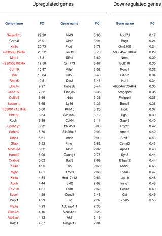
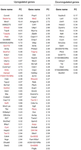
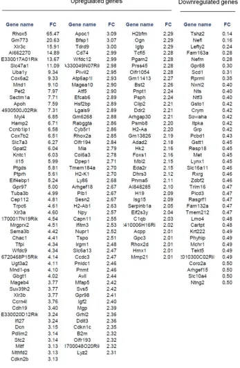
Uhrf1 cKO and controls and performed RNAseq (Figure 4-7A). We observed 68 upregulated
genes and 30 downregulated genes (Fold change > 2 and p-value < 0.05) (Figure 4-7B, Table 1).
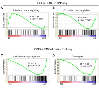
Of these, 52 genes were derepressed (Table 1, highlighted in red). We defined a derepressed