Fetal ECG Extraction from Maternal ECG
Using Extended Kalman Filter
Jeethu Krishna.T1, Lekha.N.M2 , Nisha Narayanan3
PG Student [Instrumentation & Control Systems], Dept. of EEE, JCET, Palakkad, Kerala, India
Project Manager, NeST, Eranamkulam, Kerala, India
Assistant professor, Dept. of EEE, JCET, Palakkad, Kerala, India
ABSTRACT: Congenital heart defects are among the most common birth defects and the leading cause of birth defect related deaths. Most cardiac defects have some manifestation in the morphology of cardiac electrical signals. This can recorded by electrocardiography and are believed to contain much more information as compared with other conventional sonographic methods. The Fetal Electrocardiogram (FECG) contains precise information about the fetal condition such as heart rate, waveform and dynamic behaviour which are convenient in determining the fetal life, development, maturity and existence of congenital diseases which could assist doctors in making more appropriate decisions during pregnancy. But fetal heart signals appear with undesirable disturbance. The FECG signal has to be separated from noisy environment in order to monitor both mother and child. In this research the objective is to develop a technique for extracting the fetal ECG from maternal ECG using Extended Kalman filter.
KEYWORDS: Fetal Heart defect, ECG,FECG, Fetal Monitoring, Extended Kalman Filter, Heart rate, R Peak.
I. INTRODUCTION
Congenital heart defects are heart problems present at birth. They happen when the heart does not develop normally before birth. These Heart defects are the leading cause of birth defect-related deaths. Every year, about one out of 125 babies are born with some form of congenital heart defects. The defect may be so slight that the baby appears healthy for many years after birth, or so severe that its life is in immediate danger. Heart defects originate in early stages of pregnancy when the heart is forming and they can affect any of the parts or functions of the heart. Doctors usually do not know the cause of congenital heart defects, but they do know of some conditions that increase a child's risk of being born with a heart defect such as inherited disorder, diabetes in mother, environmental factors such as infections or drug misuse. They can also occur due to specific fetal positioning that chokes the umbilical cord.
The regular monitoring of the fetal heart and the early detection of cardiac abnormalities can help obstetrics and pediatric cardiologist to prescribe proper medications in time. They can also consider the necessary precautions during delivery or after birth. Most cardiac defects have some manifestation in the morphology of cardiac electrical signals, which are recorded by electrocardiography and are believed to contain much more information as compared with conventional sonographic methods. However, due to the low SNR of fetal electrocardiogram (ECG) recorded from the maternal body surface, the application of fetal electrocardiography has been limited to heart-beat analysis and invasive ECG recordings during labor. The reason for low SNR is that the noninvasive fetal electrocardiogram (ECG) is contaminated by fetal brain activity, myographic signals from the mother and fetus, movement artifacts and multiple layers of different dielectric biological media through which the electrical signals must pass.
II. ELECTROCARDIOGRAM
performed for diagnostic or research purposes on human heart. The ECG records the electrical activity of the heart, where each heart beat is displayed as a series of electrical waves characterized by peaks and valleys. Any ECG gives two kinds of information. One, the duration of the electrical wave crossing the heart which in turn decides whether the electrical activity is normal or slow or irregular and the second is the amount of electrical activity passing through the heart muscle which enables to find whether the parts of the heart are too large or overworked.
The ECG signal measured from the patient results in a periodic waveform with multiple apexes called the PQRST-complex. In some cases we also use another peak called U. The performance of ECG analyzing system depends mainly on the accurate and reliable detection of the QRS complex, as well as T- and P waves. The apexes in the PQRST-complex are labeled with P, Q, R, S and T, which are commonly used in medical ECG terminology. Each apex results from ionic current exchanges in the heart causing muscle contractions and relaxations. All the muscle contractions and relaxations in the heart begin at the sinoatrial (SA) node, which is a specialized cell that regulates the heart beat. The SA node produces electrical impulses, which spread radially throughout the whole heart. As the electrical impulses traverse through the heart, different muscle groups in the heart contract in a sequential manner to produce the PQRST-complex waveform.The atria contraction produces the P wave and the ventricle contraction produces the QRS complex and the T wave.
Reliable and vital information about the condition of the fetus during pregnancy and labor is given by FECG, which is nothing but biomedical signal that gives electrical representation of Fetus heart beat from the recordings on the mother’s abdomen. The FECG signal is a comparatively weak signal and often embedded in AECG and noise. The FECG lies in the range from 1.3 to 3.5 Hz and sometimes it is possible for the mother and some of the FECG signals to be closely overlapping. The FECG monitoring enables accurate measurement of fetal cardiac performance including transient or permanent abnormalities of rhythm. For early stage diagnostic of fetus health and to know its status, sometimes the FECG is the only information source. The FECG is very much related to the mother ECG i.e., MECG, containing the same basic waveforms including the P wave, the QRS complex, and the T wave.
III. FETAL MONITORING
There are several fetal monitoring techniques to find the fetal heart rate. These are mainly classified as non invasive monitoring which is also known as external monitoring and invasive monitoring which is also known as internal monitoring. The external monitoring is the one which the done by placing the electrode on the abdomen surface region of a pregnant woman. In the internal monitoring the electrodes are placed inside the uterus of the mother on the scalp of the fetus during labour. The internal monitoring of fetal ECG is more accurate because of the recording electrode placed on the fetus scalp but, it can be done just during delivery. The external monitoring can be done during all gestation week and during delivery. But the external monitoring the electrodes are placed on the abdomen region of the pregnant woman, they record both maternal electrocardiogram (MECG) and the fetal electrocardiogram (FECG), also in this case it may contain a relatively large amount of noise.
In the external fetal monitoring means that the fetus heartbeat is detected by placing a small round ultrasound disc with ultrasound gel on the abdomen and held in place by a lightweight stretchable band or belt. A pair of belts is wrapped around the mother’s abdomen. In this one belt detects the fetal heartbeat and the other belt monitors the mother’s contractions.
In internal monitoring a small electrode is attached on the baby's scalp to monitor the baby's heartbeat directly. This electrode provides a constant report of the baby’s heartbeat. Uterine contractions can also be measured with an internal device. But the internal monitoring can be only done after the bag of water is broken.
IV. EXTENDED KALMAN FILTER
The Extended Kalman Filter was developed for non-linear discrete-time processes. , the purpose of the Extended Kalman Filter is to estimate unmeasured states As with the original Kalman , the Extended Kalman Filter uses a 2 step predictor-corrector algorithm. The first step involves projecting both the most recent state estimate and an estimate of the error covariance from the previous time period forwards in time to compute a predicted or a-priori estimate of the states at the current time. The second step involves correcting the predicted state estimate calculated in the first step by incorporating the most recent process measurement to generate an updated or a-posteriori state estimate.
For the Extended-Kalman Filter, mathematically, the predictor step is given by,
= ( , , ) (1)
= + (2)
And the corrector step is given by,
= ( + ) (3)
= + ( – (, , ) ) (4)
= (I- ) (5)
In the above equations Pk is an estimate of the covariance of the measurement error and Kk is called the Kalman gain.
After both the prediction and correction steps have been performed then is the current estimate of the states and can be calculated directly from it. Both and Pk are stored and used in the predictor step of the next time
period.
The Kalman filter and its variants such as the extended Kalman filter is one of the most celebrated and popular data fusion algorithms in the field of information processing. The most famous early use of the Kalman filter was in the Apollo navigation computer that took Neil Armstrong to the moon, and brought him back. Today, Kalman filters are at work in every satellite navigation device, every smart phone, and many computer games.
V.PROPOSED SYSTEM
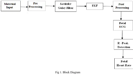
The figure 1 shown below is the Block Diagram of the proposed system. In this the input signal is taken as maternal input. This maternal input is the combination of maternal and fetal ECG. This signal is then given to the pre processing where some amount of denoising occurs. Then it is given to the Savitzky Golay Filter for smoothening. After the smoothening the signal is given to the Extended Kalman Filter for the extraction of the noisy fetal ECG. This ECG is given to the post processing. After the post processing we will get the recovered Fetal ECG. This fetal ECG is used to find the R-peak and the fetal heart rate.
1.Maternal Input
2.Pre-Processing
The fetal ECG signal is corrupted by different types of noise hence, noise reduction represents another important objective of ECG signal processing. . It should be performed only when the desired information remains undistorted.
Fig 1. Block Diagram
3.Savitzky Golay
Savitzky-Golay smoothing filters also called digital smoothing polynomial filters or least squares smoothing filters are typically used to smooth out a noisy signal whose frequency span is large. S-G filters does the job of smoothening the noisy data by performing a least square fitting of a frame of data to a polynomial of given degree.
4.Extended Kalman Filter
In this project we are taking the case of an abdominal signal which is the mixture of mECG and one fECG. In this raw recording ,the maternal ECG is mostly considered as the dominant ECG and fetal ECG is considered as the concurrent ECG and other noises as a unique Gaussian noise. Using this extended Kalman filter the maternal ECG is extracted from the raw recording. This extracted signal is subtracted from the original signal. After this we will get a signal which combination of the fetal ECG and the residual signal.
.
5.Post-Processing
In this project the post processing is done by using wavelet. Here this post processing is used to eliminate the noise from the signal which contains the noisy fetal ECG.
6.Fetal ECG
7.R-Peak Detection
R wave is one of the most important sections of ECG. It has an essential role in diagnosis of heart rhythm irregularities and also in determining heart rate variability (HRV).
8.Fetal Heart Rate
The normal fetal heart rate is between 120 to 160 beats per minute (BPM), but can vary. Fluctuations of the fetal heart rate usually associated with fetal movement during different periods of the day are common. Although in the healthy fetus the heart rate is usually regular, a beat-to-beat variation of approximately 5 to 15 beats per minute can be allowed.
VI. EXPERIMENTAL RESULT
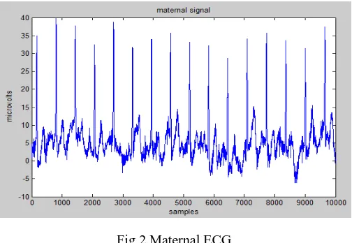
In the experimental result the figure 2 is the ECG of the mother abdomen. As we can see it consists of several noises. Due to this noise it is difficult to know the reliable information about the fetal development, any kind of distress etc.So our aim is to get the fetal ECG from this maternal abdominal ECG.
Fig 2.Maternal ECG
Figure 3 shows the output of preprocessing. In this the first plot shows the maternal signal with the denoised signal. The second one shows the denoised signal and the third plot shows the difference between the actual and the denoised signal.
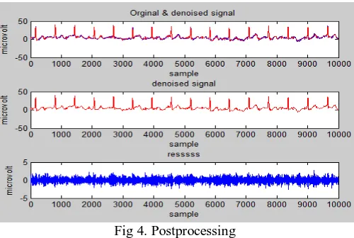
Figure 4 shows the post processing output. In this the second plot shows the signal after post processing.
Fig 4. Postprocessing
Figure 5 shows the fetal ECG with peak detection. By this R peak we can find the beats per minute of the fetal.
Fig 5.Fetal ECG with R Peak
VII. CONCLUSION
The fetal ECG provides a lot of necessary information to physicians without breaking up of the membrane which protects the child. It provides reliable information on fetal status and the detection of abnormalities which will help the doctors. The objective of this thesis has been to provide a method of FECG extraction using Extended Kalman Filter based on non invasive abdominal measurements. In this research work I have used three different abdominal ECG signal from PhysioNet database. But the problem with the abdominal signal is that along with fetal ECG it also consists of a maternal ECG and other noise. In this research work I have used Extended Kalman filter to extract the maternal ECG and wavelet method is used for the denoising. From the result we can see that using this algorithm most amount of noise is removed. This will be very helpful for the physicians for making proper precautions during the delivery and also helps to prescribe proper medications in time of pregnancy. In future we can make a real time noise removal.
REFERENCES
[1] S. Cuomo, G. De Pietro, R. Farina2, A. Galletti, and G. Sannino “A Novel O(n) Numerical Scheme for ECG Signal Denoising” ICCS 2015 International Conference On Computational Science, Procedia Computer Science, Volume 51, Pages 775–784, 2015
[2] Naveen Ku. Dewangan1, S. P. Shukla “A Survey on ECG Signal Feature Extraction and Analysis Techniques “International Journal Of
Innovative Research In Electrical, Electronics, Instrumentation And Control Engineering,Vol. 3, Issue 6, June 2015
[4] Mohammed AlMahamdy, H. Bryan Riley “Performance Study of Different Denoising Methods for ECG Signals “ The 4th International Conference on Current and Future Trends of Information and Communication Technologies in Healthcare (ICTH-2014),Procedia Computer Science 37, 325 – 332, 2014
[5] S.Subhashini , D.J J agannath , A.Immanuel Selvakumar “Extricating Non Invasive Fetal ECG by Adaptive Optimization Technique” 2014 International Conference On Electronics and Communication System ,lCECS- 2014
[6] Huawen Yan, Hongxing Liu, Xiaolin Huang, Ying Zhao, Junfeng Si and Tiebing Liu, “Invariant heart beat span versus variant heart beat intervals and its application to fetal ECG extraction” Yan et al. BioMedical Engineering OnLine ,2014
[7] Winnie Rachel Cherian, D.J.Jagannath, A.Immanuel Selvakumar, “Comparison of Algorithms for Fetal ECG Extraction “ International Journal
of Engineering Trends and Technology (IJETT) Volume 9 Number 11 - Mar 2014.