Diabetic Retinopathy and Detection of Optical
Disc in Fundus Image
Gayatri Madale, Prof. Rahul Mulajkar
M.E. Student, Dept. of Electronics and Telecommunications, JCOE, Pune, India Professor, Dept. of Electronics and Telecommunications, JCOE, Pune, India
ABSTRACT: now a day’s diabetic retinopathy (DR) has turned out to be not kidding disease among diabetic patients. It would recognize at the early stage else it will causes to aggregate visual impairment .The paper proposes to programmed ID of exudates pathologies in retinopathy fundus pictures a novel strategy is computational insight method. To concentrate components of fundus picture like blood vessels, optical nerve, red sores and white sores together with surface element examination utilizes the remarkable execution of morphological administrators.
KEYWORDS: diabetic retinopathy, green channel, red sores, white sores, optic nerve, exudates
I. INTRODUCTION
Diabetic retinopathy is the genuine disease in diabetic patients in this world. It would be distinguish early stage to prevention from visual deficiency. Diabetic retinopathy is also called diabetic eye illness, is when harm strikes the retina because of diabetes. It can in the long run prompt blindness. It influences up to 80 percent of individuals who have had diabetes for a long time or more. At minimum 90% of new cases could be decreased if there were appropriate treatment and checking of the eyes. The longer a man has diabetes, the higher his or her odds of creating diabetic retinopathy.
To recognize diabetic retinopathy (DR), check red sores, white sores, exudates, optical nerve, microaneurysms, hemorrhages. In the main stage which is called non-proliferative diabetic retinopathy (NPDR) there are no manifestations, the signs are not obvious to the eye and patients will have 20/20 vision. The best way to recognize NPDR is by fundus photography. In which microaneurysms (minute blood-filled lumps in the vein dividers) can be seen.
Accordingly consistent screening of diabetic retina is essential yet it is financially savvy and impractical for each patient. With a specific end goal to suit the screening and annual reviews imperative of an extensive number of patients, an automated screening device is a valuable extra in diabetes clinics. At present, there are a few strategies which can accurately analyse particular DR related injuries
II. LITERATURE SURVEY
Year 1996, M J Cree design a digital image processing system has been developed to quantify and monitor the pre-scene of microaneurysms in retinal fluoresce in angiograms. Year 2005, Meindert Niemeijer design a red lesion detection method is presented based on a hybrid approach, the first contribution is a new red lesion candidate detection system based on pixel Classification. Year 2015, Deepthi K Prasad this paper proposes the use of morphological operations and segmentation techniques for the detection of blood vessels, exudates and microaneurysms.
Year 2016, Sohini Roychowdhury this paper presents a novel generalized method that finds optimal image-based feature sets that reduce computational time complexity while maximizing overall Classification accuracy for detection
III. OVERALLAPPROACH
In this paper we propose to design automated detection of diabetic retinopathy using feature extraction from the fundus image. Here feature extraction is done using MATLAB. The extracted features would be blood vessels, exudates, optical nerve, microaneurysms.
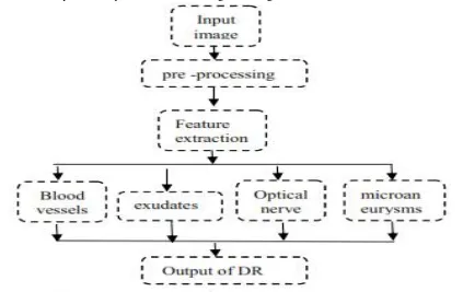
There are separate steps carried out to extract features from fundus images. Output of paper is depends on the success rate of each step. These steps are listed in following block diagram.
Figure 1. Block Diagram of Lesion Detection Algorithm
The calculation utilized for DR is successive execution of the morphological operations. Last outcome is the combination of each progression done for each element.
IV. PROPOSED METHOD
A. Image acquisition
The optical plan of fundus cameras depends on the rule of monocular circuitous ophthalmoscopy. A fundus camera gives an upright, amplified perspective of the fundus. A run of the mill camera sees 30 to 50° of retinal range, with an amplification of 2.5x, and permits some adjustment of this relationship through zoom or assistant focal points from 15°, which gives 5x amplification, to 140° with a wide edge focal point, which minifies the picture considerably
B. Pre-processing
(a) (b)
Figure.2 pre-processed image (a) original image (b) green channel image
C. Blood vessel detection
The input at this step is green plane image then applying the canny edge detection method which is use a multi-stage algorithm to detect a wide range of edges in image. On Detected edge now applying the morphological opening which is erosion followed by dilation. After subtracted eroded image from original image we extract border or boundaries of blood vessels.
Adaptive histogram equalization is carried out to improve contrast and correct uneven illumination. Also the thresholding is done and median filtering is used to remove salt and paper noise from image.
(a) (b) (c)
Figure.3 Blood vessel detection (a) input image (b) adaptive histogram equalization image (c) blood vessel extracted image
D. Exudates segmentation
Exudates are small yellow-white patches with sharp margins and different shapes. Exudates are one of the early occurring lesions. Morphological closing operation is perform Morphological closing operation is nothing but dilation followed by erosion operation is used to detect exudates. Dilation in gray scale enlarges brighter regions and closes small dark regions. Canny edge detection is used to highlight strong and weak fine blood vessel. Adaptive histogram is used to make exudates more visible.
(a) (b)
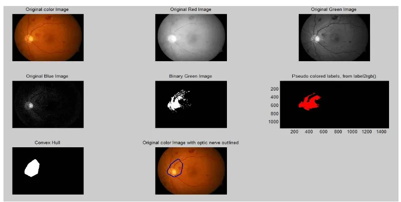
E. Optical nerve detection
The optical disc is the brighter part of the normal eye. Optic nerve is to transfer visual information from the retina to the vision centers of the brain via electrical impulses. Glaucoma is one of the most common illnesses affecting the optic nerve. Glaucoma is caused by high intraocular pressure, or high pressure in the fluid that is inside the eye.
Canny edge detection method detects blurred edges by preserving all local maxima the gradient, through this it detects optimally the boundaries of features.
Figure 5. Optical nerve detected image
F. Red lesion detection
Microaneurysms (MAs) are central dilatations of retinal vessels and show up as red dabs in retinal fundus images. As canny edge detection is used in blood vessel it is also used in red lesion detection. Red lesions are nothing but the microaneurysms detection. Morphological opening operation is used which is nothing but the erosion followed by the dilation. Finally edge detected image is subtracted from original image i.e. red plane image
(a) (b)
Figure.6 Microaneurysms extracted image (a) input image (b) microaneurysms detected image
V. CONCLUSION
VI. ACKNOWLEDGMENT
The author would like to thank all those who provides the data for detecting and extracting the features which are responsible for diabetic retinopathy
REFERENCES
[1] Lama Seoud, Thomas Hurtut, Jihed Chelbi, Farida Cheriet, and J. M. Pierre Langlois, “Red Lesion Detection Using Dynamic Shape Features for Diabetic Retinopathy Screening,” IEEE transactions on medical imaging, vol. 35, no. 4, April 2016
[2] M. Sridevi Mahe swari , Adarsh Punnolil, “A Novel Approach For Retinal Lesion Detection In Diabetic Retinopathy Images”, International Journal of Innovative Research in Science, Engineering and Technology Volume 3, Special Issue 3, March 2014
[3] Arulmozhivarman Pachiyappan, Undurti N Das, Tatavarti VSP Murthy, and Rao Tatavarti, “Automated diagnosis of diabetic retinopathy and glaucoma using fundus and OCT images”, https://www.ncbi.nlm.nih.gov/pmc/articles/PMC3477058/
[4] Swati Gupta* and Karandikar AM, “Diagnosis of Diabetic Retinopathy using Machine Learning”, Volume 3 • Issue 2 • 1000127 ISSN: JRD [5] Meera Walvekar1, Geeta Salunke2, “ Detection of Diabetic Retinopathy with Feature Extraction using Image Processing ”,ISSN 2250-2459, ISO 9001:2008 Certified Journal, Volume 5, Issue 1, January 2015
[6] Deepthi K Prasad,Vibha L,Venugopal K R, “Early Detection of Diabetic Retinopathy from Digital Retinal Fundus Images”, 2015 IEEE Recent Advances in Intelligent Computational Systems (RAICS) | 10-12 December 2015 | Trivandrum
[7] Asiri Wijesinghe,N. D. Kodikara,Damitha Sandaruwan, “Autogenous Diabetic Retinopathy Censor for Ophthalmologists - AKSHI”,
[8] Md. Jahiruzzaman, A. B. M. Aowlad Hossain, “Detection and Classification of Diabetic Retinopathy Using K-Means Clustering and Fuzzy Logic”, ICCIT 21-23 December 2015
[9] Snehal Dilip Kasurde, Prof.S.N.Randive, “An Automatic Detection of Proliferative Diabetic Retinopathy”, 2015 International Conference on Energy Systems and Applications (ICESA 2015) Dr. D. Y. Patil Institute of Engineering and Technology, Pune, India 30 Oct - 01 Nov, 2015 [10] Amanjot Kaur, Prabhpreet Kaur, “An Integrated Approach for Diabetic Retinopathy Exudate Segmentation by Using Genetic Algorithm and Switching Median Filter”,2016 IEEE.
[11] poonam,poonam yadav,neelam ruhil, “ blood vessel detection for diabetic retinopathy”,2016 IEEE.
[12] Priyadarshini Patil, Pooja Shettar, Prashant Narayankar,Mayur Patil, “An Efficient Method of Detecting Exudates in Diabetic Retinopathy : Using Texture Edge Features”, ICACCI sept 21-24 2016,jaipur,india