_____________________________________________________________________________________________________
*Corresponding author: E-mail: [email protected];
(Past name: British Journal of Medicine and Medical Research, Past ISSN: 2231-0614, NLM ID: 101570965)
Evaluating Electrical Activity of Tibialis Anterior
Muscle and Balance in Hemiparetic Patients
Following Central and Peripheral Electrical
Stimulation - Protocol for a Randomized,
Double-blinded, Clinical Trial
Aline Marina Alves Fruhauf
1*, Fabiano Politti
1, Camila Cardoso da Silva
1,
David Correa Alves
1, João Carlos Ferrari Corrêa
1and Fernanda Ishida Corrêa
11
Graduate Program in Rehabilitation Sciences, Universidade Nove de Julho, Rua Adolpho Pinto 109, Barra Funda, São Paulo, Brazil.
Authors’ contributions
This work was carried out in collaboration among all authors. Authors FIC and AMAF designed the study. The data collection, interventions and recruitment of the participants were performed by authors CCS and DCA. The manuscript was prepared by authors FIC and AMAF, which was revised and edited by authors FP and JCFC. All authors read and approved the final manuscript.
Article Information
DOI: 10.9734/JAMMR/2019/v30i830218 Editor(s): (1)Dr. Jera Kruja, Neurology, University of Medicine, Tirana, Albania. Reviewers: (1)Waleed Morshed, Prince Sattam Bin Abdulaziz University, Saudi Arabia. (2)M. Rajajeyakumar, The Tamil nadu Dr. M.G.R. Medical University, India. (3)Bo Yuc, Shanghai Jiao Tong University, China. Complete Peer review History:http://www.sdiarticle3.com/review-history/50902
Received 15 June 2019 Accepted 25 August 2019 Published 06 September 2019
ABSTRACT
Concomitant transcranial direct current stimulation (tDCS) is suggested to enhance the functional effects of other physical rehabilitation methods in individuals with motor impairment stemming from a chronic cerebrovascular disease. Thus, the primary aim of the proposed study is to analyze the electrical activity of the tibialis anterior (TA) muscle of the paretic limb in stroke survivors following an intervention involving the combination of tDCS over the motor cortex and peripheral electrical stimulation (PES) administered over the paretic TA. The secondary objective is to analyze the effect on dynamic balance.
Methods: Thirty-six adult stroke survivors will be randomized into three groups: 1) Active tDCS + active PES; 2) Sham tDCS + active PES and 3) Active tDCS + sham PES. TDCS active will be positioned bilateral over the primary motor cortex of the damaged hemisphere (C1 or C2) and the cathode will be positioned over the primary motor cortex of the undamaged hemisphere (C1 or C2) with a current of 2 mA for 20 minutes. For sham tDCS, will follow the same standarts, however, the equipment will be switched on for only 20 seconds. PES will be administered to the paretic TA at 50 Hz for 30 minutes. Evaluations: the median frequency and root mean square (RMS) of the paretic TA will be analyzed using electromyography (EMG) and dynamic balance will be evaluated using the Mini-Balance Evaluation System (Mini-BESTest) at baseline (pre-intervention), after 10 treatment sessions at a frequency of five times a week for two weeks (post-intervention) and 30 days after the end of the interventions (follow up).
Discussion: PES has proven to facilitate the conduction of sensory-motor afferences to the cerebral cortex in stroke survivors. Combining PES with tDCS, which has a direct effect on increasing cortical excitability, could favor motor acquisition and neuronal plasticity in this population.
Keywords: Electromyography; balance; hemiparesis; peripheral electrical stimulation; tibialis anterior; transcranial direct current stimulation.
ABBREVIATIONS
BDI :Beck Depression Inventory;
EMG :Electromyography;
Hz :Hertz;
Mini-BES Test: Mini-balance Evaluation System; PES :Peripheral electrical stimulation;
RMS : Root mean square;
SENIAM :Surface Electromyography for the Non-Invasive Assessment of Muscles;
TA :Tibialis anterior;
tDCS : Transcranial direct current stimulation;
1. INTRODUCTION
The physiopathology of cerebrovascular accident (stroke) is governed by the leakage of blood or restricted blood flow in a given area of the brain. According to data from the World Health Organization, stroke is the third major cause of morbidity, mortality, and Disability-adjusted years of life in the world [1]. In Brazil, it is the leading cause of death and acquired physical disability, with an annual incidence of 108 cases per 100 thousand inhabitants [2].
Difficulty performing hip flexion, knee flexion and dorsiflexion of the foot are among the disabilities commonly found in stroke survivors. In some individuals, the ankle remains in the extended position, which is denominated equinus foot, characterized by hypertonia of the gastrocnemius and soleus (triceps surae) muscles and a reduction in or absence of strength in the tibialis anterior (TA) muscle [3]. This situation affects the
adequate support of the feet on the ground, which makes the individual distribute his/her weight more to the non-paretic side as a compensatory mechanism [4]. Consequently, the individual experiences a reduction in postural control static or dynamic and gait velocity, leading to greater insecurity, a risk of falls and functional limitations [4].
Many clinical trials have been conducted to minimize those dysfunctions by using peripheral electrical stimulation (PES) [5]. Combined PES with exercises based on the Bobath concept in 40 stroke survivors and found an increase in dorsiflexion range of motion, a reduction in spasticity of the plantar flexors and gain in TA muscle strength [6]. PES used on the TA of 15 individuals with hemiparesis stemming from a stroke combined with active contraction of the dorsiflexors in the standing position on a dynamic platform for 30 minutes, followed by 15 minutes of gait training focused on ankle control, resulting in a reduction in dynamic spasticity of the plantar flexors, an increase in dorsiflexor strength and improved gait symmetry [7]. PES combined with ankle strength and proprioception training or ankle stretching and proprioception training in 11 individuals with hemiparesis stemming from a stroke and found that the former combination
resulted in positive effects on balance
performance [8].
in the literature on the ideal parameters (duration/number of applications, pulse, intensity, and frequency) for neurological diseases, and better results are achieved when combined with other forms of rehabilitation.
TDCS consists of a low-intensity electrical current generally administered over the scalp using two electrodes of different polarity (anode and cathode). The current is able to penetrate the skull and produce modulating effects on the neural membrane, either increasing (anodal stimulation) or diminishing (cathodal stimulation) cortical excitability [10].
In this context, researchers have proposed investigating the combination of PES and other forms of electrical stimulation to enhance its effects, such as transcranial direct current stimulation (tDCS). Kwon et al. [11] evaluated the activity of the primary motor cortex (M1) using magnetic resonance imaging in two healthy individuals during a session of anodal tDCS over M1 combined with PES of the wrist extensors and found an increase in M1 activity. Rizzo et al. (2014)[12] investigated the motor evoked potential in 10 young healthy individuals after 10, 20, 30 and 60 min of anodal or cathodal tDCS over M1 combined with repetitive PES over the left median nerve and found that anodal stimulation + repetitive PES led to an increase in the motor evoked potential up to 60 minutes after stimulation. In a study involving 20 stroke survivors in the subacute phase, Sattler et al. (2015) [13] evaluated the effect of anodal tDCS over M1 combined with PES over the radial nerve for five consecutive weeks and found a significant increase in motor function of the hand up to one month after treatment. However,
Fruhauf et al. (2018)[14] evaluated the
immediate effect of tDCS combined with PES on the electrical activity of the paretic TA muscle and balance in 30 stroke survivors and found no effect after the administration of the two techniques combined. The researchers suggest that this may have occurred because only a single session was used, implying that longer treatment with the combination of the techniques could achieve different results. No clinical studies were found investigating the combination of PES and tDCS for more than one treatment session with the aim of assessing the electrical activity of the TA muscle and functional balance in stroke survivors.
When combined with other forms of treatment, tDCS has been demonstrated to enhance the
effects of physical therapy [15]. Dutta et al. (2014)[16] studied the effect of tDCS over the primary motor cortex and cerebellum combined with ankle training involving biofeedback in healthy individuals to improve myoelectrical control of the TA muscles and found that anodal stimulation over M1 resulted in the optimization in terms of the onset and end of electrical activity in the muscles. Madhavan et al. (2011) [17] found an increase in motor evoked potential for 15 minutes and immediately after the end of ankle dorsiflexion training combined with tDCS over M1 in stroke victims. Sohn et al. (2013) [18] investigated the effect of tDCS over the damaged M1 in 11 individuals with hemiparesis and found significant increases in quadriceps strength and static postural stability.
These interactions Central and Peripheral stimulation may have benefits regrading function, especially in cases of neurological disorders, as tDCS enhances cortical excitability, while PES triggered ascending sensory-motor information. Therefore, the present protocol proposes the investigation of the effects of tDCS combined with PES in individuals with hemiparesis stemming from a stroke on the electrical activity of the TA muscle and dynamic balance, being these factors one the of components important to functional independence.
1.1 Objective Primary
The primary aim of the proposed study is to analyze the electrical activity of the tibialis anterior (TA) muscle of the paretic limb in stroke survivors following an intervention involving the combination of tDCS over the motor cortex and
peripheral electrical stimulation (PES)
administered over the paretic TA.
1.2 Objective Secondary
The secondary objective is to analyze the effect on dynamic balance in stroke survivors following an intervention involving the combination of tDCS over the motor cortex and peripheral electrical stimulation (PES) administered over the paretic TA.
2. METHODS
2.1 Study Design
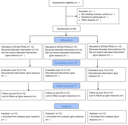
(Fig.1). This protocol received approval from the Human Research Ethics Committee of University Nove de Julho, São Paulo, Brazil (certificate
number: 2.015.168) in compliance with
Resolution 466/12 of the Brazilian National Board of Health. Written informed consent will be obtained from each participant and approval of the register of Clinical Trials: NCT03008720.
The participants will be recruited from the physical therapy clinics (waiting list) of University Nove de Julho, São Paulo, Brazil. Evaluations primary (electromyography-EMG of TA) and
secondary Dynamic Balance (Mini-Balance
Evaluation System- Mini BESTest) will be performed on three occasions: 1) baseline (pre-intervention) 2), after ten treatment sessions
(post-intervention) and 3) 30 days after the end of the sessions (follow up).
2.1.1 Eligibility criteria
The following are the inclusion criteria:
hemiparesis stemming from a stroke in the chronic stage (six months after stroke);2 TA muscle weakness > 1 and < 5 on the Medical Research Council scale (this scale grades muscle power on a scale of 0 to 5 in relation to the maximum expected for that muscle. In a recent comparison to an analogue scale the MRC scale is more reliable and accurate for clinical assessment in weak muscles); [19] adults (> 20 years of age) with independent gait (with or without a gait assistance device); agreement
Fig. 1. Flowchart of study
Assessed for eligibility (n= )
Excluded (n= )
Not meeting inclusion criteria (n= )
Declined to participate (n= )
Other reasons (n= )
Analysed (n=12)
Excluded from analysis (give reasons) (n= )
Evaluation post 10 (n=12)
Discontinued intervention (give reasons) (n= )
Allocated to tDCSa+FESa (n= 12) Received allocated intervention (n=12) Did not receive allocated intervention
(give reasons) (n= )
Evaluation post 10 (n=12) Discontinued intervention (give reasons) (n= )
Allocated to tDCSs+FESa (n=12) Received allocated intervention (n=12) Did not receive allocated intervention
(give reasons) (n= )
Analysed (n=12)
Excluded from analysis (give reasons) (n= )
Allocation
Analysis
Evaluation post 10th
Randomized (n=36)
Allocated to tDCSa+FESs (n= 12) Received allocated intervention(n=12) Did not receive allocated intervention
(give reasons) (n= )
Follow-up post 30 (n=12) Lost to follow-up (give reasons) (n=)
Evaluation post 10 (n=12) Discontinued intervention (give reasons) (n= )
Follow-Up postmth
Follow-up post 30 (n=12) Lost to follow-up (give reasons) (n=)
Follow-up post 30 (n=12) Lost to follow-up (give reasons) (n=)
Analysed (n=12)
to participate through the signing of a statement of informed consent. The following are the exclusion criteria: positive cutoff point for cognitive impairment on the Mini Mental State Examination (less than 11 points; corrected for schooling);[20] diagnosis of severe depression (Beck Depression Inventory);[21] active ankle mobility less than 5 degrees (determined using a
universal goniometer); [22] participants
presenting grade 5 spasticity in triceps suralis muscle (Ashworth Scale); [23] need for the use of orthopedic insoles or rigid braces; use of botulinum toxin in the lower limbs; severe visual
impairment (confirmed by ophthalmological
exams); contraindication for tDCS (history of seizures, tumors at stimulation site; metal implants in skull [all confirmed by medical exams]); skin lesion at application site of tDCS or PES (visual inspection by therapist); anesthesia or hyperesthesia at central or peripheral stimulation site (physical evaluation of surface sensitivity using a esthesiometer); diagnosis of deep vein thrombosis (confirmed by medical exam); diagnosis of degenerative disease or polyneuropathy (confirmed by medical exam); undergoing physical therapy or alternative therapy during the development of the study or in the one-month period after the 10 treatment sessions.
2.1.2 Sample size
The sample size was calculated using the G*Power program. Based on the results of a study by Sabut et al. (2010),[24] the calculation was performed considering mean and standard deviation root mean square (RMS) values for the experimental group before and after PES (60 ± 6 and 110 ± 11, respectively), α = 0.05, β = 0.2 (80% power) and a 0.94 effect size. Twelve individuals were determined for each group (total sample: 36 individuals).
2.1.3 Randomization
The allocation of the 36 participants (12 per group) will be randomized and counterbalanced using a randomization table in ExcelTM with codes for the combinations of the two central (active or sham) and two peripheral (active or
sham) stimulations [14]. A researcher not
involved in the evaluations or treatment will be responsible for the randomized allocation of the participants to the three groups:
1- Active tDCS + active PES over paretic TA; 2- Sham tDCS + active PES over paretic TA; 3- Active tDCS + sham PES over paretic TA.
2.1.4 Blinding
The Neuro Conn DC-STIMULATOR PLUS device has settings that enable the selection of the active stimulation mode or sham mode by entering codes. A researcher not involved in the treatment or evaluations will program the equipment with the code to which the patient was allocated. The type of stimulation (active or sham) will not be perceptible by visual cues or the external functioning of the device. Therefore, neither the researcher who will place the equipment on the patient nor the patient will know which treatment he/she is receiving (double-blind study).
2.2 Assessment Procedures
The evaluation of the tibial anterior muscle will be made by electromyographic analysis and all individuals will be seated on a chair with a backrest, with knees flexed at 90 degrees and ankle in the neutral position [14]. Dynamic Balance analysis will be performed by Mini BESTest Scale (consists of 14 functional tasks). Already, confounding variables will be collected in order to prevent potential factors such as depression or severe motor impairment from influencing intervention responses.
2.3 Electromyography of Tibialis Anterior Muscle
The EMG data of TA muscle activity will be analyzed by the amplitude/power of the signal (RMS) and muscle fiber recruitment rate (median frequency) captured using the electromyograpy (EMG SYSTEM® BRAZIL), consisting of an A/D converter with 16 bits of resolution, six channels and data transmission. The EMG signals will be pre-amplified with a gain of 1000 fold, a common rejection mode ratio > 100 dB and filtered using a 20-450 Hz bandpass filter, with a sampling frequency of 1 kHz. The data will subsequently
be coded using routines developed in MATLAB®
version R2010a (The MathWorks Inc., Natick, Massachusetts, USA).
Two disposable surface electrodes (Ag/AgCl – Medical Trace®) measuring 10 mm in diameter will be positioned over the skin (previously cleaned with 70% alcohol) in the region of the TA, following the guidelines of the Surface
Electromyography for the Noninvasive
Assessment of Muscles(SENIAM) [25]. For each
(maximum active dorsiflexion) for 10 seconds following a verbal command, followed by rest for 2-3 minutes between each reading. Next, the
participant will perform five consecutive
concentric contractions (isotonic) of the TA three times, with 2-3 minutes of rest between each reading [14].
No previous study has been conducted to determine the reliability of this tool for the population of stroke survivors, but this instrument has demonstrated solid, effective results in the investigation of muscle actions in this group of patients [26,27].
2.4 Mini-Balance Evaluation System
(Mini-BESTest)
Dynamic balance will be evaluated using the Mini-BESTest, which consists of 14 tasks distributed among four domains: (1) anticipatory postural adjustments (transition from sitting to standing position; standing on the tips of the toes; one-legged stance); (2) postural responses (four different direction of body movement: anterior, posterior and side-to-side); (3) sensory orientation (feet together on a stable surface with eyes open; feet together on an unstable surface with eyes open; leaning with eyes closed) and (4) gait stability (walking with change in velocity; horizontal movement of the head; around obstacles; turning on one's own axes; and with and without a cognitive dual task) [28].
Each item is scored on a two-point scale from
zero (worst performance) to two (best
performance). The maximum score is 28 points (domain 1= 6 points; 2= 6 points; 3= 6 points and 4= 10 points) [28]. This instrument has high reliability for the evaluation of balance in stroke survivors (ICC > 0.90) [29].
2.5 Determination of Potential
Confounding Factors
2.5.1 Depressive symptoms
Depressive symptoms will be evaluated and graded with regard to severity using the Beck Depression Inventory (BDI), [30] which is a self-administered questionnaire composed of 21 items. Each item is scored from 0 to 3 points. The total ranges from 0 to 63 points and is interpreted as follows: 0 to 10 indicates the absence of depression; 11 to 18 = mild depression; 19 to 29 = moderate depression; and 30 to 63 = severe depression. The BDI score will
be determined on three occasions (pre-intervention, post-intervention and 30-day follow up) and used as a covariant to determine whether motor recovery is independent of possible mood-related effects [31]. The reliability of the BDI is 0.89, and this measure has been used in studies that have shown good clinical results [32].
2.5.2 Fugl-meyer scale
The measures proposed on the Fugl-Meyer Scale are based on the neurological examination and sensory-motor activity of the upper and lower limbs to determine selective activity and synergic patterns in patients who have suffered a stroke. This is an accumulative numeric scoring system used to evaluate range of motion, pain, sensitivity, upper and lower limb motor function, balance, coordination and velocity, totaling 226 points [33]. A three-point ordinal scale is used for each item: 0 – not performed; 1 – partially performed; and 2 – fully performed. The scale has a total of 100 points for normal motor function, in which the maximum score is 66 for the upper limbs and 34 for the lower limbs [33]. The score is interpreted as follows: < 50 points = severe motor impairment; 50-84 = marked impairment; 85-95 = moderate impairment; and 96-99 = mild impairment. The Fugl-Meyer Scale will be used in this study for the characterization of the individuals considering demographic aspects, degree of global motor impairment and specific motor impairment of the lower limbs. In the literature, this scale has high reliability (ICC = 0.99 and 0.98, respectively) for the evaluation of stroke survivors [34].
2.6 Assessment Interventions
For both interventions, the patient will be seated on a chair with a backrest and knees flexed at 90° and ankle in the neutral position [14]. Treatment will consist of 10 sessions (five per week for two weeks). PES will last 30 minutes per session,[5] the first 20 minutes of which will be combined with tDCS [14].
2.6.1 Transcranial direct current stimulation
The one-channel unipolar DC Stimulation Plus
(NeuroConn®) will be used. Stimulation will be
administered through two silicone/carbon
positioned over the primary motor cortex of the undamaged hemisphere (C1 or C2) – both at a distance of 2 cm from Cz based on the map of the 10-20 International Electroencephalogram System [35]. Central stimulation with tDCS will occur concomitantly to peripheral stimulation (first 20 minutes of PES) with a current of 2 mA [36].
Sham stimulation will involve the same
procedures as active stimulation, but the stimulator will only be switched on for the first 20 seconds, after which the current will be reduced to zero. All patients will be informed that they may feel a mild initial tingling that may disappear or may continue throughout the 30 minutes of treatment. This is considered a valid control procedure for the use of tDCS [37].
2.6.2 Determination of potential side effects
Possible adverse effects stemming from
noninvasive brain stimulation will be determined using the tDCS – Side Effects Questionnaire (version translated into Portuguese) after each session with tDCS [38].
2.6.3 Peripheral electrical stimulation
The two-channel QUARK® FES VIF 995 DUAL will be used for PES. Two self-adhesive rubber electrodes measuring 5 x 9 cm will be positioned on the motor point and belly of the paretic TA muscle [14]. PES will be performed with a pulse width of 250 µs and a frequency of 50 Hz. The intensity will be increased until reaching the motor threshold (20-30% of maximum voluntary contraction) [14]. The stimulation cycles will be 1:2 (six seconds on and 12 seconds off) [13] combined with active contraction of the TA every six seconds for 30 minutes [14]. Sham stimulation will involve the same procedures as active PES, but the electrodes will be positioned in the tibial region (bone portion) [39].
2.7 Statistical Analysis
Descriptive data, characteristics of the sample (gender, age, type of stroke [ischemic or hemorrhagic], damaged hemisphere [right or left], time elapsed since the stroke event, Fugl-Meyer lower limb score, Beck Depression Inventory (BDI), use of controlled medications and associated comorbidities will be expressed as mean and standard deviation values or median and interquartile range.
The Shapiro-Wilk test will be used to determine the normality of the data (EMG and
Mini-BesTest). Repeated-measures ANOVA will be used for the comparison parametric data and the Kruskal-Wallis will be used for nonparametric data. The effect size will also be determined for the comparison of evaluation times (pre-intervention, post-intervention and 30-day follow-up). A (P = < 0.05 will be considered indicative of statistical significance. All analyzes will be processed using the IBM SPSS program v.19.
3. DISCUSSION
Considering that, after brain injury, functions such as the ability to ambulate can be substantially modified due to several changes, among them the inability to properly move the ankle and knowing that the ankle is of
fundamental importance especially in the
mechanisms of balance, mobility and adequate plantar distribution; to prove the efficiency of treatments currently available for this purpose become important.
It is known that the PES has already presented promising results. However, understanding the tDCS efficiency, still little studied for this purpose,
becomes necessary. Additionally, authors
suggest that tDCS can facilitate central nervous system modulation, favoring increased local synaptic efficacy and cortical excitability in humans; promoting short-term or long-term
cerebral neuroplasticity. Therefore, the
hypothesis of this research would be that tDCS associated PES in individuals with hemiparesis due to stroke may potentiate TA muscle activity and functional balance, promoted and expected by PES stimulation, especially in the chronic phase of the disease (when installed the mechanisms of maladaptive plasticity).
4. CONCLUSION
This article presents a detailed description of a prospective, randomized, controlled, double-blind trial designed to demonstrate the effects of the
combination of transcranial direct current
stimulation and peripheral electrical stimulation on electrical activity of the tibialis anterior muscle
and postural control in individuals with
hemiparesis stemming from a stroke. The results will be published and the evidence could contribute to the rehabilitation of this population.
5. LIMITATIONS
combined with neuronavigation equipment would offer better interpretation of the investigated variables. However, availability will not be possible, but there are validated and reliable functional resources such as EMG and the Mini- BESTest scale that will be used for collection and proper analysis of the results in this stroke individuals.
TRIAL STATUS
At the time of manuscript submission, we were recruiting patients. The study in question is expected to be completed in December 2019.
AVAILABILITY OF DATA AND
MATERIALS
Data sharing is not applicable to this article because no datasets were generated or analyzed during the present study.
CONSENT AND ETHICAL APPROVAL
This protocol received approval from the Human Research Ethics Committee of University Nove de Julho, São Paulo, Brazil (certificate number: 2.015.168) in compliance with Resolution 466/12 of the Brazilian National Board of Health. Written informed consent will be obtained from each participant.
Participating volunteers must accept the study consent form (attached document), which ensures the confidentiality of data, free access to the final data, explanations of any nature related to treatment and compensation for those suffering from participation in trials. The results of this study will be published in a journal of interest in the field of physical therapy and rehabilitation.
ACKNOWLEDGMENTS
The authors are grateful to University Nove de Julho for supporting the present study.
COMPETING INTERESTS
The authors declare that they have no competing interests.
REFERENCES
1. Murray CJL, Vos T, Lozano R, Naghavi M,
Flaxman AD, Michaud C. Disability-adjusted life years (DALYs) for 291 diseases and injuries in 21 regions, 1990-2010: A systematic analysis for the global
burden of disease study 2010. Lancet. 2012;2197-223.
2. Bensenor IM, et al. Prevalence of stroke
and associated disability in Brazil: National Health Survey – 2013. Arq. Neuro-Psiquiatr. São Paulo. 2015;73:9.
3. Andrews AW, Bohannon RW. Distribution
of muscle strength impairments following stroke. Clin. Rehabil. 2000;14:79.
4. Azevedo ERFBM, Macedo LS, Paraízo
MFN, Oberg TD, Lima NMFV, Cacho
EWA. Correlação do déficit de
equilíbrio, comprometimento motor e
independência funcional em indivíduos hemiparéticos crônicos. Acta Fisiátrica. 2008;15(4):225-8.
5. Howlett OA, Lannin NA, Ada L, Mckinstry
C. Functional electrical stimulation
improves activity after stroke: A systematic review with meta-analysis. Arch. Phys. Med. Rehabil. 2015;934-43.
6. Bakhtiary AH, Fatemy E. Does electrical
stimulation reduce spasticity after stroke? A randomized controlled study. Clin. Rehabil. 2008;418-25.
7. Cheng JS, Yang YR, Cheng SJ, Lin
PY, Wang RY.
Effects of combining electricstimulation wit h active ankle dorsiflexion while stadng on a rocker board: A pilot study for subjects with spastic foot after stroke. Arch. Phys. Med. Rehabil. 2010;505-12.
8. Kyunghoon, Lee S, Donghoon, Sik K. The
effects of ankle joint muscle strengthening, and proprioceptive exercise programs
accompanied by functional electrical
stimulation on stroke patients’ balance. J. Phys. Ther. Sci. 2015;2971–2975.
9. Rushton DN. Functional Electrical
Stimulation and rehabilitation- an
hypothesis. Medical Engineering &
Physics. 2003;75–78.
10. Bikson M, Datta A, Rahman A, ScaturroJ.
Electrode montages for tDCS and weak transcranial electrical stimulation: Role of “return” electrode’s position and size. Clin. Neurophysiol. 2010;121(12):1976– 1978.
11. Kwon et al. Cortical Activation by
Transcranial Direct Current Stimulation and Functional Electrical Stimulation in Normal Subjects: 2 Case Studies. J. Kor Soc Phys Ther. 2011;23(1):77-82.
12. Rizzo V, Terranova C, Crupi D,
Sant'angelo A, Girlanda P, Quartarone A.
Increased transcranial direct current
peripheral electrical nervestimulation. Brain Stimul. 2014;113-211.
13. Sattler V. et al. Anodal tDCS Combined
With Radial Nerve Stimulation Promotes Hand Motor Recovery in the Acute Phase After Ischemic Stroke, Neurorehabilitation and Neural Repair. 2015;29(8):743–754.
14. Fruhauf AMA, Politti F, Dal Corso S, et al.
Immediate effect of transcranial direct
current stimulation combined with
functional electrical stimulation on activity of the tibialis anterior muscle and balance of individuals with hemiparesis stemming from a stroke. J Phys Ther Sci. 2017; 29(12):2138-2146.
15. Holgado, Darías et al. The effects of
transcranial direct current stimulation on objective and subjective indexes of exercise performance: A systematic review and meta-analysis. Brain Stimulation:
Basic, translational, and Clinical
Research in Neuromodulation. 2019;
12(2):242–250.
16. Dutta A, Paulus W, Nitsche MA.
Facilitating myoelectric-control with
transcranial direct current stimulation: a preliminary study in healthy humans.
Journal of Neuro Engineering and
Rehabilitation. 2014;11:13.
17. Madhavan S, Weber KA, Stinear JW.
Non-invasive brain stimulation enhances Wne motor control of the hemiparetic ankle: Implications for rehabilitation. Exp. Brain Res. 2011;9–17.
18. Sohn MK, Sung JJ, Yeong W. Effect of
Transcranial Direct Current Stimulation on Postural Stability and Lower Extremity Strength in Hemiplegic Stroke Patients. Ann. Rehabil. Med. 2013;759-765.
19. Hermans G, et al. Interobserver
agreement of Medical Research Council sumscore and handgrip strength in the intensive care unit. Muscle Nerve. 2012; 45(1):18-25.
20. Maheshwari SG, IqbaL M, Hashmi SFA,
Devrajani B. Stroke patients; assessment of cognitive impairment in. Professional Med. J. 2015;541- 545.
21. Beck AT, Ward CH, Mendelson M, Mock J,
Erbaugh J. An inventory for measuring depression. Arch. Gen. Psychiatry. 1961; 4:561–71.
22. Madhavan S, Weber KA, Stinear JW.
Non-invasive brain stimulation enhances Wne motor control of the hemiparetic ankle: implications for rehabilitation. Exp. Brain Res. 2011;9–17.
23. Ashword B. Preliminary trial of carisoprodol
in multiplesclerosis. Practitioner. 1964; 540–2.
24. Sabut R. Kumar PK, Lenka MM. Surface
EMG Analysis of Tibialis Anterior Muscle in Walking with FES in Stroke Subjects. Conf Proc IEEE Eng Med Biol Soc. 2010;5839-42.
25. Hermens HJ, Freriks B, Disselhorst- Klug
C, Rau G. Development of
recommendations for SEMG sensors and
sensor placement procedures. J.
Electromyography and Kinesiology. 2000; 361–74.
26. Suhaimi R, et al. Analysis of EMG-based
Muscles Activity for Stroke Rehabilitation. 2014 2nd International Conference on Electronic Design, ICED; 2014.
DOI: 10.1109/ICED.2014.7015792
27. Tania FC, et al. Electromyographic
analysis of spastic muscles in hemiparetic patients before and after physical therapy intervention. Ter Man. 2012;10(48):148-153.
28. Charlotte SLT, Lin RL, Raymond CK,
Chung MYC. Psychometric Properties of the Mini-Balance Evaluation Systems Test
(Mini-BESTest) in Community-dwelling
Individuals with Chronic Stroke. Phys Ther. 2013;93(8):1102-1115.
29. Carla B, Lívia CM , Fátima RP.
Electromyographic analysis of spastic muscles in hemiparetic patients before and after physical therapy intervention. Ter Man. 2012;10(48):148-153.
30. Beck AT, Ward CH, Mendelson M, Mock J,
Erbaugh J. An inventory for measuring depression. Arch. Gen. Psychiatry. 1961; 4:561–7.
31. Kroenke K, Spitzer RL, Williams JB. The
PHQ-9: validity of a brief depression severity measure. J Gen Intern Med. 2001;16(9):606-613.
32. An TG, Kim SH, Kim KU. Effect of
transcranial direct current stimulation of stroke 528 patients on depression and quality of life. J Phys Ther Sci. 2017; 29(3):505-507.
33. Fugl-meyer AR, Jaasko L, Leyman I,
Olsson S, Steglind S. The post-stroke
hemiplegic patient: A method for
evaluation of physical performance. Scand. J. Rehab. Med. 1975;13-31.
34. MAKI T, et al. Estudo de confiabilidade da
35. Edwin HFA, Tjitske AB. Transcranial Direct Current Stimulation of the Leg Motor
Cortex Enhances Coordinated Motor
Output During Walking With a Large Inter-Individual Variability. Brain Stimulation 2016;9:182–190.
36. Adeyemo BO, Simis M, Macea DD, Fregni
F. Systematic review of parameters of
stimulation, clinical trial design
characteristics, and motor outcomes in non-invasive brain stimulation in stroke. Front. Psychiatry. 2012;3:88.
37. Miranda PC, Lomarev M, Hallett M.
Modeling the current distribution
during transcranial direct current
stimulation. Clinical Neurophisiology. 2006; 1623–1629.
38. Xu J, Fregni F, Brody AL, Rahman AS.
Transcranial direct current stimulation reduces negative affect but not cigarette craving in overnight abstinent smokers. Front Psychiatry. 2013;20:4-12.
39. Ambrosini E, Ferrante S, Pedrocchi
A, Ferrigno G, Molteni F. Cycling induced by electrical stimulation improves motor
recovery in post-acute hemiparetic
patients: A randomized controlled
trial. 2011;1068-73.
_________________________________________________________________________________
© 2019 Fruhauf et al.; This is an Open Access article distributed under the terms of the Creative Commons Attribution License (http://creativecommons.org/licenses/by/4.0), which permits unrestricted use, distribution, and reproduction in any medium, provided the original work is properly cited.
Peer-review history: