DEVELOPMENT OF SPECIFIC ANTIBODY
FRAGMENTS FOR THE DETECTION AND
TREATMENT OF MELANOMA
Joy Odili MB ChB MRCS
A thesis submitted in accordance with the requirements for the examination for the degree of Doctor of Medicine (MD)
University o f London
April 2002
RAFT Institute for Plastic Surgery Division of Molecular Biology
All rights reserved
INFO RM A TIO N TO ALL U SER S
The quality of this reproduction is dependent upon the quality of the copy submitted.
In the unlikely event that the author did not send a complete manuscript
and there are missing pages, these will be noted. Also, if material had to be removed, a note will indicate the deletion.
uest.
ProQuest U 643259
Published by ProQuest LLC(2016). Copyright of the Dissertation is held by the Author.
All rights reserved.
This work is protected against unauthorized copying under Title 17, United States Code. Microform Edition © ProQuest LLC.
ProQuest LLC
789 East Eisenhower Parkway P.O. Box 1346
ABSTRACT
The incidence of Malignant Melanoma continues to rise. Melanoma is potentially curable by surgery if caught early. However once the disease has spread the response to adjuvant therapy is poor. The combination of high prevalence and therapeutic resistance has led to an interest in other methods for detecting melanoma at earlier time points, when treatments may affect patient outcome and survival.
Immunoscintigraphy is the use o f radiolabelled tumour-specific antibodies to detect disease. Previous work has centred on the use o f the whole monoclonal antibody LHM2, against the high molecular weight melanoma-associated antigen (HMW-MAA). Single chain Fv fragments (scFv) have been developed from this molecule. In this thesis optimal conditions for storage and stability o f the scFvs were explored. Some of the scFvs were selected by rounds of phage display. The clones seemed to have better properties with each round of selection on phage, and the role of Darwinian “selection of the fittest” was explored. Some scFvs were genetically modified by the addition of cysteine residues to form multimeric constructs for improved tumour targeting. We were unable to show improved tumour targeting in vivo when the multimers were compared to their monomeric equivalents, and possible reasons for this are discussed.
One of the antibody fragments RAFTS was modified for therapy by conjugation with Staphyllococcal protein A. This clone was shown to retarget IgG and complement to melanoma in vitro. In vivo the construct behaved more like the larger LHM2 antibody than the parent (RAFTS scFv), consistent with IgG recruitment in the murine model.
for his love, patience, and understanding
to Michael
and
to my parents
ACKNOWLEDGMENTS
Many colleagues and co-workers have contributed to this thesis:
I would like to thank Professor Sanders for entrusting me with this exciting project, Jorg Kupsch for his endless vision and tireless supervision, and George Wilson for his guidance at the Gray Laboratory. I would also like to thank Stephen Hamilton and Norbert Kang my predecessors. I thank Elizabeth Clayton for persevering and successfully staining my paraffin sections, and Paul Richmond for reviewing my difficult histopathological slides. I humbly thank, my fellow surgical research fellows for their support, advice, jokes, and good spirit, all the scientists and sandwich students, Clare Linge, and the administrative staff in RAFT for supporting me and keeping me stimulated for two years. I wish to thank all the staff at the Animal House for their excellent animal husbandry. Finally, I wish to thank Mr Grobbelaar for being my mentor, and Mr Grover for creating mighty footsteps in which I have tried to follow.
The research and data management represented in this thesis would not have been possible without the financial support and generous assistance of The RAFT Institute o f Plastic Surgery, a fellowship from the Royal College o f Surgeons, and grants from the BUPA foundation and the Smith’s Charity.
I would like to thank Dr Judy Evans for believing in me and convincing me that there was more to melanoma than “mole-picking”.
ABSTRACT
2
ACKNOWLEDGEMENTS
4
TABLE OF CONTENTS
5
LIST OF CHAPTERS
6
LIST OF FIGURES
12
LIST OF TABLES
14
1. INTRODUCTION
17
2. MATERIALS AND METHODS
54
3. ANTIBODY PREPARATION
85
4. FORMATION OF MULTIMERS
117
5. THERAPEUTIC CONSTRUCTS
159
6. IMMUNOHISTOCHEMISTRY USING LHM2 Mab
188
7. SUMMARY & FUTURE DIRECTIONS
224
8. REFERENCES
237
LIST OF CHAPTERS
1. INTRODUCTION...17
1.1 THE CLINICAL PROBLEM... 18
1.1.1 H isto ry...18
1.1.2 E pidem iology o f M elanom a...19
1.1.3 A etiology o f M elanom a...21
1.1.4 M etastatic D ise a se...22
1.1.5 Staging P atients a n d Predicting Survival....22
1.1.6 The Effects o f D etection an d Treatment on S u rvival...25
1.2 DIAGNOSING METASTATIC DISEASE... 26
1.2.1 P hysical E xam ination...26
1.2.2 L aboratory Tests...2 7 1.2.3 R adiological Tests....2 7 1.2.4 P reoperative L ym phoscintigraphy...29
1.2.5 Sentinel Lymph N ode B iopsy (SLNB)...30
1.3 HISTOPATHOLOGY AND PROGNOSIS... 30
1.3.1 H istopathology o f M elanom a...30
1.3.2 Im m unohistochem istry...32
1.3.3 H M W -M AA...32
1.3.4 HMW-MAA Expression in N orm al Tissues...33
1.3.5 HMW-MAA Expression on M elanoma C e lls...33
1.4 RADIO-IMMUNOSCINTIGRAPHY...35
1.4.1 Introduction...35
1.4.2 M elanom a a s a Target f o r R IS...35
1.4.3 Com parison to Other Imaging M o d a lities...36
1.4.4 Clinical Experience...3 7 1.4.5 Size M odification to Im prove Tumour T argetin g...38
1.4.6 A ntibody Fragments in Im m unoscintigraphy...41
1.4.7 Im proving ScFv Solubility an d Stability...42
1.4.8 Tackling Renal Accum ulation...43
1.4.9 Im proving Tumour L o ca lisa tio n...45
1.4.10 Phage D isplay an d the Darwininan Selection o f "Happy " C lo n es...46
1.5 MELANOMA THERAPY... 47
1.5.1 S u rg ery...47
1.5.2 Chem otherapy and Im unotherapy...48
1.5.3 R ad io th era p y...49
1.5.4 Vaccines...50
1.5.5 M onoclonal A ntibodies a n d A ntibody Fragm ents in M elanoma T herapy...51
1.6 CONCLUSIONS... 52
1.7 A IM S ...52
1.7.1 Clinical A im s...53
1.7.2 H yp o th esis...53
2.2 MOLECULAR BIOLOGY TECHNIQUES...56
2.2.1 P lasm id Preparation...56
2.2.2 Purification o f Plasm ids a n d DNA F ragm ents...57
2.2.3 Polym erase Chain Reaction (PGR)...58
2.2.4 Restriction D ig ests...59
2.2.5 Ligation o f DNA Fragm ents...59
2 .2.6 B acterial Culture M edia a n d A gar P la tes...59
2 .2.7 B acterial Transformation...60
2.2.8 E lectrophoresis Buffers a n d A garose G els...60
2.2.9 C olony L ift...60
2.2.10 Technique o f B acterial C ulture...62
2.3 PROTEIN PURIFICATION TECHNIQUES...63
2.3.1 Storage o f B acterial C lo n es...63
2.3.2 P reparation o f B acterial Supernatants...64
2.3.3 Protein C oncentration...64
2.3.4 Biologic LP Platform f o r Column Chrom atography...64
2.3.5 Purification o f a ll scF vs (except R3ZZ) by Im m obilised M etal-ion Affinity C hrom atography (IM AC)...65
2.3.6 Purification o f R3ZZ scFv b y IgG sepharose column...66
2.3.7 A nalysis o f scF vs by G el F iltration...66
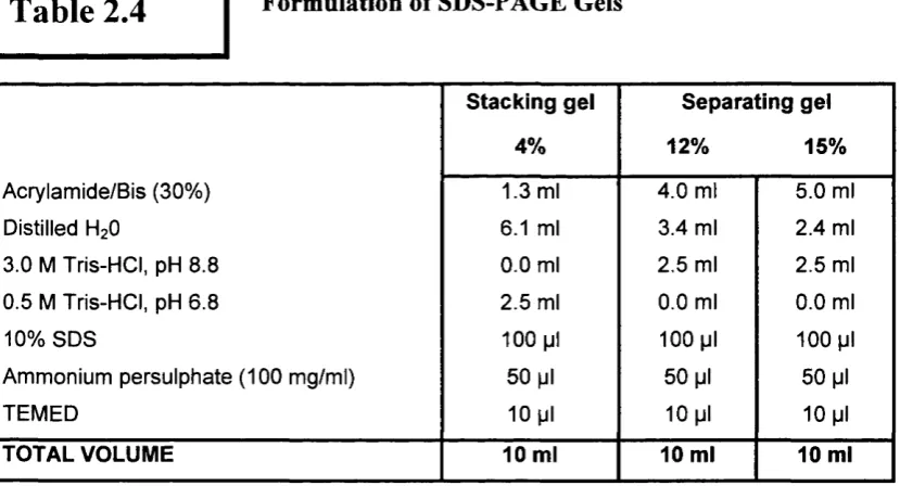
2.4 SDS-PAGE AND WESTERN BLOT ANALYSIS... 67
2.4.1 G els...67
2.4.2 B u ffers...67
2.4.3 SDS-PA GE A n a ly sis...68
2.4.4 W estem -blot A n a lysis...69
2.5 TISSUE CULTURE...70
2.5.1 M aintenance o f A 3 7 5 M C e ll L in e...71
2.5.2 P reparation o f C ell ELISA (Enzyme Linked Immunosorbent Assay) P la te s...71
2.5.3 Technique o f C ell ELISA...72
2.5.4 M aintenance o f 9E10 H ybridom a C ell Line...72
2.6 RADIOLABELLING TECHNIQUES... 74
2.6.1 B u ffers...74
2.6.2 lodination o f Antibodies...75
2.6.3 Separation o f Unincorporated Iodine-1 2 5...75
2.6.4 Estimation o f Incorporation o f Iodine-125...76
2.7 DETERMINATION OF ANTIBODY AFFINITY... 76
2.7.1 P reparation o f A ntibody a n d Human M elanoma C e lls...76
2.7.2 Experim ental Technique...76
2.7.3 Affinity D eterm ination...77
2.8 ANIMAL MODEL...77
2.8.1 P reparation o f Tumour C e lls...78
2.8.2 Tumour Production in the M ic e...78
2.9 ANTIBODY BIODISTRIBUTION AND PHARMACOKINETICS...79
2.9.1 D esign o f E xperim ents...79
2.9.2 Injection o f R adio-labelled A n tib o d y...80
2.9.4 Sample Analysis...81
2.10 HISTOLOGY TECHNIQUES... 82
2.10.1 Preparation o f Tissue Sections...82
2.10.2 LHM2 Staining o f Paraffin Sections...82
2.10.3 SlOO Staining o f Paraffin Sections...84
3. ANTIBODY PREPARATION...85
3.1 INTRODUCTION...86
3.1.1 Rationale...86
3.1.2 Design o f scFvs...88
3.1.3 Solubility Studies...93
3.2 METHODS...95
3.2.1 Design and Choice o f scFvs...95
3.2.2 Production and Purification o f scF vs...95
3.2.3 Effect o f Storage Temperature...96
3.2.4 Effect o f IMA C Elution Buffer on Stability...96
3.2.5 Confirmation o f Epitopic Specificity...97
3.2.6 Affinity Determination...97
3.3 RESULTS...98
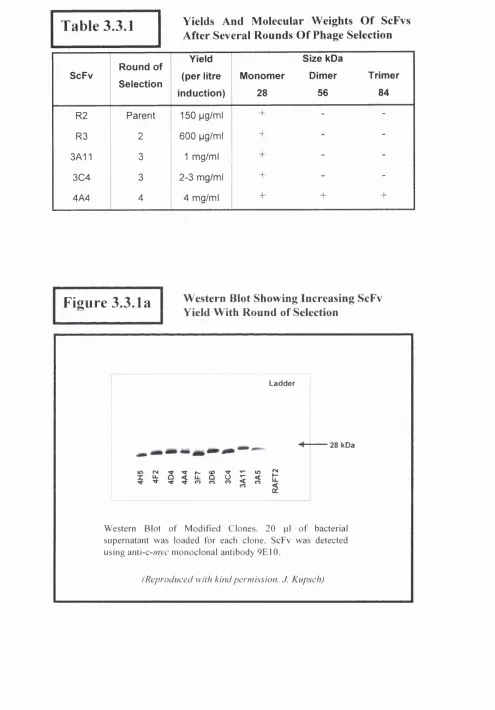
3.3.1 ScFv Yield and Molecular Weight Determination...98
3.3.2 Optimising Storage Temperature...101
3.3.3 Effect o f IMAC Elution Buffer on Stability...102
3.3.4 Confirmation o f Epitopic Specificity...105
3.3.5 Affinity Estimation...106
3.4 DISCUSSION... 106
3.4.1 A nalysis o f Results...106
3.4.2 "Rational" Approaches To Stability Engineering...109
3.4.3 Evolutionary Approaches For Stability Engineering...I l l 3.4.4 Bacterial Expression...112
3.4.5 Vector Expression...113
3.4.6 A Iternative Formats...114
3.5 CONCLUSIONS... 115
4. FORMATION OF MULTIMERS...117
4.1 INTRODUCTION... 118
4.2 MULTIMERISATION STRATERGIES... 120
4.2.1 Shortening o f ScFv Peptide Linker....120
4.2.2 Disulphide Bond Formation with Cysteine Residues...121
4.2.3 Naturally Evolved Multimers...122
4.2.4 Aims...123
4.3 MATERIALS AND METHODS (IN VITRO)... 123
4.3.1 Design and Choice o f Multimers...123
4.3.2 Production and Purification o f Multimers...126
4.3.3 Confirmation o f Epitopic Specificity...127
4.4.1 Production an d Purification o f ScF vs...128
4.4.2 Radiolabelling....128
4.4.3 Biodistribution and Pharm acokinetics...129
4.5 IN VITRO RESULTS...129
4.5.1 Selection an d Purification o f ScFvs Expressed in E. C oli Origam i...129
4.5.2 Selection an d Purification o f TOPI 0 cys-ScFvs...131
4.5.3 Selection an d Purification o f 3C 4 a n d 4A4 ScFvs...132
4.5.4 M olecular Weight Estimation o f S cF vs...132
4.5.5 Confirmation o f Epitopic S pecificity...134
4.5.6 Affinity D eterm ination...136
4.5.7 Effect o f Temperature and Mouse Serum on S tability...136
4.6 IN VIVO RESU LTS...138
4.6.1 Radiolabelling....138
4.6.2 In vivo Studies...139
4.7 DISCUSSION...150
4.7.1 A nalysis o f R esu lts...150
4.7.2 M ultivalency an d Affinity...152
4.7.3 D isulphide B ond Formation Using A dditional Cysteine R esidu es...153
4.7.4 The Use o f Origam i Strain o f E. C oli f o r Multim erisation...154
4.7.5 Alternative M ultimerisation Stratergies: A ltered Linker Length...155
4.7.6 F actors Influencing D im er to M onom er Transition...156
4.7.7 Thermal S ta b ility...157
4.8 CONCLUSIONS... 158
5. THERAPEUTIC ScFv CONSTRUCTS... 159
5.1 INTRODUCTION... 160
5.1.1 Effector Functions...161
5.1.2 B ispecific A ntibody F ragm ents...163
5.1.3 Staphyllocaccal Protein A...164
5.1.4 A im s...165
5.2 METHODS...165
5.2.1 D esign o f R3ZZ...165
5.2.2 Production an d Purification o f R3ZZ....167
5.2.3 IgG B in din g...168
5.2.4 Com plem ent Binding....168
5.2.5 Radiolabelling....169
5.2.6 In vivo stu d ies...169
5 .2.7 Tumour Penetration Studies...169
5.3 RESULTS...170
5.3.1 Yield a n d M olecular W eight...170
5.3.2 M elanom a Binding....171
5.3.3 IgG B in din g...173
5.3.4 Com plem ent Binding....174
5.3.6 In vivo Studies...
5.3.7 Tumour Penetration Studies...180
5.4 DISCUSSION...181
5 .4 .1 A nalysis o f R esu lts...181
5.4.2 Im proving R3ZZ ScFv D etection in Immunohistochemistry...183
5.4.3 The Use o f Frozen Sections...185
5.4.4 A lternatives to Im m unohistochem istry...186
5.5 CONCLUSIONS...186
6. IMMUNOHISTOCHEMISTRY USING LHM2 MAb... 188
6.1 INTRODUCTION...189
6.1.1 M elanocytic Lesions a n d M elanom a...189
6.1.2 Common A c q u ire d N a e v i...190
6.1.3 Spitz N aevi (Spindle or E pithelioid C ell Tumour; Juvenile M elanom a)...191
6.1.4 D ysplastic N a e v i...192
6.1.5 M alignant M elanom a...193
6.1.6 D esm oplastic M elanom as...197
6.2 IMMUNOHISTOCHEMICAL MARKERS... 198
6.2.1 S I 00 P ro tein...198
6.2.2 H M B -45...199
6.2.3 Antibodies to M elan-A...200
6.2.4 Tyrosinase...200
6.2.5 O ther M arkers...200
6.2.6 Prognostic M a rk ers...201
6.2.7 M elanoma A n tigen s...201
6.2.8 Rationale F or Use o f LHM2 M onoclonal A n tib o d y...202
6.3 MATERIALS AND METHODS... 203
6.3 .1 Lesions Stu died...203
6.3.2 Im m unohistochem istry...203
6.3.3 A nalysis o f R e su lts...204
6.4 RESULTS... 205
6.4.1 Benign M elanocytic L esio n s...205
6.4.2 Spitz N a evi...205
6.4.3 D ysplastic N a e v i...207
6.4.4 P rim ary M e la n o m a s...208
6.4.5 M etastatic M elanom as...209
6.4.6 Corresponding Prim ary a n d M etastatic M elanom as Stained with LHM2 M ab...211
6.4.7 D esm oplastic M elanom as...214
6.4.8 A dditional O b serva tio n s...215
6.5 DISCUSSION... 216
6.5.1 LHM2 Im m unoreactivity...216
6.5.2 Background Im m unoreactivity...217
6.5.3 Staining Intensity and Prognostic Im plications...217
6.5.4 D esm oplastic M elanom as...219
6.5.5 M elanom a A n tig en s...210
6.5.6 ScFvs in Immunohistochemistry...221
7.2 ANTIBODY PREPARATION...226
7.2.1 Future D irections...2 2 6 7.3 MULTIMERISATION STRATEGIES...227
7.3 .1 Future D irections...2 2 7 7.4 THERAPEUTIC CONSTRUCTS... 228
7.4.1 Future D irections...228
7.5 IMMUNOHISTOCHEMISTRY...230
7.5 .1 Future D irections...230
7.6 ALTERNATIVE STRATEGIES... 2 3 1 7.6.1 Improving Tumour Targeting Using Antibody C ocktails...231
7.6.2 Therapeutic ScFvs: A lternatives To Staphylococcal Protein-A...233
7.6.3 Radioim m unotherapy...234
7.6.4 Pretargeting S tra teg ies...234
7.6.5 PEG ylation o f S cF vs...235
7.7 CONCLUSIONS... 236
8. REFERENCES... 237
8.1 REFERENCES... 238
8.2 REFERENCED WEBSITES...283
LIST OF FIGURES
Chapter 1 Introduction
Figure 1.1.2a Cutaneous Melanomas
Figure 1.1.2b Melanoma Lifetime Risk
Figure 1.4.5a The Y-shaped Configuration o f a Whole Antibody Molecule
Figure 1.4.5b Antibody Fragments
Chapter 2 Materials and Methods
Figure 2.2 Colony Lift
Figure 2.4.4 Semi-Dry Western Blotting Unit
Figure 2.5.3 Technique o f Cell ELISA
Figure 2.8 Balb/c nu Mouse Bearing Human Melanoma Xenograft
Figure 2.10.2 Staining Technique Using Renal Epithelial Membrane
Chapter 3 Antibody Preparation
F igure 3.1.2a Chain-Shuffling to Humanise Mouse ScFvs
F igure 3.1.2b A ntibody Phage Display
Figure 3.1.2c Folycloning Region inpU C l 19 His 6 mycXba
Figure 3.1.2d ScFv in p U C lI9 His 6 myc Xba
F igure 3.1.2e The Evolutionary Selection o f ScFvs
Figure 3.1.3 Reactivity of'^^I-Labelled ScFv Containing Imidazole
Figure 3.3.1a Western Blot Showing Increasing ScFv Yield With Round o f Selection
Figure 3.3.1b Cell ELISA o f Phage-selected ScFvs
F igure 3.3.1c Elution Profile o f ScFvs Obtained by Direct ELISA Following Gel Filtration
Figure 3.3.2a Effect o f Storage Temperature on Stability and Melanoma Binding o f RAFTS ScFv
Figure 3.3.2b Coomassie Gels Showing the Effect o f Storage Temperature on Stability
Figure 3.3.3a Effect o f Removal o f IMAC Elution Buffer on 4A4 ScFv Concentration
Figure 3.3.3b Effect o f Desalting 4A4 ScFv (Removing IMAC Elution Buffer) on Melanoma Binding
Figure 3.3.3c Effect o f IMAC Elution Buffer on 4A4 ScFv Concentration
Figure 3.3.4 Competition Cell ELISA Demonstrating Shared Epitope o f ScFvs and LHM2 IgG
Chapter 4 Formation of Multimers
Figure 4.1 Diagram o f Recombinant Antibody Fragments
F igure 4.3.1 Formation o f Cysteine-containing ScFvs
Figure 4.5.1a Western Blot o f ScFvs Transformed Into Origami Strain o f E. Coli
Figure 4.5.4b Figure 4.5.5 Figure 4.5.7a Figure 4.5.7b Figure 4.5.7c Figure 4.6.2a Figure 4.6.2b Figure 4.6.2c
C h ap ter 5
Figure 5.1.1 Figure 5.1.2 Figure 5.2.1a Figure 5.2.1b Figure 5.3.1 Figure 5.3.2a Figure 5.3.2b Figure 5.3.3 Figure 5.3.4 Figure 5.3.5 Figure 5.3.6a Figure 5.3.6b Figure 5.3.7
C h ap ter 6
Figure 6.1.2 Figure 6.1.3 Figure 6.1.4 Figure 6.1.5a Figure 6.1.5b Figure 6.4.1 Figure 6.4.2 Figure 6.4.3 Figure 6.4.4 Figure 6.4.5a Figure 6.4.5b
Elution Profile o f 4A4 and 3C4 scFvs Following Gel Filtration
Competition Cell ELISA Demonstrating Shared Epitope o f scFvs and LHM2 IgG Effect o f High Temperature and Mouse Serum on Melanoma Binding o f ScFvs Effect o f High Temperature and Mouse Serum on 3A lle y s ScFv Stability Effect o f High Temperature and Mouse Serum on 4A4 & 3C4 ScFv Stability Histograms o fB 4 & B4(cys )scFvs
Histograms o f 3A l l & 3A lle y s scFvs Histograms o f 3C4 & 4A4 scFvs
Effect of Coadm inistration of L-lysine on Biodistribution of scFv
Mechanisms o f Antibody-Mediated Killing Recombinant Antibody Fragments Design o f R3ZZ ScFv
Z Domain o f Staphylococcal Protein A Molecular Weight Determination o f R3ZZ Human Melanoma Binding o f R3ZZ
Human Melanoma Binding o f R3ZZ in the Presence o f IgG (Mouse and Human) Human and Mouse IgG Binding by R3ZZ ScFv
R3ZZ ScFv Binding to Complement (Clq) Immunoreactivity o f R3ZZ ScFv
Pharmacokinetics o f R3ZZ in Balb\c nu mice Biodistribution of R3ZZ ScFv in Balb\c nu mice
Paraffin sections o f Melanoma Xenograft Stained to Detect R3ZZ ScFv
Histopathology and Im m unostaining
Common Acquired Naevi Spitz Naevi
Dysplastic Naevi
Clinical Subtypes o f Primary Melanoma
Lymphatic Spread o f Metastatic Melanoma to Axilla
Immunoreactivity o f Benign Naevi with SlOO and LHM2 Antibodies Immunoreactivity o f Spitz Naevi with SlOO and LHM2 Antibodies Immunoreactivity o f Dyaplastic Naevi with SlOO andLHM 2 Antibodies Immunoreactivity o f Primary Melanomas with SlOO and LHM2 Antibodies Immunoreactivity o f Metastatic Melanomas with H&E Staining, SlOO and LHM2 Antibodies
Figure 6.4.5c
Figure 6.4.6a
Figure 6.4.6b
Figure 6.4.7
Figure 6.4.8
Immunoreactivity o f Metastatic Melanomas with H&E Stain and SlOO Antibody (Higher Magnification)
Relationship Between Antigen Expression (Staining Intensity) and Outcome Correlation Between The Two Groups and Outcome (Disease-Free Interval) Immunoreactivity o f Desmoplastic Melanomas with SlOO and LHM2 Antibodies Incidental Staining o f Normal Tissues with LHM2 Mab
LIST OF TABLES
Chapter 1 Introduction
Table 1.1.5a Melanoma Survival by Stage
Table 1.1.5b Melanoma TNM Classification
Table 1.2.3 Relative Costs o f Imaging Techniques
Table 1.5.2 Survival o f Patients With Advanced Melanoma
Chapter 2 Materials and Methods
Table 2.1 Antibodies Used in This Thesis
Table 2.4 Formulation o f SDS-PAGE Gels
Chapter 3 ScFv Development and Purification
T able 3.3.1 Yields and Molecular Weights o f ScFvs After Several Rounds o f Phage Selection
Chapter 4 Single Chain Fv Affinity Maturation
Table 4.5.4 Molecular Weight Estimation By Low-Pressure Gel Filtration
Table 4.5.6 Antibody Affinities
Table 4.6.1 Characteristics of^^^I-Radiolabelled ScFvs
Table 4.6.2a % ID/gfor I-Labelled B4 & B4cys(or) ScFvs
Table 4.6.2b %oID/gfor I-Labelled 3A I I &3A I leys ScFvs
Table 4.6.2c %oID/gfor ‘^^I-Labelled 3C4 & 4A4 ScFvs
Table 4.6.2d T:NT fo r I Labelled B4 & B4cys(or) ScFvs
Table 4.6.2e T:NTfor I Labelled 3A11 & 3A I leys ScFvs
% ID/gfor ‘^^I-LabelledLHM2 Mab, R3ZZ& R TiN Tfor ‘^^I-LabelledLHM2 Mab, R3ZZ & RA
Histopathology and Immunostaining
Clark’s Levels o f Melanoma Invasion Breslow’s Staging o f Melanoma Thickness Tissues Demonstrating SlOO Immunoreactivity
With LHM2
Table 6.4.8 Positive Staining o f all Specimens Stained with SlOO and LHM2
Table 5.3.6a
Table 5.3.6b
Table 5.3.6c
Chapter 6
Table 6.1.5a
Table 6.1.5b
Table 6.2.1
Table 6.4.6a
“In the present state o f our knowledge,
when the disease appears in several parts o f the body,
physic will, Ifear, uniformly fa il and surgery will be foiled.
Introduction
1.1 THE CLINICAL PROBLEM
1.1.1 History
The first description o f melanoma was made by John Hunter in 1787. He described a case of a recurrent mass behind the angle of the jaw in a young man, calling it a ^"cancerous fungous excrescence^". The first series of patients with melanoma was reported in 1857 by
William Norris, who noted a number o f features (the association with fair hair and complexion, as well as the familial component) of the disease that have remained valid today. Pemberton in 1858 first described wide and deep excision o f melanoma, but it was William Handley in 1907 who recommended wide excision of the primary lesion, regional lymph node dissection, and amputation o f extremities for advanced cases. Such recommendations still hold true today [Neville 1998].
For patients with métastasés there was no effective treatment once radical surgery was no longer an option. It was not until the 1940s that studies were designed to determine more precise and tailored methods o f treatment. The observation that antibodies could target tumour cells in patients selectively paved the way for antibody-based anti-cancer strategies. Interest in detection, evaluation and advanced treatment of cancers using radiolabelled antibodies has steadily increased since the pioneering work of Pressman during the 1940s and early 1950s [Pressman 1948; Pressman 1953]. Pressman et al
to develop radioimmunopharmaceutical agents for the detection and treatment of malignant melanoma.
Figure 1.1.2a
Cutaneous MelanomasSuperficial spreading melanoma exhibiting asymmetr), irregular borders, and variegation in colour.
Acral lentiginous melanoma occurring on the sole of the foot. This lesion also has an irregular border.
1.1.2 Epidemiology of Melanoma
Introduction
on the head and neck. Thick lesions, especially in men, have increased significantly with time, and these lesions are contributing to the mortality associated with the disease [Morton 1993a; Kim 1999; Marghoob 2000; SEER Report, National Cancer Institute website 2001]. It may soon become an overwhelming burden for the challenged healthcare service.
Figure 1.1.2b
Melanoma Lifetime RiskRigel at al, N Y U M e lan o m a C o o p erative Group, 2001
USA Lifetime Risk Invasive MM
1/50
1/75 1/71
1/150
1/250
1/600 1/1500
2001
(Adapted from American Cancer Society Website 2001)
Clinical studies have emphasised that melanoma is more aggressive in Blacks than in Caucasians [Krementz 1976; Reintgen 1982]. The main reason for the poorer prognosis in Blacks is thought to be the advanced stage o f disease at the time o f initial presentation [Krementz 1976; Vayer 1993; Bellows 2001]. This may be due to lack of knowledge and low-level o f awareness of melanoma, or the propensity to overlook a dark lesion on dark skin. This highlights the importance o f educational programmes, a high index of suspicion, coupled with a scrupulous examination and a low threshold for excising suspicious pigmented lesions.
1.1.3 Aetiology of Melanoma
Both environmental and genetic factors have been associated with an increased predisposition towards malignant melanoma [Whiteman 1998; Kefford 1999]. Exposure to ultraviolet radiation from the sun is the most avoidable cause of melanoma. [Armstrong 1993; Weinstock 1998a; Weinstock 1998b]. This association was first clearly demonstrated in the 1950s when Lancaster described the link between incidence of melanoma and latitude for white European populations across the globe [Lancaster 1956]. Radiation in the ultraviolet B range (280 to 320nm) is thought to be the most important contributor [Koh 1990]. Acute intermittent sun exposure has been shown to play a much greater role than cumulative sun exposure [Elwood 1997]. This contrasts with the more common basal and squamous cell carcinomas, where cumulative sun exposure is directly related to risk [Gilchrest 1999; Weinstock 1999]. The problem o f Ozone depletion leading to increasing UVB levels may lead to an increase in melanoma and other skin cancers in the future though no direct evidence exists as yet. Similarly, some preliminary evidence has linked melanoma to sun beds and tanning parlours [Swerdlow 1998]. Sunburn in childhood also appears to be an important factor in developing melanoma [Zanetti 1992; Autier 1998]. Appreciating the role o f sun exposure in melanoma risk is critical for prevention strategies.
Introduction
people diagnosed with cutaneous melanoma have multiple primary cutaneous melanomas. Strong evidence for a genetic link comes from association with Atypical Mole Syndrome (formerly Dysplastic Naevus Syndrome) [Lynch 1980]. Individuals with this syndrome have 10% risk of developing melanoma [Greene 1985a; Greene 1985b; Greene 1999]. Other well-defined factors include multiple melanocytic naevi, atypical/dysplastic naevi, family history o f melanoma, and light skin and hair which are independent risk factors, compounded by sun exposure [Rhodes 1987; Conrad 1999; Naldi 2000]. Their occurrence increases the lifetime risk o f melanoma dramatically, in some studies to as high as 50%, and the average age at diagnosis in such families is 10 years younger than in control populations [Conrad 1999].
1.1.4 Metastatic Disease
Melanoma is potentially curable if caught early. However, once there is metastasis, response to available treatments is poor. Melanoma is relatively resistant to chemotherapy, with response rates o f 20-40% [Legha 1989; McClay 1992]. In a large number of patients with malignant melanoma, metastasis may well have occurred by the time of diagnosis [Balch 1982; Akslen 1987]. The most important determinants of survival for these patients are site of metastasis (i.e. local, regional, distant), the number o f sites involved, and the involvement of visceral organs [Soong 1998]. For nodal disease metastasis can be confirmed by fine-needle aspiration or open biopsy. If there is a high index of suspicion then elective lymph node dissection (ELND) is carried out without a biopsy. The goals of ELND are curative, local disease control or palliation and staging, but at present ELND does not seem to provide any survival benefits [Cole 1996; Piepkom 1997; Hochwald
1998].
1.1.5 Staging Patients and Predicting Survival
trials and treatments are to be compared. Melanoma staging is generally described using the AJCC (American Joint Committee on Cancer) system, which has recently been updated [Balch 2001a; Balch 2001b]. The new system now incorporates clinical and pathologic factors that more accurately reflect the biology o f the disease. Clinical staging includes microstaging o f the primary melanoma and clinical/radiological evaluation for métastasés. Pathological staging includes microstaging of the primary melanoma and pathological information about the regional lymph nodes after partial or complete lymphadenectomy. Staging involves the TNM classification where the thickness (T), number o f metastatic nodes (N), and presence of métastasés (M) are correlated with survival (Tables 1.1.5 a and 1.1.5b). Information regarding ulceration and micro- or macrometastases are now also required. This allows identification of patients who as low, intermediate, or high risk.
Tumour thickness and ulceration are the most powerful predictors o f survival in patients with localised melanomas (stages I and II) [Balch 2001a]. The level o f invasion, as measured by Breslow’s microstaging method [Morton 1993a], has a significant impact only for patients with thin (<1 mm) melanomas. The number o f metastatic nodes, tumour burden (microscopic or macroscopic) and presence/absence of melanoma ulceration are the most powerful predictors of survival in patients with nodal métastasés (stage III). Sentinel node biopsy provides vital information regarding the state of the sentinel nodes [Morton 1991; Essner 1999] and has thus led to an alteration in the way melanoma patients are now staged. The anatomical site of distant métastasés is the most significant predictor of survival in patients with distant métastasés (stage IV), with non-visceral métastasés associated with a better survival compared to visceral métastasés.
Table 1.1.5a
Melanoma Survival by StageIntroduction
Stage Disease
Spread 1-year
Survival (%)
5-year 10-year
l & l l Local 90-100 4 5 - 9 5 3 2- 8 8
III Nodal mets 71- 96 2 7 - 7 0 18- 63
IV Distant mets 41 -60 10- 19 6 - 1 6
(Adaptedfrom Balch, 2001b)
Table 1.1.5b
Melanoma TNM ClassificationT classification Thickness (mm) Ulceration Status
T1 < 1.0 a; without ulceration and level ll/ll 1
b: with ulceration and level IVA/
T2 1.01 - 2 . 0 a: without ulceration; b: with ulceration
T3 2.01 - 4 . 0 a: without ulceration; b: with ulceration
T4 >4.0 a: without ulceration; b: with ulceration
N classification No of Metastatic Nodes Nodal Metastatic Mass
N1 1 a: micrometastases
b: macrometastases
a: micrometastases
N2 2 - 3 b: macrometastases
c: in transit met(s) / satellite(s)
without metastatic nodes
N3 ^ 4 metastatic nodes, or matted nodes, or in transit met(s) / satellite with metastatic node(s).
M classification Site Serum LDH
M ia Distant skin, subcutaneous, or nodal métastasés Normal
M ib Lung métastasés Normal
M ic All other visceral métastasés Normal
Any distant metastasis Elevated
A number o f clinical features serve as prognostic indicators. Advancing age is an independent prognostic factor for all patient groups, and correlates with thicker lesions and shortened survival [Cohen 1987; Austin 1994; Balch 2000; Balch 2001a], Sex (males with poorer prognosis than females) and anatomic site (trunk and head and neck sites with poorer prognosis than extremities) of the primary melanoma correlate significantly with survival [Balch 2001a]. Seventy-three percent of lower extremity melanomas occur in women and 66% o f chest and back lesions occur in men. 52% o f melanomas occur on upper and lower extremities, whilst 47% occur on trunk or head and neck. Survival rates are better for extremities than for trunk melanomas [Balch 2001a]. Elevated serum lactate dehydrogenase (LDH) levels are also associated with a poor prognosis [Garg 1979; Finck 1983; Sirott 1993; Franzke 1998; Deichmann 1999]. Other pathological factors associated with poor prognosis include absence of lymphocytic reaction, decreased or absent pigmentation, and melanoma regression [McGovern 1975; Reed 1975; Park 1993; Australian Cancer Network, NHMRC Guidelines 2002]. Regressed melanomas are more likely to be missed leading to late diagnosis, and hence poor outcome [Barr 1994; Prehn 1996].
1.1.6 The Effects of Detection and Treatment on Survival
Introduction
melanoma. As most of these studies are carried out in advanced disease, it may be that earlier detection and treatment may affect survival.
Whilst it may be said that the benefits gained fi*om detection of early disease appear minimal [Thomas 2000], it may also be argued that there is a need to stratify patients both for entry into clinical trials and ultimately to guide clinical therapy. Furthermore the value of prognosis as related to patient well-being cannot be ignored. There is thus an increasing need to develop efficient methods of accurate detection o f metastatic spread. Bender et al
have shown that melanoma imaging using tumour specific antibodies can improve patient survival [Bender 1997]. Advances being made in the field of tumour imaging will enable the precise determination o f disease extent, enabling patients to benefit fi"om adjuvant therapy.
1.2 DIAGNOSING METASTATIC DISEASE
The ideal diagnostic technique used should have high sensitivity and specificity whilst allowing easy whole body tumour detection. Clinical examination remains the most reliable method of detecting recurrence and métastasés. Chest radiography, computer tomography and ultrasound are used as a complement to clinical diagnosis, with MRI being used less frequently. PET scanning and sentinel node biopsy (SLNB) are becoming more widely used although at present are principally involved in patient trials.
1.2.1 Physical Examination
provides no information regarding micro-metastases, which may be present at the initial consultation, or thereafter at subsequent follow-ups [Schmelter 1986].
1.2.2 Laboratory Tests
An isolated elevation o f serum alkaline phosphatase or LDH level suggests liver involvement, and is presumptive evidence o f metastatic disease [Garg 1979; Finck 1983]. This factor is among the most predictive independent factors o f diminished survival when analysed in a multivariate analysis, even after accounting for site and number of métastasés [Sirott 1993; Franzke 1998; Deichmann 1999]. Other tests are being evaluated but have not yet reached the point of having routine clinical application.
1.2.3 Radiological Tests
1.2.3.1. Plain X-ray
The chest x-ray is useful for screening purposes. Its interpretation however relies upon differentiation between the radiodensities of the various tissues, and is thus used primarily in the detection o f bone or lung metastasis. Most studies evaluating its role have failed to demonstrate a clear use, but ease of use and familiarity have helped to maintain this investigation as routine in many centres [Simeone 1977; Curtis 1982].
1.2.3.2. Ultrasound (US) and Computerised Tomography (CT)
Introduction
frequencies used for diagnosis [Farges 1985]. However its value in terms o f whole body imaging remains extremely limited.
1.2.3.3. Magnetic Resonance Imaging
Magnetic resonance imaging (MRI) is a non-invasive technique avoiding exposure to ionising radiation. It produces superior soft tissue resolution and has been shovm to distinguish melanoma from other tissues [Takahashi 1992; Premkumar 1996]. However, cost (Table 1.2.3) and availability, as well as the discomfort and anxiety associated with being in the scanner for long periods of time, preclude the use of MRI for whole body imaging.
1.2.3.4. Positron Emission Tomography
Table 1.2.3
Relative Costs of Imaging TechniquesExamination Cost (£)
Chest X-ray £34 single film
£51 multiple views
Computerised Tomography (CT)
£250 single region
£350 whole body survey
£80 contrast and rescan
Magnetic Resonance Imaging (MRI)
£400 single region
£650 two or more body parts
£100 contrast and rescan
Positron Emission Tomography (PET) £500 head
£700 whole body survey
Ultrasound £85 single region
£130 whole abdomen
Bone scan £200 whole body survey
Single Photon Emission CT (SPECT) £120 single region
Lymphoscintigraphy (LSG) £120 single region
Immunoscintigraphy (ISG) £200 whole body
Costs include interpretation of the images by a radiologist. Costs are correct for April 2002. Prices courtesy of Department of Radiology and Department of Nuclear Medicine, Mount Vernon Hospital, Northwood, United Kingdom.
1.2.4 Preoperative Lymphoscintigraphy
Introduction
1.2.5 Sentinel Lymph Node Biopsy (SLNB)
The initial route o f metastasis in most patients with melanoma is via the lymphatics to the regional nodes [Barnhill 2000]. Elective lymph node dissection (ELND) for patients with clinical stage I melanoma remains controversial as most o f these patients do not have nodal métastasés, are unlikely to benefit from the operation and may suffer troublesome postoperative oedema o f the limbs [Cole 1996]. SLNB permits intraoperative identification of the lymph node nearest the site o f the primary melanoma on the direct drainage pathway. The sentinel node is the most likely site o f early métastasés and can be removed for immediate intraoperative study by histology to identify clinically occult melanoma cells. This technique identifies, with a high degree o f accuracy, patients with early stage melanoma who have nodal métastasés and are likely to benefit from ELND [Morton 1991]. Morton, in his series demonstrated that the sentinel node histology accurately reflects the pathology o f the remainder o f the lymph node basin [Morton 1992]. This work has been reproduced and further substantiated by other studies [Reintgen 1983; Ross 1993; Reintgen
1994]. The incidence o f regional nodal recurrence can be as low as 3.2% after SLNB [Gogel 1998].
1.3 HISTOPATHOLOGY AND PROGNOSIS
1.3.1 Histopathology of Melanoma
Histopathological factors have been identified as being o f prognostic significance. These include tumour infiltrating lymphocytes, tumour regression and tumour cell mitotic rate [Ronan 1987; Clark 1989; Vollmer 1989]. In a study o f 534 stage I melanoma patients Heenan et al noted that the presence o f spindle cells in the primary lesion predicted a more favourable prognosis, whereas a polypoid or verrucous tumour configuration correlated with a worse outcome [Heenan 1991]. Melanoma is a highly ‘immunogenic’ tumour [Bystryn 1992; Crowley 1993] and therefore it might be expected that the host immune response would correlate with a favourable outcome [Tefany 1991; Soong 1992]. A strong anti-tumour response in the form of intense lymphocytic infiltration is often seen at histological examination [Crowley 1990], and spontaneous remission of melanoma has been described [McGovern 1975; Bodurtha 1976; Menzies 1997] and observed in up to 0.4% [Baldo 1992]. Vitiligo is a favourable prognostic sign in metastatic melanoma and is likely to represent autoimmune destruction o f melanocytes [Nordlund 1983; Bystryn 1987]. However others have suggested that the presence o f an inflammary response may represent a more aggressive tumour, thus adversely affecting outcome [Mascaro 1987].
The anti-tumour response seen with melanoma is at least in part due to the wide range of tumour-associated antigens found on melanoma cells. Over 40 melanoma-associated antigens (MAAs) have been described [Kang 1997] and show varying degrees o f tumour specificity. Several o f these exhibit consistent expression across all stages of the disease [Herlyn 1988] and thus make ideal candidates for antibody targeting studies.
In the past few years a number o f biomolecular factors have been implicated in prognosis. Many o f these factors can be related to histopathological attributes. For example tumour thickness and mitotic rate reflect the degree of cellular proliferation, representing a balance between cell division and apoptosis. Thickness will also be influenced by the degree to which tumour cells can invade the dermis. These processes are governed by cytokines and other immunoregulatory molecules, some of which have been shown to have an effect on prognosis [Smolle 1996]. Other biomolecular factors studied include p53 and c-myc.
Grover et al showed that overexpression of c-myc predicted metastatic potential and poor short-term survival [Grover 1997]. However, this finding has not been consistent with some authors finding no significant difference between biological behaviour and c-myc
Introduction
1.3.2 Immunohistochemistry
Antigens are commonly used in the diagnosis of histologically different types o f melanoma and thus play an extremely important role. The development o f antibodies directed against melanoma-associated antigens has greatly aided this process. The most commonly used reagent is the antibody that reacts with S I00. Although highly specific for melanoma, it is also expressed in a wide range o f tissues including neurons, macrophages, dendritic cells, thyroid and renal carcinoma [Takahashi 1984]. HMB-45 antibody is more melanoma specific but less sensitive than S I00, and is used mainly as a confirmatory antibody [Gown 1986]. Melanoma-associated antigens such as protein S I00 or HMB-45 play an important role as melanoma markers in the histological differential diagnosis o f melanoma. Serological tumour markers such as S I00 protein have acquired increasing importance recently, particularly in assessing the course o f malignant melanoma and monitoring treatment [Brochez 2000].
Novel markers o f prognosis are required which are more melanoma specific. Whole monoclonal antibodies directed against high molecular weight melanoma-associated antigen (HMW-MAA) may have a role in immunohistochemistry and as melanoma prognostic markers [Kageshita 1992]. The use o f tissue micro-arrays, which has removed the tedium of examining hundreds o f melanoma specimens, should assist greatly in assessing these antibodies.
1.3.3 HMW-MAA
in immunoscintigraphy and immunotherapy is known [Buraggi 1985; Mittelman 1990; Siccardi 1990]. Its expression in melanomas may provide an important tool in terms of prognosis and disease progression. Monoclonal antibodies directed against this antigen have been produced by hybridoma techniques [Reisfeld 1987; Kupsch 1995], making it a promising target for radio-immunodetection and targeted therapy as well as a useful tool for the pathological diagnosis o f melanoma. As a diagnostic tool it would be highly melanoma specific, and thus could potentially be used as an alternative to S I00 and HMB- 45 in immunohistochemistry.
1.3.4 HMW-MAA Expression in Normal Tissues
HMW-MAA has been extensively studied as a potential antigen for tumour targeting. Immunohistochemical studies have shown its expression to be largely restricted to melanoma cells. In fact, HMW-MAA has been found on cultured melanocytes but is not seen in normal epidermal melanocytes [Hellstrom 1983]. It has also been detected at low levels on some small blood vessels [Hellstrom 1983; Schlingemann 1990] and guinea pig studies have shown low levels o f expression in hair follicles and some basal kératinocytes [Liao 1987]. A minor degree o f expression on human basal kératinocytes has also been reported [Kupsch 1995]. While these data show this antigen to be only relatively
melanoma-specific, cross reactivity with normal tissues in these studies has been very low. Indeed, Pluschke’s study revealed very little HMW-MAA expression in normal tissues by RNA blot [Pluschke 1996]. A low level o f binding to blood vessels is unlikely to interfere greatly with melanoma targeting. These data therefore suggest that a radioimmunopharmaceutical directed at HMW-MAA will bind to melanoma in a reliable fashion with very little cross-reactivity with normal tissues.
1.3.5 HMW-MAA Expression on Melanoma Cells
Introduction
determining the amount of targeting molecule that accumulates in and around the tumour. For immunoscintigraphy to be successful, uniformity o f antigen expression within a given tumour deposit would not be absolutely necessary, as any “hotspot” would be considered suspicious and warrant further investigation. In contrast uniformity of expression would be important for therapeutic studies where it is important to target as many tumour cells as possible. Fortunately, the heterogeneity of antigenic expression for HMW-MAA in metastatic melanoma deposits is relatively low in comparison to other tumour-associated antigens [Natali 1985; Kageshita 1991]. There is also low heterogeneity o f HMW-MAA expression in different deposits within the same individuals [Natali 1985]. The expression is also similar in both primary and metastatic cutaneous melanomas [Kageshita 1991], with the exception being acral lentiginous melanomas, where the difference in expression is of prognostic significance [Kageshita 1992].
In vitro studies on the number o f antigenic sites per cell have shown that HMW-MAA is present at levels of between 2x10"^ and 1.8x10^ molecules per melanoma cell [Giacomini 1985; Shockley 1990; Kang 1999]. The density o f HMW-MAA on a wide variety o f both cultured melanoma cells and melanoma specimens is comparable to that of other potential target antigens such as p97 (5x10"^ to 5x10^ molecules per melanoma cell) [Houghton
1.4 RADIO-IMMUNOSCINTIGRAPHY
1.4.1 Introduction
Radio-immunoscintigraphy (RIS) is the use of radiolabelled tumour-specific antibodies or their fragments to detect tumour. The antibodies act as carriers o f radionuclides that enable external imaging of disease throughout the body by means of gamma camera imaging [Larson 1984]. Melanoma seems ideally suited to antibody-mediated targeting as a number of melanoma antigens have been well characterised [Carrel 1980; Houghton 1982; Reisfeld 1982; Houghton 1986; Herlyn 1988]. Monoclonal antibodies (Mabs) against melanoma antigens have been labelled with radioisotopes and shown to localise to human melanoma [Buraggi 1984; Carrasquillo 1984; Buraggi 1986]. The majority of these studies used Mabs directed against high molecular weight melanoma-associated proteoglycan (HMW-MAA) [Kang 1998]. HMA-MAA is a well-characterised melanoma surface antigen with limited expression by normal tissues [Kupsch 1999], and low heterogeneous expression between patients [Natali 1985]. It has also been used as a target for melanoma therapy [Mittelman 1995a]. It is expressed in up to 90% of cutaneous melanomas, and with similar frequency in both primary and metastatic lesions [Kageshita 1991].
1.4.2 Melanoma as a Target for RIS
Introduction
hyperthermic isolated lymph node perfusion for patients with in-transit métastasés) or aggressive biochemotherapy. Bender et al have been able to show that repeated administration of their antibody fragment targeting HMW-MAA almost doubles survival in melanoma patients [Bender 1997]. This is thought to be due short-term blockade o f tumour progression caused by an antibody-induced immune response. Similarly, anti-idioptypic antibodies can be used to treat patients with melanoma, thereby prolonging patient survival [Mittelman 1995b]. Further extension of radiolabelled antibodies towards treatment of disease would offer an improved method of delivering high doses o f radiation to lesions throughout the body.
1.4.3 Comparison To Other Imaging Modalities
Currently, CT scans, radionuclide bone scans, and MRI are used to evaluate the extent of disease. The tests are usually conducted when specific symptoms require investigation. However these diagnostic modalities are not specific for melanoma, and biopsy remains the only definitive means of identifying the cause of a lesion. The sensitivity o f ISG using anti-HMW-MAA antibody preparations for the detection o f known metastasis is around 75%, comparing favourably with standard diagnostic methods [Kang 1998]. ISG is able to survey the entire body in a single procedure, detecting a substantial number of clinically occult metastasis [Siccardi 1990].
survival benefit if it is administered when tumour deposits are small [Moertel 1994; Glimelius 1992].
1.4.4 Clinical Experience
Despite the initial promise o f ISG its use for melanoma detection in patients has not been routine. Numerous studies have been published showing a wide variation in the overall sensitivity for melanoma métastasés, ranging from 48% [Jaffe 1988] to 98% [Santos 1995]. Investigations have mainly focussed on two antigens, HMW-MAA (48 studies) and p97 (10 studies) [Kang, MD thesis 1997]. Sensitivity appears to be largely independent o f the antigen studied.
One o f the main limitations to the routine use o f ISG for metastatic melanoma is non specific accumulation of IgG (Mab) in normal organs. Binding o f IgG to Fc receptors present on normal tissues is a factor in increasing non-specific accumulation [Herlyn 1983; Bucheegger 1986; Herlyn 1986]. These receptors are naturally present on macrophages (especially in the lungs, kidneys, and liver) as well as other cells involved in antibody metabolism and clearance [Carone 1979; Mizoguchi 1979; Sancho 1984]. This in turn results in a reduction in the tumour to normal tissue contrast achieved and the efficiency of imaging or therapeutic studies. In addition, this obscures the detection o f métastasés located in those tissues [Siccardi 1990]. Non-specific accumulation would also result in toxicity to tissues such as the kidneys, should a toxic agent be attached to the antibody for the purpose of therapy.
Introduction
contrast would take 6-7 days to develop and is thus incompatible with the use o f ^^‘"Tc- labelling, with a half-life of 6 hours.
Monoclonal antibodies of murine origin elicit immune responses in human subjects [Rosen 1987; Schulz 1988; Van Kroonenburgh 1988]. Multiple administrations o f murine Mabs can result in the development o f human-anti-mouse antibodies (HAMA) in > 90% of patients. This results in the formation and deposition of immune complexes in normal tissues (lung, liver, spleen, kidney) [Torres 1993]. The HAMA response frequently results in the accelerated clearance o f Mabs from the blood [Dillman 1994]. This hampers efficient tumour localisation and limits the number o f doses that can be administered. Antibody fragments are less immunogenic [Reynold 1989] and the production o f humanised Mabs and antibody fragments should eliminate this problematic HAMA response characteristic of murine antibodies [Clackson 1991; Winter 1994]. Antibody fragments have therefore formed the focal point for improving the tumour to normal tissue contrast in melanoma antibody localisation studies.
1.4.5 Size Modification to Improve Tumour Targeting
Antibodies can be altered in size as a means to increase tumour specificity, increase tumour penetration and accelerate plasma clearance [Plückthun 1997; Adams 1998a; Colcher 1999]. Numerous variations of the antibody molecule have been described and some are discussed here briefly. The whole monoclonal antibody is a large Y-shaped molecule o f approximately 150 kDa, with two antigen-binding sites located at the tips o f the outstretched arms (Figurel.4.5a). The stem o f the Y is known as the Fc region and provides the effector arm o f the molecule. The Fc region activates complement and is recognised by the Fc receptor on phagocytic cells such as neutrophils and macrophages. It can also bind to natural killer cells and initiate antibody-dependent cell mediated cytotoxicity (ADCC).
composed of three constant domains and one V domain. Light chains (25-27 kDa) have one constant and one V domain. Within the V domains there are hypervariable loops of amino acids known as complementarity determining regions (CDRs) separated by relatively constant framework regions. There are three hypervariable loops for each V- domain. The pairing of heavy and light chains brings a total of six CDRs together to create a single hypervariable surface. This forms the antigen-binding site at the tip of the Fab arms. The complete antibody is shown in Figure 1.4.5a.
Figure 1.4.5a
The Y-shaped Configuration of a Whole Antibody MoleculeAntigen Binding Sites
Variable region
Constant region (Fc)
H = Heavy chain L = Light chain
^ = Disulphide bonds
Pepsin digestion of whole monoclonal antibody produces F(ab' ) 2 fragments. They consist
of the two antigen binding fragments (Tabs) of the whole IgG molecule linked by disulphide bonds formed in the hinge region that connects the Fab and Fc parts of the antibody [Milenic 1991; Molthoff 1992]. The absence of the Ch2 and Ch3 domains reduces their immunogenicity and gives F(ab') 2 fragments a molecular weight of 100 kDa.
Infrochiction
only the Fab region of the whole molecule. This fragment retains the full antigen specificity of the parent and possesses only a single antigen-binding site (Figure 1.4.5b).
Figure 1.4.5b
Antibody FragmentsFab
50 kDa
IgG
150 kDa
Fv
25 kDa
ScFv
28 kDa
segments from a specific hybridoma [Huston 1988]. Figure 1.4.5b describes the fragmentation of the whole antibody to ultimately give the scFv fragment.
1.4.6 Antibody Fragments in Immunoscintigraphy
Reduction in background accumulation, and improved tumour targeting has been achieved using scFvs [Colcher 1990; Colcher 1998]. The lower molecular weight o f scFvs allows rapid plasma clearance and improved tumour penetration, resulting in higher tumour-to- normal tissue ratios at early time points in vivo [Begent 1996]. The reasons for the faster dynamics is not clearly understood but most probably relates both to the smaller physical size o f the molecule and to the fact that they lack the Fc portion o f the antibody, which promotes uptake and metabolism by phagocytic cells [Holliger 1997].
The problem of immunogenicity is also reduced because immunogenic protein domains that are not required for antigen binding are absent. The human anti-mouse antibody (HAMA) response seen in 50-90% of cases with whole immunoglobulin is reduced as these molecules lack the highly immunogenic Fc portion [Harwood 1994; Moffat 1996]. This HAMA response is a direct result of using mice as the hosts to produce Mabs. The production of humanised scFv fragments is further expected to reduce this problem.
ScFvs show significantly lower background accumulation in blood-rich organs but high renal accumulation remains. The kidney is the principle organ o f excretion, and the kidney accumulation is consistent with glomerular filtration and reabsorption o f scFv in the renal tubules, which is a common problem with proteins o f similar molecular size [Takakura 1990]. Renal uptake does not appear to interfere with tumour imaging in patients but would limit the capability of detecting small tumours near the kidney [Begent 1996].
Introduction
the kidneys, liver or bone marrow, which were the tissues exposed to the highest amounts of activity.
1.4.7 Improving ScFv Solubility and Stability
The production o f scFv as recombinant protein can result in reduced yield [Anand 1991; Harper 1999] and poor solubility. ScFv yields have been noted to vary greatly depending on the individual molecule [Deng 1994; Kipriyanov 1997; Merk 1999]. ScFvs can be recovered from bacterial cytoplasm, periplasm or supernatant [Plückthun 1991]. In addition, much of the protein may be produced in a non-native, unfolded form which is insoluble or does not retain the antigen-binding capability o f the folded peptide. The use of plasmids incorporating a secretion leader allows the bacteria to export the protein into the supernatant in the properly folded state, though often in much reduced amounts [Harper 1999]. Alternatively, periplasmic or intracellular non-native (unfolded) protein can be isolated and attempts made at refolding in vitro [Whitlow 1991; Skerra 1993; Kipriyanov
1995]. Whilst this technique can significantly increase yields, it can be a time-consuming and difficult process. Large-scale fermentation systems are capable o f increasing scFv yield over 10-fold [King 1993; Carter 1997], and could thus be used to increase the amounts of scFv produced.
To use recombinant molecules such as scFvs in clinical practice, one must not only demonstrate the efficacy of the molecule in targeting but also develop a method of production and purification capable o f producing large amounts o f stable, soluble clinical grade material. The problems o f yield, purification and solubility are discussed further in Chapter 3.
1.4.8 Tackling Renal Accumulation
Renal accumulation o f antibodies and small peptide molecules is largely a function of the kidney’s ability to filter and reabsorb peptides falling below a threshold of around 60 kDa in size [Rennke 1978; Silbemagl 1988]. These molecules are filtered through the glomerular basement membrane and then actively reabsorbed in the proximal convoluted tubule by pinocytosis. Under physiological conditions, little or no protein or peptide material is lost in the urine. Following pinocytosis, peptides within the proximal tubular cells are transferred to lysosomes and digested by proteolytic enzymes. The breakdown products (including any radiolabelled fractions) are then primarily returned to the bloodstream though some will be excreted in urine. Similarly, iodinated tyrosine is rapidly released into circulation whilst radiometal-chelated amino acids are trapped in the lysosomes and remain in the kidney [Duncan 1993; Duncan 1997].
High renal uptake o f antibody fragments can increase their clearance, but also results in a reduction of their bioavailability. There are thus a number o f approaches to reducing the amount o f renal accumulation: using cationic amino acids, covalent modification of antibody fragments, charge alteration and pretargeting.
1.4.8.1 Cationic Amino acids