Page 1 of 13 Review Article
Complications on minimally invasive oblique lumbar interbody
fusion at L2–L5 levels: a review of the literature and surgical
strategies
Javier Quillo-Olvera, Guang-Xun Lin, Hyun-Jin Jo, Jin-Sung Kim
Department of Neurosurgery, Seoul St. Mary’s Hospital, College of Medicine, The Catholic University of Korea, Seoul, South Korea
Contributions: (I) Conception and design: All authors; (II) Administrative support: All authors; (III) Provision of study materials or patients: All authors; (IV) Collection and assembly of data: All authors; (V) Data analysis and interpretation: All authors; (VI) Manuscript writing: All authors; (VII) Final approval of manuscript: All authors.
Correspondence to: Jin-Sung Kim, MD, PhD. Department of Neurosurgery, Seoul St. Mary´s Hospital, College of Medicine, The Catholic University of Korea, 222 Banpo Daero, Seocho-gu, Seoul, 137-701, Seoul, South Korea. Email: mddavidk@gmail.com.
Abstract: Fusion is the cornerstone in the treatment of an unstable degenerative lumbar spinal disease. Various techniques have been developed. Amongst these techniques exists the oblique lumbar interbody fusion (OLIF), which is the ante-psoas approach. Adequate restoration of disc height with large cages placed in the intervertebral space, indirect decompression, and correction of sagittal and coronal alignment can be achieved with OLIF procedure with the advantage of minimal risk for the psoas muscle and lumbar plexus. Nevertheless, this technique entails complications directly associated with the anatomical location where the fusion takes place. This surgical area is a window between the left lateral border of the aorta, or the left common iliac artery, and the anterior belly of the left psoas muscle. Vascular complications associated with the injury of the main vessels, segmental artery or iliolumbar vein of the lumbar spine have been reported, as well as urologic lesions due to ureter transgression, amongst others. Although these complications have been described in the literature, an article that complements this information with technical advice for its avoidance is yet to be published. This article is a review of the most frequent complications associated with the OLIF procedure in L2–L5 lumbar levels, as well as a description of technical strategies for the prevention of such complications.
Keywords: Complications; degenerative lumbar spinal diseases; lumbar interbody fusion; minimally invasive; oblique lumbar interbody fusion (OLIF)
Submitted Nov 04, 2017. Accepted for publication Jan 12, 2018. doi: 10.21037/atm.2018.01.22
View this article at: http://dx.doi.org/10.21037/atm.2018.01.22
Introduction
Fusion techniques for cases of lumbar spinal instability are diverse, among which are: posterior lumbar interbody fusion and transforaminal lumbar interbody fusion (PLIF/ TLIF), anterior lumbar interbody fusion (ALIF), lateral transpsoas lumbar interbody fusion (LLIF), and oblique lumbar interbody fusion (OLIF). LLIF includes direct lateral interbody fusion (DLIF) or extreme lateral interbody fusion (XLIF), which are same regarding transpsoas
Advantages and disadvantages of the lumbar fusion techniques
It is of great importance to understand the pros and cons provided by the current surgical options for lumbar interbody fusion. The PLIF requires an extensive dissection of the paraspinal tissue as well as prolonged soft tissue retraction (13). Other complications include significant perioperative bleeding, postoperative radiculopathy secondary to the prolonged retraction of the dural sac, dural tear, and postoperative muscular atrophy caused by denervation during the approach (4,14). Harms et al. (15), described the TLIF as an alternative to PLIF. Subsequently, Foley et al. (16), published results applying a tubular retractor to PLIF, TLIF, and posterolateral fusion (16,17).Minimally invasive TLIF is associated with less damage to the paravertebral muscles, especially to multifidus muscle, shorter length of hospital-stay, lesser perioperative bleeding, and a reduced rate of procedure-related infections. However, it is also associated with
greater perioperative radiation exposure as well as difficulty
for lordosis restoration and coronal balance correction (2,16,18). ALIF was described by Capener (19) in 1932
and modified by Mayer (5) in 1997. This procedure offers
an anterior access at L4–L5 and L5–S1 levels, allowing for a complete discectomy, better end-plate preparation, direct insertion of the cage, and adequate distraction for the development of lordosis. Moreover, this approach does not damage the posterior vertebral elements so that it can result in less postoperative axial back pain and a reduced possibility of adjacent segmental disease (ASD) (20).The complications described in the ALIF include damage to the abdominal viscera and anterior lumbar vessels, retrograde ejaculation, intestinal adhesions, and abdominal hernia (21-23).Ozgur et al. (24),described the XLIF in 2006. The intervertebral space is reached laterally using an expandable tubular retractor to be located in the retroperitoneal space through the psoas muscle. This type of fusion allows for the placement of a large cage in the intervertebral space, specifically in the apophyseal ring where the bone is strongest, improving the intervertebral height and the correction of the deformity (25). However, various studies have reported the rates of complications lie between 6.2% to 52% (26-28). Another study published the neurological complications associated with the minimally invasive lateral lumbar interbody fusion (MIS-LLIF), which include: plexus injuries (13.28%), sensory deficits (0–75%: permanent in 62%), motor deficits (0.7–33.6%), and anterior thigh
pain (12.5–25%) (29). LLIF should be performed under transoperative neurophysiological monitoring to prevent lesions to the lumbar plexus while the tubular retractor is being placed through the psoas muscle (30).Lastly, it is not feasible to approach L5–S1 level and L4–L5 level with high iliac crest (28). The anterior oblique retroperitoneal approach was described by Mayer in 1997 (5). The OLIF is being used more extensively (31). Similar to the XLIF,
with the OLIF, can be placed a significant graft or cage in
the intervertebral space, restoring disc height and achieving indirect decompression including in patients with severe lumbar spinal stenosis without injuring the psoas muscle nor the lumbar plexus (32). Its utility for correcting deformity has also been reported (7). Additionally, the use of transoperative neurophysiologic monitoring is not necessary (3).
Complications associated with OLIF as reported on the literature
found on 3 of 21 patients, 2 of which presented leg paresthesia and 1 with a local hematoma. Not one patient within this group presented a persistent complication. In a study directed by Abe et al. (10), in 155 patients, a complication incidence of 48.3% was reported. Intraoperative complications were reported in 44.5% of the cases. Postoperative complications were only seen in 4.7%. The most common complication was the endplate fracture followed by the transitory weakness of the psoas muscle and transient neurological symptoms, lesion to the segmental
artery, and superficial surgical site infection, as well as 1 case
of each of the following: ureter, radicular, and cauda equina injury. The complications reported by Kaiser et al. (33), in 51 patients were: 3.9% perioperative and 17.6% immediately postoperative. The perioperative complications described in this study were: vascular laceration and dural
[image:3.595.49.540.338.651.2]tear. Whereas the immediate postoperative complications were: transitory ileus, retroperitoneal hematoma, urinary tract infection, wound infection, and worsening of radiculopathy. Kim et al. (34), retrospectively evaluated the results of 29 patients operated with the OLIF technique. The reported complications were the subsidence of the cage on 8 of the 37 levels and four patients with alleviated lumbar plexopathy within four weeks after surgery. Injury to the sympathetic chain was documented using digital infrared thermal imaging, which was reported in four patients. There also exist reports on isolated intraoperative complications on OLIF. Chang et al. (35), described a case of a ventral dural tear during end-plate preparation and Lee et al. (36), reported an intraoperative ureter injury. With this information, interest should be taken in vascular, peritoneal, and urinary tract injury during OLIF (37). Table 1 summarized the complications reported in the literature.
Table 1 Complications reported in the literature
Author Type of study Sample size
(patients) Complications reported (N = patients)
Follow-up (months)
Mayer (5) 1997 Prospective 20 NR 10.4
Kaiser et al., (33) 2002 Retrospective 51 Iliac vein laceration =2, dural tea r=1, transient ileus =5,
retroperitoneal hematoma =1, worsening radiculopathy =1, urinary tract infection =1, wound infection =1, retrograde ejaculation =17
NR
Saraph et al., (38) 2004
Retrospective 23 Vascular injuries =2, paralytic ileus =2, superficial wound infection =1, sympathetic dysfunction =2, lateral femoral cutaneous nerve injury =1
66
Patel et al., (39) 2010 Retrospective 23 Lateral wall denervation =1, sympathectomy effect =1, transient intercostal neuralgia =1, transient male sexual dysfunction =1, symptomatic pseudarthrosis =1
56.4
Silvestre et al., (1) 2012
Retrospective 179 Incisional pain =4, sympathetic chain injury =3, neurological deficit =2, Iliac vein laceration =1, Iliac vein laceration and bilateral deep venous thrombosis =1, Iliolumbar vein laceration =1, peritoneal laceration =1, postoperative peripheral ischemia in lower extremities =1, transient psoas paresis =1, transient groin numbness =1, pseudarthrosis =1, minor complications =3
11.2
Hynes (40) 2014 Retrospective 186 Groin and anterior thigh numbness =30, psoas weakness =12, vascular injury =2
14
Fujibayashi et al., (32) 2015
Prospective 28 Hip flexor weakness =2, thigh pain/numbness =6, resolved 3 months after surgery
15
Ohtori et al., (37) 2015 Prospective 35 Cage subsidence =1, quadriceps weakness =1, thigh pain =1, thigh numbness =3, segmental artery injury =1
7
Table 1 (continued)
Author Type of study Sample size
(patients) Complications reported (N = patients)
Follow-up (months)
Ohtori et al., (7) 2015 Prospective 12 Cage subsidence =1, thigh pain =1, thigh numbness =2 14.5
Molloy et al., (41) 2016
Prospective 64 Transient motor electrophysiology deficits =3, revision procedure =3, wound complications =2, CSF leak =4, ileus =8, pulmonary embolism =3, catheter =3
21.6
Kim et al., (34) 2016 Prospective 29 Cage subsidence =8/37 levels, lumbar plexopathy =4, sympathetic chain injury =4
24
Gragnaniello et al., (42) 2016
NR 21 Weakness of hip flexion =2, EHL weakness =1, lateral cutaneous nerve palsy =2, sympathetic chain symptoms =1, psoas abscess =1
8.57
Mehren et al., (11) 2016
Retrospective 812 Infection =5, hematoma =11, paralytic ileus =2, iliac vein injury =2, aortic injury =1, irritation of the ilioinguinal and genitofemoral nerve =1, irritation of the lumbar plexus =2
NR
Sato et al., (43) 2017 Prospective 20 Cage subsidence =2, thigh pain =1, thigh numbness =1, segmental artery injury =1
12
Woods et al., (9) 2017*
Retrospective 137 Cage subsidence =4.4, postoperative ileus =2.9, vascular injury =2.9, blood transfusion =1.5, retrograde ejaculation =0.7, superior mesenteric arterial syndrome =0.7
6
Jin et al., (6) 2017 Retrospective 21 Leg paresthesia =2, local hematoma =1, abdominal ileus =4 NR
Abe et al., (10) 2017 Retrospective 155 Spinal nerve injury =1, cauda equina injury =1, transient thigh pain/ numbness, psoas weakness =21, endplate injury =29, segmental artery damage =4, other vessels =2, ureteral injury =1, pleural laceration =2, peritoneal laceration =3, breakage of the LIF cage =2, surgical instrument failure =2, surgical site infection =3, reoperation =3, postoperative death =1
NR
Fujibayashi et al., (12) 2017
Retrospective 1003 Sensory nerve injury =35, psoas weakness =30, vertebral body fracture =22, motor nerve injury =10, anterior longitudinal ligament rupture =6, surgical site infection =4, pleural laceration =5, segmental artery injury =7, peritoneum laceration =8, cage malpositioning =3, retroperitoneal hematoma =3, ureteral injury =3, abdominal wall hernia =2, ileus =1, major vascular injury =1, posterior conversion =1
NR
Chang et al., (35) 2017
Case report 1 Ventral dural injury =1 24
Lee et al., (36) 2017 Case report 1 Ureter injury =1 2
*, complications reported as (%). NR, not reported; EHL, extensor halluces longus muscle; CSF, cerebrospinal fluid.
How to avoid complications in OLIF at L2–L5 levels
Preoperative planning
Magnetic resonance imaging (MRI) and computed tomography (CT) should be carefully reviewed before the procedure (3,6,9,44). The surgical planning guided
[image:4.595.49.549.103.574.2]lumbar artery (aorta or left iliac artery) (Figure 1). Liu et al. (3), recommended opting for a different lumbar fusion technique when this distance is less than 1 cm, due to the risk of vascular damage and a greater retraction of the psoas muscle and lumbar plexus. Also, it is important to take into account the area of which can be obtained using a gentle retraction of the anterior belly of the psoas muscle (45). Therefore, we could get not only a fixed OLIF corridor
from the CT/MRI images but also flexible OLIF corridor
by retraction of the psoas muscle.
Incision and dissection of the abdominal wall
The patient should be placed in a right lateral position, to expose the left side, and fastened with adhesive drapes to avoid manipulation of the surgical field during the
procedure. The mild flexion of the left hip will help relax
the left psoas muscle (46). Molinares et al. (47), published that the patient in lateral decubitus over the flexed table and the time lapsed on this position are directly proportional to the postoperative neuropraxia. Therefore, is recommended to flex the surgical table during OLIF lightly. With the use of the fluoroscope and true lateral projections, it is advisable to project on the skin the anterior, posterior and, midpoint of the intervertebral space that will be fused (48). We consider that in this way guidance is given to the surgeon during the approach. The skin incision is localized anterior to the index level and can measure between 2.5 and 4 cm long per level, and also it can be performed in oblique or transversal
fashion (1,9,11). The abdominal skin tends to be flexible
[image:5.595.344.517.83.245.2]and can be displaced with ease to reach two levels with an incision between 3 to 5 cm long (5,11,49). Incision have to be planned concerning preoperative images, taking advantage of the obliquity of the approach (33,43) (Figure 2). The patient´s wrong position, as well as a deficient fluoroscopic technique, can lead to a wrongly placed incision or the need to make a bigger approach. After correctly performing the incision, it is recommended to directly visualize the external and internal oblique muscles and the transverse muscle. The dissection should be blunt, with the same direction as the fibers in each muscular layer. Mirilas et al. (50), described four nerves that can be possibly found upon this dissection, these being the subcostal, iliohypogastric, ilioinguinal, and lateral femoral cutaneous nerves. With an adequate technique, these four nerves can be preserved. The iliohypogastric and ilioinguinal nerves tend to be seen underneath the internal oblique muscle. If they are found, they can be mobilized to avoid injury. It is also recommended to dissect the transversalis fascia as laterally as possible to evade the peritoneum. Extended muscular dissections should be prevented, and there should be precaution taken on the muscular closure of the abdominal wall as to avoid paresthesias, dysesthesias, and abdominal wall paresis after the procedure (3,51).
Figure 1 Preoperative measuring of the surgical corridor. Axial view on computed tomography (CT) scan of an L4–L5 level with the patient in the right lateral decubitus position.
[image:5.595.81.252.85.245.2]Dissection of the retroperitoneal space
The dissection of the retroperitoneal space should be done with the purpose of placing the tubular retractor over the disc. The anatomical limits of the retroperitoneal space are: the psoas muscle and the spine medially, peritoneum and abdominal viscera anteriorly, quadratus lumborum muscle and the iliac muscles posteriorly, diaphragm superiorly, and pelvis being the inferior limit, respectively (28). After identifying the peritoneum and exposing the retroperitoneal fatty tissue, a blunt dissection using an
index finger is recommended; using back-and-forth and
up-and-down movements until the anterior psoas border and intervertebral space is felt (Figure 3). The structures found during the trajectory towards this point should be retracted towards the midline. The adhesions that can occasionally be found between the peritoneum and the anterior border
of the psoas muscle should be gently relieved (45). Some authors recommend palpating the lateral margin of the anterior longitudinal ligament as a reference of medial limit (11).
Psoas muscle mobilization
[image:6.595.127.469.84.430.2]and the L4–L5 as being the narrowest. Thus, there should be precaution upon approaching this level. We suggest a meticulous dissection of the anterior belly of the psoas muscle that does not go beyond the median coronal plane as to avoid injury to the genitofemoral nerve (which runs on the anterolateral surface of the psoas muscle), lumbar plexus, and the muscle itself (3,11,42,52). Also, it should be taken into account that the prolonged retraction of the psoas muscle against the transverse process jeopardizes the
lumbar plexus (42,52).
Tubular retractor placement
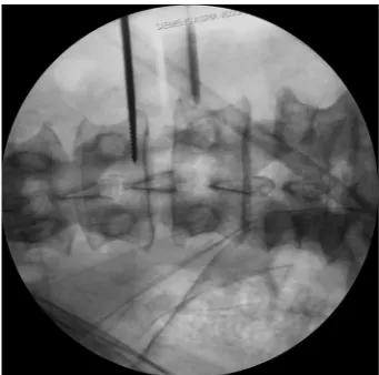
[image:7.595.81.251.232.395.2]Tubular retractor should be placed on an oblique trajectory, and the vertex of the retractor should be centered to the intervertebral disc (44). To get this, the initial needle should be positioned correctly. We suggest that the initial needle should always be introduced protected underneath using the other hand’s index finger to avoid injuring structures during its trajectory (Figure 5). Recognizing the intervertebral space and discriminating between the disc and vertebral body structures should always be palpated using the index finger that protects the introduction of the initial needle, that way any potential laceration to the segmental lumbar artery can be avoided. The needle should be inserted into the junction of the anterior third with the middle third of the intervertebral space in the lateral fluoroscopic projection. This advice could facilitate the oblique placement of the tubular retractor and will also aid in the orthogonal maneuver for the cage placement. The lateral and anteroposterior projections guarantee security within the tubular retractor placement. Direct vision is necessary during this step; there should not be any structures left under the valves of the tubular retractor because of potential risk to the ureter, sympathetic chain, or vascular structures (Figure 6).
[image:7.595.128.496.469.657.2]Figure 5 Lateral intraoperative fluoroscopic projections showing the proper placement of the tubular retractor. The tip of retractor is located in the midpoint of the disc.
Vascular lesion
These can be presented due to lesions on the segmental lumbar vessels, main vessels such as renal vein or artery, or major vessels that rest on the anterior surface of the spine (53). The renal vessels are found anterior to the L1–L2 intervertebral space, and so the surgeon should be able to anticipate this situation when approaching with OLIF at higher levels such as L1–L2 and L2–L3 (3). The segmental lumbar arteries are direct branches of the
aorta and thus supply blood to the caudal portion of the intervertebral foramen. These arteries run in a dorsolateral direction over the surface of the vertebral bodies. Orita et al. (53) described their location and angles based on magnetic resonance imaging. The angles on L1 and L3 are significantly acute (≤90 degrees) and substantially obtuse (>90 degrees) on L4 and L5. The lesion on the segmental vessels is associated to a laceration usually caused by the
pin fixation of the tubular retractor. We recommend great
caution upon the fixation of the tubular retractor at the
L4–L5 space and if possible to avoid the fixation on L5 to
prevent laceration to the iliolumbar vein (46). Moreover, the localization of these vessels should be prognosticated with the use of preoperative MRI. Palpation of the vertebral body with the initial needle while searching for the intervertebral body have to be avoided. Tubular retractor pin has to be inserted the most proximal to the endplates (53) (Figure 7). Damage to the great vessels that are found anteriorly to the lumbar spine is associated with its mobilization (3). This mobilization can be avoided through the use of a careful preoperative assessment through imaging and with the minimal medial exposure during the procedure (53).
Ureter lesion
[image:8.595.80.251.243.412.2]Various authors coincide in noting that the level of greater risk for a ureter lesion is L2–L3 (45). The tubular retractor can overlap the ureter, specifically at superior lumbar Figure 6 AP intraoperative fluoroscopic image. Retractor’s blade
is fixed with the pin in the most proximal part of the L3 inferior endplate.
[image:8.595.115.483.482.680.2]levels. Therefore, ureter can be easily injured during any stage of the retroperitoneal corridor dissection and the placement of the tubular retractor. Fujibayashi et al. (54), proposed the use of dual-phase contrast-enhanced CT and reconstructed 3D images to know its preoperative anatomy. In this study, the ureter was localized anterior to the psoas muscle in 90.4% of the cases, and laterally to the vertebral body in 16% of the cases. Some surgical strategies to avoid a ureter injury could be the complete retraction of the retroperitoneal fatty tissue before starting the discectomy, the anterior mobilization of the ureter and, the inspection of the intervertebral space through the tubular retractor without seeing structures underneath the valves. Lastly, the possibility of a ureter lesion should be considered when
faced with perioperative hematuria or non-specific signs and
symptoms in the postoperative scenario, such as abdominal pain, fever, vomit, ileum, leukocytosis, or abdominal distention (36,54).
Sympathetic chain lesion
The sympathetic chain can be found in the anterior third of the vertebral body (11). However, in spite of it being a frequently described complication in the literature, there is no technical advice reported for the preservation of its integrity (1,6,8,34). Kim et al. (34), reported an incidence of sympathetic chain injury of 13.4% in 29 patients underwent OLIF L4–L5. We believe that sympathetic chain injury symptoms are underestimated because in some patients the thermal discrepancy in both legs is minimal and the symptoms
are mild or reversible. Some authors suggest anterior mobilization after liberating the delicate communicating branches (11,42,52). The lesion to the sympathetic chain can be documented through physical exploration and the use of digital infrared thermal imaging (34). We recommend taking full advantage of the oblique vector that this approach offers so that the tubular retractor is placed posterior to the sympathetic chain and thus diminishing its manipulation.
Complications associated with the discectomy and the preparation of the endplates
[image:9.595.129.471.86.251.2]A repeated fluoroscopic image-based control is recommended during the release of the contralateral annulus and should be done cautiously. Blunt surgical tools in this step are recommended to avoid a contralateral psoas muscle and lumbar plexus injury (10). Another technical advice is being able to understand the orthogonal maneuver that is executed in the discectomy, the endplates preparation, and the intervertebral cage placement. This maneuver refers to the 90-degree angle that is formed with the instruments when placed perpendicularly to the sagittal plane of the vertebral body. The maneuver initiates with the placement of the tubular retractor in an oblique fashion, and posteriorly during the endplates preparation, there is a 90-degree angle correction of the instruments (Figure 8). This confers to a proper control of the cage placement and the possibility of placing it more posteriorly than what the DLIF approach can offer (6) (Figure 9). Chang et al. (35), published a case where there was a ventral dural sac tear on Figure 8 Intraoperative technique to demonstrate proper preparation of endplates and avoid over-preparation. (A) AP C-arm view shows no gap between contrast medium and endplates; (B) lateral C-arm view showing the location where the cage will be inserted.
an OLIF approach. This complication was due to disorientation in part of the surgeon upon fluoroscopic control during the endplates preparation as well as a failed orthogonal maneuver. Thus, the irruption to the spinal canal can be avoided by following the recommendations mentioned (10). In some cases, such as large disc herniation or disc herniation beyond the PLL
indirect decompression might not be sufficient. There have been
reports on endoscopic assistance for the OLIF to discectomy. The direct decompression through the conventional discectomy on OLIF is not recommended as it conveys a risk of spinal canal irruption (55,56).
Subsidence
Currently, no literature specifically talks about how to prevent subsidence in OLIF. However, some reports describe that the incidence oscillates between 0.3% and 22% on XLIF. The subsidence can be solely radiographic,
which is defined as a postoperative finding in images. When
the subsidence is clinical, it is associated with axial pain and recurrent neurological symptoms related to the loss of indirect decompression. The iatrogenic subsidence occurs when the endplates are damaged during their preparation, placement of the cage or immediately after so, and it tends to
be perioperative, associated with a deficient technique (57).
Hence, subsidence depends on multiple factors related to the technique, implant material, and bone quality of the patient.
Some specific situations reported in the literature are
over-distraction, multilevel fusion, and small cages. Also, there have been reports citing that the superior lumbar vertebrae endplates are more susceptible to suffering subsidence in respect to the inferior vertebrae endplates and that the superior endplate is weaker than the inferior endplate in all lumbar vertebrae (25,57-60). When the cage is being placed, it is important to have in account that the endplate is most resistant peripherally and weaker centrally. In this way, large and wide cages that have bilateral contact with the periphery of the endplates have a diminished risk of subsidence (3,59). It is recommended to avoid an aggressive end-plate preparation (61). We suggest filling out the intervertebral
space with contrast medium and taking fluoroscopic images
on the anteroposterior and lateral projections with the objective of evidencing an adequate endplates preparation and also avoiding an aggressive preparation (Figure 10). At last, the cage placement on the middle third of the intervertebral
space on the lateral projection of the fluoroscope, as well as the bilateral transpedicular fixation with screws can assist in
[image:10.595.73.260.85.269.2]avoiding the implant subsidence. Figure 9 OLIF surgical corridor is schematized in the axial view
of lumbar MRI. The yellow block exemplifies the tubular retractor. The red area is the space obtained with retraction of psoas muscle to the medial coronal plane (red arrows). OLIF, oblique lumbar interbody fusion.
[image:10.595.102.231.367.550.2]Conclusions
MIS-OLIF, for levels L2–L5, is a technique that has proven to have encouraging outcomes. However, there is still a need for high evidence-based, and larger sample sized studies to establish its feasibility entirely. Although its advantages concerning the lateral direct transpsoas (LLIF, DLIF o XLIF) technique are evident and the associated complications are few when they do present they can be catastrophic.
Acknowledgements
None.
Footnote
Conflicts of Interest: The authors have no conflicts of interest
to declare.
References
1. Silvestre C, Mac-Thiong JM, Hilmi R, et al.
Complications and morbidities of mini-open anterior retroperitoneal lumbar interbody fusion: oblique lumbar interbody fusion in 179 patients. Asian Spine J 2012;6:89-97.
2. Mobbs RJ, Phan K, Malham G, et al. Lumbar interbody fusion: techniques, indications and comparison of
interbody fusion options including PLIF, TLIF, MI-TLIF, OLIF/ATP, LLIF and ALIF. J Spine Surg 2015;1:2-18. 3. Liu L, Liang Y, Zhang H, et al. Imaging anatomical
research on the operative windows of oblique lumbar interbody fusion. PLoS One 2016;11:e0163452.
4. Phan K, Maharaj M, Assem Y, et al. Review of early clinical results and complications associated with oblique lumbar interbody fusion (OLIF). J Clin Neurosci 2016;31:23-9. 5. Mayer HM. A new microsurgical technique for minimally
invasive anterior lumbar interbody fusion. Spine (Phila Pa 1976) 1997;22:691-9.
6. Jin J, Ryu KS, Hur JW, et al. Comparative study of the difference of perioperative complication and radiologic results: MIS-DLIF (minimally invasive direct lateral lumbar interbody fusion) versus MIS-OLIF (minimally invasive oblique lateral lumbar interbody fusion). Clin Spine Surg 2018;31:31-6.
7. Ohtori S, Mannoji C, Orita S, et al. Mini-open anterior retroperitoneal lumbar interbody fusion: oblique
lateral interbody fusion for degenerated lumbar spinal kyphoscoliosis. Asian Spine J 2015;9:565-72.
8. Li JX, Phan K, Mobbs R. Oblique lumbar interbody fusion: technical aspects, operative outcomes, and complications. World Neurosurg 2017;98:113-23. 9. Woods KR, Billys JB, Hynes RA. Technical description of
oblique lateral interbody fusion at L1-L5 (OLIF25) and at L5-S1 (OLIF51) and evaluation of complication and fusion rates. Spine J 2017;17:545-53.
10. Abe K, Orita S, Mannoji C, et al. Perioperative complications in 155 patients who underwent oblique lateral interbody fusion surgery: perspectives and
indications from a retrospective, multicenter survey. Spine (Phila Pa 1976) 2017;42:55-62.
11. Mehren C, Mayer HM, Zandanell C, et al. The oblique anterolateral approach to the lumbar spine provides access to the lumbar spine with few early complications. Clin Orthop Relat Res 2016;474:2020-7.
12. Fujibayashi S, Kawakami N, Asazuma T, et al.
Complications associated with lateral interbody fusion: nationwide survey of 2998 cases during the first two years of its use in Japan. Spine (Phila Pa 1976) 2017;42:1478-84. 13. Fan SW, Hu ZJ, Fang XQ, et al. Comparison of paraspinal
muscle injury in one-level lumbar posterior inter-body fusion: modified minimally invasive and traditional open approaches. Orthop Surg 2010;2:194-200.
14. Wang YP, An JL, Sun YP, et al. Comparison of outcomes between minimally invasive transforaminal lumbar interbody fusion and traditional posterior lumbar intervertebral fusion in obese patients with lumbar disk prolapse. Ther Clin Risk Manag 2017;13:87-94. 15. Harms JG, Jeszenszky D. Die posteriore, lumbale,
interkorporelle fusion in unilateraler transforaminaler technik. Oper Orthop Traumatol 1998;10:90-102. 16. Foley KT, Holly LT, Schwender JD. Minimally invasive
lumbar fusion. Spine (Phila Pa 1976) 2003;28:S26-35. 17. Oppenheimer JH, DeCastro I, McDonnell DE. Minimally
invasive spine technology and minimally invasive spine surgery: a historical review. Neurosurg Focus 2009;27:E9. 18. Phan K, Rao PJ, Kam AC, et al. Minimally invasive versus open transforaminal lumbar interbody fusion for treatment of degenerative lumbar disease: systematic review and meta-analysis. Eur Spine J 2015;24:1017-30.
19. Capener N. Spondylolisthesis. Br J Surg 1932;19:374-86. 20. Choi KC, Kim JS, Shim HK, et al. Changes in the adjacent
21. Phan K, Thayaparan GK, Mobbs RJ. Anterior lumbar interbody fusion versus transforaminal lumbar interbody fusion – systematic review and meta-analysis. Br J Neurosurg 2015;29:705-11.
22. Kim JS, Choi WG, Lee SH. Minimally invasive anterior lumbar interbody fusion followed by percutaneous pedicle screw fixation for isthmic spondylolisthesis: minimum 5-year follow-up. Spine J 2010;10:404-9.
23. Kim JS, Kang BU, Lee SH, et al. Mini-transforaminal lumbar interbody fusion versus anterior lumbar interbody fusion augmented by percutaneous pedicle screw
fixation: a comparison of surgical outcomes in adult low-grade isthmic spondylolisthesis. J Spinal Disord Tech 2009;22:114-21.
24. Ozgur BM, Aryan HE, Pimenta L, et al. Extreme lateral interbody fusion (XLIF): a novel surgical technique for anterior lumbar interbody fusion. Spine J 2006;6:435-43. 25. Lowe TG, Hashim S, Wilson LA, et al. A biomechanical study of regional endplate strength and cage morphology as it relates to structural interbody support. Spine (Phila Pa 1976) 2004;29:2389-94.
26. Rodgers WB, Gerber EJ, Patterson J. Intraoperative and early postoperative complications in extreme lateral interbody fusion: an analysis of 600 cases. Spine (Phila Pa 1976) 2011;36:26-32.
27. Tormenti MJ, Maserati MB, Bonfield CM, et al. Complications and radiographic correction in adult scoliosis following combined transpsoas extreme lateral interbody fusion and posterior pedicle screw instrumentation. Neurosurg Focus 2010;28:E7. 28. Regev GJ, Kim CW. Safety and the anatomy of the
retroperitoneal lateral corridor with respect to the minimally invasive lateral lumbar intervertebral fusion approach. Neurosurg Clin N Am 2014;25:211-8. 29. Epstein NE. Extreme lateral lumbar interbody fusion:
Do the cons outweigh the pros? Surg Neurol Int 2016;7:S692-700.
30. Cheng I, Acosta F, Chang K, et al. Point-counterpoint: the use of neuromonitoring in lateral transpsoas surgery. Spine (Phila Pa 1976) 2016;41:S145-51.
31. Choma TJ, Mroz TE, Goldstein CL, et al. Emerging techniques in degenerative thoracolumbar surgery. Neurosurgery 2017;80:S55-S60.
32. Fujibayashi S, Hynes RA, Otsuki B, et al. Effect of indirect neural decompression through oblique lateral interbody fusion for degenerative lumbar disease. Spine (Phila Pa 1976) 2015;40:E175-82.
33. Kaiser MG, Haid RW Jr, Subach BR, et al. Comparison
of the mini-open versus laparoscopic approach for anterior lumbar interbody fusion: a retrospective review. Neurosurgery 2002;51:97-103; discussion 103-5. 34. Kim JS, Choi WS, Sung JH. 314 Minimally invasive
oblique lateral interbody fusion for L4-L5: clinical outcomes and perioperative complications. Neurosurgery 2016;63:190-1.
35. Chang J, Kim JS, Jo H. Ventral dural injury after oblique lumbar interbody fusion. World Neurosurg 2017;98:881. e1-881.e4.
36. Lee HJ, Kim JS, Ryu KS, et al. Ureter injury as a complication of oblique lumbar interbody fusion. World Neurosurg 2017;102:693.e7-693.e14.
37. Ohtori S, Orita S, Yamauchi K, et al. Mini-open anterior retroperitoneal lumbar interbody fusion: oblique lateral interbody fusion for lumbar spinal degeneration disease. Yonsei Med J 2015;56:1051-9.
38. Saraph V, Lerch C, Walochnik N, et al. Comparison of conventional versus minimally invasive extraperitoneal approach for anterior lumbar interbody fusion. Eur Spine J 2004;13:425-31.
39. Patel NP, Birch BD, Dement SE, et al. The mini-open anterolateral approach for degenerative thoracolumbar disease. Clin Neurol Neurosurg 2010;112:853-7. 40. Hynes RA. Oblique lateral interbody fusion (OLIF)
technique and complications in 457 levels L1-S1. International Society for the Advancement of Spine Surgery 2014 Meeting. 2014. Available online: http:// www.isass.org/abstracts/isass14_oral_posters/isass14-77- Oblique-Lateral-Interbody-Fusion-(OLIF)-Technique-and-Complications-in.html
41. Molloy S, Butler JS, Benton A, et al. A new extensile anterolateral retroperitoneal approach for lumbar interbody fusion from L1 to S1: a prospective series with clinical outcomes. Spine J 2016;16:786-91.
42. Gragnaniello C, Seex K. Anterior to psoas (ATP) fusion of the lumbar spine: evolution of a technique facilitated by changes in equipment. J Spine Surg 2016;2:256-65. 43. Sato J, Ohtori S, Orita S, et al. Radiographic evaluation
of indirect decompression of mini-open anterior retroperitoneal lumbar interbody fusion: oblique lateral interbody fusion for degenerated lumbar spondylolisthesis. Eur Spine J 2017;26:671-8.
lateral position: an anatomic study. J Neurosurg Spine 2014;21:785-93.
46. Hynes RA. Oblique lateral approach to the lumbar spine (L2-L5). In: Watkins III RG, Watkins IV RG, editors. Surgical approaches to the spine. New York: Springer; 2015;223-34.
47. Molinares DM, Davis TT, Fung DA, et al. Is the lateral jack-knife position responsible for cases of transient neuropraxia? J Neurosurg Spine 2016;24:189-96. 48. Mehren C, Korge A. Minimally invasive anterior
oblique lumbar interbody fusion (OLIF). Eur Spine J 2016;25:471-2.
49. Sofianos DA, Briseño MR, Abrams J, et al. Complications of the lateral transpsoas approach for lumbar interbody arthrodesis: a case series and literature review. Clin Orthop Relat Res 2012;470:1621-32.
50. Mirilas P, Skandalakis JE. Surgical anatomy of the retroperitoneal spaces, part IV: retroperitoneal nerves. Am Surg 2010;76:253-62.
51. Korenkov M, Rixen D, Paul A, et al. Combined abdominal wall paresis and incisional hernia after laparoscopic cholecystectomy. Surg Endosc 1999;13:268-9.
52. Gragnaniello C, Seex KA. Anterior to psoas fusion of the lumbar spine. Neurosurg Focus 2013;35:Video 13. 53. Orita S, Inage K, Sainoh T, et al. Lower lumbar segmental
arteries can intersect over the intervertebral disc in the oblique lateral interbody fusion approach with a risk for arterial injury: radiological analysis of lumbar segmental arteries by using magnetic resonance imaging. Spine (Phila
Pa 1976) 2017;42:135-42.
54. Fujibayashi S, Otsuki B, Kimura H, et al. Preoperative assessment of the ureter with dual-phase contrast-enhanced computed tomography for lateral lumbar interbody fusion procedures. J Orthop Sci 2017;22:420-4. 55. Heo DH, Choi WS, Park CK, et al. Minimally
invasive oblique lumbar interbody fusion with spinal endoscope assistance: technical note. World Neurosurg 2016;96:530-6.
56. Kim JS, Seong JH. Endoscope-assisted oblique lumbar interbody fusion for the treatment of cauda equina syndrome: a technical note. Eur Spine J 2017;26:397-403. 57. Satake K, Kanemura T, Yamaguchi H, et al. Predisposing
factors for intraoperative endplate injury of extreme lateral interbody fusion. Asian Spine J 2016;10:907-14.
58. Hou Y, Luo Z. A study on the structural properties of the lumbar endplate: histological structure, the effect of bone density, and spinal level. Spine (Phila Pa 1976) 2009;34:E427-433.
59. Le TV, Baaj AA, Dakwar E, et al. Subsidence of polyetheretherketone intervertebral cages in minimally invasive lateral retroperitoneal transpsoas lumbar interbody fusion. Spine (Phila Pa 1976) 2012;37:1268-73. 60. Grant JP, Oxland TR, Dvorak MF. Mapping the structural
properties of the lumbosacral vertebral endplates. Spine (Phila Pa 1976) 2001;26:889-96.
61. Uribe JS. Neural anatomy, neuromonitoring and related complications in extreme lateral interbody fusion: video lecture. Eur Spine J 2015;24 Suppl 3:445-6.