3058
Screening And Antibiotic Profile of Uropathogens with
Reference To Extended-spectrum Beta-lactamases
(ESBL)
Summera Rafiq
1*, Soifiat Abdou Soumara
2,
Sk. Jasmine Shahina
31* Head & Associate Professor, Department of Microbiology, Justice Basheer Ahmed Sayeed College for Women, Chennai-18.
2 Department of Microbiology, Justice Basheer Ahmed Sayeed College for Women, Chennai-18.
3 Associate Professor, Department of Microbiology, Justice Basheer Ahmed Sayeed College for Women,Chennai-18. * Corresponding author email: [email protected]
Abstract-The most common human bacterial infections are urinary tract infections with global infection rates nearing 180 million per year. These infections occur due to bacterial colonization of any tissue along the urinary tract, from the urethral opening to the kidneys. The emergence of antibiotic resistance in the management of urinary tract infections possesses a serious public health issue. In the present study 22 bacterial isolates were obtained from 15 urine sample collected from patients suspected with urinary tract infection. 9/22 (41%) were found to be gram positive cocci and 13/22 (59%) were found to be gram negative bacteria. Among the gram positive bacteria isolated the most predominant organism was found to be Staphylococcus aureus (56%) followed by Staphylococcus epidermidis (33%) and Enterococcus faecalis (11%). The most predominant gram negative bacteria were found to be E.coli (38%) followed by Klebsiella pneumoniae and Acinetobacter baumanii (23%), Pseudomonas aeruginosa and Proteus vulgaris (8%). 69% of gram negative bacteria were found to be ESBL positive. 54% of gram negative bacterial isolates were found to be positive for inducible AmpC β-lactamase.
Keyword:
ESBL, UTI, AmpC, MRSA.
1. INTRODUCTION
The most common bacterial infections are urinary tract infections (UTIs). UTIs accounts for 35% of nosocomial infections making them the most common hospital-acquired infection, and they are the second most common cause of bacteraemia in hospitalized patients (Stamm, 2002). UTI causes bacterial infections of one or more parts of urinary system after bacteria overcome the natural host defence mechanism (Al-Dujiaily, 2000). Bladder infection or cystitis is the infection of lower urinary tract and infections in the upper urinary tract is known as kidney infection or pyelonephritis. Urgency of urination, dysuria, pyuria, irritation of urinary tract, discomfortable pressure, bloody urine which may have a strong smell and tiredness are associated with cystitis. Fever and flank pain as well as the symptoms of cystitis are significant in kidney infections (Lane and Takhar, 2011).
These infections are caused by both Gram-negative which includes a large number of aerobic bacilli such as Escherichia sp, Klebsiella sp, Enterobacter sp, Citrobacter sp, Proteus sp, Serratia
sp, Salmonella sp and Pseudomonas sp. and Gram-positive bacteria which includes Staphylococcus sp, Streptococcus sp and Enterococcus sp. Among this 80-90% of UTI are caused by E. coli (Rushton, 1997) and in ambulatory patients and of nosocomial infections, Klebsiella pneumoniae, Proteus mirabilis, Staphylococcus aureus, Enterococcus faecalis are the most frequently isolated (Andreu et al., 2008).
One of the greatest discoveries of modern medicine has been antibiotics, but the development of microbial resistance towards antibiotics increased due to availability and increased use of antibiotics (Gottlieb and Nimmo, 2011). Developing countries show increasing amounts of antimicrobial resistance (Sadeghabadi et al., 2014). Antimicrobial resistance is increasingly a global threat for public health and a serious threat to modern medicine according to the World Health Organization in 2014.
2. MATERIALS AND METHODS
a) Collection of urine sample:
3059 patients suspected with urinary tract infections. The
specimens were labelled, transported to laboratory in ice packs and were processed within one hour for aerobic bacterial culture. Urine was observed by naked eyes for the altered colour, presence of turbidity and deposits.
b) Isolation and identification of bacterial isolates:
10 ml of the urine sample was transferred into sterile centrifuge tubes and centrifuged at 3000 rpm for 5 minutes. The supernatant was discarded and the sediment was inoculated on various media which included- Blood agar, MacConkey’s agar, cetrimide agar and nutrient agar. The plates were incubated at 37oC for 24 hrs. Colony morphology of the isolates was observed on following growth of the organisms. For pure culture single colony was further streaked on nutrient agar and incubated for 24 hours at 37°C. Bacterial isolates were identified based on the standard biochemical methods.
Antibiotic sensitivity testing
Once the bacterial isolate was identified, the antibiotic sensitivity testing was carried out by Kirby Bauer disc diffusion method (Bauer et al., 1996) for the following antibiotics- (in µg/disc)- ceftazidime (30mcg), ceftazidime/clavulanic acid (30 mcg/ 10 mcg), imipenem (30mcg), cefoxitin (30 mcg), ceftriaxone (30mcg), cefotaxime (30mcg), gentamicin, erythromycin, clindamycin, ofloxacin and teicoplanin.
Cefoxitin Disc diffusion Method organism were inoculated into Muller Hinton broth and incubated at 37°C for 3hrs.The turbidity of the growth was compared with 0.5 McFarland and lawn culture
ESBL screening by Disc diffusion test:
This test requires the use of ceftazidime (30mcg) alone and in combination with clavulanic acid (10mcg). Ceftazidime and with ceftazidime/clavulanic acid (30 mcg/10 mcg) discs were placed with distance between the two discs 10mm edge to edge on MHA
plate inoculated with standard inoculum (0.5 McFarland) of the test organism to form a lawn culture and was incubated overnight at 37oC. An increase in the zone diameter by > 5 mm of ceftazidime versus its zone when tested in combination with clavulanic acid was considered as an ESBL producer. (Jayakumar, 2007).
Disc Antagonism Test (AmpC β-lactamase inducibility) β-lactamase inducibility was recognized by isolates showing blunting of ceftazidime zone of inhibition adjacent to cefoxitin disc and were considered screen positive. (Sanders et al., 1982)
3. RESULTS
Prevalence of Bacterial Isolates from Urine Sample:
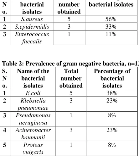
22 bacterial isolates were obtained from 15 urine sample collected from patients suspected with urinary tract infection. 9/22 (41%) were found to be gram positive cocci and 13/22 (59%) were found to be gram negative organisms. (Table 1 & 2)
Table 1: Prevalence of gram positive bacteria, n=9 S.
3060
Antibiotic sensitivity testing:
Gram positive bacteria exhibited 100% sensitivity towards teicoplanin, while they showed 63% resistance towards erythromycin followed by gentamicin (47%), clindamycin (36%) and ofloxacin (23%). Gram negative bacterial isolates exhibited 100% sensitivity towards imipenem. The isolates were found to be highly resistant to ceftazidime (78%) followed by cefotaxime (68%), cefoxitin (65%) and ceftriaxone (61%).
MRSA screening by cefoxitin disc diffusion method:
A total of 8 Staphylococcus sps were isolated in the present study. Out of the 8 isolates, 5 (63%) were found to be Staphylococcus aureus and 3 (37%) were Staphylococcus epidermidis. All the Staphylococcal isolates were subjected to MRSA screening by cefoxitin disc diffusion method. Among 5 Staphylococcus aureus, 2(40%) were found to be MRSA and 3 (60%) were found to be MSSA, while all the Staphylococcus epidermidis were found to be sensitive to methicillin.
Screening for ESBL production among gram negative organisms:
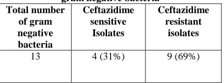
A total of 13 gram negative isolates were obtained in the present study. Out of the 13 isolates, 9 (69%) were found to be positive for the production of ESBL by disc diffusion method using ceftazidime and with ceftazidime/clavulanic acid. (Table 3)
Table 3: Ceftazidime antibiotic sensitivity pattern of gram negative bacteria
Total number of gram negative bacteria
Ceftazidime sensitive
Isolates
Ceftazidime resistant
isolates
13 4 (31%) 9 (69%)
Screening for AmpC β-lactamase production:
The disc antagonism test was done to detect inducible AmpC β-lactamase for all the isolates. 7/13 (54%) isolates were found to be positive for inducible AmpC β-lactamase. The isolates showed blunting of the ceftazidime zone of inhibition adjacent to the cefoxitin disc.
AmpC β-lactamase screening of gram negative bacteria
Total number of gram negative bacteria
Cefoxitin sensitive isolates
Cefoxitin resistant isolates
13 6 (46%) 7 (54%)
4. DISCUSSIONS
One of the most important causes of morbidity in the general population and the second most common cause of morbidity among hospital visitors is the urinary tract infection. UTI is also the most common causes of nosocomial infection among hospitalized patients (Ronald and Puttulo, 1991). UTI increases in men due to prostate enlargement and neurogenic bladder with advancing age (Liperky, 1989). Recurrent UTI are common and can lead to irreversible damage to the kidneys, resulting in renal hypertension and renal failure in server cases (New, 1992). Women are more prone to develop UTI in the community. About 20% of the women experience a single episode of UTI during their lifetime, and 3% of women had more than one episode of UTI per year (Gebre-Selassie, 1998). Pregnant women are more susceptible to the infection (Van Nostrand et al., 2000). UTI associated with catheter is a severe problem with about 10% of the patients developing bacteriuria (Srinivassa et al., 1999).
In the present study 22 bacterial isolates were obtained from 15 urine sample collected from patients suspected with urinary tract infection. 9/22 (41%) were found to be gram positive cocci which included Staphylococcus aureus, Staphylococcus epidermidis and Enterococcus faecalis and 13/22 (59%) were found to be gram negative organisms which included E.coli, Klebsiella pneumoniae, Pseudomonas aeruginosa, Acinetobacter baumanii and Proteus vulgaris. Among the gram negative bacteria, E.coli was the most common pathogen isolated predominantly. The isolation rate of urinary pathogens of the present study is found to be consistent with reports of the studies Chandra et al., 2013 and Savitha, 2011.
3061 cefotaxime (68%), cefoxitin (65%) and ceftriaxone
(61%). Amoxycillin with clavulanic acid which are commonly used antibiotics showed poor in-vitro sensitivity against majority of the organisms isolated in this study.
In the present study, a total of 5 Staphylococcus aureus isolates were obtained. Of the 5 isolates, 2 (40%) were found to be MRSA and 3 (60%) were found to be MSSA. The incidence of MRSA varies according to the region, 25% in Western part of India (Patel et al., 2010) to 50% in South India (Gopalakrishnan et al., 2010). The prevalence of MRSA in a study from Chennaiwas reported as 40-50 per cent. The prevalence of MRSA in the present study was found to be 40% which was in agreement with Patel and Gopalakrishnan.
ESBL producing E. coli isolates are frequently found to be resistant to other antibiotics, in particular fluoroquinolones (Lautenbach et al., 2001). Studies in some places like in Nagpur showed 50% of ESBL producers and in another study done in 2005, from New Delhi, showed 68.78 % of the strains of gram negative bacteria to be ESBL producers and this was in agreement with our study. Studies in other places like in Varanasi by Upadhyay et al.,(2010) showed the prevalence of ESBL producers to be 3.3% and Rodrigues et al.,(2004) in a study showed 5.9% of bacterialisolates harbouring ESBLs in Mumbai, which was less in comparison with our study. In the present study, a total of 13 gram negative isolates were obtained. Out of the 13 isolates, 9 (69%) were found to be positive for the production of ESBL by disc diffusion method using ceftazidime and with ceftazidime/clavulanic acid.
It is necessary to identify AmpC β-lactamase producing bacteria as they can cause major therapeutic failure if they remain undetected. AmpC β-lactamase producing organisms are increasing and they poses a major therapeutic challenge due to treatment failure (Arora and Bal, 2005), and have been responsible for several nosocomial outbreak. The disc antagonism test was done to detect inducible AmpC β-lactamase in all the isolates. In the present study, 7/13 (54%) isolates were found to be positive for inducible AmpC β-lactamase. The isolates showed blunting of the ceftazidime zone of inhibition adjacent to the cefoxitin disc. Our study exhibited more positive when compared with reports from Aligarh by Shahid et al.,(2004) in 2004 who reported 20% AmpC positivity, from Kolkata by Arora et al. in 2005 to be 17.3% (Arora and Bal,2005) and from Varanasi to be 22% (Bhattacharjee et al.,2008).
5. CONCLUSION
Our study shows high prevalence of ESBL producers among E.coli and Klebsiella pneumoniae. Gram positive bacteria showed high sensitivity towards teicoplanin, while gram negative bacteria towards imipenem. AmpC β-lactamase detection using disc antagonism method should be done for an effective UTI treatment. As different centres show variations among antibiotic resistance rates, it will be helpful that every region perform surveillance studies to determine local antibiotic resistance rates for the development of treatment protocols.
REFERENCE
[1] Stamm, W. E. (2002). Scientific and clinical challenges in the management of urinary tract infections. The American journal of medicine, 113(1), 1-4.
[2] Al-Dujiaily, A. A. (2000). Urinary tract infection during pregnancy in Tikrit. Medical Journal of Tikrit, 6(3), 220-4.
[3] Lane, D. R., & Takhar, S. S. (2011). Diagnosis and management of urinary tract infection and pyelonephritis. Emergency Medicine Clinics, 29(3), 539-552.
[4] Rushton, H. G. (1997). Urinary tract infections in children: epidemiology, evaluation, and management. Pediatric Clinics of North America, 44(5), 1133-1169.
[5] Andreu, A., Planells, I., & Spanish Cooperative Group for the Study of Antimicrobial Sensitivity of Urinary Pathogens. (2008). Etiology of low urinary infection acquired in the community and resistance of Escherichia coli to first-line antimicrobials. National multicentric study. Clinical Medicine, 130 (13), 481-486. [6] Gottlieb, T., & Nimmo, G. R. (2011). Antibiotic
resistance is an emerging threat to public health: an urgent call to action at the Antimicrobial Resistance Summit 2011. Med J Aust, 194(6), 281-3.
[7] Sadeghabadi, A. F., Ajami, A., Fadaei, R., Zandieh, M., Heidari, E., Sadeghi, M., Ataei, B. & Hoseini, S. G. (2014). Widespread antibiotic resistance of diarrheagenic Escherichia coli and Shigella species. Journal of research in medical sciences: the official journal of Isfahan University of Medical Sciences, 19(Suppl 1), S51.
3062 [9] Jayakumar, S., & Appalaraju, B. (2007).
Prevalence of multi and pan drug resistant Pseudomonas aeruginosa with respect to ESBL and MBL in. Indian J Pathol Microbiol, 50(4). [10] Sanders, C.C; Sanders, W.E; Goering, H.V.(1982).
In vitro antagonism of β lactam antibiotics by cefoxitin. J Antimicrob Chemother. 21.968- 75. [11] Ronald, A. R., & Pattullo, A. L. (1991). The
natural history of urinary infection in adults. The Medical clinics of North America, 75(2), 299-312. [12] Lipsky, B. A. (1989). Urinary tract infections in
men: epidemiology, pathophysiology, diagnosis, and treatment. Annals of internal medicine, 110(2), 138-150.
[13] New CH. Urinary tract infection. Am J Med 1992; 4A (supp 1): 63-7.
[14] Gebre-Selassie, S. (1998). Asymptomatic bacteriuria in pregnancy: epidemiological, clinical and microbiological approach. Ethiopian medical journal, 36(3), 185-192.
[15] Van Nostrand, J. D., Junkins, A. D., & Bartholdi, R. K. (2000). Poor predictive ability of urinalysis and microscopic examination to detect urinary tract infection. American journal of clinical pathology, 113(5), 709-713.
[16] Srinivassa, H., Parija, S. C., Bhattacharya, S., & Sehgal, R. (1999). Incidence of ciprofloxacin resistance in urinary isolates. Eastern Nepal J Comm Dis, 31, 45-47.
[17] Chandra Mondal, K., Kumar Maji, S., Maity, C., Kumar Halder, S., Paul, T., & Kumar Kundu, P. (2013). Studies on drug sensitivity and bacterial prevalence of UTI in tribal population of paschim Medinipur, West Bengal, India. Jundishapur Journal of Microbiology, 6(1), 42-46.
[18] Savitha, T. 2011. Urinary tract infection among patients at G.G.Hospital & Medical College, Jamnagar. Int. J. Curr. Res., 2(1): 067- 072. [19] Manges, A. R., Natarajan, P., Solberg, O. D.,
Dietrich, P. S., & Riley, L. W. (2006). The changing prevalence of drug-resistant Escherichia coli clonal groups in a community: evidence for community outbreaks of urinary tract infections. Epidemiology & Infection, 134(2), 425-431.
[20] Kahan, N. R., Chinitz, D. P., Waitman, D. A., Dushnitzky, D., Kahan, E., & Shapiro, M. (2006). Empiric treatment of uncomplicated urinary tract infection with fluoroquinolones in older women in Israel: another lost treatment option?. Annals of Pharmacotherapy, 40(12), 2223-2227.
[21] Patel, A. K., Patel, K. K., Patel, K. R., Shah, S., & Dileep, P. (2010). Time trends in the epidemiology
of microbial infections at a tertiary care center in west India over last 5 years. J Assoc Physicians India, 58(Suppl), 37-40.
[22] Gopalakrishnan, R., & Sureshkumar, D. (2010). Changing trends in antimicrobial susceptibility and hospital acquired infections over an 8 year period in a tertiary care hospital in relation to introduction of an infection control programme. J Assoc Physicians India, 58(Suppl), 25-31.
[23] Lautenbach, E., Patel, J. B., Bilker, W. B., Edelstein, P. H., & Fishman, N. O. (2001).
Extended-spectrum β-lactamase-producing
Escherichia coli and Klebsiella pneumoniae: risk factors for infection and impact of resistance on outcomes. Clinical Infectious Diseases, 32(8), 1162-1171.
[24] Upadhyay, S., Sen, M. R., & Bhattacharjee, A. (2010). Presence of different beta-lactamase classes among clinical isolates of Pseudomonas aeruginosa expressing AmpC beta-lactamase enzyme. The Journal of Infection in Developing Countries, 4(04), 239-242.
[25] Rodrigues, C., Joshi, P., Jani, S. H., Alphonse, M., Radhakrishnan, R., & Mehta, A. (2004). Detection of-lactamases in nosocomial gram negative clinical isolates. Indian journal of medical microbiology, 22(4), 247.
[26] Arora, S., & Bal, M. (2005). AmpC beta-lactamase producing bacterial isolates from Kolkata hospital. Indian Journal of Medical Research, 122(3), 224-233.
[27] Shahid, M., Malik, A., Agrawal, M., & Singhal, S. (2004). Phenotypic detection of extended-spectrum and AmpC β-lactamases by a new spot-inoculation method and modified three-dimensional extract test: comparison with the conventional three-dimensional extract test. Journal of Antimicrobial Chemotherapy, 54(3), 684-687.