R E S E A R C H
Open Access
Fucoidan from
Fucus vesiculosus
suppresses
hepatitis B virus replication by enhancing
extracellular signal-regulated Kinase
activation
Huifang Li
1, Junru Li
1, Yuan Tang
1, Lin Lin
1,2, Zhanglian Xie
3, Jia Zhou
1, Liyun Zhang
1, Xiaoyong Zhang
3,
Xiaoshan Zhao
4, Zhengliang Chen
1,5and Daming Zuo
1,5*Abstract
Background:Hepatitis B virus (HBV) infection is a serious public health problem leading to cirrhosis and hepatocellular carcinoma. As the clinical utility of current therapies is limited, the development of new therapeutic approaches for the prevention and treatment of HBV infection is imperative. Fucoidan is a natural sulfated polysaccharide that extracted from different species of brown seaweed, which was reported to exhibit various bioactivities. However, it remains unclear whether fucoidan influences HBV replication or not.
Methods:The HBV-infected mouse model was established by hydrodynamic injection of HBV replicative plasmid, and the mice were treated with saline or fucoidan respectively. Besides, we also tested the inhibitory effect of fucoidan against HBV infection in HBV-transfected cell lines.
Results: The result showed that fucoidan fromFucus vesiculosusdecreased serum HBV DNA, HBsAg and HBeAg levels and hepatic HBcAg expression in HBV-infected mice. Moreover, fucoidan treatment also suppressed intracellular HBcAg expression and the secretion of the HBV DNA as well as HBsAg and HBeAg in HBV-expressing cells. Furthermore, we proved that the inhibitory activity by fucoidan was due to the activation of the extracellular signal-regulated kinase (ERK) pathway and the subsequent production of type I interferon. Using specific inhibitor of ERK pathway abrogated the fucoidan-mediated inhibition of HBV replication.
Conclusion:This study highlights that fucoidan might be served as an alternative therapeutic approach for the treatment of HBV infection.
Keywords:Fucoidan, Hepatitis B virus, Extracellular signal-regulated kinase, Interferon, Anti-viral effect
Background
Hepatitis B virus (HBV) infection is a global public health problem. The infection can lead to acute and chronic hepatitis, which makes liver prone to develop cirrhosis and hepatocellular carcinoma [1]. Although an effective vaccine has been used for 20 years in many countries, there are still about 350 million chronic carriers of HBV worldwide, and approximately 1 million people die from
HBV-related liver diseases each year [2]. The currently ap-proved agents for the treatment of HBV infection include immunomodulatory agents, such as interferon-α (IFN-α) and pegylated IFN-α, and oral nucleoside/nucleotide ana-logues, like lamivudine, adefovir, telbivudine, entecavir and tenofovir [3, 4]. However, the major drawback of nu-cleoside/nucleotide analogues therapies is the develop-ment of drug resistance mutations with long-term treatment [3], and the disadvantages to using interferon-α are its various side effects and inconvenience of adminis-tration [4]. Therefore, exploring and developing new anti-HBV agents different from nucleoside/nucleotide ana-logues and interferon are urgently needed.
* Correspondence:zdaming@smu.edu.cn 1
Department of Immunology, School of Basic Medical Sciences, Southern Medical University, Guangzhou, Guangdong 510515, China
5Guangdong Provincial Key Laboratory of Proteomics, Southern Medical
University, Guangzhou, Guangdong 510515, China
Full list of author information is available at the end of the article
Fucoidan is a natural sulfated polysaccharide that ex-tracted from different species of brown seaweed [5], which was reported to exhibit numerous biological activities, in-cluding bacterial, oxidant, coagulant, anti-viral, anti-tumor, and anti-inflammatory effects [6–10]. These functional properties of fucoidan make it an attract-ive candidate for the development of biomaterials and drugs. Fucoidan isolated from Fucus vesiculosus is com-posed of 44.1% fucose, 26.3% sulfate, 31.1% ash, and a small amount of aminoglucose, which is commercially available now [5]. It has undergone extensive in vitro, in vivo as well as human clinical testing, and has also been shown to have a range of biological properties, including beneficial effects in the areas of inflammation, immune modulation, and enzyme inhibition [11]. There are several literatures concerning the anti-viral bioactivity of fucoidan isolated from Fucus vesiculosus[11–13]. Beress et al., de-scribed an anti-human immunodeficiency virus (HIV) ac-tivity of some fractions of self-produced Fucus vesiculosus fucoidan [12]. Besides, fucoidan displayed antiviral activ-ities against influenza A infection by different subtypes [14, 15]. A recent clinical study showed that treatment with fucoidan suppressed the expression of hepatitis C virus (HCV) replicon, accompanied by limited serum alanine aminotransferase levels, in HCV-infected patients [16]. However, it remains to be studied whether and how fucoi-dan inhibits HBV replication in vivo and in vitro.
In this study, we determined that fucoidan isolated fromFucus vesiculosuseffectively inhibited HBV replica-tion in an HBV replicareplica-tion mouse model in vivo and in HepG2.2.15 cells in vitro. Furthermore, we provided the evidence that fucoidan activated mitogen-activated pro-tein kinases (MAPKs) extracellular signal-regulated kin-ase 1/2 (ERK1/2) pathway and subsequently promoted the expression of IFN-α, leading the decreased produc-tion of HBV DNA and proteins. These data suggest the possibility of using fucoidan as an alternative therapeutic strategy for HBV infection.
Methods
Animal
C57BL/6 mice (male, 6–8 weeks old) were purchased from the Laboratory Animal Center of Southern Medical University (Guangzhou, China). The HBV infection mouse model was established as described previously [17]. Briefly, 10 μg of pHBV1.3 plasmid, carrying a ter-minally redundant (1.3-fold) replication-competent HBV genome, was injected into the tail vein of mice with in 5 s in a volume of PBS equivalent to 8% of the mouse body weight. For fucoidan treatment, HBV-infected mice were intraperitoneally injected with 100 mg fucoidan at 0, 1, 3, 5, and 7 days post-infection. The levels of HBV DNA, HBsAg and HBeAg in the serum and the
expression of HBcAg in liver tissues of mice were de-tected at the indicated time point.
Reagents and antibodies
Fucoidan fromFucus vesiculosuswas purchased from Sigma (St. Louis, Mo, USA.). Human IFN-αELISA kit and mouse IFN-α ELISA kit were purchased from Cloud-Clone Corp (Wuhan, China). The primary antibodies were purchased from Cell Signaling Technology (Danvers, MA, USA), in-cluding the antibodies against phospho-ERK1/2, ERK1/2, phpspho-p38, p38, phospho-JNK, JNK, phospho-IRF3, IRF3 and phospho-IRF7, IRF7, and GAPDH. The antibodies against HBsAg and HBcAg were obtained from Thermo Fisher (MS-314, RB-1413, Thermo Fisher, IL, U.S.A.). ERK inhibitor U0126 was from Cell Signaling Technology.
Cell culture and transfection
The HBV-producing cell line HepG2.2.15 were main-tained in RPMI 1640 medium supplemented with 10% fetal bovine serum and 400 μg/mL of G418, and incu-bated at 37 °C in an atmosphere of 5% CO2. HepG2 cells were seeded in 6-well plates at a density of 6 × 10⁵cells per well and were transfected with 3 μg of plasmid pHBV1.3 with PEI (Polysciences, Warrington, PA, USA) following the manufacturer’s instruction.
Cytotoxicity assay
HepG2.2.15 cells (1 × 10⁴ cells/well) were cultured in 96-well plates and incubated with varying concentrations of fucoidan for 24 or 48 h. Cytotoxic effect of fucoidan on HepG2.2.15 cells was measured via CCK-8 assays (Dojindo Laboratories, Kumamoto, Japan) following the manufacturer’s instruction. The absorbance at 450 nm was measured using a microplate reader.
Determination of HBsAg and HBeAg
HepG2.2.15 cells were seeded in 6-well plates at a density of 1 × 10⁶cells per well and cultured for 12 h, then treated with indicated concentrations of fucoidan for another 24 h. The levels of HBsAg and HBeAg in the supernatants were determined using ELISA kits (RongSheng Bioengineering, Shanghai, China) according to the manufacturer’s instruc-tion. The absorbance at 450 nm was measured by a micro-plate reader.
Western blot analysis
antibodies, followed by incubation with the horseradish peroxidase-conjugated secondary antibody for 1 h at room temperature. Finally, the membranes were washed three times, and detection of the target protein was conducted with enhanced chemiluminescence (Thermo Fisher).
Southern blot analysis
The isolation of HBV DNA replication intermediates were performed as previously described [18]. Samples were electrophoresed into agarose gels and blotted onto Hybond-XL membranes (GE Healthcare, Marlborough, MA, USA). Then, the membranes were probed with a DIG-labeled specific full-length HBV riboprobe, generated using the PCR DIG Probe Synthesis Kit (Roche, Basel, Switzerland), and visualized with CDP-Star using an Ima-geQuant LAS 2000 mini system (GE Healthcare).
HBV DNA isolation and real-time PCR
Intracellular nucleocapsid-associated viral DNA was ex-tracted from HepG2.2.15 cells. SYBR Green quantitative RT-PCR was performed to determine the gene expres-sion level using a 7900HT fast real-time PCR system (Applied Biosystems, San Francisco, CA, USA), accord-ing to the protocols provided with the SYBR Premix EX Taq (TaKaRa). The primers used to amplify were: HBV DNA (forward 5′-ACC AAT CGC CAG TCA GGA AG-3′and reverse 5′-ACC AGC AGG GAA ATA CAG GC-3′); GAPDH (forward 5′-CAT CAC TGC CAC CCA GAA GAC TG-3′and reverse 5′-ATG CCA GTG AGC TTC CCG TTC AG-3′). The levels of target gene were normalized with respect to GAPDH gene expression.
Northern blot analysis
Total RNA was isolated from HepG2.2.15 cells using Trizol reagent (Invitrogen). For Northern blotting, 30μg total RNA was electrophoresed in a 1% formaldehyde agarose gel containing 5% formaldehyde and blotted onto Hybond-XL membranes (GE Healthcare, Marlbor-ough, MA, USA). Then, the membrane was probed with a DIG-labeled specific full-length HBV riboprobe, gener-ated using the PCR DIG Probe Synthesis Kit (Roche, Ba-sel, Switzerland), and visualized with CDP-Star using an ImageQuant LAS 2000 mini system (GE Healthcare). Fi-nally, the blot was stripped and rehybridized with a DIG-labeled GAPDH probe for normalization.
Statistical analysis
The experimental data were evaluated by calculating the mean ± SD. One-way ANOVA was used for compari-sons among multiple groups. Differences between two groups within experiments were analyzed by Student’st -test. All the experiments were repeated at least three times independently. Values ofp< 0.05 were considered statistically significant.
Results
Inhibitory activity of fucoidan on HBV replication in vivo
We first investigated whether fucoidan can inhibit HBV replication in vivo. C57BL/6 mice were hydrodynamic-injected with 10 μg of pHBV1.3 plasmid through the tail vein. Meanwhile, the mice were intraperitoneally administered with 100 mg of fucoidan at 0, 1, 3, 5, and 7 days post-infection. Mouse serum was collected at in-dicated time points after the injection. Serum HBV DNA was analyzed by using real-time PCR, and HBsAg/HBeAg levels were analyzed by using ELISA. As shown in Fig. 1a, serum HBV DNA level was signifi-cantly reduced after injection of fucoidan. Moreover, fucoidan administration also suppressed HBsAg and HBeAg secretion (Fig. 1b and c). Immunohistochemical analysis showed that fucoidan suppressed the HBcAg expression in the liver tissues from HBV-infected mice (Fig. 1d). These results indicated that fucoidan could inhibit HBV replication in vivo.
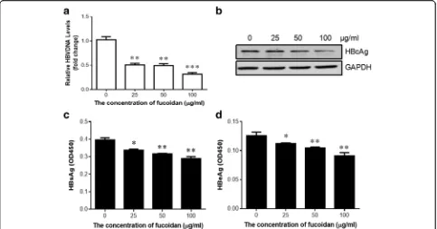
Fucoidan inhibited the HBV replication in vitro
Next, we evaluated the effect of fucoidan on HBV replica-tion in vitro. CCK8 assay showed that fucoidan was not cytotoxic at concentrations lower than 200 μg/ml on HepG2.2.15 cells (Fig. 2a). Real-time PCR revealed that fucoidan treatment reduced the levels of HBV DNA repli-cative intermediates in a dose-dependent manner (Fig. 2b). Additionally, the inhibitory effect of fucoidan on HBV DNA replicative intermediates was confirmed by southern blotting analysis (Fig. 2c). To explore whether the observed reduction of HBV DNA replication in HepG2.2.15 cells by fucoidan was due to its impact on HBV RNA synthesis. Northern blotting was performed to measure the intracel-lular HBV RNA levels after fucoidan treatment. The results showed that treatment with different concentrations of fucoidan induced a dose-dependent decrease in the levels of HBV RNA in HepG2.2.15 cells (Fig.2d). Secretion of HBsAg and HBeAg in the culture supernatants was also significantly decreased in the fucoidan-treated cells com-pared to the control cells (Fig. 2e and f). Western blotting showed that fucoidan markedly limited the expression of HBsAg and HBcAg in HepG2.2.15 cells (Fig. 2g).
Fig. 2Fucoidan suppresses HBV replication in the HepG2.2.15 cell line.aCells were incubated with varying concentrations of fucoidan for 24 or 48 h. Cytotoxic effect of fucoidan on HepG2.2.15 cells was measured via CCK-8 assays.b-gHepG2.2.15 cells were treated with the indicated concentrations of fucoidan for 24 h.b,cThe HBV DNA replicative intermediates were determined by quantitative PCR (b) and Southern blotting (c).dNorthern blotting was performed to measure the intracellular HBV RNA levels.e,fThe levels of HBsAg (e) and HBeAg (f) in the cell culture medium were analyzed via ELISA.
gThe expressions of HBsAg and HBcAg in HepG2.2.15 cells were examined by western blotting analysis. HepG2 cells was served as a negative control. *
p< 0.05, **p< 0.01, ***p< 0.001, compared to the group without fucoidan treatment. All the experiments were repeated at least three times independently
together, these data suggested that fucoidan repressed HBV replication in vitro.
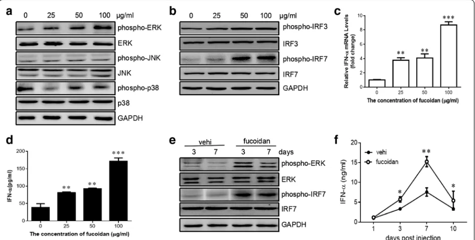
Fucoidan enhanced the activation of ERK pathway and facilitated the production of type I interferon
After determination of the effect of fucoidan in sup-pressing HBV replication in vivo and in vitro, we next moved on to elucidate the underlying mechanism. It has been reported that activation of MAPK pathway could lead to the suppression of HBV replication [19]. We, therefore, sought to determine whether the MAPK members (i.e. ERK1/2, p38, and JNK) were activated in HepG2.2.15 cells by fucoidan treatment. The level of phosphorylated ERK was significantly increased after stimulation with 100μg/ml of fucoidan, while the phos-phorylation of p38 and JNK was comparable between the cell with or without fucoidan treatment (Fig. 4a).
To determine whether fucoidan has the capacity to regulate type I interferon response, we examined the ef-fect of fucoidan on the activation of interferon regula-tory factor 3 (IRF3) and IRF7 in HepG2.2.15 cells. The result showed that fucoidan significantly promoted the phosphorylation of IRF3 and IRF7 in the HBV-transfected cells (Fig. 4b). Also, fucoidan enhanced the production of IFN-α at both mRNA level and protein level (Fig. 4c and d). To support these findings, we next investigated whether fucoidan has the capacity to
regulate type I interferon response in vivo. Western blot-ting showed more phosphorylated IRF7 and ERK in the liver of mice with fucoidan treatment than that of con-trol group (Fig. 4e). Meanwhile, decreased level of IFN-α was detected in the serum samples from fucoidan-treated HBV-infected mice compared to those from con-trol mice. (Fig. 4f ). These results demonstrated that fucoidan selectively activated ERK pathway and subse-quently enhanced type I interferon response in vitro and in vivo.
Blockage of ERK activation abolished the inhibitory activity of fucoidan on HBV replication
To validate the impact of ERK activation on fucoidan-mediated anti-viral effect, HepG2.2.15 cells were treated with U0126 to block ERK activation prior to incubation with 100 μg/ml of fucoidan. The result indicated that 2 μM of U0126 is sufficient to block ERK activation (Fig. 5a). After ERK inhibition, fucoidan-mediated en-hancement of expression of IFN-α was significantly inhibited at either mRNA level or protein level (Fig. 5b and c). In the culture supernatants, the level of HBV-DNA (Fig. 5d) and secretion of HBsAg and HBeAg (Fig. 5e and f ) were comparable between the control cells and fucoidan-treated cells with U0126 pretreatment. The data indicated that fucoidan-mediated inhibition of HBV replication was disturbed after pretreatment with ERK
inhibitor. The western blotting analysis of the expression of HBcAg in the cells supported the above results (Fig. 5g). Together, these results strongly indicated that the fucoidan-mediated suppression of HBV was dependent on ERK activation.
Discussion
Fucoidan, a natural sulfated polysaccharide that exists mainly in the cell wall matrix of various species of brown seaweed, has been shown to possess anti-cancer, anti-oxidant, anti-obesity and anti-diabetic effects [5, 20]. In this study, we demonstrated that fucoidan effect-ively inhibits HBV replication, and its mechanism par-tially occurs through the activation of ERK pathway and increased production of type I interferon.
It has been suggested that fucoidan has antivirus activ-ity against many virus, such as HIV [12], herpes simplex virus (HSV) [13], influenza A virus [14], and newcastle disease virus [21]. Mori et al., previously found that fucoidan fromCladosiphon okamuranushas HCV repli-cation suppressive effects in the cellular analysis [16]. After oral administration of fucoidan, HCV-infected pa-tients showed a lower level of serum alanine aminotrans-ferase, correlated with a significant decrease in HCV RNA. Therefore, we expected that fucoidan might have
potentially beneficial effects in patients with chronic hepatitis B virus infection. To our knowledge, this study reported for the first time that fucoidan effectively inhib-ited HBV replication in vivo and in vitro. We found that treatment with fucoidan inhibited the total HBVRNA and the replication of HBV DNA replicative intermedi-ates. Moreover, fucoidan treatment also reduced the amount of HBV core protein and the secretion of HBsAg and HBeAg. Given that the brown seaweeds containing fucoidan are widely consumed as part of the regular diet in East Asia (particularly China, Japan, and Korea), fucoidan is expected to have less drug-resistance com-pared to currently available anti-HBV agents. However, the mechanism of the effect of fucoidan on HBV gene expression should be elucidated with a variety of studies in vitro and in vivo, which remain need further investigation.
Activation of the MEK-ERK pathway has been re-ported to enhance the replication of viruses, such as HIV [22], influenza [23], and HSV [24]. By contrast, in the case of HBV, activation of MEK-ERK signaling led to the inhibition of HBV replication [19]. Several previous researches have pointed out that fucoidan could upregu-late the phosphorylation of ERK in immune cells in re-sponse to diverse pro-inflammatory signals [25, 26]. Our
Fig. 4Fucoidan promotes the activation of ERK pathway and enhances the production of type I interferon in vitro and in vivo.a-dHepG2.2.15 cells were incubated with indicated concentrations of fucoidan for 24 h.aPhospho-ERK, total ERK, phospho-JNK, total JNK, phospho-p38, and total p38 were determined by western blotting.bPhospho-IRF3, total IRF3, phospho-IRF7, and total IRF7 were examined by western blotting.c
The production of IFN-αin the culture supernatant was evaluated by ELISA analysis.dThe mRNA level of IFN-αin cells was determined by quantitative RT-PCR.e,fC57BL/6 mice were hydrodynamically injected with 10μg of pHBV1.3 plasmids through the tail vein. The mice were intraperitoneally adminis-trated with 100 mg fucoidan at 0, 1, 3, 5, and 7 days post-infection.ePhospho-ERK, total ERK, phospho-IRF7, and total IRF7 in the liver tissue of infected mice were examined by western blotting at the indicated days post-infection.fThe production of IFN-αin the serum was evaluated by ELISA analysis. *
data here demonstrated that fucoidan also increased the levels of phosphorylated ERK in hepatocytes and simul-taneously suppressed HBV replication. When the specific inhibitors of ERK were added, both ERK phosphorylation and the anti-HBV activity of fucoidan were blocked. Taken together, the results revealed that fucoidan activated MEK-ERK signaling pathway contributes to the inhibition of HBV replication. It remains to be determined how fucoidan activates ERK in the hepatocyte.
In the early phase of HBV infection, type I IFN also controls viral infection by directly inducing antiviral in-fection in cells [2, 27]. IRF3 and IRF7, which are highly homologous, has been shown to play a role in the tran-scriptional activation of virus-inducible cellular genes, especially type I IFN gene expression [28]. Upon viral in-fection, IRF3 and IRF7 reside in the cytosol and under-goes serine phosphorylation in its C-terminal region, allowing their homo- or heterodimerization and nuclear translocation. The dimers then translocate to the nu-cleus and activate the transcription of type I IFN genes [28]. Fucoidan was thought to inhibit virus infection via blocking virus adsorption to the cell surface and subse-quent virus entry [29]. Our current study provided the evidence that fucoidan enhanced the activation of IRF3 and IRF7 in the condition of HBV infection, which ex-pands our understanding of the mechanism of fucoidan-mediated antiviral effect. Indeed, Wang et al., observed
that virus infection of primary mouse embryo fibroblasts elicited ERK signaling, which was integrated into IRF3/7 activation and type I interferon induction [30]. Interest-ingly, the inhibitor of ERK U0126 attenuated fucoidan inducing IFN-α production, which suggests that fucoi-dan promotes type I interferon immune responses by ac-tivating ERK.
Conclusion
We conclude that fucoidan inhibits HBV replication in vivo and in vitro. Our results demonstrated that fucoi-dan suppresses HBV gene expression by activating the ERK signaling pathway and subsequently enhancing the production of type I interferon. This new mechanism suggests another possible approach to inhibit HBV repli-cation. Furthermore, either fucoidan alone or fucoidan combined with other established anti-HBV agents may serve as a new therapeutic strategy for HBV prevention and treatment.
Abbreviations
ERK:Extracellular signal-regulated kinase; HBcAg: Hepatitis B core antigen; HBeAg: Hepatitis B e antigen; HBsAg: Hepatitis B surface antigen;
HBV: Hepatitis B virus; HCV: Hepatitis C virus; HIV: Human immunodeficiency virus; HSV: Herpes simplex virus; IFN: Interferon; IRF: Interferon regulatory factor; MAPK: Mitogen-activated protein kinase
Acknowledgments
Not applicable
Funding
This work was supported in part by National Natural Science Foundation of China (grant no.: 81671568), Science and Technology Planning Project of Guangdong Province (grant no.: 2016A020215106), and Science and Technology Planning Project of Guangzhou (grant no.: 201607010195).
Availability of data and materials
All data generated or analysed during this study are included in this published article.
Authors’contributions
DZ and ZC designed research. HL, YT, LL, JL, and LZ performed experiments. HL, YT, JZ, YZ, Xiaoyong Zhang, Xiaoshan Zhao and DZ analyzed data. DZ, HL, and ZC wrote the manuscript. All authors have read and approved final manuscript.
Ethics approval
All animal experiments in this study were approved by the Welfare and Ethical Committee for Experimental Animal Care of Southern Medical University.
Consent for publication
Not applicable
Competing interests
The authors declare that they have no competing interest.
Publisher’s Note
Springer Nature remains neutral with regard to jurisdictional claims in published maps and institutional affiliations.
Author details
1
Department of Immunology, School of Basic Medical Sciences, Southern Medical University, Guangzhou, Guangdong 510515, China.2Department of
Dermatology, Zhujiang Hospital, Southern Medical University, Guangzhou, Guangdong 510282, China.3State Key Laboratory of Organ Failure Research,
Guangdong Provincial Key Laboratory of Viral Hepatitis Research, Department of Infectious Diseases, Nanfang Hospital, Southern Medical University, Guangzhou, Guangdong 510515, China.4School of Traditional Chinese Medicine, Southern Medical University, Guangzhou, Guangdong 510515, China.5Guangdong Provincial Key Laboratory of Proteomics, Southern Medical University, Guangzhou, Guangdong 510515, China.
Received: 27 June 2017 Accepted: 12 September 2017
References
1. Ganem D, Prince AM. Hepatitis B virus infection–natural history and clinical consequences. N Engl J Med. 2004;350(11):1118–29.
2. Dandri M, Locarnini S. New insight in the pathobiology of hepatitis B virus infection. Gut. 2012;61(Suppl 1):i6–17.
3. Fung J, Lai CL, Seto WK, Yuen MF. Nucleoside/nucleotide analogues in the treatment of chronic hepatitis B. J Antimicrob Chemother. 2011;66(12): 2715–25.
4. Perrillo R. Benefits and risks of interferon therapy for hepatitis B. Hepatology. 2009;49(5 Suppl):S103–11.
5. Li B, Lu F, Wei X, Zhao R. Fucoidan: structure and bioactivity. Molecules. 2008;13(8):1671–95.
6. Ale MT, Mikkelsen JD, Meyer AS. Important determinants for fucoidan bioactivity: a critical review of structure-function relations and extraction methods for fucose-containing sulfated polysaccharides from brown seaweeds. Mar Drugs. 2011;9(10):2106–30.
7. Zapopozhets TS, Besednova NN, Loenko IN. Antibacterial and
immunomodulating activity of fucoidan. Antibiot Khimioter. 1995;40(2):9–13. 8. Wang J, Zhang Q, Zhang Z, Zhang J, Li P. Synthesized phosphorylated and
aminated derivatives of fucoidan and their potential antioxidant activity in vitro. Int J Biol Macromol. 2009;44(2):170–4.
9. Durig J, Bruhn T, Zurborn KH, Gutensohn K, Bruhn HD, Beress L. Anticoagulant fucoidan fractions from Fucus Vesiculosus induce platelet activation in vitro. Thromb Res. 1997;85(6):479–91.
10. Alekseyenko TV, Zhanayeva SY, Venediktova AA, Zvyagintseva TN, Kuznetsova TA, Besednova NN, Korolenko TA. Antitumor and antimetastatic activity of fucoidan, a sulfated polysaccharide isolated from the Okhotsk Sea Fucus Evanescens Brown alga. Bull Exp Biol Med. 2007;143(6):730–2.
11. Zayed A, Muffler K, Hahn T, Rupp S, Finkelmeier D, Burger-Kentischer A, Ulber R. Physicochemical and biological characterization of Fucoidan from Fucus Vesiculosus purified by dye affinity chromatography. Mar Drugs. 2016; 14(4):79.
12. Beress A, Wassermann O, Tahhan S, Bruhn T, Beress L, Kraiselburd EN, Gonzalez LV, de Motta GE, Chavez PI. A new procedure for the isolation of anti-HIV compounds (polysaccharides and polyphenols) from the marine alga Fucus Vesiculosus. J Nat Prod. 1993;56(4):478–88.
13. Baba M, Snoeck R, Pauwels R, de Clercq E. Sulfated polysaccharides are potent and selective inhibitors of various enveloped viruses, including herpes simplex virus, cytomegalovirus, vesicular stomatitis virus, and human immunodeficiency virus. Antimicrob Agents Chemother. 1988;32(11):1742–5. 14. Wang W, Wu J, Zhang X, Hao C, Zhao X, Jiao G, Shan X, Tai W, Yu G.
Inhibition of influenza a virus infection by Fucoidan targeting viral neuraminidase and cellular EGFR pathway. Sci Rep. 2017;7:40760. 15. Synytsya A, Bleha R, Synytsya A, Pohl R, Hayashi K, Yoshinaga K, Nakano T,
Hayashi T. Mekabu fucoidan: structural complexity and defensive effects against avian influenza a viruses. Carbohydr Polym. 2014;111:633–44. 16. Mori N, Nakasone K, Tomimori K, Ishikawa C. Beneficial effects of fucoidan in
patients with chronic hepatitis C virus infection. World J Gastroenterol. 2012; 18(18):2225–30.
17. Cao L, Wu C, Shi H, Gong Z, Zhang E, Wang H, Zhao K, Liu S, Li S, Gao X, et al. Coexistence of hepatitis B virus quasispecies enhances viral replication and the ability to induce host antibody and cellular immune responses. J Virol. 2014;88(15):8656–66.
18. Pang J, Zhang G, Lin Y, Xie Z, Liu H, Tang L, Lu M, Yan R, Guo H, Sun J, et al. Transforming growth factor beta-activated kinase 1 transcriptionally suppresses hepatitis B virus replication. Sci Rep. 2017;7:39901.
19. Zheng Y, Li J, Johnson DL, Ou JH. Regulation of hepatitis B virus replication by the ras-mitogen-activated protein kinase signaling pathway. J Virol. 2003; 77(14):7707–12.
20. Fitton JH, Stringer DN, Karpiniec SS. Therapies from Fucoidan: an update. Mar Drugs. 2015;13(9):5920–46.
21. Elizondo-Gonzalez R, Cruz-Suarez LE, Ricque-Marie D, Mendoza-Gamboa E, Rodriguez-Padilla C, Trejo-Avila LM. In vitro characterization of the antiviral activity of fucoidan from Cladosiphon okamuranus against Newcastle disease virus. Virol J. 2012;9:307.
22. Yang X, Gabuzda D. Regulation of human immunodeficiency virus type 1 infectivity by the ERK mitogen-activated protein kinase signaling pathway. J Virol. 1999;73(4):3460–6.
23. Pleschka S, Wolff T, Ehrhardt C, Hobom G, Planz O, Rapp UR, Ludwig S. Influenza virus propagation is impaired by inhibition of the Raf/MEK/ERK signalling cascade. Nat Cell Biol. 2001;3(3):301–5.
24. Smith KD, Mezhir JJ, Bickenbach K, Veerapong J, Charron J, Posner MC, Roizman B, Weichselbaum RR. Activated MEK suppresses activation of PKR and enables efficient replication and in vivo oncolysis by Deltagamma(1)34. 5 mutants of herpes simplex virus 1. J Virol. 2006;80(3):1110–20.
25. Kim BS, Kang HJ, Park JY, Lee J. Fucoidan promotes osteoblast differentiation via JNK- and ERK-dependent BMP2-Smad 1/5/8 signaling in human mesenchymal stem cells. Exp Mol Med. 2015;47:e128.
26. Sapharikas E, Lokajczyk A, Fischer AM, Boisson-Vidal C. Fucoidan stimulates Monocyte migration via ERK/p38 signaling pathways and MMP9 secretion. Mar Drugs. 2015;13(7):4156–70.
27. Caselmann WH, Meyer M, Scholz S, Hofschneider PH, Koshy R. Type I interferons inhibit hepatitis B virus replication and induce hepatocellular gene expression in cultured liver cells. J Infect Dis. 1992;166(5):966–71. 28. Honda K, Takaoka A, Taniguchi T. Type I interferon [corrected] gene
induction by the interferon regulatory factor family of transcription factors. Immunity. 2006;25(3):349–60.
29. Kim SY, Joo HG. Evaluation of adjuvant effects of fucoidan for improving vaccine efficacy. J Vet Sci. 2015;16(2):145–50.