R E S E A R C H
Open Access
Prognostic analysis of gastric gastrointestinal
stromal tumor with synchronous gastric cancer
Mi Lin, Jian-Xian Lin, Chang-Ming Huang
*, Chao-Hui Zheng, Ping Li, Jian-Wei Xie, Jia-Bin Wang and Jun Lu
Abstract
Background:Many patients with gastric gastrointestinal stromal tumor (GIST) and synchronous gastric cancer have been described, most in single case studies. We retrospectively investigated the clinicopathologic features and prognostic effects of gastric GIST in patients with synchronous gastric cancer.
Methods:The study enrolled 170 patients with gastric GIST, who had undergone complete surgical resection (R0) from January 2000 to December 2011. Forty-two patients had synchronous gastric cancer (CA Group), whereas 128 did not (Non-CA Group). The clinicopathologic features and potential prognostic factors in the two groups were compared.
Results:Patients in the CA Group had more obvious symptoms, but a lower rate of preoperative diagnosis of gastric GIST (P<0.05). The two groups differed significantly in gender, age, greatest tumor diameter, risk
stratification, tumor-associated ulcers, and CD117 and CD34 expression (P<0.05 each). Univariate analysis showed that age, risk stratification, postoperative oral imatinib and synchronous gastric cancer were predictive factors of survival (P<0.05). Cox regression analysis showed that risk stratification, postoperative oral imatinib and synchronous gastric cancer were independent predictors of survival (P<0.05). Stratified analysis showed that the 5-year overall survival rate was lower in patients with synchronous gastric cancer than in those without synchronous gastric cancer.
Conclusions:Gastric GIST with synchronous gastric cancer had a lower rate of preoperative diagnosis, with correct diagnosis often missed. Survival, however, depended primarily on the gastric cancer.
Keywords:Gastrointestinal stromal tumor, Synchronous gastric cancer, Risk stratification, Prognosis
Background
Gastrointestinal stromal tumor (GIST) is the most com-mon mesenchymal tumor of the gastrointestinal tract, with the most frequent site being the stomach. Since the first report of synchronous epithelial and stromal tumors in the stomach in 2000, [1] many patients with gastric GIST and synchronous gastric cancer have been de-scribed, most in single case studies [2-8]. However, little is known about the synchronous GIST and gastric can-cer. Its clinicopathologic characteristics and prognostic factors are unclear. We therefore retrospectively com-pared clinicopathologic findings and prognostic factors in patients with primary GIST with those in patients with primary GIST and synchronous gastric cancer.
Methods
Between January 2000 and December 2011, 194 patients diagnosed with primary gastric GIST underwent surgi-cal treatment at the Affiliated Union Hospital of Fujian Medical University, Fuzhou, China. Patients were included if their diagnosis of GIST was confirmed pathologically after surgery and if they underwent initial complete surgi-cal resection (R0) for GIST and/or gastric cancer at our hospital. Patients were excluded if they had malignancies other than gastric cancer along with gastric GIST; if they had distant metastases before surgery; or if their patho-logical diagnosis was incomplete. Of the 170 patients enrolled, 42 had synchronous gastric cancer (CA Group), and 128 did not (Non-CA Group).
Combinations of abdominal ultrasonography, com-puted tomography/magnetic resonance imaging, gastros-copy/endoscopic ultrasound were used for diagnosis of
* Correspondence:hcmlr2002@163.com
Department of Gastric Surgery, Fujian Medical University Union Hospital, No.29 Xinquan Road, Fuzhou 350001, Fujian Province, China
GIST/gastric cancer and for assessment of resectability. Metastatic disease was evaluated by computed tomography of the thorax, abdomen and pelvis and/or chest radiog-raphy. The surgical resection (enucleation, wedge resection, segmental resection and total/subtotal organ resection) of the GIST was performed according to the tumor site and size. All patients with gastric cancer underwent a D2 lymphadenectomy as described by the Japanese Classifica-tion of Gastric Carcinoma (JCGC) [9]. The risk stratifica-tion of GIST was according to the proposed modificastratifica-tion of the NIH consensus classification for GIST [10]. The TNM stage of gastric cancer was based on the 7th edition of UICC/TNM system [11]. Patients classified as inter-mediate risk or high risk were suggested to receive 400 mg of imatinib orally after the operation, taken once daily with food, in the form of 100-mg capsules. The therapy was usually given for about 2 years for the intermediate risk and 3 years for the high risk.
The patients were followed up by trained investigators by mail, email, telephone, visits to patients or consulta-tions at the outpatient clinic. The last follow-up date was February 2013. Survival duration was defined as the interval between the date of operation to the date of last contact, date of death, or date on which survival infor-mation was collected (due, for example, to loss of con-tact or death from other causes).
Statistical analysis
All statistical analysis was performed using the Statistical Package for the Social Sciences (SPSS), version 18.0 for Windows (SPSS Inc, Chicago, USA). Measurement data were reported as means ± standard deviations, while enu-merated data were assessed using the Chi-square or Fisher’s exact test. Kaplan-Meier curves were used to estimate overall survival time, with univariate comparisons between groups through the log-rank test. Multivariate analysis using the Cox model was used to evaluate independent predictors of survival. APvalue <0.05 was considered sta-tistically significant.
Ethical approval
Ethics Committee of Fujian Medical University Union Hospital approved this retrospective study. Written consent was given by the patients for their informa-tion to be stored in the hospital database and used for research.
Results
Clinicopathologic features
In the 170 patients, there were 93 males and 77 females, with a male to female ratio of 1.21:1. The mean age at diag-nosis of GIST was 61.1 ± 12.0 years. For GIST, 52 patients were classified as very low risk, 58 as low risk, 29 as inter-mediate risk, and 31 as high risk. In the CA Group, the
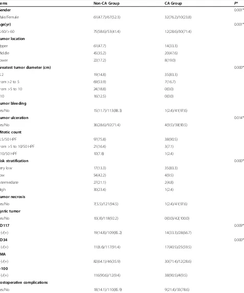
staging of the synchronous gastric cancer was as follows: 14 patients were classified as Stage IA, 8 as Stage IB, 5 as Stage IIA, 1 as Stage IIB, 7 as Stage IIIA, 4 as Stage IIIB, and 3 as Stage IIIC. The histological subtype of the gastric cancer was as follows: 6 patients were classified as well differentiated, 21 as moderately differentiated, 10 as poorly differentiated and 5 as signet ring cell (SRC) histology. Compared with the Non-CA Group, the CA Group had a higher percentage of males, was older in age, and had a lower frequency of ulcer, a smaller greatest tumor diam-eter, lower risk stratification, and lower positivity rates for CD117 and CD34, with all of these differences being sta-tistically significant (Table 1).
Diagnosis
Of the 146 (85.9%) symptomatic patients, 97 had abdom-inal pain, 38 had abdomabdom-inal tenderness, 33 had black stool, 32 had abdominal distension, 30 had weight loss, 18 had eructation, 16 had anorexia, 16 had sour regurgi-tation, 16 had hematemesis, 14 had an abdominal mass, 11 had a loss of strength, 11 had dysphagia, 7 had vomit-ing and 6 had nausea. The proportion of patients with symptoms was significantly higher in the CA than in the Non-CA Group (P <0.05) (Table 1). Of the 97 patients preoperatively diagnosed with gastric GIST, 50 were diag-nosed by computed tomography, 38 by abdominal ultra-sonography, 8 by magnetic resonance imaging, 2 by gastroscopy, and 37 by endoscopic ultrasound, while 8 pa-tients were confirmed to have the disease by endoscopic biopsy pathology. GISTs in the remaining 73 patients were detected incidentally during surgery or by postoperative analysis of resected specimens, with patients being subse-quently diagnosed with gastric GIST by postoperative pathology. Of the 128 patients in the Non-CA group, 88 tumors (68.8%) were identified before surgery but not confirmed by pathology, 8 (6.3%) were confirmed before surgery and 32 (25.0%) were confirmed after surgery. In the CA group, however, only 1 tumor (2.4%) was detected before surgery, whereas 41 (97.6%) were confirmed after surgery. The rate of preoperative diagnosis was signifi-cantly lower in the CA than in the Non-CA Group (2.4% versus 97.6%,P= 0.000) (Table 2).
Long-term surgical outcomes
Table 1 Clinicopathologic features of all patients (cases (%))
Items Non-CA Group CA Group P*
Gender 0.001*
Male/Female 61(47.7)/67(52.3) 32(76.2)/10(23.8)
Age(yr) 0.001*
≤60/> 60 75(58.6)/53(41.4) 12(28.6)/30(71.4)
Tumor location
Upper 61(47.7) 14(33.3)
Middle 45(35.2) 20(47.6)
Lower 22(17.2) 8(19.0)
Greatest tumor diameter (cm) 0.000*
≤2 19(14.8) 35(83.3)
From >2 to 5 69(53.9) 7(16.7)
From >5 to 10 24(18.8) 0(0.0)
>10 16(12.5) 0(0.0)
Tumor bleeding
Yes/No 15(11.7)/113(88.3) 1(2.4)/41(97.6)
Tumor ulceration 0.014*
Yes/No 36(28.6)/92(71.4) 4(9.5)/38(90.5)
Mitotic count
≤5/50 HPF 97(75.8) 38(90.5)
From >5 to 10/50 HPF 21(16.4) 3(7.1)
>10/50 HPF 10(7.8) 1(2.4)
Risk stratification 0.000*
Very low 17(13.3) 35(83.3)
Low 54(42.2) 4(9.5)
Intermediate 27(21.1) 2(4.8)
High 30(23.4) 1(2.4)
Tumor necrosis
Yes/No 7(5.5)/121(94.5) 1(2.4)/41(97.6)
Cystic tumor
Yes/No 10(.8)/118(92.2) 0(0.0)/42(100.0)
CD117 0.009*
(−)/(+) 19(14.8)/109(85.2) 14(33.3)/28(66.7)
CD34 0.000*
(−)/(+) 11(8.6)/117(91.4) 17(40.5)/25(59.5)
SMA
(−)/(+) 82(64.1)/46(35.9) 30(71.4)/12(28.6)
S-100
(−)/(+) 116(90.6)/12(9.4) 38(90.5)/4(9.5)
Postoperative complications
Yes/No 18(14.1)/110(85.9) 9(21.4)/33(78.6)
Univariate and multivariate survival analysis
Univariate analysis showed that patient age, risk stratifi-cation, postoperative oral imatinib and synchronous gastric cancer were predictive factors of survival (P<0.05; Table 3). Cox regression analysis showed that risk stratifi-cation, postoperative oral imatinib and synchronous gas-tric cancer were independent predictors of OS (P <0.05; Table 4).
Survival analysis based on risk stratification
The 5-year survival rates were significantly lower among patients with synchronous gastric cancer than among pa-tients without synchronous gastric cancer, both among patients stratified as being at very low risk/low risk (60.2% versus 98.6%, P <0.05) and among those stratified as
Table 2 Diagnosis of all patients (cases (%))
Items Non-CA Group CA Group P*
Symptom 0.012*
Symptomatic 105(82.0) 41(97.6)
Asymptomatic 23(18.0) 1(2.4)
Diagnosis 0.000*
Preoperative 96(75.0) 1(2.4)
Postoperative 32(25.0) 41(97.6)
*Analysis of variance.P<0.05 is significant. CA Group, gastric GIST patients with synchronous gastric cancer; Non-CA Group, gastric GIST patients without synchronous gastric cancer.
Figure 1Kaplan-Meier estimates of overall survival rates. Kaplan-Meier estimates of overall survival rates relative to the presence or absence of synchronous gastric cancer in patients with gastrointestinal stromal tumor (GIST) (n = 170,χ2 = 22.508,P= 0.000). CA Group, gastric GIST patients with synchronous gastric cancer; Non-CA Group, gastric GIST patients without synchronous gastric cancer.
Table 3 Univariate analysis of variables associated with survival in 170 patients with gastric gastrointestinal stromal tumor (GIST)
Items Cases (5-year
survival rate,%) P *
Gender
Male/Female 93(77.1)/77(88.2)
Age (yr) 0.020*
≤60/>60 87(89.3)/83(74.2)
Tumor location
Upper/Middle/Lower 75(84.0)/65(76.3)/30(92.1)
Greatest tumor diameter (cm)
≤2/>2 to 5/>5 to 10/>10 54(77.3)/76(92.5)/24(78.4)/ 16(63.3)
Tumor bleeding
Yes/No 16(100.0)/154(80.1)
Tumor ulceration
Yes/No 40(92.3)/130(78.4)
Mitotic count (/50 HPF)
≤5/>5 to 10/>10 135(86.1)/24(67.9)/11(70.7)
Risk stratification 0.004*
Very-low 52(76.2)
Low 58(94.2)
Intermediate 29(96.6)
High 31(58.4)
CD117
(−)/(+) 33(64.8)/137(85.8)
CD34
(−)/(+) 28(71.2)/142(84.3)
SMA
(−)/(+) 112(82.2)/58(82.9)
S-100
(−)/(+) 154(82.3)/16(84.4)
Tumor necrosis
Yes/No 8(87.5)/162(81.7)
Cystic tumor
Yes/No 10(100.0)/160(81.6)
Synchronous gastric cancer 0.000*
Yes/No 42(57.8)/128(90.1)
SRC in synchronous gastric cancer
Yes/No 5(40%)/37(59.2)
Postoperative complications
Yes/No 27(83.8)/141(82.2)
Postoperative oral imatinib 0.009*
Yes/No 53(97.0)/117(77.3)
SRC, signet ring cell.
being at intermediate risk/high risk (33.3% versus 98.1%, P<0.05) (Figure 2).
Discussion
The incidence of GIST is only approximately 10 to 20 cases per million per year, [12-16] with gastric GIST be-ing the most common type. Although GISTs are rare, the proportion of GIST patients who present synchron-ously with other malignancies is not low. In particular, the combination of gastric GIST and synchronous gastric cancer is relatively common. An analysis of 14 studies found that 4.5% to 33% of patients had GIST simultan-eously with other neoplasms [17]. In our series we found that 42 of 170 (24.7%) patients with gastric GISTs pre-sented with synchronous gastric cancers. Gastric GISTs accompanied by synchronous gastric cancer have spe-cific pathological features. For example, 14 of 15 gastric GISTs with synchronous gastric cancer were smaller than 2.0 cm in size, with the fifteenth being 2.5 cm; moreover, almost all of these tumors were stratified as very low or low risk [2]. Similarly, we found that most of the gastric GISTs in patients with synchronous gastric cancer were small and of very low or low risk of malignancy. Moreover, only one of the 42 patients (2.4%) found to have gastric GIST with synchronous gastric cancer was diagnosed pre-operatively, with all others detected incidentally during surgery or in postoperative pathology, a finding in agree-ment with previous results [1,2,18].
Clinical manifestations of gastric GIST were nonspecific, with some patients having no clinical manifestations when the tumor was small. The preoperative diagnosis of GIST depended mainly on imaging modalities, such as com-puted tomography and endoscopy [19,20]. In patients with simultaneous gastric cancer and gastric GIST, the symp-toms of gastric GIST were often masked by the clinical symptoms of gastric cancer. Most of these patients had small GISTs (<2.0 cm) and saw a doctor for the symptoms of gastric cancer. Moreover, since most gastric GISTs were submucosal, muscular, or subserosal, patients often could not be preoperatively diagnosed by endoscopic bi-opsy. Furthermore, many clinicians lack the knowledge of
Table 4 Multivariate analysis of factors prognostic of survival in patients with gastric gastrointestinal stromal tumor (GIST) and synchronous gastric cancer
Parameters β SE Wald P* RR 95% CI
Age −0.596 0.481 1.531 0.551 0.215-1.416
Synchronous gastric cancer −2.296 0.602 14.571 0.000* 0.101 0.031-0.327
Risk stratification 24.190 0.000*
Very low versus high −2.504 0.607 17.001 0.000* 0.082 0.025-0.269
Low versus high −2.544 0.682 13.895 0.000* 0.079 0.021-0.299
Intermediate versus high −2.638 1.060 6.191 0.013* 0.071 0.009-0.571
Postoperative oral imatinib 2.213 1.045 4.489 0.034* 9.146 1.180-70.864
*Analysis of variance.P<0.05 is significant.
multiple primary tumors and are satisfied with a diagnosis of gastric cancer alone, resulting in a low rate of preopera-tive diagnosis of gastric GIST.
Interestingly, we found that the gastric GIST patients with synchronous gastric cancer were older in age com-pared with those without synchronous gastric cancer, and that was a predictive factor of survival. However, the age was not an independent predictor of OS. We specu-lated that the finding might be associated with the high incidence of gastric cancer in older patients. In addition, the elderly might be with some change of gene expression profile and a lower immunity, resulting in more easily suffering from the synchronous tumors. Further stud-ies are needed on the gene expression in primary tumor cells from older and younger patients and signal transduc-tion may also provide us with some clues to this finding.
Common immunohistochemistry included CD117, CD34, SMA, S-100 of GIST were analyzed in our study, where we found statistically different positive rates of CD117 and CD34 between groups. However, further prognosis ana-lysis suggested this finding was not related to prognosis. We found that the gastric GIST with synchronous gastric cancer had a lower positive rate of CD117 and CD34 based on the large sample. This was a finding not encoun-tered before in the literature. It might be worth forming a base of classification of GIST tumors according to it. More research is needed.
Previously, GIST was associated with a poor prognosis, with 5-year OS rates after R0 resection ranging from 28% to 65%, [21-25] and another study reporting that patients with gastric GIST had a 5-year OS rate of 42% [26]. Add-itional studies, improvements in surgical skill, and the intro-duction of the molecular targeted drug imatinib have significantly improved the prognosis of patients with GIST, with a study in 2010 reporting a 5-year OS rate in 187 patients with gastric GIST being 75.9% [27]. Few studies to date have assessed the prognosis of patients with syn-chronous gastric GIST and gastric cancer. A study of 22 patients with gastric GIST and synchronous gastric cancer who underwent surgical treatment found that the 5-year OS rate was 57.8%, with a median survival time of 36 months [28]. We found that the 5-year OS rate in patients was significantly lower in gastric GIST patients with than without gastric cancer. Furthermore, risk strati-fication and the presence of synchronous gastric cancer were independent predictors of survival. The prognosis of gastric GIST patients was reported to be poorer for those with synchronous gastric cancer than for those without synchronous gastric cancer, regardless of risk stratification [4]. Similarly, we found that the 5-year OS rates were sig-nificantly lower in patients with synchronous gastric can-cer than in those without synchronous gastric cancan-cer, whether patients were stratified into the very low/low risk or intermediate/high risk groups. Thus, because of smaller
tumor size, lower risk stratification and lower recurrence risk after complete resection, the prognosis of gastric GIST patients with synchronous gastric cancer was good, with GIST itself having little effect on patient prognosis. The main cause of poor prognosis in these patients was advanced synchronous gastric cancer, suggesting that ac-tive treatment of the synchronous gastric cancer would improve long-term survival of these patients.
Some studies revealed that SRC histology was associated with worse survival than non-SRC [29-31]. In our study, there were 5 patients with SRC carcinoma in the syn-chronous gastric cancer. To study the importance of SRC histology on survival, univariate analysis was done in the gastric GIST patients with SRC or with non-SRC, with a result of no significant differences. But the result might be of limitation of the small sample. Previous reports of the prognosis of patients with SRC were controversial. Some studies reported better 5-year survival rates in SRC than in other cell types in early gastric cancer [32,33]. However, others reported no significant differences when the stage of gastric cancer matched [34]. It has also been suggested that SRC histology is an independent predictor of poor prognosis in gastric cancer [29].
All GISTs are regarded as having malignant poten-tial. Moreover, in patients with gastric GIST and syn-chronous gastric cancer, larger sized GISTs and higher risk stratification were associated with a high recurrence rate and poor prognosis, even after complete resection of the GIST [35]. Consequently, gastric GIST should be re-moved when incidentally discovered during surgery for gastric cancer; when necessary, targeted therapy should be considered.
Conclusions
Gastric GIST with synchronous gastric cancer had a lower rate of preoperative diagnosis, with correct diag-nosis often missed. Survival, however, depended primar-ily on the gastric cancer, suggesting that active treatment of the synchronous gastric cancer would improve long-term survival of these patients. Moreover, gastric GISTs should be removed when incidentally discovered during surgery for gastric cancer; when necessary, targeted ther-apy should be considered.
Consent
Written informed consent was obtained from the patient for the publication of this report and any accompanying images.
Abbreviations
GIST:gastrointestinal stromal tumor; OS: overall survival; SRC: signet ring cell.
Competing interests
Authors’contributions
ML, JXL and CMH conceived the study and participated in its design and coordination. ML, JXL, PL, JWX, JBW and JL helped to collect data. ML performed the statistical analysis. ML and JXL drafted the manuscript. CMH and CHZ helped revise the paper critically for important intellectual content. All authors read and approved the final manuscript.
Received: 2 August 2013 Accepted: 18 January 2014 Published: 31 January 2014
References
1. Maiorana A, Fante R, Cesinaro AM, Fano RA:Synchronous occurrence of epithelial and stromal tumors in the stomach: a report of 6 cases. Arch Pathol Lab Med2000,124:682–686.
2. Yan Y, Li Z, Liu Y, Zhang L, Li J, Ji J:Coexistence of gastrointestinal stromal tumors and gastric adenocarcinomas.Tumour Biol2013,34:919–927. 3. Firat Ö, Çalişkan C, Karaca C, SEZAK M, ÖZÜTEMİZ Ö, ERSİN S, GÜLER A:
Coexistence of gastric cancer and gastrointestinal stromal tumor: report of two cases.Turk J Gastroenterol2010,21:302–304. 4. Lee FY, Jan YJ, Wang J, Yu CC, Wu CC:Synchronous gastric
gastrointestinal stromal tumor and signet-ring cell adenocarcinoma: a case report.Int J Surg Pathol2007,15:397–400.
5. Lin YL, Tzeng JE, Wei CK, Lin CW:Small gastrointestinal stromal tumor concomitant with early gastric cancer: a case report.World J Gastroenterol
2006,12:815–817.
6. Kleist B, Lasota J, Miettinen M:Gastrointestinal stromal tumor and gastric adenocarcinoma collision tumors.Hum Pathol2010,41:1034–1039. 7. Katsoulis IE, Bossi M, Richman PI, Livingstone JI:Collision of
adenocarcinoma and gastrointestinal stromal tumour (GIST) in the stomach: report of a case.Int Semin Surg Oncol2007,4:2.
8. Yamamoto D, Hamada Y, Tsubota Y, Kawakami K, Yamamoto C, Yamamoto M: Simultaneous development of adenocarcinoma and gastrointestinal stromal tumor (GIST) in the stomach: case report.World J Surg Oncol
2012,10:1–4.
9. Japanese Gastric Cancer Association:Japanese classification of gastric carcinoma: 3rd English edition.Gastric Cancer2011,14:101–112. 10. Joensuu H:Risk stratification of patients diagnosed with gastrointestinal
stromal tumor.Hum Pathol2008,39:1411–1419.
11. Sobin LH, Wittekind C, Gospodarowicz M:TNM classification of malignant tumors (UICC).7th edition. New York: Wiley-Blackwell; 2009:73–77. 12. Nilsson B, Bümming P, Meis-Kindblom JM, Odén A, Dortok A, Gustavsson B,
Sablinska K, Kindblom LG:Gastrointestinal stromal tumors: the incidence, prevalence, clinical course, and prognostication in the preimatinib mesylate era.Cancer2005,103:821–829.
13. Monges G, Bisot-Locard S, Blay JY, Bouvier AM, Urbieta M, Coindre JM, Scoazec JY:The estimated incidence of gastrointestinal stromal tumors in France. Results of PROGIST study conducted among pathologists. Bull Cancer2010,97:E16–E22.
14. Espinosa I, Lee CH, Kim MK, Rouse BT, Subramanian S, Montgomery K, Varma S, Corless CL, Heinrich MC, Smith KS:A novel monoclonal antibody against DOG1 is a sensitive and specific marker for gastrointestinal stromal tumors.Am J Surg Pathol2008,32:210–218.
15. Goettsch WG, Bos SD, Breekveldt-Postma N, Casparie M, Herings R, Hogendoorn PC:Incidence of gastrointestinal stromal tumours is underestimated: results of a nation-wide study.Eur J Cancer2005, 41:2868–2872.
16. Tzen CY, Wang JH, Huang YJ, Wang MN, Lin PC, Lai GL, Wu CY, Tzen CY: Incidence of gastrointestinal stromal tumor: a retrospective study based on immunohistochemical and mutational analyses.Digest Dis Sci2007, 52:792–797.
17. Agaimy A, Wünsch PH, Sobin LH, Lasota J, Miettinen M:Occurrence of other malignancies in patients with gastrointestinal stromal tumors. Semin Diagn Pathol2006,23:120–129.
18. Chan CH, Cools-Lartigue J, Marcus VA, Feldman LS, Ferri LE:The impact of incidental gastrointestinal stromal tumours on patients undergoing resection of upper gastrointestinal neoplasms.Can J Surg2012,55:366–370. 19. Gold JS, DeMatteo RP:Combined surgical and molecular therapy: the
gastrointestinal stromal tumor model.Ann Surg2006,244:176–184. 20. Nowain A, Bhakta H, Pais S, Kanel G, Verma S:Gastrointestinal stromal
tumors: clinical profile, pathogenesis, treatment strategies and prognosis.J Gastroenterol Hepatol2005,20:818–824.
21. DeMatteo RP, Lewis JJ, Leung D, Mudan SS, Woodruff JM, Brennan MF: Two hundred gastrointestinal stromal tumors: recurrence patterns and prognostic factors for survival.Ann Surg2000,231:51–58. 22. Joensuu H:Sunitinib for imatinib-resistant GIST.Lancet2006,368:1303–1304. 23. van der Zwan SM, DeMatteo RP:Gastrointestinal stromal tumor: 5 years
later.Cancer2005,104:1781–1788.
24. Ng E, Pollock RE, Munsell MF, Atkinson EN, Romsdahl MM:Prognostic factors influencing survival in gastrointestinal leiomyosarcomas. Implications for surgical management and staging.Ann Surg1992, 215:68–77.
25. Ranchod M, Kempson RL:Smooth muscle tumors of the gastrointestinal tract and retroperitoneum: a pathologic analysis of 100 cases. Cancer1977,39:255–262.
26. Wong N, Young R, Malcomson R, Nayar A, Jamieson L, Save V, Carey F, Brewster D, Han C, Al-Nafussi A:Prognostic indicators for gastrointestinal stromal tumours: a clinicopathological and immunohistochemical study of 108 resected cases of the stomach.Histopathol2003,43:118–126. 27. Huang H, Liu YX, Zhan ZL, Liang H, Wang P, Ren XB:Different sites and
prognoses of gastrointestinal stromal tumors of the stomach: report of 187 cases.World J Surg2010,34:1523–1533.
28. Liu YJ, Yang Z, Hao LS, Xia L, Jia QB, Wu XT:Synchronous incidental gastrointestinal stromal and epithelial malignant tumors.World J Gastroenterol2009,15:2027–2031.
29. Piessen G, Messager M, Leteurtre E, Jean-Pierre T, Mariette C:Signet ring cell histology is an independent predictor of poor prognosis in gastric adenocarcinoma regardless of tumoral clinical presentation.Ann Surg
2009,250:878–887.
30. Li C, Kim S, Lai JF, Hyung WJ, Choi WH, Choi SH, Noh SH:Advanced gastric carcinoma with signet ring cell histology.Oncol2007,72:64–68. 31. Kim JP, Kim SC, Yang HK:Prognostic significance of signet ring cell
carcinoma of the stomach.Surg Oncol1994,3:221–227. 32. Jiang CG, Wang ZN, Sun Z, Liu FN, Yu M, Xu HM:Clinicopathologic
characteristics and prognosis of signet ring cell carcinoma of the stomach: results from a Chinese mono-institutional study.J Surg Oncol
2011,103:700–703.
33. Kunisaki C, Shimada H, Nomura M, Matsuda G, Otsuka Y, Akiyama H: Therapeutic strategy for signet ring cell carcinoma of the stomach. Br J Surg2004,91:1319–1324.
34. Zhang M, Zhu G, Zhang H, Gao H, Xue Y:Clinicopathologic features of gastric carcinoma with signet ring cell histology.J Gastrointest Surg Off J Soc Surg Aliment Tract2010,14:601–606.
35. Hassan I, Shyyan R, Dozois EJ, Smyrk TC, Okuno SH, Schleck CD, Hodge DO, Donohue JH:Surgically managed gastrointestinal stromal tumors: a comparative and prognostic analysis.Ann Surg Oncol2008,15:52–59.
doi:10.1186/1477-7819-12-25
Cite this article as:Linet al.:Prognostic analysis of gastric gastrointestinal
stromal tumor with synchronous gastric cancer.World Journal of Surgical
Oncology201412:25.
Submit your next manuscript to BioMed Central and take full advantage of:
• Convenient online submission
• Thorough peer review
• No space constraints or color figure charges
• Immediate publication on acceptance
• Inclusion in PubMed, CAS, Scopus and Google Scholar
• Research which is freely available for redistribution