Pseudoreversion Analysis Indicates a Direct Role of Cell Division Genes in
Polar Morphogenesis and Differentiation
in
Caulobacter crescentus
Jurg
M.
Sommer'
and
Austin Newton
Department of Molecular Biology, Lewis Thomas Laboratory, Princeton University, Princeton, New Jersey 08544
Manuscript received April 25, 199 1 Accepted for publication July 9, 199 1
ABSTRACT
A pseudoreversion analysis was used to examine the role of cell division genes in polar morpho- genesis in Caulobacter crescentus. Extragenic suppressors of temperature sensitive mutations in pleC, a pleiotropic gene required for cell motility, formation of polar 4CbK bacteriophage receptors, and stalk formation, were isolated. These suppressors, which restored motility at 37", simultaneously conferred a cold sensitive cell division phenotype and they were mapped to the three new cell division genes diuJ, diuL and diuK. The cold-sensitive mutations in diuL, and to a lesser extent diuJ, exhibited a relatively narrow range of suppression. The cold-sensitive cell division mutation in diuK, by contrast, suppressed all pleC mutations examined and behaved as a classical bypass suppressor. The direct role of this cell division gene in the regulation of motility is suggested by the observation that diuK341
mapped to the same locus as pleD301, a pleiotropic mutation that prevents loss of motility and stalk formation. These results provide strong evidence that the cell division and developmental pathways are interconnected and they support our earlier conclusion that cell division is required for the regulation of polar morphogenesis and differentiation in C . crescentus.
A
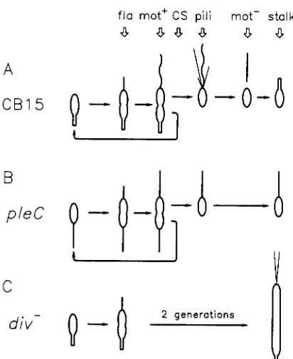
SYMMETRIC cell division is a central mecha- nism for the formation of differentiated cell types, and the pattern of division in Caulobacter cres- centus has provided a useful model system for studying the mechanisms regulating this developmental process (SHAPIRO 1985; NEWTON 1989; NEWTON and OHTA 1990). These aquatic bacteria divide unequally to produce two different cells, the nonmotile stalked cell and a new, motile swarmer cell with a single polar flagellum. Formation of the swarmer cell and its dif- ferentiation into a stalked cell result from a series of discrete morphological events at the stalk-distal pole of the dividing cell (see cell cycle in Figure 1). The first of these is flagellum assembly, which depends on over 50 flagellin Cfla) genes (NEWTON and OHTA1990; NEWTON 1989). A subsequent sequence of events includes activation of flagellum rotation (gain
of motility), biosynthesis of polar bacteriophage recep tors, pili formation, loss of motility, and stalk forma- tion (SOMMER and NEWTON 1988). A pleiotropic gene required for these latter events, pleC, is the subject of the pseudoreversion analysis described in this paper.
We have proposed that the temporal and spatial control of developmental events in C. crescentus is ultimately regulated by steps in the cell division cycle (HUGUENEL and NEWTON 1982). The requirement of chromosome replication for flagellum formation a p pears to be exerted at the level of flagellar Cfla) gene expression (SHEFFREY and NEWTON 1981) and the
' Present address: Department of Pharmaceutical Chemistry, School of Pharmacy, University of California, San Francisco, California 94143. Genetics 1 4 9 623-630 (November, 1991)
analysis of cell division mutants has suggested that completion of early cell division steps are required for gain of motility and stalk formation at the flagellated cell pole (HUGUENEL and NEWTON 1982; see Figure 1). More recent studies have also indicated a require- ment of cell separation, the last step in cell division, for polar morphogenesis: cells blocked at this step gain motility normally, but they fail to assemble pili (SOMMER and NEWTON 1988).
T o determine if the requirement of cell division for polar morphogenesis reflects the direct involvement of cell division genes, we have undertaken a pseudo- reversion analysis of pleiotropic, developmental mu- tations in pleC. The similarities in phenotypes between
pZeC mutants that assemble nonmotile flagella and do not form stalks (SOMMER and NEWTON 1989; FUKUDA, IBA and OKADA 1977; Figure 1B) and the polar mor- phology of some filamentous cell division mutants that also assemble nonmotile flagella and do not form stalks at the new cell pole (HUGUENEL and NEWTON
1982; Figure 1 C) suggested to us that pZeC might play a key role in coupling polar morphogenesis to cell division. Motile revertants of temperature sensitive
(Ts) pleC mutants were isolated at the nonpermissive temperature of 37 O and then screened for extragenic
suppressors that simultaneously conferred a cold-sen- sitive (Cs) cell division phenotype at 24 O
.
The assump-fla mot' CS pili mot- stalk
a
8.3a
a
a
A
C B 1 5
B
pleC
C
div-
FIGURE 1 .-C. crescentus cell cycle. A, The sequence of develop mental events in the wild type strain GB15 includes flagellum formation (fla), activation of flagellum rotation (mot+), cell separa- tion (CS), pili formation (pili), loss of motility (mot-), and stalk formation. B, pleC mutants assemble inactive flagella (designated as straight lines), they are mot-, bacteriophage &bK resistant and fail
to form a stalk, but they divide normally. C, Cells blocked early in cell division (diu-) also assemble inactive flagella and do not form a stalk at the cell pole.
function. It should be possible to identify such muta- tions as extragenic suppressors.
Pseudoreversion studies have been extremely useful
in the study of how genes function in complex regu- latory pathways. This approach was used to identify interacting gene products in bacteriophage P22 as-
sembly (JARVICK and BOTSTEIN 1975), to analyze the
yeast cell cycle (MOIR a n d BOTSTEIN 1982) and cyto- skeleton (NOVICK, OSMOND and BOTSTEIN 1989), and
to study the allele-specific interaction of che a n d J a
genes in Escherichia coli (PARKINSON et al. 1983) and in Salmonella typhimurium (YAMAGUCHI et al. 1986). I n previous pseudoreversion studies revertants have
been examined at the second temperature for a phe-
notype similar to that conferred by the original mu- tation. O u r analysis differs in that revertants of the original nonmotile strain were examined for the ac- quisition of a new phenotype, namely a defect in an essential cell division function. This procedure al- lowed us to identify Cs mutations in three previously unidentified cell division genes, divJ, divK, a n d divL.
O u r results indicate that the cell division and devel- opmental pathways are interconnected and they sup-
port a model for developmental regulation of C . cres-
centus in which cell division genes play a direct role in polar morphogenesis and cell differentiation (HUGUE- NEL and NEWTON 1982).
MATERIALS AND METHODS
Strains and growtb conditions: Wild-type C. crescentus
strain CB15 (American Type Culture Collection 19089) and motility and cell division mutants were routinely grown at 30" in peptone-yeast extract (PYE; POINDEXTER 1964) me- dium. All pleC point mutations used in this study were isolated after UV-light irradiation (SOMMER and NEWTON 1989). These strains, as well as the divJ, divK, and divL mutants, are listed in Table 1.
Isolation of cold-sensitive suppressor mutations: Strain
PC5262 carrying the Ts pleC319 allele (SOMMER and NEW-
TON 1989) was inoculated into motility agar [% PYE
+
0.35% agar (Difco)] and grown at 37". Cells picked from individual motile flares produced from spontaneous rever- tants were patched onto PYE plates, grown overnight at 37", and replica printed at 24". Colonies that grew poorly at 24" were purified at 37" and analyzed by phase contrast microscopy for defects in cell division after an extended period of growth at 24". Each of the original motile rever- tants was also screened for Cs motility defects.
Genetic mapping: ThepleC319 allele in each Cs revertant
was replaced with the ple+ allele by transduction with a bacteriophage &r30 lysate (ELY, CROFT and GERARWT 1984) made on Tn5 insertion mutant PC5347. This strain contains zhf340::Tn5 which is 30% linked to the wild type pleC locus. Twenty to 40 kanamycin resistant (Km') recom- binants were then screened for Cs defects in cell division and motility; presence of the pleC+ allele was verified by back crossing the linked Tn5 insertion into strain CB15. With the exception of strain PC5318 (pleC319, divK341), presumptive pleC+ recombinants were recovered from all crosses that displayed altered cell division or motility phe- notypes (see Figure 2 below) at a frequency expected for replacement of the pleC319 allele by pleC+. Tn5 insertions linked to the Cs suppressor mutations were isolated using a pool of bacteriophage 4Cr30 lysates made on strain CB15 cells containing random Tn5 insertions (orgy lysate), as described previously (OHTA, MASUREKAR and NEWTON
1990). Recombinants of the original Cs revertants that had been transduced with the lysate were selected for Km' and screened for loss of the cell division defect (SOMMER and NEWTON 1989). Recombinants containing the Tn5 insertion linked to the wild-type allele of the Cs suppressor were verified by back crossing the Tn5 insertion into the original Cs revertant strain and screening Km' recombinants for loss of the Cs division phenotype. The Tn5 insertion in a cold sensitive recombinant was then used to move the suppressors allele into the wild-type background. These crosses are illustrated in Table 1 by the construction of strains PC5343 and PC5432 which contain insertion element zzz354::Tn5 linked to dzvJ332.
Phenotype assays: Motility was assayed by swarm for-
mation in motility agar. Cs recombinants were usually ana- lyzed by phase contrast microscopy to distinguish between nonmotile strains and cell division mutants, both of which produce tight colonies on motility agar. Cell morphology was always assayed by phase contrast microscopy. Stalk formation was determined by phase contrast microscopy on a smear of cells immediately after drying at 500X magnifi- cation without a coverslip and confirmed in some instances by electron microscopy. Bacteriophage +CbK resistance was assayed by suspending parts of a colony in a loop of PYE broth and streaking it across the phage on a fresh PYE plate.
RESULTS
625
TABLE 1 Bacterial strains
Strain Genotype Source or construction"
~
CB15 Wild type ATCC 19089
PC5225 pleC301::Tn5 SOMMER and NEWTON (1989)
PC5253 pleC358::Tn5 This work
PC5255 pleC314 SOMMER and NEWTON (1989)
PC5258 pleC315 This work
PC5262 pleC319 SOMMER and NEWTON ( 1 989)
PC5264 pleC321 SOMMER and NEWTON (1989)
PC5267 pleC322 This work
PC5270 pleC323 SOMMER and NEWTON (1 989)
PC5281 pleC330 SOMMER and NEWTON (1989)
PC5282 pleC331 This work
PC5283 pleC343 This work
PC5287 pleC335 This work
PC5302 pleC319, pleD301, zzz-351::Tn5 Orgy lysate X PC53 16 + Km'[ Div+] PC5309 pleC358::Tn5, pleD301, divJ332 g(PC5253) X PC53 16 + Km'[Ple-]
PC53 10 pleC358::Tn5, pleD301 g(PC5309) X CB15 + Km'[mot+]
PC53 15 pleC319, divJ331 This work
PC53 16 pleC319, pleD301, divJ332 SOMMER and NEWTON (1989)
PC53 17 pleC319, divJ333 This work
PC53 18 pleC319, divK341 This work
PC53 19 pleC319, divL342 This work
PC5320 pleC319, divL343 This work
PC5321 pleC319, divJ334 This work
PC5323 pleC319, divJ335 This work
PC5325 pleC319, divJ336 This work
PC5326 pleC319, divJ337 This work
PC5327 pleC319, divJ338 This work
PC5343 pleC319, pleD301, zz%-354::Tn5, divJ332 b(PC5344) X PC5316 + Km'[Div-] PC5344 pleC319, pleD301, zzz-354::Tn5 Orgy lysate X PC53 16 + Km'[Div+] PC5345 pleC319, pleD301, divJ332, zhf341::Tn5 SOMMER and NEWTON ( 1 989)
PC5347 zhJ340:Tn5 Orgy lysate X PC5262 + Km'[Ple+]
PC5353 divJ332, hunEl12::Tn5 g(SC1588) X PC5316 + Km'[Ple+]
PC5346 pleC319, pleD301, divJ332, hunEl12::Tn5 SOMMER and NEWTON (1 989)
PC5385 pleC319, rrr-353::Tn5 Orgy lysate X PC5319 + Km'[Div+]
PC5386 pleC319, divL342, zzz-353::Tn5 g(PC5385) X PC5319 + Km'[Div"]
PC5432 zzr354::Tn5, divJ332 g(PC5343) X CB15 + Km'[Div-]
SC1588 hunElI2::Tn5, str152 ELY, CROFT and GERARDOT ( 1984)
* Allele divJ332 is referred to as divJ302 in SOMMER and NEWTON (1989). Abbreviations: Km', resistance to kanamycin; mot+, motile; ple-, @CbK resistant and mot-; pie+, gCbK sensitive and mot+; Div, defective in cell division.
strain PC5262 at
37".
The revertants were then screened for second site mutations that suppressed the nonmotile (mot-) phenotype of the pleC319 allele at37" and simultaneously conferred a Cs phenotype at
24" (MATERIALS AND METHODS). Eleven
Cs
revertantswere identified and all of them produced long straight, largely unpinched filaments after prolonged incubation at 24" (Table 1; strains PC5315-PC5327).
A similar frequency of
Cs
strains was found among the revertants of T s strain PC5255 (pleC314 SOMMERand NEWTON 1989). None of the 500 motile rever- tants of the pleC314 and pleC319 mutants isolated at
37" was found to be Cs for motility alone.
We examined the phenotypes of revertant strains that contained a suppressor mutation in each of the three linkage groups defined below (divJ, divK and
d i d ) in greater detail at the nonpermissive tempera- ture. The rate of DNA synthesis in these strains con-
tinued at the wild type rate after shifting the cells from
37"
to 24" (data not shown), as measured by the incorporation of ['Hlguanosine (OSLEY and NEW-TON 1980). Although the Cs mutants did not form
colonies at 24", the cells continued to grow at this temperature for several generations as measured by the increase of optical density. The cell division de- fects were rapidly reversed when the cells were shifted back to 37". Based on this analysis (OSLEY and NEW-
TON 1980), we concluded that the primary effect of
the suppressors is on cell division.
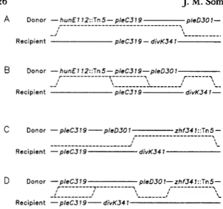
Mapping suppressor mutations to divJ, divK and
divL: The pleC319 allele was replaced by the pleC+
allele in the 1 1 Cs revertants using a Tn5 insertion element tightly linked to the PleC locus (MATERIALS AND METHODS). All Km' recombinants isolated from
J. and A.
A Donor - h u n E 7 1 2 : : T n 5 - p / e C 3 7 9
"""""""""""""""""
pleD3O 1
-
"
- - -
-- -
-., ,
- J
Recipient p l e C 3 1 9 - divK34 1
Donor - h u n E l 1 2 : : T n 5 - p / e C 3 1 9 - p / e D 3 0 1 - - -
-
- - --
T""""". r - - - y I
" """""_I """"I
Recipient pleC379 di~K341-
"
-
c
Donor - p / e C 3 1 9 - p l e D 3 0 1 z h f 3 4 7 : : T n 5 -"""""""""""-I " """"""""""" I
I ,
Recipient -p/eC319 divK34 7
D Donor - p / e C 3 1 9
r""""-,"""""y
, , , , p / e D 3 O l - ~ h f 3 4 1 : : T n 5 - ,"""""", """""" " """-; "
Recipient -p/eC379-ddivK341
FIGURE 2.-Mapping of divK341 to the pleD locus. The dashed lines indicate the crossover events required to produce nonmotile kanamycin resistant recombinants, depending on the relative order of PleD and diuK. A total of 160 kanamycin-resistant recombinants were scored from each of these two crosses for motility. An addi- tional 160 kanamycin-resistant recombinants from a similar cross in which a T n 5 insertion in pleC (PC5310, pleC358::Tn5, pleD301)
was used as the selectable marker when transduced into strain PC53 18 were screened for motility. All 480 recombinants were found to be motile at 37", indicating recombination had not oc- curred between the pleD301 and divK341 alleles.
extragenic and unlinked to pleC. The suppressor (divK341) in one revertant
(PC5318)
was also extra- genic, but linked to pleC (see below). T n 5 insertions linked by transduction to each of theCs
suppressors were isolated (MATERIALS AND METHODS) and used in phage &r30 crosses to place these cell division genes in three linkage groups. No genetic linkage between the genes identified here and previously published cell division genes (OSLEY and NEWTON1980)
was ob- served.divK: The
Cs
suppressor in strainPC53
18,
which is50% linked to pleC301::TnS
(PC5225),
was desig- nated as divK341 (see Figure2).
This linkage is ap- proximately the same as that observed between pleC and pleD301, a mutation reported to act as a bypass suppressor of the pleC motility defect (SOMMER andNEWTON
1989),
but not to confer a cell division phenotype. The pleD mutant, which is blocked at a step required for loss of motility, retains motility throughout the cell cycle and does not form stalks(SOMMER and NEWTON
1989).
We originally at- tempted to order the two suppressors genetically using three factor crosses. The donor strain contained the pleC319 and pleD301 mutations along with either the T n 5 insertion hunEll2::Tn5 (Figure2,
A and B) or zhf-341 ::Tn5 (Figure2, C
and D) which are located on either side of the pleClpleD gene cluster (SOMMERand NEWTON
1989)
and used as the selected markers. The recipient strainPC53
18 also contained pleC319 along with divK341. At37"
both pleD301 and div-K341 suppress the nonmotile phenotype of pleC319. Because pleC319 was present in both the donor and recipient strain, nonmotile pleD+, divK+ recombinants would be expected if recombination occurred be- tween the divK341 and pleD3Ol alleles. In none of the
480
recombinants examined from these and similar crosses (see Figure2
legend), however, did we recover a nonmotile strain (pleC319, pleD+, divK+). The fail- ure to observe recombination between divK341 and pleD3Ol suggests that the two mutations are very closely linked and possibly allelic.divL: A second linkage group was identified using insertion zzz-353::Tn5
(PC5385),
which is approxi- mately30%
linked to theCs
suppressors in strainsPC5319
andPC5320.
The two revertant alleles were designated divL342 and divL343, respectively. The divJ and divL loci are unlinked by transduction.divJ: zzz-351 ::Tn5
(PC5302)
was shown to be40%
linked to suppressor divJ332 in strain
PC53
16
and the linked Tn5 insertion used to transduce the divJ+ allele into the other pleC revertants. Analysis of the recom- binants showed thatCs
suppressors in seven strains(PC5315
[divJ331],PC5317
[divJ333],PC5321
[divJ334],
PC5323
[divJ335],PC5325
[divJ336],PC5326
[divJ337] andPC5327
[divJ338]; Table1)
were also approximately
40%
linked to zzz-351 ::Tn5, which allowed us to assign them to the divJ locus. The recent isolation of a cloned C. crescentus DNA frag- ment which complements all of these mutations fur- ther confirms that the suppressors map to the same locus (E. NINFA, J. M. SOMMER and A. NEWTON,unpublished).
Characterization of the cold-sensitive suppres- sors: The Tn5 insertions linked to divJ (zzz-351 ::Tn5 or zzz-354::Tn5), divL (zzz-353::Tn5) and divK ( z h .
341::Tn5) were used to back-cross the suppressor mutations into strain
CB15
to assess the phenotypes of the suppressor in the wild type genetic background (Figure3,
first row) and into the pleC319 mutant background to confirm the mapping and observed phenotypes of the original revertant strains (Figure 3, second row). Recombinant strains were examined both at37"
and24"
for motility and cell division and scored as described in the legend to Figure3.
Open boxes indicate essentially wild-type motility or cell division, filled boxes a defective motility or cell divi- sion phenotype, and shaded boxes an intermediate defect.All of the suppressors displayed the same motility and cell division phenotypes when they were back crossed into the pleC319 genetic background that we had observed in the original pseudorevertants (Figure
FIGURE 3.-Motility and cell division phenotypes of divJ, diuK and divL mutations. T n 5 insertions linked to the diuJ, divK and d i d alleles were used to transduce the suppressors into strains CB15 and PC5262 (pleC3ZS). Motility and cell division morphology were scored by light microscopy of cells taken from motility agar. Open boxes indicate wild type motility or cell division (more than 50% normal motility or less than 5% filamenting cells), filled boxes indicate cells defective in motility or cell division (uniformly nonmotile cells or very long filamentous cells), and shaded boxes represent an intermediate defect (a mixture of partially motile single cells and filaments of up to 5 cell lengths). The cell division phenotype could also be scored by the inability of defective mutants (closed boxes) or impaired ability of partially defective mutants to form colonies at the nonpermissive temperature (shaded boxes). Filamentous cells displayed no motility at 24" or 37" and in cultures containing mixed filaments and single cells, motility was always scored on single cells.
notype identical to that observed in the pleC319 back- ground and the divJ mutations conferred a partial defect in cell division at 24". Thus, the Cs cell division phenotype is a characteristic of the extragenic s u p pressors; only in the case of the divJ mutations was the phenotype more extreme in the presence of the mutant pZeC allele. All of the divJ alleles conferred substantially less motility at 37" in the pleC+ back- ground than in the pleC319 background, and most strikingly, divJ332 was completely nonmotile at 37"
in the wild-type background.
Pattern of pleC suppression
by
divK, divL and divJ alleles: T h e specificity of the suppressors was examined in more detail by transducing them into strains with different pleC point mutations (SOMMERand NEWTON 1989) using the linked T n 5 insertions (Figure 4). Km' recombinants were selected at 37"
and the double mutants were scored for motility and cell division at 24" and 37" as described in Figure 3.
Suppression of pleC3OI :: T n 5 was tested by transduc- ing the T n 5 into the original Cs pleC319 revertants
by selecting for Km'.
d i v K 3 4 l : T h e divK34I allele was introduced into the pleC mutant backgrounds by selection for the Km' marker of the z h f 3 4 1 : : T n 5 insertion that is 44%
linked to the divK and 30% linked to pleC (SOMMER
and NEWTON 1989). Because the pZeC marker would be lost in about one-third of the crosses, w e analyzed at least 20 Km' recombinants for each pleC mutant tested. A total of 29 different pleC mutations, includ- ing the 1 1 shown in Figure 4, were examined. divK341
completely suppressed the motility defect of all pleC
mutations examined, including the T n 5 insertion
pleC30I ::Tn5, and the patterns of motility and cell division in all of the mutant pleC backgrounds (Figure
4) were identical to that observed originally in pleC319
(Figure 3). These results led us to conclude that
divK341 acts as a bypass suppressor of pleC mutations. T h e suppression of pleC301 ::Tn5 also suggested that
pZeC is not essential for motility.
divL : Suppressors divL342 and divL343 displayed a much narrower range of specificity than the divK
FIGURE 4.--Suppression of pleC mutations by divJ, divL, and
diuK alleles. divJ332, diuJ335, divL342, divL343 and diuK34Z were transduced into different pleC point mutants and pleC3OZ ::Tn5 was transduced into the suppressor strains as described in the text. The resulting double mutants were analyzed for motility and cell division as described in Figure 3.
allele. Other than the original mutation pleC319, divL343 fully suppressed the motility defect of only 2
of the 6 pleC point mutations examined at 37", while
divL342 fully suppressed the motility of 5 of the 1 1
point mutations. When both motility and cell division were examined at 24" and 37" the patterns of suppression were quite diverse, particularly with
divL343: each of the pleC point mutations examined produced a different suppression pattern (Figure 4).
Although this type of result is normally associated with the behavior of interaction suppressors, the
divL342 and divL343 alleles also suppressed the mo- tility defect of pZeC30I ::Tn5 (Figure 4), which is dis- cussed below (Discussion).
stored motility at 37" to most of the pleC mutations examined, including the T n 5 insertion mutation
pleC301 ::Tn5 (Figure 4). None of the divJ mutations examined, including those shown in Figure 3, sup- pressed pleC322 and pleC343 (Figure 4)) however. Also, the 6 divJ mutant alleles displayed less motility at 37" in the pleC+ than in the original pleC319
background (Figure 3). Thus, the mechanism of suppression by the divJ mutations is not clear from these results (see DISCUSSION).
T h e two pleC mutations (pleC322 and pleC343) that were not suppressed for motility at 37 O by the Cs divJ
and divL alleles (Figure 4) were shown to revert to
mot+ at approximately the same frequency as other
pleC mutants (data not shown). This result indicates that the mutations are not deletions and that the
pleC322 and pleC343 strains used in these experiments do not contain additional motility mutations that would have interfered with the suppression tests. Fur- thermore, in a more recent pseudoreversion study of
pleC we have isolated T s mutations in divL that com- pletely suppress the mot defects of pleC322 and ple- C 3 4 3 at 24" (G. HECHT and A. NEWTON, unpub- lished).
Revertants of divJ map to pleC: T h e behavior of the divJ locus was further investigated by isolating suppressors of divJ332. T o d o this we took advantage of the nonmotile phenotype conferred by divJ332 at
37" in a pleC+ background (Figure 3). Independent
mot' revertants were isolated at the high temperature from strain PC5353, which contains divJ332 and a T n 5 insertion ( h u n E l l 2 : : T n 5 ) closely linked to the
pleC locus (SOMMER and NEWTON 1989). Bacterio- phage 4Cr30 lysates were prepared on each revertant, the pleC region transduced into strain CB15 by select- ing for Km', and 10 recombinants scored for motility and phage resistance at 37
"
and 24". Eight of the 48mot+ revertants of strain divJ332 were found to be outside suppressors and to map to pleC as judged by their close linkage to h u n E l 1 2 : : T n 5 , their nonmotile, stalkless phenotype, and their resistance to phage (bCbK (data not shown); this conclusion can be con- firmed by complementation of the suppressor alleles with a pleC+ clone. T h e isolation of suppressors of
divJ332 that map back to pleC argues at a minimum that the divJ mutations analyzed here do not act as informational suppressors.
DISCUSSION
Pseudoreversion analysis has been applied previ- ously in genetic studies of yeast, bacteria, and bacte- riophage development (JARVICK and BOTSTEIN 1975; MOIR and BOTSTEIN 1982; NOVICK, OSMOND and
BOTSTEIN 1989; PARKINSON et al. 1983; YAMAGUCHI
et ad. 1986). We have extended and modified this approach to the study of developmental regulation in
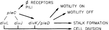
@ RECEPTORS
f PILI /yT:(Ll;y OFF
d i v L d i v J d i v K / p / e D
-
STALK FORMATION1 I I
-
CELL DIVISIONFIGURE 5.-Working model for organization and function of cell division genes in the pleC/pleD-dependent pathway. The proposed functional relationship of the genes is based on the analysis of pleC suppression by the p l e D , diuJ, diuK and d i d mutations described in the text (see DISCUSSION). The pattern of pili assembly generally paralleled that shown for phage sensitivity (data not shown). These results suggest that the developmental pathway branches to phage receptor and pili formation at the diuJ/pleC/divL step(s) and to flagellum activation at or after divK/pleD step.
C. crescentus. Starting with a Ts mutation in pleC, a gene required for motility, polar phage receptor site formation and stalk formation, we have isolated sup- pressors that restored the mot+ phenotype of a pleC
mutation at 37" and simultaneously conferred a Cs defect in cell division. Genetic analysis of eleven such suppressors showed that they retained a partial Cs phenotype in the absence of the pleC mutations and mapped to three previously unidentified genes, divJ, divK and divL. Thus, the selection of conditional pseudorevertants of developmental mutants of C . cres- centus is a fruitful approach to the identification of new cell division genes.
We have reported in an earlier study that steps in the DNA synthetic (SHEFFREY and NEWTON 1981)
and cell division (HUGUENEL and NEWTON 1982; SOM-
MER and NEWTON 1988) pathways are required for flagellum formation, motility and spatial localization of polar structures, and on the basis of these results we have proposed that development in C . crescentus is coupled to steps in the cell division cycle (HUGUENEL
and NEWTON 1982). T h e isolation of Cs cell division mutations as suppressors of pleiotropic developmental mutations in pleC now provides direct evidence for this conclusion; the results indicate that the cell divi- sion and developmental pathways are interconnected and the different behaviors of the suppressors map- ping to divK on the one hand and to divJ and divL on the other suggest that more than a single mechanism may be involved.
Results obtained with the divK allele are the easiest
to interpret. In contrast to other suppressors, divK341
suppressed all PZeC mutations tested and it behaved like a classical bypass suppressor at 37" (Figure 4).
These findings suggest that cell division gene divK
acts after pleC in the pathway regulating cell motility and stalk formation, as diagrammed in Figure 5.
pleD301, like divK341, acts as a bypass suppressor of
in the cell cycle at 37", while strains containing ple- 0301 fail to turn off cell motility, do not form stalks, and are motile throughout the entire cell cycle (SOM-
MER and NEWTON 1989). divK341 and pleD301 could
not be separated by recombination (Figure 2) and if the two mutations prove to be allelic when examined
by genetic complementation, then cell division gene divK (divK/pleD) could play a central role in switching motility on and off at the appropriate times during the cell cycle (see Figure 5).
Mutations in divL and divJ also suppress the defects in motility and stalk formation conferred by pleC mutations, but they restore the mot+ phenotype to only a subset of the pleC mutants examined. The divL alleles divL342 and divL343 displayed a narrow and varied pattern of suppression, particularly when scored for both the mot and div phenotypes at 24"
and 37" (Figure 4). This narrow range of suppression is one characteristic of allele specificity that is fre- quently associated with interaction suppressors. The behavior of the divJ alleles is more difficult to inter- pret because they suppressed the motility defect at 37" of all but two of the pleC point mutations exam- ined. At the same time, however, the Cs diu mutation divJ332, which suppressed the motility defect of Tn5 insertion in pleC, was unique in conferring a T s mot- phenotype in strains containing the wild-type, pleC+ allele (Figure 4).
The ability of divL and divJ mutations to suppress the motility defects of pleC3Ol ::Tn5 (Figure 4) was unexpected, although we have no direct evidence that the Tn5 insertion is a null mutation. Interaction sup- pressors are known, however, that suppress null mu- tations. In Escherichia coli suppressors of TnlO inser- tions in dnaQ have been mapped to the alpha subunit gene of DNA polymerase 111 (LANCY et al. 1989), and suppressors of deletions in the maltose binding protein gene malE have been isolated in the inner membrane protein genes malF and malG (TREPTOW and SHUMAN
1988). The protein subunits of DNA polymerase 111, as well as components of the maltose transport system, are known to physically interact.
Isolation and study of the divJ, divL and pleC gene products will be required to elucidate the mechanism of suppression, but the results obtained here do sug- gest several different possibilities. Among them are a transient interaction in which the gene products are part of a single transduction pathway that coordinates cell division and polar morphogenesis or, alterna-
tively, a physical interaction between PleC and the cell division gene products, We have proposed previ- ously that targeting of polar structures in Caulobacter depends on organizational centers localized at the cell poles (HUGUENEL and NEWTON 1982), and we specu- late that some of the genes identified in this study encode proteins that are laid down at the division site
as proposed for proteins in the E . coli septalsome (GILL and SALMOND 1987; HOLLAND and JONES 1985). A biochemical analysis will be necessary to explore pos- sible interactions between products of the PleC, divJ and divL genes and their localization within the cell. A working model for the functional organization of the cell division and developmental genes examined in this study is shown in Figure 5. This scheme, which is independent of the precise mechanism of gene function, proposes that the cell division pathway is interconnected with the pleC-PleD-dependent pathway (SOMMER and NEWTON 1989) to control develop- mental events, including bacteriophage sensitivity, pili formation, cell motility and stalk formation. The sup- pressor results show that cell division genes divL and divJ modulate the activity of the pleC gene, which is required in turn for formation of 4CbK receptors and pili, as well as the activity of divK/pZeD in regulating motility and stalk formation. divK/pleD is placed after pleC and shown to be required for regulation of motility because pleD301 acts as a bypass suppressor of the motility defect, but not the 4CbK resistance phenotype conferred by pleC mutations.
In summary, we have isolated suppressors in three new cell division genes of C. crescentus. Our analysis of these Cs mutations suggests that the cell division and developmental pathways in these bacteria are interconnected and they support a model proposed previously (HUGUENEL and NEWTON 1982) that cell division genes play a direct role in the regulation of polar morphogenesis and differentiation.
We thank NORIKO OHTA and many other colleagues for their comments on the manuscript. J.M.S. was supported in part by U.S. Public Health Service predoctoral training grant GM 07388 from the National Institutes o f Health. This work was also supported by grant MV-386 from the American Cancer Society.
LITERATURE CITED
ELY, B., R. H. CROFT and C. J. GERARDOT, 1984 Genetic mapping
ofgenes required for motility in Caulobacter crescentus. Genetics
FUKUDA, A., H. IBA and Y. OKADA, 1977 Stalkless mutants of
Caulobacter crescentus. J. Bacteriol. 131: 280-287.
GILL, D. R., and G. P. C. SALMOND, 1987 The Escherichia coli cell division proteins FtsY, FtsE and FtsX are inner membrane- associated. Mol. Gen. Genet. 2 1 0 504-508.
HOLLAND, I. B., and C. JONES, 1985 The role of the FtsZ protein (SfiB) in UV-induced division inhibition and in the normal
Escherichia coli cell division cycle. Ann. Inst. Pasteur/Microbiol.
HUGUENEL, E. D., and A. NEWTON, 1982 Localization of surface structures during procaryotic differentiation: role of cell divi- sion in Caulobacter crescentus. Differentiation 21: 71-78. JARVICK, J., and D. BOTSTEIN, 1975 Conditional-lethal mutations
that suppress genetic defects in morphogenesis by altering structural proteins. Proc. Natl. Acad. Sci. USA 72: 2738-2742. LANCY, E. D., M. R. LIFSICS, D. G. KEHRFS and R. MAURER,
1989 Isolation and characterization of mutants with deletions in dnaQ the gene for the editing subunit of DNA polymerase 111 in Salmonella typhimurium. J. Bacteriol. 171: 5572-5580.
108: 523-532.
MOIR, D., and D. BOTSTEIN, 1982 Determination of the order of gene function in the yeast nuclear pathway using cs and ts mutants. Genetics 100 565-577.
NEWTON, A., 1989 Differentiation in Caulobacter flagellum devel- opment, motility and chemotaxis, pp. 199-220, in Genetics
of Bacterial Diuersity, edited by D. A. HOPWOOD, and K. F.
CHATER. Academic Press, London.
NEWTON, A, and N. OHTA, 1990 Regulation of the cell division cycle and differentiation in bacteria. Annu. Rev. Microbiol. 44:
NOVICK, P., B. C. OSMOND and D. BOTSTEIN, 1989 Suppressors of yeast action mutations. Genetics 121: 659-674.
OHTA, N., M. MASUREKAR and A. NEWTON, 1990 Cloning and cell cycledependent expression of DNA replication gene dnaC
from Caulobacter crescentus. J. Bacteriol. 172: 7027-7034. OSLEY, M. A., and A. NEWTON, 1980 Temporal control of the
cell cycle in Caulobacter crescentus: roles of DNA chain elonga- tion and completion. J. Mol. Biol. 138 109-128.
PARKINSON, J. S., S. R. PARKER, P. B. TALBERT and S. E. HOUTS, 1983 Interactions between chemotaxis genes and flagellar genes in Escherichia coli. J. Bacteriol. 155: 265-274.
POINDEXTER, J. S., 1964 Biological properties and classification 689-7 19.
of the Caulobacter group. Bacteriol. Rev. 28: 231-295. SHAPIRO, L., 1985 Generation of polarity during Caulobacter cell
differentiation. Annu. Rev. Cell Biol. 1: 173-207.
SHEFFREY, M., and A. NEWTON, 1981 Regulation of periodic protein synthesis in the cell cycle: control of initiation and termination of flagellar gene expression. Cell 24: 49-57. SOMMER, J. M., and A. NEWTON, 1988 Sequential regulation of
developmental events during polar morphogenesis in Cnulobac- ter crescentus: assembly of pili on swarmer cells requires cell separation. J. Bacteriol. 1 7 0 409-415.
SOMMER, J. M., and A. NEWTON, 1989 Turning off flagellum rotation requires the pleiotropic gene pleD: pleA, pleC, and
pleD define two morphogenic pathways in Caulobacter crescen-
tus. J. Bacteriol. 171: 392-401.
TREFTOW, N. A., and H. A. SHUMAN, 1988 Allele-specific malE
mutations that restore interactions between maltose-binding protein and the inner membrane components of the maltose transport system. J. Mol. Biol. 2 0 2 809-822.
YAMAGUCHI, S., S. -I. AIZAWA, M. KIHARA, M. ISOMURA, C. J. JONES
and R. M. MACNAB, 1986 Genetic evidence for a switching and energy-transducing complex in the flagellar motor of Sal- monella typhimurium. J. Bacteriol. 168: 1172-1 179.