_____________________________________________________________________________________________________
*Corresponding author: E-mail: dranilgupta1964@hotmail.com;
(Past name: British Journal of Medicine and Medical Research, Past ISSN: 2231-0614, NLM ID: 101570965)
Histopathological Spectrum of Hysterectomy
Specimens
Anil Kumar Gupta
1*, Isha Gupta
1and Anil Kumar Suri
11Department of Pathology, NC Medical College, Israna, Panipat, India.
Authors’ contributions
This work was carried out in collaboration among all authors. Author AKG designed the study, performed the statistical analysis, wrote the protocol and wrote the first draft of the manuscript. Authors IG and AKS managed the analyses and literature search of the study. All authors read and approved the final manuscript.
Article Information
DOI: 10.9734/JAMMR/2020/v32i630440
Editor(s):
(1) Dr. Chan-Min Liu, Xuzhou Normal University, China.
Reviewers:
(1)Rana Choudhary, Wockhardt Hospital, India. (2)Elisabete Gonçalves, ULSAM, Portugal. (3)Stalin Chandrasekaran, India. Complete Peer review History:http://www.sdiarticle4.com/review-history/56839
Received 02 March 2020 Accepted 07 May 2020 Published 13 May 2020
ABSTRACT
Aims: Aim of this retrospective study, was to analyze the histopathological spectrum among women who have underwent hysterectomies at our institution.
Study Design: All hysterectomies were included in this study. Except 7 vaginal hysterectomies, all were abdominal hysterectomies. Clinical history and other relevant data were obtained from the records of Department of Pathology of medical college hospital. Formalin fixed paraffin embedded tissue sections stained with haematoxylin and eosin were examined and analysed for histopathological diagnosis.
Place and Duration of Study: Study was conducted at NC medical College, Panipat India during calendar year 2019.
Methodology: A total of 480(n=480) hysterectomies received during the study period. Mean age of the patient was 40.02 years ranging from 30 to 66 years. 176 (36.5%) hysterectomy specimens did have unilateral salpingo-oophorectomy and 51 (10.6%) with bilateral salpingo-oophorectomy.
Results: Histopathologically, most common findings in hysterectomy specimens were chronic cervicitis in 396(82.5%), uterine fibroids in 251 (52.29%), adenomyosis in 24(5.0%) and endometrial hyperplasia in 18(3.75%). A total of 144 patients (30.0%) showed corpus luteum in
their ovaries; of which cystic changes occurred in 28 (5.8%) and hemorrhagic luteal cysts in 13(2.70%) patients. Fallopian tubes, were pathologically unremarkable; 7(1.45%) patients had paratubal cysts. Ovarian neoplasms were accounted for only 1(0.2%) patient.
Conclusion: Histopathological diagnosis in present study were of benign in nature, requiring no further treatment and management beyond hysterectomy. Hysterectomies were performed in patients to improve the quality of life, to alleviate symptoms and occasionally as a life-saving measure.
Keywords: Hysterectomy; histopathology; leiomyoma; endometrium.
1. INTRODUCTION
Hysterectomy is commonest surgical procedures in females next to cesarean sections and accounted for about 600,000 surgery yearly in US [1]. Around 60% of major surgical procedures in gynecology department were hysterectomy [2]. Various terminology is used depending on the type of surgical procedure performed e.g. hysterectomy (removal of uterus and cervix), or supra-cervical hysterectomy (hysterectomy without removal of the cervix) or hysterectomy with salpingo-oophorectomy (hysterectomy with removal of adnexa either unilateral or bilateral).
2. MATERIALS AND METHODS
This study, was carried out on hysterectomy specimens received in the Department of Pathology during calendar year 2019. All hysterectomy specimens were included in this study. All were abdominal hysterectomies except 7 vaginal in patients with uterine prolapse. After the specimens received in the dept of pathology, detailed history and findings of specimens were
noted; specimens were kept in 10% formal saline for fixation, followed by gross examination on next day; findings noted and sections taken. One section was taken, each from the ectocervix, endocervix, endometrium, endomyometrial junction and myometrium, respectively. Atleast, one tissue bit was taken each from ovary and fallopian tube. Additional tissue were taken in case of grossly visible pathology including fibroid, cyst,etc. The tissue bits were processed in histopathology section, paraffin blocks were made and microsections cut. Microsections were studied after staining with Hematoxylin and Eosin stain. Histopathological findings were noted and analyzed for various pathology in hysterectomy specimens.
3. RESULTS
A total of 480(n=480) hysterectomy specimens received during the study period. 176 (36.5%) hysterectomy specimens did have unilateral salpingo-oophorectomy and 51
(10.6%) with bilateral salpingo-oophorectomy (Fig. 1).
Fig. 1. Nature of hysterectomy specimens uterus with
unilateral adnexa
37%
uterus with Bilateral
adnexa 10% uterus without
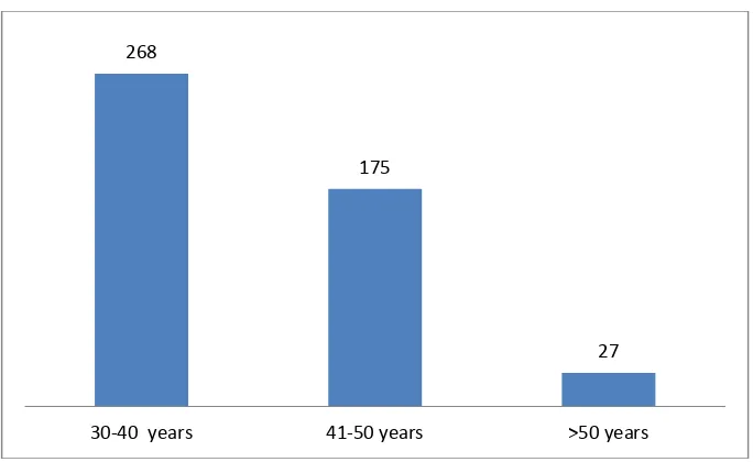
Mean age of the patient was 40.02 years ranging from 30 to 66 years. A total of 268 (55.8%) patients in our study underwent hysterectomies in the age group of 30 to 40 years (Fig. 2).
Chronic cervicitis was the commonest findings in 396 (82.5%) uterine cervix (Fig. 3), additionally 54 (11.25%) patients had nabothian cysts .Cervical erosion and metaplastic changes, each accounted for 7(1.45%) patients (Table 1).
Uterine body showed physiological endometrium in 431(89.79%) patients, adenomyosis in
24(5.0%) and endometrial hyperplasia in 18 (3.75%) patients, either alone or in association with each other. The other less common
histopathological findings in uterine cavity were atrophied endometrium in 14 (2.9%)
patients, endometrial polyp in 8 (1.66%) (Table 1).
Leiomyoma in the myoemtrium accounted for 251 patients (52.29%) (Fig. 4). There was focal adenomyosis in 24 (5.0%) cases. Some of the specimens show more than one lesions, of which coexistence of adenomyosis and leiomyoma was the most common.
Fig. 2. Age at hysterectomy
Fig. 3. Chronic cervicitis (H&E, 40x) 268
175
27
Table 1. Histopathological spectrum in hysterectomies
Histopathologic diagnosis
Number of patients (n)
Percentage (%)
Cervix
Chronic cervicitis 396 82.5
Nabothian cyst 54 11.25
Cervical erosion 7 1.45
Metaplastic changes 7 1.45
Endometrium
Physiological 431 89.79
Endometrial hyperplasia 18 3.75
Atrophic endometrium 14 2.90
Endometrial polyp 8 1.66
Myometrium
Leiomyoma 251 52.29
Adenomyosis 24 5.00
Ovaries
Corpus luteum 144 30.0
Cystic changes in corpus 28 5.80
Hemoorhhage in corpus 13 2.70
Serous cystadenoma 1 0.02
Endometriosis 15 3.1
Fallopian tubes
Paraovarian/paratubal cyst 7 1.45
Fig. 4. Leiomyoma (H&E, 40x)
Histopathologically, 144 (30.0%) patients showed corpus luteum in their ovaries; of which 28 (5.8%) were luteal cyst and 13(2.7%) became hemorrhagic luteal cysts (Fig. 5). Endometriosis is found in 15 (3.1%) patients in present study.
Fig. 5. Hemorrhagic luteal cyst (H&E, 40x)
Fig. 6. Paratubal cyst (H&E, 40x)
4. DISCUSSION
Most common surgical approach was abdominal (n=473, 98.55%), followed by vaginal (n = 7, 1.45%). Most of the hysterectomies (478) were done as elective surgical procedure. Mean age of women undergoing hysterectomy was 40.02 years. In one of study from Saudi Arabia [3], the patients’ ages ranged from 30-76 years, with an average age of 52.4 years and corresponds to present study. Two patients in our study underwent for hystrectomy following postpartum hemorrhage (PPH) with atonia. Among the 268 patients in our study who underwent hysterectomies in the age group of 30 to 40
years, all of them had completed their families and their clinical symptoms were not resolved with other medical therapies.
leiomyoma was 11.5 cm. Malignancy in leiomyoma was although reported [7] but not seen in present study. Uterine leiomyomas are an important health concern in about 70–80% of women in USA [8]. It was observed that, by age of 35 years, 60% black women and 40% white women develop uterine leiomyoma detectable by imaging; the incidence increases with growing age [8]. Leiomyoma is also one of cause of infertility, probably produce mechanical obstruction in uterine cavity, damage to utero-tubal junctions, or affect movement in uterine cavity, and implantation of ova in the endometrium.
Endometrium remains the most sensitive indicator of ovarian functions. In present study, functional endometrial changes were most common and accounted for 89.7% cases; out of which nonsecretory phase accounting for 86.25% , secretory endometrium in 2.29%, and hormonal imbalance in 1.25% of the cases. Comparative studies revealed functional endometrial in 53.8% patients [9]; out of which secretory phase accounting for 71.5%, proliferative phase 22.5%, and hormonal imbalance 3% of the cases.[9]. Various studies also noted endometritis (7.8%), endometrial hyperplasia (5.8%), partial hydatidiform mole (2.3%), complete mole (2.1%) and malignant neoplastic lesions in (3.9%) [9]. In present study, simple endometrial hyperplasia was found in 3.75% patients and comparable to other study from India where endometrial hyperplasia accounted for 5.5%. [10]; but relatively on lower side compared to other studies, varies from 7% to 20% [11].
Association of adenomyosis with uterine leiomyomata was found in 12.2% of one study [12] Concomitant benign ovarian cysts may influence the leiomyomatous growth through production of estrogen and progesterone besides other co-factors. [13]. Endometrial hyperplasia was found in 13% to 44% patients [14,15], endometrial polyp in 4-25% [16,14] and chronic endometritis in 12% each. Although endometrial polyps and endometrial hyperplasia show a lower figure in our study and accounted for 1.66% and 3.75% respectively.
In present study, endometriosis is found in 15 (3.1%) patients. Endometriosis is estrogen-dependent, chronic disorder characterized by the presence of ectopic functional endometrium outside the uterine cavity, affects 10% – 15% of all women during their reproductive age (18 – 49 years) [17]. The most common sites for ectopic
endometrial implantation are in adjacent pelvic organs [18], usually in the ovaries and fallopian tubes; but can implant in distant organ such as the lung, liver, pleura, brain and skin [19]. Endometriosis is postulated as an inflammatory reaction with an abnormal immune response. Various cytokines and inflammatory mediators and growth factors e.g. tumor necrosis factor-a (TNFa), interleukins(IL), platelet activating factors, fibroblast growth factors(FGF), macrophage-derived growth factor(MDGF), vascular endothelial growth factor(VEGF), angiogenesis factor, fibronectin has all been implicated and found in higher concentration at ectopic site of endometriosis [20,21,22]. These mediators facilitate implantation of ectopic endometrium. One of author postulated that detection of endometriosis had been a significant predictor of chronic endometritis [23].
Adenomyosis is another common debilitating gynecological disorder in females, and found in 24 (5%) patients in present study. Presence of adenomyosis in present study was less as compared to other study [7]. Adenomyosis is defined as presence of endometrial tissue deep into the myometrium; exhibits ectopic, non-neoplastic, endometrial glands and stroma in myometrium [24]. Adenomyotic foci are common in fourth and fifth decade, more in multiparous [25], frequently associated with uterine leiomyoma and endometrial hyperplasia. Downward extension of the endometrium is influnced by estrogen hormone receptors on epithelial-mesenchymal cells [26]. Mutations of somatic ERα gene have also been identified in adenomyosis [24]. The clinical features includes enlargement of uterus, abnormal uterine bleeding, dysmenorrhea, dyspareunia, and pelvic inflammation, pain and irritability during menstrual period [27]. Clinical features in adenomyosis is often masked by signs and symptoms of other illness. Other pathology like leiomyoma, endometrial hyperplasia, and chronic endometritis may hinder diagnosis of adenomyosis and thus often delayed its treatment.
pedunculated or sessile with flat surface attached with enodmetrium. The endometrium may vary from normal cycling endometrium to hyperplasia in the presence of endometrial polyps. Chromosomal rearrangements (translocation) in the stromal cells have also been implemented for development of endometrial polyps [28]. Another risk factor for endometrial polyps is increased production of endogenous estrogen or exogenous administration of estrogen. Tamoxifen, an estrogen agonist used to treat breast cancer, is known with increased risk of endometrial polyp, endometrial hyperplasia and endometrial cancer [29]. Similarly, women on hormone replacement therapy have been associated with increase risk of endometrial polyps. [30]; due to the continuous estrogenic stimulation of the endometrium. Similarly obesity is associated with increased endogenous estrogen production .About 1.0%, endometrial polyp may become hyperplastic with endometrial hyperplasia or results in malignant transformation [31]. Microscopically, endometrial polyp shows dense stroma of fibrous tissue. Glands are dilated, spaced, irregular in size and shape. Stroma contains thick-walled stromal blood vessels [32].
Nabothian cysts are common in cervix of an adult, are translucent or opaque, may enlarge in size upto 3 to 4 cm in diameter. Inflammation and reparative changes at squamocolumnar junction may block glands opening, results in retention cyst or Nabothian cysts. They are generally asymptomatic and require no treatment [33]. Chronic cervicitis may results with an infectious or noninfectious etiology. Chlamydial cervicitis is often asymptomatic, thus its screening is recommended for all sexually active females of less than 25 years and older women with risk factors [34]. Cervicitis due to HSV shows multiple small vesicular or ulcerative lesions. Appropriate testing of causative agent and treatment is essential for suspected infectious cervicitis [33]. Various studies showed cervical intraepithelial neoplasia as high as 2.6% [6] but in the present study, no such lesion was noted.
Adnexa in present study, histopathologically showed corpus luteum in 144 (30.0%) patients; with evidence of luteal cysts in 28 (5.8%) and hemorrhagic luteal cysts in 13(2.70%) patients. Due to complex embryologic and histogenetic, ovaries may have variety of physiological and pathological cysts and tumour. Small simple ovarian cysts(< 5.0 cm) are usually physiological and resolve in subsequent menstrual cycles.
Larger cysts (>5.0 cm) require regular follow up or surgical intervention. About 50% of simple cysts resolve with time and about 30% remain static [35]. Of all ovarian tumours, 90% are benign, [36] and remain asymptomatic, as evident in present study or present with pain, abdominal swelling, pressure effects, menstrual irregularities or an abnormal cervical smear. In present study, corpus luteal cysts were most common (30.0%) physiological cysts, most are asymptomatic. Follicular cysts were accounted in 2.29% patients as a result of non rupture of the dominant follicle [37].
Incidental finding of benign serous cystadenoma is found in one patient 0.2%) in present study, is much less compared to other Indian studies [38].
Fallopian tubes, were pathologically unremarkable in our study, but 7(1.45%) patients had paraovarian cysts. Paraovarian cysts represent approximately 10% of adnexal masses in various studies, more common in childbearing women [39]. Paraovarian cysts originate from embryologic remnants from Müllerian or Wolffian structures. These cysts are common in adult, hormone sensitive and remain asymptomatic. Rarely they can be associated with torsion of fallopian tubes, hemorrhage, rupture and infection [40].
5. CONCLUSION
Most histopathological findings of hysterectomy specimens in present study were of benign in nature, requiring no further treatment. Evidence of simple endometrial hyperplasia without cytological atypia require no further intervention beyond hysterectomy. Hysterectomy is done commonly in most of patients, to improve their quality of life and sometimes as a lifesaving measure. Hysterectomy , like other surgeries , is also associated with a surgical risk and complications .Indication for hysterectomy , thus should be evaluated carefully prior to surgery;alternative modalities should be explored before sacrificing uterus of a patients [2]. The possible hormonal and physiological changes after hysterectomy should be observed and managed.
CONSENT
Written informed consent was obtained from the patients for publications, whenever
ETHICAL APPROVAL
Authors have obtained all necessary ethical approval from institutional ethical committee.
COMPETING INTERESTS
Authors have declared that no competing interests exist.
REFERENCES
1. Wechter JM, Wu ME, Geller EJ, Nguyen
TV, Visco AG. Hysterectomy rates in the United States, 2003, Obstetrics and Gynecology. 2007;110(5):1091– 1095.
2. Pandey D, Sehgal K, Saxena A, Hebbar S, Nambiar J, Bhat RG. An audit of indications, complications and justification of hysterectomies at a teaching hospital in India. International Journal of Reproductive Medicine.
Available:http://dx.doi.org/10.1155/2014/27 9273
3. Alturkustani M, Al-Maghrabi H. The effects of delayed formalin fixation on endometrial pathology in hysterectomy specimens. Int J Clin Exp Pathol. 2019;12(8):3134-3139. 4. Broder MS, Kanouse DE, Mittman BS,
Bernstein SJ. The appropriateness of recommendations for hysterectomy, Obstetrics and Gynecology 2000;95(2): 199–205.
5. Leung PL, Tsang SW, Yuen PM. An audit on hysterectomy for benign diseases in public hospitals in Hong Kong, Hong Kong Medical Journal. 2007;13(3):187–193. 6. Mahajan G, Kotru M, Batra M, Gupta A,
Sharma S. Usefulness of histopathological examination in uterine prolapse specimens. Aust N Z J Obstet Gynaecol. 2011;51:403‑5.
7. Muezzinoglu B, Doger E, Yildiz DK. The pathologic spectrum of prolapse uteri: Histopathologic evaluation of hysterectomy specimens. J Gynecol Surg 2005;21: 133‑5
8. Baird DD, Dunson DB, Hill MC, Cousins D, Schectman JM. High cumulative incidence of uterine leiomyoma in black and white women: ultrasound evidence. Am J Obstet Gynecol. 2003;188:100–107.
9. Abdullahi YM, MA Ajani MA, Iyapo O, Aramide KO, Okolo CA, Akang EEA. Morphological pattern of endometrial
biopsies in southwestern Nigeria. Ann Ibd. Pg Med. 2016;14:103-109.
10. Chhabra S, Sonak M, Prem V, Sharma S. Gynaecological malignancies in a rural institute in India. J Obstet Gynaecol. 2002; 22(4):426-429.
11. Ikeme ACC, Ezegwui HU. Histological analysis of endometrial curettings performed for infertility in Nigeria. Journal of Obstetrics & Gynaecology. 2004;24(8): 914-915
12. Rizvi G, Pandey H, Pant H, Chufal SS, Pant P. Histopathological correlation of
adenomyosis and leiomyoma in
hysterectomy specimens as the cause of abnormal uterine bleeding in women in different age groups in the Kumaon region: A retroprospective study. J Midlife Health. 2013;4:27-30.
13. Reis FM, Bloise E, Ortiga-Carvalho TM. Hormones and pathogenesis of uterine fibroids. Best Pract Res Clin Obstet Gynaecol. 2016;34:13-24.
14. Ali A. Incidence of adenomyosis in hysterectomies. Pakistan Journal of Medical Research. 2005;44:38-40.
15. Taran FA, Stewart EA, Brucker S. Adenomyosis: Epidemiology, risk factors, clinical phenotype and surgical and interventional alternatives to hysterectomy. Geburtshilfe und Frauenheilkd. 2013;73: 924-931.
16. Sawke NG, Sawke GK, Jain H.
Histopathology findings in patients presenting with menorrhagia: A study of 100 hysterectomy specimen. J Midlife Health. 2015;6:160-163.
17. Fuldeore MJ, Soliman AM. Prevalence and symptomatic burden of diagnosed endometriosis in the United States: national estimates from a cross-sectional survey of 59,411 women. Gynecol Obstet Invest. 2017;82:453-61.
18. Boesgaard-Kjer D, Boesgaard-Kjer D, Kjer JJ. Primary umbilical endometriosis (PUE). Eur J Obstet Gynecol Reprod Biol. 2017; 209:445.
19. Kyamidis K, Lora V, Kanitakis J. Spontaneous cutaneous umbilical endometriosis: report of a new case with immunohistochemical study and literature review. Dermatol Online J. 2011;17:5. 20. Lebovic D, Mueller M, Hornung D, Taylor
R. Immunology of endometriosis. Immunol Allergy Clin North Am. 2002:22:585–598. 21. Siristatidis C, Nissotakis C, Chrelias C,
Immuno-logical factors and their role in the genesis and development of endometriosis. J Obstet Gynecol Re. 2006;32:162–170. 22. Berbic M, Schulke L, Markham R,
Tokushige N, Russell P, et al. Macrophage expression in endometrium of women with and without endometriosis. Hum Reprod. 2009;24:325–332.
23. Takebayashi A, Kimura F, Kishi Y, Ishida M, Takahashi A, et al. the association between endometriosis and chronic endometritis. PLOS ONE. 2014:9(2): e88354.
DOI:10.1371/journal.pone.0088354 24. Jiang JF, Sun AJ, Xue W, Deng Y, Wang
YF. Aberrantly expressed long noncoding RNAs in the eutopic endometria of patients with uterine adenomyosis. Eur J Obstet Gynecol Reprod Biol. 2016;199:32-37.
25. Bodur S, Dundar O, Pektas MK, Babayigit MA, Ozden O, Kucukodacı Z. The clinical significance of classical and new emerging determinants of adenomyosis. Int J Clin Exp Med. 2015;8:7958-7964.
26. Chen YJ, Li HY, Huang CH, Twu NF, Yen MS, Wang PH, et al. Oestrogen-induced epithelial-mesenchymal transition of endometrial epithelial cells contributes to the development of adenomyosis. J Pathol. 2010;222:261-270.
27. Ellenson LH, Pirog EC. Endometriosis and Adenomyosis. In: Kumar V, Abbas AK, Aster JC, editors. Robbins and Cotran Pathologic Basis of Disease. 9th ed. Philadelphia (US): Elsevier Saunders. 2015;1010-1012.
28. Dal Cin P, DeWolf F, Klerckx P, et al. The 6p21 chromosome region is nonrandomly involved in endometrial polyps. Gynecol Oncol. 1992;46(3):393–396.
29. McGurgan P, Taylor LJ, Duffy SR, et al. Does tamoxifen therapy affect the hormone receptor expression and cell proliferation indices of endometrial polyps? An immunohistochemical comparison of endometrial polyps from postmenopausal
women exposed and not exposed to tamoxifen. Maturitas. 2006;54(3):252–259. 30. Inceboz US, Nese N, Uyar Y, et al.
Hormone receptor expressions and proliferation markers in postmenopausal endometrial polyps. Gynecol Obstet Invest. 2006;61(1):24–28.
31. Giordano G, Gnetti L, Merisio C, et al. Postmenopausal status, hypertension and obesity as risk factors for malignant transformation in endometrial polyps. Maturitas. 2007;56(2):190–197.
32. Nogueira AA, Dos Reis FJC, Silva JCRE, et al. Endometrial polyps: A review. J Gynecol Surg. 2007;23(3):111–116. 33. Casey PM, Long ME, Marnach ML.
Abnormal Cervical Appearance: What to Do, When to Worry? Mayo Clin Proc. 2011;86(2):147-151.
34. Centers for Disease control (CDC) Website. Chlamydia-CDC facts sheet. Available:http://www.cdc.gov/std/chlamydi a/stdfact-chlamydia.htm. Accessed Ap 18, 2020
35. Ferrera PC, Kass LE, Verdile VP. Torsion of the fallopian tube. Am J Emerg Med. 1995;13:312–4.
36. Demont F, Fourquet F, Rogers M, Lansac J. Epidemiology of apparently benign ovarian cysts. J Gynecol Obstet Biol Reprod. 2001;30:8–11.
37. Soutter P, Girling J, Haidopoulos D. Benign tumors of the ovary. In: Shaw R, Soutter WP, Stanton S, eds. Gynecology. 3rd ed. 2003;665–676.
38. Shraddha SO, Sridevi TA, Renukadein TK, Gowri R, Binayah D, Indra V. Ovarian masses: Changing histopathological trends. J Obstet Gynaecol India. 2015;65 (1):34–38.
39. Khawaja B, Qazi RZ. Twisted right fimbrial (paraovarian) cyst. Journal of Surgery Pakistan (International). 2009;14:182–3. 40. Zanetto U, Downey G. Benign tumours of
the ovary. In: Shaw R, Luesley D, Monga A, eds. Gynecology. 4th ed. Churchill Livingstone: Elsevier. 2011;671–672. _________________________________________________________________________________ © 2020 Gupta et al.; This is an Open Access article distributed under the terms of the Creative Commons Attribution License (http://creativecommons.org/licenses/by/4.0), which permits unrestricted use, distribution, and reproduction in any medium, provided the original work is properly cited.
Peer-review history: