ISSN: 2319-8753
I
nternational
J
ournal of
I
nnovative
R
esearch in
S
cience,
E
ngineering and
T
echnology
(An ISO 3297: 2007 Certified Organization)
Vol. 3, Issue 3, March 2014
Copyright to IJIRSET www.ijirset.com 10129
Effect of starvation on haemopoietic organs
of fish Labeo boga
Dr. Sheetu Raina1, Dr. Anupriya Sachar2
Ph.D., Deptt. Of Zoology, University of Jammu, J&K, India1, 2
Abstract: The present study examined the histological response induced by food deprivation in a fish, Labeo boga
inhabiting Jammu waters. Stress of prolonged starvation results in alterations in the histological architecture of the haemopoietic organs viz., liver, head kidney and spleen leading to their dysfunctioning. Liver exhibited necrosis, distended sinusoids besides total degeneration. Kidney Showed tubular vacuolation and necrosis while necrosis and deposition of haemosiderin pigments were the causatives observed among splenic tissue during the experimental period of 60 days.
Keywords: Stress, Haemopoietic tissue, Starvation, Labeo boga.
I.INTRODUCTION
Starvation is one of the important causative of fish mortality in nature and in aquaculture [1]. Long-term starvation can cause severe deformity in vital organs [2] and even mortality of fish. Starvation also exhibits a widespread histological degeneration in the haemopoietic organs of fish which can bring about alterations in their cellular architecture. Haemopoietic organs (liver, head kidney and spleen) have been reported to be the most sensitive tissues to be affected by starvation [3]. Structural alterations/degeneration of haemopoietic organs impairs its functional capacity (haemopoiesis) which may prove even fatal for the survival of fish. Thus histopathological alterations in haemopoietic organs can be utilized as tools in order to get a clear idea about the extent an organism is affected at tissue or cellular level [4 and 5]. Presently the exposure of fish Labeo boga to starvation period of 60 days has been observed to result in various histological alterations in the haemopoietic organs viz., liver, head kidney and spleen. Starvation may disrupt the haemopoeitic tissue by disintegrating the functional and structural properties which may directly affect the metabolism of fish and can definitely diminish its health fitness.
The paper is organized as follows. Section II describes the methodology adopted for the research work. Section III represents the results of effect of starvation on liver, head kidney and spleen in the form of a, b and c section. Section IV and Section V reflects the discussion and conclusion part of the research work respectively.
II.MATERIALS AND METHODS
a) Sampling site
The fish, L. boga (Ham.) for present study were netted from Nagrota stream of River Tawi. The area is located about 8-12 kms from Jammu city, J&k, India.
b) Histological analysis
After acclimatization, healthy specimens of Labeo boga (size ranging between 10-15cm) were divided into control and fasting group in triplicates. Each group contained ten individuals. Control groups were fed with commercially available pellet fish feed twice a day whereas the starved groups were deprived of food for an experimental period of 60 days. The test fishes were dissected open in ringer solution and their liver, head kidney and spleen were fixed in Bouin’s fixative. After post fixation treatment and routine dehydration and clearing, these tissues were embedded in histowax of 54-56ºC.
ISSN: 2319-8753
I
nternational
J
ournal of
I
nnovative
R
esearch in
S
cience,
E
ngineering and
T
echnology
(An ISO 3297: 2007 Certified Organization)
Vol. 3, Issue 3, March 2014
Copyright to IJIRSET www.ijirset.com 10130
III. RESULTS a) Liver
The microscopic examination of the histology of liver in control group showed the presence of hepatocytes (H), blood sinusoids (S) and melanomacrophage centres (MMCs) (Fig. 1).
Liver of starved fish depicted i) necrosis of hepatocytes and distension of sinusoids (DS) after 10 days of the experiment (N) (Fig. 2), ii) vacuolation of hepatocytes (V) (Fig. 3) after 20 days of the experiment iii) Massive distension of sinusoids and vacuolation was also observed after 40 days of the experiment (Fig. 4) iv) extensive vacuolation and sinusoidal distension resulted in overall degeneration of cellular architecture (DCA) of liver tissue and loss of hepatocellular mass by 50th day of starvation (Fig. 5) v) after 60 days of starvation, liver tissue depicted degeneration (TD) to such an extent that the hepatocytes were totally lost and the tissue was left with extensive distended sinusoids resulting in its decrease (Fig. 6).
b) Head Kidney
The histological examination of kidneys of control fish showed the presence of renal tubules (RT). The interstices of renal tubules were enriched with the haemopoietic tissue (HT) (Fig. 7)
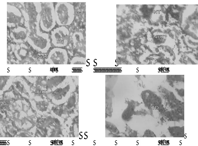
Kidneys of starved fish after 10 days of starvation exhibited i) mild tubular vacuolation (TV) (Fig. 8) ii) tubular vacuolation in kidney became more pronounced by day 20th of starvation (Fig. 9) iii) haemopoietic tissue was also observed to exhibit degenerative changes (Fig. 10), after 30 days of starvation period, iii) kidney tissue showed the initiation of process of necrosis of tubules (NT) and haemopoietic tissue (NHT) (Fig. 10) iv) necrotic tubules and haemopoietic tissue of kidney was observed to undergo acute degeneration from 50th day of starvation (Fig. 11), v) total degeneration (TD) of kidney was observed after 60 days of starvation, where tubules were seen to lose their normal architecture and the haemopoietic tissue appeared almost negligible (Fig. 12).
c) Spleen
The microscopic examination of histology of spleen of control fish showed the presence of red pulp (RP) and white pulp (WP). Splenic tissue also represented the dark staining melanomacrophage centres (MMCs) (Fig. 13)
Histology of spleen in starved fish depicted i) initiation of mild necrosis (N) and presence of accumulated haemosiderin pigments (HP) (Fig. 14) by day 10th of starvation which got more pronounced at the end of 20 days of starvation (Fig. 15) ii) After 30 days of starvation, spleen tissue was seen to exhibit intense necrosis and even initiation of vacuolation (V) (Fig. 16) iii) after 50 days of starvation, acute degenerative changes (DC) were observed in splenic tissue (Fig. 17) iv) these changes became more and more acute and ultimately by day 60th day, spleen exhibited degeneration (TD) with almost total loss of normal cellular architecture (Fig. 18).
IV. DISCUSSION
Various histological alterations in the liver of starved fish exhibited necrosis, distension of sinusoids, vacuolation and degeneration of cellular architecture of liver tissue. Necrosis is an advanced and irreversible stage of degeneration and is characterized by dead
ISSN: 2319-8753
I
nternational
J
ournal of
I
nnovative
R
esearch in
S
cience,
E
ngineering and
T
echnology
(An ISO 3297: 2007 Certified Organization)
Vol. 3, Issue 3, March 2014
Copyright to IJIRSET www.ijirset.com 10131
vacuolation of liver tissue thereby impairing its synthetic machinery and thereby may create an imbalance in the rate of synthesis and release of substances in the systemic circulation. Under the influence of prolonged starvation, therefore, it appears that liver hepatocytes lose their integrity (due to necrosis and vacuolation) and hence the energy reserves in the form of glycogen and lipids stored in hepatocytes may apparently get depleted. Since glycogen and lipids stored in liver hepatocytes are the only source of energy for the fish during starvation, their pronounced depletion provoked by starvation, lands the fish in extremely worst condition. Similar viewpoint has been put forth by Gisbert et al. [12] and Rios et al. [13] in fish liver tissue following starvation. Liver tissue of starved fish also depicted distension of sinusoids which became more massive as the experimental prolonged further. Distension of sinusoids seemingly may be due to the mechanical obstruction of blood supply to liver tissue.
Therefore it is the combined effect of necrosis, vacuolation and distension of the sinusoids which seemingly appear to result in overall degeneration of the cellular architecture of liver tissue towards the end of the starvation period. The total degeneration of the liver tissue may definitely results in disruption of various metabolic processes including erythropoiesis. All these histopathological observations indicate that exposure of fish Labeo boga to prolonged starvation caused destructive changes in its liver tissue ultimately disrupting the functional efficiency of liver.
Presently, microscopic examination of the head kidney of starved fish showed tubular vacuolation which became more pronounced with the advancement of the starvation period ultimately leading to necrosis of tubules and even haemopoietic tissue. The kidney tissue was observed to undergo total degeneration with loss of normal architecture of tubules and haemopoietic tissue by the end of starvation period. Such histological variations observed in kidney tissue are in conformity with the findings of Velmurugan et al. [14], Mohamed [15] and Prashanth [16] in fishes when exposed to different periods of starvation. . Degenerative changes in kidney tissue as observed presently can be causative of different excretory disorders and internal exaution. Moreover continuous decline in the volume of haemopoietic tissue of the kidney can lead to impairment of haemopoiesis. This may result in disruption in the blood forming efficiency of head kidney and hence inhibits further release of normal erythrocytes into the general circulation. Thus prolonged starvation degrades the normal architecture of the fish kidney thereby affecting its functional efficiency which has a direct influence on physiology of fish. Spleen of the starved fish exhibited conspicuous histological alterations viz., necrosis, appearance of haemosiderin pigments, vacuolation and total degeneration of the splenic tissue following prolonged starvation. Necrosis of splenic tissue followed by Vacuolation surfaced due to starvational stress. Spazier et al. [17] also observed necrosis and vacuolation in splenic tissue of European eel Anguilla anguilla following stress resulting in impairment of normal physiology of fish. Haemosiderin is one of the breakdown products of Hb from senescent and degenerated erythrocytes [18]. Deposition of haemosiderin leads to pathological state known as haemosiderosis. Hibiya [19] also reported haemosiderosis and attributed it to an increased rate of erythrocyte destruction in the spleen resulting eventually in a decreased number of mature erythrocytes in general circulation. Similar deposition of haemosiderin pigments has been observed in the spleen of presently studied starved fishes also.
Under the influence of prolonged starvation, the histological architecture of splenic tissue manifested severe degeneration ultimately affecting its blood cell forming capacity i.e. both erythropoiesis as well as leucopoiesis.
V. CONCLUSION
ISSN: 2319-8753
I
nternational
J
ournal of
I
nnovative
R
esearch in
S
cience,
E
ngineering and
T
echnology
(An ISO 3297: 2007 Certified Organization)
Vol. 3, Issue 3, March 2014
Copyright to IJIRSET www.ijirset.com 10132
recommended that long term food deprivation should be avoided in aquaculture to maintain healthy conditions for the better survival of fishes.
ACKNOWLEDGEMENTS
The authors extend heartfelt thanks to the Department of Zoology, University of Jammu, Jammu for providing necessary facilities regarding the research work.
REFERENCES
[1] D. Margulies, “Assessment of the nutritional condition of larval and early juvenile tuna and Spanish mackerel (Pisces, Scombridae) in the Panama Bight”, Mar. Biol., Volume 115, pp. 317-330, 1993.
[2] E. Kjorsvik, T. Vandermeeren, H. Kryvi, J. Arnfinnson, and P.G. Kvenseth, “Early development of the digestive tract of cod larvae”, Gadius morhua
L., during start feeding and starvation", J. Fish Biol.,Volume38, pp.1-15, 1991.
[3] G.A. Bisbal and D.A. Bengtson, “Description of the starving condition in summer flounder, Paralichthys dentatus, early life history stages”, Fish Bull., Volume 93, 217-230, 1995
[4] K.S. Tilak, K. Veeraiah, and S. Vijaylakshmi, “Mixed toxicity of fenvalerate and monocrotophos to the freshwater fish Catla catla, Labeo rohita and
Cirrhinus mrigala”, J. Ecotoxicol. Environ. Monit., Volume 11, Number 3, pp.163-168, 2001.
[5] A. Sachar, “Studies on effect of organic and inorganic pollutants on haematology, blood biochemistry and immune organs in some fishes of Jammu region”. Ph.D Thesis, Department of Zoology, University of Jammu, Jammu, 2012.
[6] M. Pal, “Bioassay studies under the ecotoxicological stress of chromium tri-oxide in a freshwater food fish, Channa punctatus (Bloch)”. Ph.D. Thesis, Department of Zoology, University of Lucknow, Lucknow, 2006
[7] B. Sarkar, A. Chatterjee, S. Adhikari, and S. Ayyappan, “Carbofuran and cypermethrin induced histopathological alterations in the liver of Labeo rohita (Hamilton) and its recovery”, J. Appl. Ichthyol., Volume 21, pp.131-135, 2005.
[8]M.M.P. Camargo and C.B.R. Martinez, “Histopathology of gills, kidney and liver of a Neotropical fish caged in an urban stream”, Neotropical Ichthyology, Volume 5, Number 3, pp. 327-336, 2007
[9] T.S. Pathan, S.E. Shinde, P.B. Thete, and D.L. Sonawane, “Histopathology of liver and kidney of Rasbora daniconius exposed to paper mill effluent”, Research Journal of Biological Sciences, Volume 5, Number 5, pp. 389-39, 2010.
[10]M.V. Radhakrishnan and S. Hemalatha, “Sublethal effects of cadmium chloride to liver of freshwater fish Channa striatus (Bloch)”, American-Eurasian Journal of Toxicological Sciences, Volume 2, Number 11, pp. 54-56, 2010.
[11] S.O. Ayoola, “Histopathological effects of glyphosate on juvenile African catfish (Clarias gariepinus)”, Ameri-surasian J. Agric. Environ.Sci., Volume 4, Number 3, pp. 362-367, 2008.
[12] E. Gisbert, D.B. Conklin, and R.H. Piedrahita, “Effects of delayed first feeding on the nutritional condition and mortality of California halibut larvae”, J. Fish Biol., Volume 64, pp.116-132, 2004.
[13] F.S. Rios, L. Donatti, M.N. Fernandes, A.L. Kalinin, and F.T. Rantin, “Liver histopathology and accumulation of melanomacrophage centres in
Hoplias malabaricus after long-term food deprivation and refeeding”, Journal of Fish Biology, Volume 71, Number 5, pp. 1393-1406, 2007.
[14] B. Velmurugan, M. Selvanayagam, E.I. Cengiz, and E. Unlu, “The effects of fenvalerate on different tissues of freshwater fish Cirrhinus mrigala”, J. Environ. Sci. Heal., Volume 42, pp. 157-163, 2007.
[15] F.A.S. Mohamed, “Histopathological studies on Tilapia zilli and Soleo vulgaris from lake Qarun, Egypt”, World Journal of Fish and Marine Sciences, Volume 1, Number 1, pp.29-39, 2009.
[16] M.S. Prashanth, “Histopathological changes observed in the kidney of freshwater fish, Cirrhinus mrigala (Hamilton) exposed to cypermethrin”, Recent Research in Science and Technology, Volume 3, Number 2, pp. 59-65, 2011.
[17] E. Spazier, V. Storch, and T. Braunbeck, “Cytopathology of spleen in eel Anguilla anguilla exposed to a chemical spill in the Rhine River”, Diseases of Aquatic Organisms, Volume 14, pp.1-22, 1992.
[18] A.G. Zapata, andE.L.Cooper, “The immune system: comparative histopathology. John Wiley and Sons, Chichester, England”, Zool. Soc. Bengalis., Volume 1, pp.67-70, 1990.
ISSN: 2319-8753
I
nternational
J
ournal of
I
nnovative
R
esearch in
S
cience,
E
ngineering and
T
echnology
(An ISO 3297: 2007 Certified Organization)
Vol. 3, Issue 3, March 2014
Copyright to IJIRSET www.ijirset.com 10133
[1] [2]
[3] [4]
[5] [6]
Fig. 1 Microphotograph of liver tissue from control fish showing Hepatocytes (H), Sinusoids (S) and melanomacrophage centres (MMCs) (H&E×1000).
Fig. 2-6 Fig. 2 Microphotograph of liver tissue from Starved fish showing Necrosis (N), Distended Sinusoids (DS), Vacuolation (V), Degenerated cellular architecture (DCA) after 10, 20, 40 , 50 and 60 days of the experiment respectively.
ISSN: 2319-8753
I
nternational
J
ournal of
I
nnovative
R
esearch in
S
cience,
E
ngineering and
T
echnology
(An ISO 3297: 2007 Certified Organization)
Vol. 3, Issue 3, March 2014
Copyright to IJIRSET www.ijirset.com 10134
[9] [10]
[11] [12]
Fig. 1 Microphotograph of kidney tissue from control fish showing haemopoietic tissue (HT) and renal tubules (RT) (H&E×1000).
Fig. 7-12 Microphotograph of kidney tissue from Starved fish showing Tubular Vacuolation (TV), Necrotic Haemopoietic Tissue (NHT), Necrotic Tubules (NT), Degenerated Tubules and Haemopoietic Tissue (DHT, DT) and Total degeneration (TD) after 10, 20, 30, 50 and 60 days of the experiment respectively.
ISSN: 2319-8753
I
nternational
J
ournal of
I
nnovative
R
esearch in
S
cience,
E
ngineering and
T
echnology
(An ISO 3297: 2007 Certified Organization)
Vol. 3, Issue 3, March 2014
Copyright to IJIRSET www.ijirset.com 10135
[15] [16]
[17] [18]
Fig. 13 Microphotograph of splenic tissue from control showing Red pulp (Rp), white pulp (Wp) and melanomacrophage centres (MMCs) (H&E×1000)