Page 1 of 10
The clinical application of mesenchymal stem cells in liver
disease: the current situation and potential future
Sainan Zhang#, Ya Yang#, Linxiao Fan, Fen Zhang, Lanjuan Li
State Key Laboratory for Diagnosis and Treatment of Infectious Diseases, National Clinical Research Center for Infectious Diseases, Collaborative Innovation Center for Diagnosis and Treatment of Infectious Diseases, The First Affiliated Hospital, College of Medicine, Zhejiang University, Hangzhou 310003, China
Contributions: (I) Conception and design: L Li; (II) Administrative support: L Li; (III) Provision of study materials or patients: S Zhang, Y Yang; (IV) Collection and assembly of data: S Zhang, Y Yang; (V) Data analysis and interpretation: S Zhang, Y Yang, L Fan, F Zhang; (VI) Manuscript writing: All authors; (VII) Final approval of manuscript: All authors.
#These authors contributed equally to this work.
Correspondence to: Lanjuan Li. The First Affiliated Hospital, College of Medicine, Zhejiang University, No. 79 Qingchun Road, Shangcheng District, Hangzhou 310003, Zhejiang, China. Email: ljli@zju.edu.cn.
Abstract: Liver disease is a major health issue which present poor clinical treatment performance. Cirrhosis and liver failure are common clinical manifestations of liver diseases. Liver transplantation is recognized as the ultimate and most efficient therapy to the end stage of liver disease. But it was limited by the shortage of honor organs and high cost. Nowadays, stem cell therapy gained more and more attention due to its attractive efficacy in treating liver disease especially in cirrhosis during the clinical trials. Mesenchymal stem cell (MSC) can be differentiated into hepatocytes, promote liver regeneration, inhibit liver fibrosis and induce liver apoptosis, particularly via paracrine mechanisms. This review will highlight recent clinical applications of MSC, providing the available evidence and discussing some unsolved questions in treating liver disease.
Keywords: Mesenchymal stem cells (MSCs); liver disease; cirrhosis
Submitted Nov 14, 2019. Accepted for publication Mar 19, 2020. doi: 10.21037/atm.2020.03.218
View this article at: http://dx.doi.org/10.21037/atm.2020.03.218
Introduction
Liver disease is a major health problem and caused by various etiologies around the world. Acute liver failure
(ALF), liver cirrhosis and liver cancer are the main common
liver diseases. ALF is a fatal clinical syndrome characterized
by extensive hepatocyte necrosis and inflammatory
infiltration caused by hepatotoxic drugs, immune-mediated attacks or viral infections (1,2). In the clinical, ALF
progressed rapidly with a poor presentation to medical
treatment (3). Cirrhosis is the end stage of liver fibrosis and prone to various complications including infection, hemorrhage, hepatic encephalopathy (HE) and spontaneous peritonitis, etc. According to the latest global report, liver
cancer ranked sixth for cancer incidence and fourth for
cancer deaths in 2015 (4). Hepatocellular carcinoma (HCC) accounts for 85–90% of primary liver cancer (5). At present,
liver transplantation is recognized as the most effective treatment for advanced liver diseases. However, there is a prevailing contradiction between urgent clinical need and the shortage of donor livers. Therefore, new effective
methods for treating liver diseases is urgently needed. In recent years, mesenchymal stem cells (MSCs) have been proposed as an alternative approach to treat liver
diseases. MSCs can be defined as pluripotent cells with the capacity of self-renewal, which can give rise to many unique,
differentiated mesenchymal cell types (6). At present, the MSCs applied in clinical therapy and basic experimental
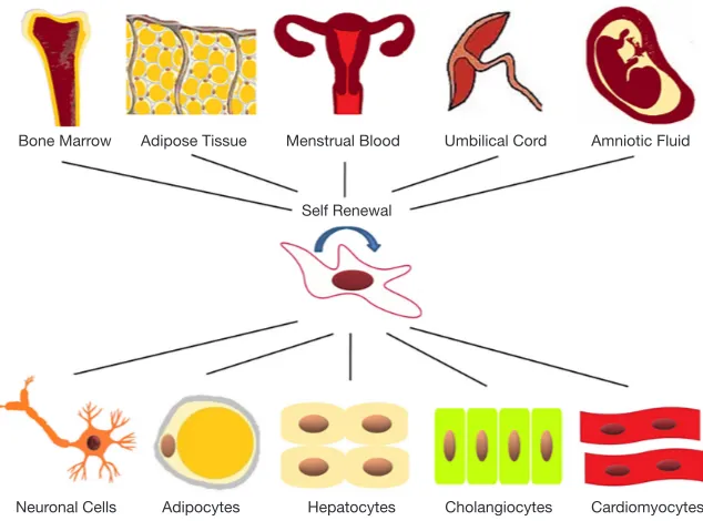
research are mainly derived from bone marrow, umbilical
cord, adipose tissue, amniotic fluid, menstrual blood, etc.
(7-9). As shown in Figure 1, MSCs have the potential to differentiate into chondrocytes, osteocytes, and adipocytes,
which show significant effect in regenerative medicine (10).
Furthermore, MSCs have low inherent immunogenicity and can modulate immune responses by interacting with various
immune cells (11). The homing capacity is the key to the effective application of MSC in clinical treatment, which was defined as blocking MSCs in the tissue vasculature and
then migrating across the endothelium (12). The present
study demonstrated that stem cell therapy was a therapeutic
strategy in liver disease.
In this review, we aim to discuss available evidence and
highlight some unsolved questions of stem cells for treating
liver disease. This work will focus on the clinical application
of MSCs in liver disease.
The mechanism of MSC in tissue repair and regeneration medicine
In previous study, stem cell therapy for liver disease has been proved to be effective in both basic and clinical
research. MSCs were mostly applied in liver cirrhosis and shown a better therapeutic effect in compensatory
period of liver disease. According to present studies,
stem cell therapy shown significant improvement in liver function through anti-apoptosis and immune regulation.
MSC can differentiate into hepatocytes in vivo and play a
key therapeutic role in the treatment of liver fibrosis by
secreting various immunomodulatory factors. After MSCs
therapy, antiapoptotic factors including hepatocyte growth factor (HGF) and insulin-like growth factor (IGF-1) were elevated with the same promotion in angiogenetic and mito-genetic factors (13).
In vitro experiments, MSCs can promote apoptosis of hepatic stellate cells and inhibit collagen synthesis.
Direct coculture of endothelial progenitor cells (EPCs)
and MSCs in vitro enhanced cell proliferation and
angiogenic capacity, PDGF and Notch signaling pathways were involved in this effect (14). In indirect coculture experiment, MSCs inhibited LX2 (hepatic stellate cell line) proliferation through secretion of inflammatory factors
[interleukin-6 (IL-6), IL-8, HGF, growth-related oncogene
and osteoprotegerin] (15). MSCs participated in the cell communication directly or indirectly through paracrine function.
[image:2.595.137.454.85.320.2]Hepatic progenitor cells (HPCs), also named hepatic stem cell, possess an extremely low percentage in adult liver. When liver injured, HPCs can differentiate into both Figure 1 MSCs derived from different tissues develop into multiple cell types in vitro. MSCs are most commonly derived from bone marrow, adipose tissue, menstrual blood, umbilical cord and amniotic fluid. MSCs can be induced different into multiple cell types such as neuronal cells, adipocytes, hepatocytes, cholangiocytes and cardiomyocytes.
Bone Marrow Adipose Tissue Menstrual Blood
Self Renewal
Neuronal Cells Adipocytes Hepatocytes Cholangiocytes Cardiomyocytes
hepatocytes and cholangiocytes.
The application of MSCs in ALF
In the clinical, ALF progressed rapidly with a poor
presentation to medical treatment (3). In recent years, the incidence of liver failure caused by herbal medicine has
increased significantly.
It has been demonstrated that excessive inflammatory
response plays a key role in pathogenesis and prognosis
of ALF (16). During the progress of ALF, the domestic Kupffer cells (KCs), dendritic cells (DCs), and natural
killer (NK) cells are highly activated, simultaneously the
monocytes/macrophagocytes and the neutrophils are recruited in liver tissue (17). Another report demonstrated
that activated KCs released high levels of TNF-α, IL-1, and IL-6 in LPS induced ALF models (18). The systemic inflammation could stimulate hepatocytes necrosis and apoptosis.
Many studies have confirmed the role of MSCs in the treatment of ALF animal models. MSCs can reduce the mortality, improve liver functions, inhibit hepatocytes apoptosis and promote proliferation (19,20). Previous
studies demonstrated that MSCs showed therapeutic
effect in ALF by immunoregulation. Firstly, MSCs can
inhibit inflammation and alleviate liver injury by regulating inflammatory cytokine levels. Chen et al. indicated that
MenSC could down-regulate the expression of TNF-α, IL-6, and IL-1β in mice models (21). Zhu et al. showed that BM-MSCs reduced the levels of TNF-α, IFN-γ and IL-4 (22).
Secondly, MSCs inhibit the levels of inflammatory cytokine
released by T cells, B cells, DCs and NKs cells (23-25). On the other hand, MSCs can enhance hepatocyte proliferation in liver failure models. Liu et al. showed that PGE2 secreted
by MSCs enhanced hepatocyte proliferation by YAP and
mTOR signaling (19). Shi et al. demonstrated that the DLL-4 secreted by Human BM-MSC promoted the proliferation
of biliary epithelium cells in ALF pigs and rats (26). Although
multiple MSCs can differentiate into hepatocyte-like cells in vitro (27-29), only few part (<4.5%) of transplanted MSCs differentiated into hepatocyte-like cells in ALF pigs (26).
Currently, the application of MSCs in ALF is limited to basic research. But it is obvious that MSCs have the capacity of promoting liver regeneration and suppressing
inflammation. Comparing to control group, BM-MSCs effectively reduced the levels of ALT and ALB after 1 week of
treatment (30). All these studies indicated the broad prospects of MSCs application in the clinical treatment of ALF.
The application of MSCs in cirrhosis
Liver fibrosis is a chronic disease caused by various etiologies, including viral infection, drug damage, alcohol abuse and immunological diseases. Chronic liver
damage leads to excessive extracellular matrix (ECM)
deposition through cycles of hepatocytes apoptosis, inflammation and repetitive damage repair. Cirrhosis is
the end stage of progressive fibrosis that lack of effective
comprehensive medical treatment. MSCs have the capacity
of differentiating into hepatocyte-like cells and secreting
factors to regulate immune. Thus, MSCs participate in tissue repair through direct and indirect approach.
It has been reported that MSCs alleviate the processes of
epithelial-mesenchymal transition (EMT) and contribute
to liver regeneration through differentiation, immune regulation and secretion (31). At present, BM-MSCs are the most widely used in clinical application.
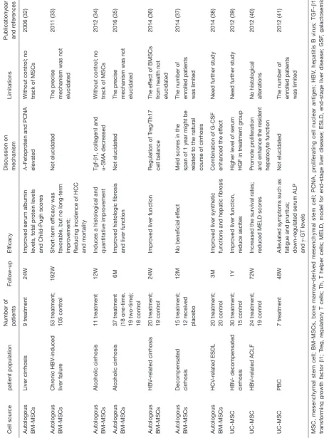
A few clinical trials have been conducted to evaluate the
curative effect of MSCs treating liver diseases (Table 1).
Autologous BM-MSCs transplantation was effective in improving liver function and Child-Pugh scores in patients with liver cirrhosis (32). Autologous BM-MSCs were investigated to improve histologic fibrosis and liver function in patients with alcoholic cirrhosis (34,35). Comparing to one-time transplantation, there was no improved results in fibrosis quantification in two-time BM-MSC transplantation (34).
After autologous BM-MSCs therapy, serum albumin levels and total protein were elevated. In SHUJI TERAI’s study, α-Fetoprotein (AFP) and proliferating cell nuclear antigen (PCNA) expression was significantly elevated in liver biopsy tissue after autologous BM-MSCs therapy (32). It was
reported that AFP and PCNA participate in the process of hepatocyte proliferation (42). MSCs can differentiate into hepatocytes, effectively promoting liver regeneration. In
another clinical trial, regulation of Treg/Th17 cell balance was
investigated in treating liver cirrhosis (36). Present evidences
demonstrated that autologous BM-MSCs can activate T
cell receptors and rebuild immunological tolerance (43). Paracrine function of MSCs regulates immune response and
promotes liver regeneration. In Mehdi Mohamad Nejad’s
research (37), autologous BM-MSC transplantation probably has no beneficial effect in decompensated cirrhotic patients. Meaningfully, repeated autologous BM-MSCs therapy improved liver function of patients with decompensated
T
able 1
MSCs in clinical trials treating liver diseases
Cell sour
ce
patient population
Number of patients
Follow-up
Ef
ficacy
Discussion on mechanism
Limitations
Publicationyear and r
efer
ences
Autologous BM-MSCs
Liver cirrhosis
9 tr
eatment
24W
Impr
oved serum albumin
levels, total pr
otein levels
and Child-Pugh scor
es
Α-Fetopr
otein and PCNA
elevated
Without contr
ol; no
track of MSCs
2006
(32)
Autologous BM-MSCs
Chr onic HBV -induced liver failur e 53 tr eatment; 105 contr ol 192W Short-term ef ficacy was
favorable, but no long-term impr
ovement;
Reducing incidence of HCC and mortality
Not elucidated
The pr
ecise
mechanism was not elucidated
2011
(33)
Autologous BM-MSCs
Alcoholic cirrhosis
11 tr
eatment
12W
Induces a histological and quantitative impr
ovement Tgf-β 1, collagen Ⅰ and
α-SMA decr
eased
Without contr
ol; no
track of MSCs
2012
(34)
Autologous BM-MSCs
Alcoholic cirrhosis
37 tr
eatment
(18 one-time, 19 two-time); 18 contr
ol
6M
Impr
oved histologic fibr
osis
and liver function
Not elucidated
The pr
ecise
mechanism was not elucidated
2016
(35)
Autologous BM-MSCs
HBV -r elated cirrhosis 20 tr eatment; 19 contr ol 24W Impr
oved liver function
Regulation of T
reg/Th17
cell balance
The ef
fect of BMSCs
fr
om health not elucidated
2014
(36)
Autologous BM-MSCs Decompensated cirrhosis
15 tr eatment; 12 r eceived placebo 12M
No beneficial ef
fect
Meld scor
es in the
span of 1 year might be related to the natural course of cirrhosis The number of enr
olled patients
was limited
2014
(37)
Autologous BM-MSCs
HCV -r elated ESDL 20 tr eatment; 20 contr ol 3M Impr
oved liver synthetic
functions and hepatic fibr
osis
Combination of G-CSF enhanced the ef
fect
Need further study
2014 (38) UC-MSC HBV - decompensated cirrhosis 30 tr eatment; 15 contr ol 1Y Impr
oved liver function,
reduce ascites
Higher level of serum HGF in tr
eatment gr
oup
Need further study
2012 (39) UC-MSC HBV -r elated ACLF 24 tr eatment; 19 contr ol 72W Incr
eased the survival rates;
reduced MELD scor
es
Pr
omote pr
oliferation
and enhance the r
esident
hepatocyte function
No histological alterations
2012 (40) UC-MSC PBC 7 tr eatment 48W
Alleviated symptoms such as fatigue and pruritus; down-r
egulated serum ALP
and
γ
-GT levels
Not elucidated
The number of enr
olled patients
was limited
2012
(41)
MSC, mesenchymal stem cell; BM-MSCs, bone marr
ow-derived mesenchymal stem cell; PCNA, pr
oliferating cell nuclear antigen; HBV
, hepatitis B virus;
TGF-β 1, transforming gr owth factor β 1; T reg, r
egulatory T cells; Th, T helper cells; MELD, model for end-stage liver disease; ESLD, end-stage liver disease; GSF
, galactosemic
fibr
oblast; UC-MSC, umbilical cor
d-derived mesenchymal stem cell; HGF
, hepatocyte gr
owth factor; ACLF
, acute-on chr
onic liver failur
e; ALP
, alkaline phosphatase;
γ
-GT
,
γ
glutamyl transferase; W
, week, M, month, Y
, year
[image:4.595.57.513.93.709.2]and hepatic fibrosis (38). The efficacy of autologous BM-MSCs was connected to age and physical condition which make a close relation to differentiative capacity (39,45). Insufficient enrolled patients may be one of the limitations
in the study. Future randomized controlled trials need to expand number of enrolled patients to confirm the efficacy of
MSCs therapy in liver disease. To be concerned, autologous
BM-MSC therapy couldn’t achieve acceptable long-term
effects on prognosis (33). The reasons for the poor long-term efficacy may be as follows: the number of autologous BM-MSCs were limited; the homing ability is poor that BM-MSCs
could not reach the effective amount in liver (46).
UC-MSC can be obtained free and amplify to a large number. It has been reported that UC-MSC transplantation
alleviate symptoms of various autoimmune diseases (47,48). In
a 1-year follow-up clinical research, UC-MSC transplantation
has been proved to be safe and could improve liver function
and reduce ascites in patients with decompensated liver
cirrhosis (39). Even in ACLF patients, Shi et al. (40) have
shown that UC-MSC transfusions significantly increased the survival rates. UC-MSCs show a more beneficial immunogenic profile and stronger overall immunosuppressive potential than BM-MSCs (49). After UC-MSC therapy, liver function was
improved, that serum albumin levels increased and bilirubin
levels decreased. UC-MSC treatment also improved liver function and patient’s quality of life in primary biliary cirrhosis
(PBC) (41). UC-MSC therapy lowered serum alkaline
phosphatase and γ-glutamyltransferase levels and alleviate
fatigue and pruritus symptom in PBC patients. Comparing
to BM-MSC, adipose-derived stromal cells (ADSCs) can be
obtained from healthy donors freely and cultured to expand sufficient numbers. During cell amplification, ADSCs could
maintain the characteristics of stem cells without losing their
differentiation capacity (50). During the past few years, ADSCs
have been used as a therapeutic strategy for tissue repair and immune modulation (51). ADSCs shown anti-inflammatory
effect and secrete various factors to promote regeneration. ADSCs have been displayed as a feasible therapeutic strategy to alleviate liver damage (11,52). But there aren’t many clinical
trials using ADSCs to treat liver disease.
Human menstrual blood-derived stem cells (MenSCs) are isolated from menstrual fluids with the advantage of simple
operation, easy obtainment, safe and painless. MenSCs
have been used to treat several diseases such as stroke,
type 1 diabetes, premature ovarian failure and myocardial infarction (53-56). Previous study described that MenSCs
can be differentiated into functional hepatocyte-like
cells (57). In mice experiment, MenSCs shown an
antifibrotic effect in liver fibrosis (9). At present, there are
few clinical applications of MenSCs in liver disease. More
clinical trials should be conducted in the future study.
The application of MSCs in liver cancer
It has been proved that MSCs have the ability of migrating and integrate into the tumor tissue (58). However, the application of multi-origin MSCs in liver cancer is
controversial. Different sources of MSCs play diverse roles
in liver cancer, which limits the application of MSCs in clinical treatment. Gardin et al. proved that AD-MSCs inhibited HepG2 and PLC-PRF-5 proliferation while
promoted apoptosis in vitro by up-regulating P53 and RB and down-regulating c-Myc and hTER (59). Another report
showed that conditioned medium (CMs) delivered from AD-MSCs inhibited hepatoma carcinoma cell proliferation and promoted death in vitro by down-regulating Akt
signaling (60).
Yet BM-MSCs showed the opposite effect in HCC. A recent work demonstrated that BM-MSCs stimulated migration and invasion of HCC cells which could be hampered by AQP1 inhibitor (61). BM-MSCs-mediated
upregulation of CXCL4 also plays critical role in promoting
HCC cell migration and invasion in vitro (62). Interestingly,
genetically modified BM-MSCs, which expressed high levels of stTRAIL, could migrate to heat-shocked HCC
cells and induce apoptosis in nude mice (63).
Despite the original effect of MSCs on tumors, MSCs
are potential tools for transporting drugs or anti-tumor virus
due to their ability of tumor chemotaxis, immunosuppressive
properties and low immunogenicity. It was reported that oncolytic measles virus infected BM-MSCs could homing to HCC tumors and transfer the virus to HCC via heterofusion to significantly inhibit tumor growth (58). What’s more, MenSCs infected with oncolytic adenovirus also showed their ability to migrate to variety kinds of tumors and suppress tumor growth in vivo. These studies show that using MSCs as gene vehicle could be a novel strategy for tumor-targeted
clinical application in the future.
The transduction of secretory substance released by MSCs
It has been reported CM from MSCs show similar protective
effects in tissue damage. CM contained paracrine soluble
factors and extracellular vesicles (EVs) promoting tissue
and transport lipids, proteins, DNA, miRNA, and non-coding RNA. EVs can be classified into three subtypes
including exosomes, microvesicles and apoptotic bodies. We
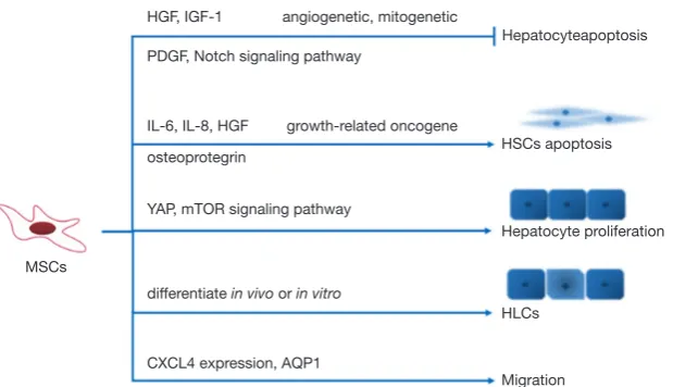
partly summarized the cytokines secreted by MSCs and the
signal transduction involved in the therapy pathway (Figure 2). In mice experiment, MenSC derived exosomes alleviated
fulminant hepatic failure induced by D-GalN/LPS (21).
MenSC derived exosomes provided an anti-apoptotic capacity with higher levels of cytokines including ICAM-1, angiopoietin-2, Axl, angiogenin, IGFBP-6, osteoprotegerin, IL-6 and IL-8 (21). MSCs exosomes were proposed as an ideal ‘cell-free’ therapeutic alternative to replace stem cell therapy. Combined with the homing capacity of stem
cells, exosomes can be used as vectors for targeted therapy.
However, exosome is currently limited to be widely used by the complicated extraction. And it is of great significance to
clarify the components of exosomes for further research.
The potential application of MSCs in bioartificial liver (BAL) system
BAL support system incorporating cell source in bioreactor
has been proposed as an effective means to treat end-stage
liver diseases. With the application of BAL, it suggested to improve the survival time of fulminant hepatic failure pigs (65). Cell source used in BAL should possess the capacity of extensive amplification and maintenance of cellular characteristics. Cell lines derived from hepatomas
and genetically engineered hepatocyte-like cells hepatoid cells have been widely used for BAL. Lv et al. study shown
that the efficacy of BAL can be enhanced by co-culturing of liver cells with MSCs (65). It suggested that MSCs can modulate cell properties to achieve a better curative effect in BAL application. Adding secretory substance released by MSCs to BAL may enhance the capacity to improve the therapeutic potential.
Safety and efficacy of MSCs treatment
Safety, especially potential of tumorigenesis is the most
concerned issue in MSCs clinical applications. However, after analyzing previous studies about chromosomal
aberrations of MSCs, Sensebe et al. argued that genomic stability of cultured adult stem cells, especially MSCs is
robust, which indicated that MSCs transplantation was less likely to cause tumor. During a follow-up of 11.5 years after BM-MSCs transplantation, neither tumor nor infections were observed among the 41 patients (66). Chen et al. indicated that MSC infusion could significantly reduce the mortality rate of ACLF patients without increasing severe
complications (67). According to another meta-analysis, BM-MSCs and UC-MSCs infusion could improve liver
function in cirrhosis patients. At the end of the first year,
there were no serious side effects or complications.
Fever (37–38 ℃) and rash are the most common adverse
events reported but recover without additional treatment
in clinical trials. Considering that MSCs has the character
of low inherent immunogenicity, it may be related to the
immune regulation function of MSCs.
Although large amount of studies has demonstrated
MSCs
HGF, IGF-1
PDGF, Notch signaling pathway
IL-6, IL-8, HGF
osteoprotegrin
YAP, mTOR signaling pathway
differentiate in vivo or in vitro
CXCL4 expression, AQP1
Migration HLCs
Hepatocyte proliferation HSCs apoptosis growth-related oncogene
angiogenetic, mitogenetic
[image:6.595.145.455.81.259.2]Hepatocyteapoptosis
the therapeutic effect of different MSCs on various liver diseases both in animal models and patients, there is currently no uniform clinical treatment guideline for
efficient therapy strategy.
Determination of cells dose and timing is urgently needed for clinical applications (68). In most clinical indications, the
dose of human MSCs is generally 1–2 million cells/kg, and never exceeds 12 million cells/kg (69). However, so far, there
are no clinical trials reporting treatment effects comparison in different cell doses. On the other hand, there are very
few related studies, especially clinical trials about timing
of MSCs transplantation in liver diseases. After comparing effect of MSCs infusion in different time points in hepatic
schistosomiasis models, El-Shennawy et al. suggested that the
earlier injection of MSCs, the better treatment effect (70).
These studies suggested necessities for in-depth research of
MSCs administration timing in patients.
Optimization and standardization of isolation, culture, expansion and delivering are also key factors to steer MSCs treatment efficacy and safety, which need undergo extensive investigations before applicated in the clinical field.
Conclusions
In present studies, stem cells applied in clinical trials varies
from different disease. Autologous BM-MSCs is the most widely used in liver diseases. To some extent, autologous
MSCs extraction from patients is still a damaging process.
The efficacy of autologous BM-MSCs is limited by physician
condition including aging differentiation and deficiency in vitality (45). Comparing to BM-MSCs, UC-MSC are free
from these limitations (49). ADSCs clinical application is limited by isolation technique. If ADSCs can be isolated more easily, they can be used in greater number than BMSCs.
In conclusion, MSCs therapy couldn’t achieve beneficial effect in decompensated cirrhosis and the long-term efficacy
is poor. By far, liver transplantation is the most effective
and the ultimate treatment for end-stage liver disease. Combined with stem cells in the treatment of liver disease, stem cell therapy can improve liver function in the short-term. Stem cell therapy can prolong the waiting time for liver transplantation in patients with end-stage liver disease and become a bridge between end-stage liver disease and
liver transplantation. Repeated stem cell therapy may be an
effective approach to achieve long-term therapeutic effects.
Further clinical trials are needed to clarify the mechanism of stem cell in liver disease.
It has great significance to further clarify the substances
secreted by MSCs, and to explore the definite substances contributing to immune regulation and tissue regeneration. To better investigate the mechanism involved in MSCs therapy, it is important to identify the protein, DNA and RNA released by MSCs. The proteomics and transcriptomics may play an important role in exploring
the under mechanism. Cell communication with the help of EVs transfects key factors to regulate cell differentiation
and induce subsequent signal transduction.
Increasing the number of cells homing to the injury site is the key point to improve the therapeutic effect of stem
cells. Studying the homing characteristics of stem cells is helpful to improve the effective therapeutic quantity of stem cells. Identifying the differentiation of stem cells in vitro micro-environment gave a direction to the targeted
regulation of stem cells.
Currently, there are no standardized protocols for stem cells clinical trials. To better analyze the difference between stem
cells, more clinical trials should be conducted in the future.
The sample size needs to be expanded and it needs to set an effective control group during the study. Therapeutic pathways
and effective time points are required to be confirmed. We
need to establish standardized protocols for clinical trials. In summary, it was confirmed that MSCs has a promising therapeutic effect in liver disease therapy. More works
are needed to clarify the underlying mechanism of stem
cell therapeutic effects. Standardized protocols should
be established according to the best time interval and
reasonable therapeutic dose. Cell type, injection route and
observation time points need to be better formulated in the clinical application of MSCs.
Acknowledgments
Funding: This work was supported by the National Natural Science Foundation of China (NSFC: 81721091).
Footnote
Provenance and Peer Review: This article was commissioned by the Guest Editor (Ling Lu) for the series “Stem Cell and
Clinical Application” published in Annals of Translational Medicine. The article was sent for external peer review organized by the Guest Editor and the editorial office.
Conflicts of Interest: All authors have completed the ICMJE
Clinical Application” was commissioned by the editorial office without any funding or sponsorship. The authors have no other conflicts of interest to declare.
Ethical Statement: The authors are accountable for all
aspects of the work in ensuring that questions related to the accuracy or integrity of any part of the work are
appropriately investigated and resolved.
Open Access Statement: This is an Open Access article
distributed in accordance with the Creative Commons Attribution-NonCommercial-NoDerivs 4.0 International License (CC BY-NC-ND 4.0), which permits the non-commercial replication and distribution of the article with
the strict proviso that no changes or edits are made and the
original work is properly cited (including links to both the
formal publication through the relevant DOI and the license).
See: https://creativecommons.org/licenses/by-nc-nd/4.0/.
References
1. Bernal W, Wendon J. Acute liver failure. N Engl J Med 2013;369:2525-34.
2. Bernal W, Auzinger G, Dhawan A, et al. Acute liver failure. Lancet 2010;376:190-201.
3. Andersson U, Wang H, Palmblad K, et al. High mobility group 1 protein (HMG-1) stimulates proinflammatory cytokine synthesis in human monocytes. J Exp Med 2000;192:565-70.
4. Fitzmaurice C, Allen C, Barber RM, et al. Global, Regional, and National Cancer Incidence, Mortality, Years of Life Lost, Years Lived With Disability, and Disability-Adjusted Life-years for 32 Cancer Groups, 1990 to 2015: A Systematic Analysis for the Global Burden of Disease Study. JAMA Oncol 2017;3:524-48.
5. El-Serag HB, Rudolph KL. Hepatocellular carcinoma:
epidemiology and molecular carcinogenesis.
Gastroenterology 2007;132:2557-76.
6. da Silva Meirelles L, Caplan AI, Nardi NB. In search of the in vivo identity of mesenchymal stem cells. Stem Cells
2008;26:2287-99.
7. Bieback K, Kern S, Kluter H, et al. Critical parameters
for the isolation of mesenchymal stem cells from umbilical
cord blood. Stem Cells 2004;22:625-34.
8. Miki T. Stem cell characteristics and the therapeutic potential of amniotic epithelial cells. Am J Reprod
Immunol 2018;80:e13003.
9. Chen L, Zhang C, Chen L, et al. Human Menstrual
Blood-Derived Stem Cells Ameliorate Liver Fibrosis in Mice by Targeting Hepatic Stellate Cells via Paracrine Mediators. Stem Cells Transl Med 2017;6:272-84.
10. Klingemann H, Matzilevich D, Marchand J. Mesenchymal Stem Cells - Sources and Clinical Applications. Transfus Med Hemother 2008;35:272-7.
11. Liu WH, Song FQ, Ren LN, et al. The multiple
functional roles of mesenchymal stem cells in participating
in treating liver diseases. J Cell Mol Med 2015;19:511-20. 12. Karp JM, Leng Teo GS. Mesenchymal stem cell homing: the devil is in the details. Cell Stem Cell 2009;4:206-16. 13. Mohammadi Gorji S, Karimpor Malekshah AA,
Hashemi-Soteh MB, et al. Effect of mesenchymal stem cells on Doxorubicin-induced fibrosis. Cell J 2012;14:142-51. 14. Liang TZ, Zhu L, Gao WL, et al. Coculture of endothelial
progenitor cells and mesenchymal stem cells enhanced
their proliferation and angiogenesis through PDGF and Notch signaling. Febs Open Bio 2017;7:1722-36. 15. Chen LJ, Zhang CF, Chen L, et al. Human Menstrual
Blood-Derived Stem Cells Ameliorate Liver Fibrosis in Mice by Targeting Hepatic Stellate Cells via Paracrine Mediators. Stem Cells Transl Med 2017;6:272-84. 16. Wyke RJ, Yousif-Kadaru AG, Rajkovic IA, et al. Serum
stimulatory activity and polymorphonuclear leucocyte
movement in patients with fulminant hepatic failure. Clin Exp Immunol 1982;50:442-9.
17. Possamai LA, Thursz MR, Wendon JA, et al. Modulation
of monocyte/macrophage function: a therapeutic
strategy in the treatment of acute liver failure. J Hepatol 2014;61:439-45.
18. Luster MI, Germolec DR, Yoshida T, et al. Endotoxin-induced cytokine gene expression and excretion in the liver. Hepatology 1994;19:480-8.
19. Liu Y, Ren H, Wang J, et al. Prostaglandin E2 secreted
by mesenchymal stem cells protects against acute liver
failure via enhancing hepatocyte proliferation. FASEB J 2019;33:2514-25.
20. Salomone F, Barbagallo I, Puzzo L, et al. Efficacy of adipose tissue-mesenchymal stem cell transplantation in rats with acetaminophen liver injury. Stem Cell Res 2013;11:1037-44.
21. Chen L, Xiang B, Wang X, et al. Exosomes derived from human menstrual blood-derived stem cells alleviate fulminant hepatic failure. Stem Cell Res Ther 2017;8:9. 22. Zhu X, He B, Zhou X, et al. Effects of transplanted
between mesenchymal stem cells and T regulatory cells is crucially important for the attenuation of acute liver injury. Liver Transpl 2018;24:687-702.
24. Zhang Y, Cai W, Huang Q, et al. Mesenchymal stem cells alleviate bacteria-induced liver injury in mice by inducing regulatory dendritic cells. Hepatology 2014;59:671-82. 25. Milosavljevic N, Gazdic M, Simovic Markovic B, et
al. Mesenchymal stem cells attenuate acute liver injury by altering ratio between interleukin 17 producing and regulatory natural killer T cells. Liver Transpl 2017;23:1040-50.
26. Shi D, Zhang J, Zhou Q, et al. Quantitative evaluation of
human bone mesenchymal stem cells rescuing fulminant
hepatic failure in pigs. Gut 2017;66:955-64.
27. Chitrangi S, Nair P, Khanna A. Three-dimensional polymer
scaffolds for enhanced differentiation of human mesenchymal
stem cells to hepatocyte-like cells: a comparative study. J Tissue Eng Regen Med 2017;11:2359-72.
28. Mou XZ, Lin J, Chen JY, et al. Menstrual blood-derived
mesenchymal stem cells differentiate into functional
hepatocyte-like cells. J Zhejiang Univ Sci B 2013;14:961-72. 29. Wang Y, Wang JL, Ma HC, et al. Mesenchymal stem
cells increase heme oxygenase 1-activated autophagy in
treatment of acute liver failure. Biochem Biophys Res
Commun 2019;508:682-9.
30. Lin BL, Chen JF, Qiu WH, et al. Allogeneic bone marrow-derived mesenchymal stromal cells for hepatitis B virus-related acute-on-chronic liver failure: A randomized controlled trial. Hepatology 2017;66:209-19.
31. Xie G, Diehl AM. Evidence for and against epithelial-to-mesenchymal transition in the liver. Am J Physiol Gastrointest Liver Physiol 2013;305:G881-90.
32. Terai S, Ishikawa T, Omori K, et al. Improved liver function in patients with liver cirrhosis after autologous bone marrow cell infusion therapy. Stem Cells 2006;24:2292-8.
33. Peng L, Xie DY, Lin BL, et al. Autologous bone marrow
mesenchymal stem cell transplantation in liver failure
patients caused by hepatitis B: short-term and long-term outcomes. Hepatology 2011;54:820-8.
34. Jang YO, Kim YJ, Baik SK, et al. Histological
improvement following administration of autologous bone marrow-derived mesenchymal stem cells for alcoholic cirrhosis: a pilot study. Liver Int 2014;34:33-41. 35. Suk KT, Yoon JH, Kim MY, et al. Transplantation with
autologous bone marrow-derived mesenchymal stem cells for alcoholic cirrhosis: Phase 2 trial. Hepatology 2016;64:2185-97.
36. Xu L, Gong Y, Wang B, et al. Randomized trial of
autologous bone marrow mesenchymal stem cells
transplantation for hepatitis B virus cirrhosis: regulation of
Treg/Th17 cells. J Gastroenterol Hepatol 2014;29:1620-8. 37. Mohamadnejad M, Alimoghaddam K, Bagheri M, et al.
Randomized placebo-controlled trial of mesenchymal stem
cell transplantation in decompensated cirrhosis. Liver Int
2013;33:1490-6.
38. Salama H, Zekri AR, Medhat E, et al. Peripheral vein infusion of autologous mesenchymal stem cells in Egyptian HCV-positive patients with end-stage liver disease. Stem
Cell Res Ther 2014;5:70.
39. Zhang Z, Lin H, Shi M, et al. Human umbilical cord
mesenchymal stem cells improve liver function and ascites
in decompensated liver cirrhosis patients. J Gastroenterol Hepatol 2012;27 Suppl 2:112-20.
40. Shi M, Zhang Z, Xu R, et al. Human mesenchymal stem cell transfusion is safe and improves liver function in acute-on-chronic liver failure patients. Stem Cells Transl Med 2012;1:725-31.
41. Wang L, Li J, Liu H, et al. Pilot study of umbilical cord-derived mesenchymal stem cell transfusion in patients with primary biliary cirrhosis. J Gastroenterol Hepatol 2013;28 Suppl 1:85-92.
42. Kayano K, Yasunaga M, Kubota M, et al. Detection
of proliferating hepatocytes by immunohistochemical staining for proliferating cell nuclear antigen (PCNA) in
patients with acute hepatic failure. Liver 1992;12:132-6.
43. Alexander T, Thiel A, Rosen O, et al. Depletion of
autoreactive immunologic memory followed by autologous hematopoietic stem cell transplantation in patients with refractory SLE induces long-term remission through de novo generation of a juvenile and tolerant immune system. Blood 2009;113:214-23.
44. Zhang W, Teng M, Liu B, et al. Repeated Autologous Bone Marrow Transfusion through Portal Vein for Treating
Decompensated Liver Cirrhosis after Splenectomy.
Gastroenterol Res Pract 2018;2018:4136082. 45. Schubert T, Xhema D, Veriter S, et al. The enhanced
performance of bone allografts using osteogenic-differentiated adipose-derived mesenchymal stem cells. Biomaterials 2011;32:8880-91.
46. Kantarcioglu M, Demirci H, Avcu F, et al. Efficacy of
autologous mesenchymal stem cell transplantation in patients
with liver cirrhosis. Turk J Gastroenterol 2015;26:244-50. 47. Sun LY, Wang DD, Liang J, et al. Umbilical Cord
Mesenchymal Stem Cell Transplantation in Severe and
48. Liu YY, Mu R, Wang SY, et al. Therapeutic potential
of human umbilical cord mesenchymal stem cells in the treatment of rheumatoid arthritis. Arthritis Research & Therapy 2010;12:13.
49. Baksh D, Yao R, Tuan RS. Comparison of proliferative
and multilineage differentiation potential of human mesenchymal stem cells derived from umbilical cord and
bone marrow. Stem Cells 2007;25:1384-92. 50. Baer PC, Geiger H. Adipose-derived mesenchymal
stromal/stem cells: tissue localization, characterization, and heterogeneity. Stem Cells Int 2012;2012:812693. 51. Frese L, Dijkman PE, Hoerstrup SP. Adipose
Tissue-Derived Stem Cells in Regenerative Medicine. Transfus
Med Hemother 2016;43:268-74.
52. Minteer D, Marra KG, Rubin JP. Adipose-derived
mesenchymal stem cells: biology and potential applications.
Adv Biochem Eng Biotechnol 2013;129:59-71. 53. Wu X, Luo Y, Chen J, et al. Transplantation of human
menstrual blood progenitor cells improves hyperglycemia by promoting endogenous progenitor differentiation in
type 1 diabetic mice. Stem Cells Dev 2014;23:1245-57. 54. Borlongan CV, Kaneko Y, Maki M, et al. Menstrual blood
cells display stem cell-like phenotypic markers and exert neuroprotection following transplantation in experimental stroke. Stem Cells Dev 2010;19:439-52.
55. Zhang Z, Wang JA, Xu Y, et al. Menstrual blood derived mesenchymal cells ameliorate cardiac fibrosis via inhibition
of endothelial to mesenchymal transition in myocardial
infarction. Int J Cardiol 2013;168:1711-4.
56. Rodrigues MC, Voltarelli J, Sanberg PR, et al. Recent progress in cell therapy for basal ganglia disorders with emphasis on menstrual blood transplantation in stroke. Neurosci Biobehav Rev 2012;36:177-90.
57. Khanjani S, Khanmohammadi M, Zarnani AH, et al. Efficient generation of functional hepatocyte-like cells from menstrual blood-derived stem cells. J Tissue Eng Regen Med 2015;9:E124-34.
58. Ong HT, Federspiel MJ, Guo CM, et al. Systemically delivered measles virus-infected mesenchymal stem cells can evade host immunity to inhibit liver cancer growth. J Hepatol 2013;59:999-1006.
59. Gardin C, Ferroni L, Bellin G, et al. Therapeutic Potential of Autologous Adipose-Derived Stem Cells for the Treatment of Liver Disease. Int J Mol Sci 2018. doi: 10.3390/ijms19124064.
60. Zhao W, Ren G, Zhang L, et al. Efficacy of mesenchymal
stem cells derived from human adipose tissue in inhibition
of hepatocellular carcinoma cells in vitro. Cancer Biother
Radiopharm 2012;27:606-13.
61. Pelagalli A, Nardelli A, Fontanella R, et al. Inhibition
of AQP1 Hampers Osteosarcoma and Hepatocellular Carcinoma Progression Mediated by Bone Marrow-Derived Mesenchymal Stem Cells. Int J Mol Sci 2016. doi: 10.3390/ijms17071102.
62. Fontanella R, Pelagalli A, Nardelli A, et al. A novel antagonist
of CXCR4 prevents bone marrow-derived mesenchymal stem cell-mediated osteosarcoma and hepatocellular carcinoma cell migration and invasion. Cancer Lett 2016;370:100-7. 63. Deng Q, Zhang Z, Feng X, et al. TRAIL-secreting
mesenchymal stem cells promote apoptosis in heat-shock-treated liver cancer cells and inhibit tumor growth in nude mice. Gene Ther 2014;21:317-27.
64. Gnecchi M, He H, Liang OD, et al. Paracrine action accounts for marked protection of ischemic heart by Akt-modified mesenchymal stem cells. Nat Med 2005;11:367-8. 65. Lv G, Zhao L, Zhang A, et al. Bioartificial liver system
based on choanoid fluidized bed bioreactor improve the
survival time of fulminant hepatic failure pigs. Biotechnol
Bioeng 2011;108:2229-36.
66. Wakitani S, Okabe T, Horibe S, et al. Safety of
autologous bone marrow-derived mesenchymal stem cell transplantation for cartilage repair in 41 patients with 45 joints followed for up to 11 years and 5 months. J Tissue Eng Regen Med 2011;5:146-50.
67. Chen B, Wang YH, Qian JQ, et al. Human mesenchymal stem cells for hepatitis B virus-related acute-on-chronic liver failure: a systematic review with meta-analysis. Eur J Gastroenterol Hepatol 2018;30:1224-9.
68. Wang J, Liao L, Tan J. Mesenchymal-stem-cell-based
experimental and clinical trials: current status and open
questions. Expert Opin Biol Ther 2011;11:893-909. 69. Galipeau J, Sensebe L. Mesenchymal Stromal Cells:
Clinical Challenges and Therapeutic Opportunities. Cell
Stem Cell 2018;22:824-33.
70. El-Shennawy SF, Abdel Aaty HE, Radwan NA, et al.
Therapeutic Potential of Mesenchymal Stem Cells on
Early and Late Experimental Hepatic Schistosomiasis Model. J Parasitol 2015;101:587-97.