CORRIGENDUM
for article in the following pages: ANTONY J. DURSTON, HANS J. JANSEN, PAUL IN DER RIEDEN and MICHIEL H.W.
HOOIVELD (2011). Hox collinearity - a new perspective. Int. J. Dev. Biol. 55: 899-908 (doi: 10.1387/ijdb.113358ad).
The authors would like to apologize for not having duly acknowledged that Fig. 2B is reproduced from Fig. 2 in
SOSHNIKOVA, N. and DUBOULE, D. (2009). Epigenetic temporal control of mouse Hox genes in vivo. Science 324:
1320-1323 and that Fig. 2C is from Fig. 7 in SPITZ, F., GONZALEZ, F. and DUBOULE, D. (2003). A global control region
defines a chromosomal regulatory landscape containing the HoxD cluster.
Cell 113: 405–417. We regret not having
requested permission either from the authors or from the publishers to use these figures. Permission from the latter
has now been granted. In addition, in the legend to Fig. 2, we have misrepresented the original legend by stating that
"Action on the posterior Hoxd genes is shown as repressing (red) and activating (green) inputs (arrows)." In the original
paper, all arrows represent enhancement. This has now been corrected in the present legend to Fig. 2. Finally, we as
authors apologize for the oversight in having reproduced most of 3 sentences from a review by YETKA S., TABIN, C. and
BARTEL, D. (2008). Nature Reviews Genetics. This appears on Int. J. Dev. Biol. page 902, right column, starting with:
"Further experiments..."
until ...
"(Gibson and Gehring, 1988)"
. The authors and publisher would like to apologize for any
inconvenience, grievance or confusion which may have been occasioned as a consequence.
Hox collinearity – a new perspective
ANTONY J. DURSTON*
,1,
HANS J. JANSEN
1, PAUL IN DER RIEDEN
2and MICHIEL H.W. HOOIVELD
31Institute of Biology, University of Leiden, Sylvius Laboratory, Wassenaarseweg, Leiden, 2Studiekring, Utrecht, Mgr. van de Weteringstraat, Utrecht and
3Research Institute BCN-BRAIN, University Medical Center Groningen, University of Groningen, The Netherlands.
ABSTRACT Hox collinearity is a spectacular phenomenon that has excited life scientists since its discovery in 1978. Two mechanisms have been proposed to explain the spatially sequential pattern of Hox gene expression in animal embryonic development: interactions among Hox genes, or the progressive opening of chromatin in the Hox clusters, from 3’ to 5’. A review of the evidence across different species and developmental stages points to the universal involvement of trans-acting factors and cell–cell interactions. The evidence focuses attention on interactions between Hox genes and on the vertebrate somitogenesis clock. These novel conclusions open new perspectives for the field.
KEY WORDS:
hox, collinearity, evolution, Xenopus, Drosophila
Introduction
Hox complexes are among the most remarkable regions of the
genome. A Hox complex consists of up to 14 transcription factor genes arranged in tandem. These genes specify patterning along body axes in all bilateria (Gehring et al., 2009; Duboule, 2007; DeRobertis, 2008). Invertebrates have a single Hox complex, or dispersed Hox genes, but tetrapod vertebrates typically pos-sess four similar Hox complexes (HoxA–D), located on different chromosomes (Duboule, 2007). (Fig. 1) The Hox complexes also contain 5 micro RNA (miRNA) genes intercalated at homologous positions (Pearson et al., 2005; Yekta et al., 2004, 2008; Woltering and Durston, 2008; Ronshaugen et al., 2005).
The 3’ to 5’ sequence of the Hox genes in a Hox cluster matches the sequence in which they act along body axes; this collinear property links clustering to function, emphasizing that Hox com-plexes are functional units or meta genes (Mainguy et al., 2007, Duboule 2007). Hox collinearity is crucial in embryogenesis and includes 3 important and interrelated properties: functional col-linearity describes the order in which Hox genes act along a body axis; spatial collinearity refers to the spatial order in which the
Hox genes are expressed, and temporal collinearity is the time
sequence in which they are expressed (Box 1). The organization of Hox complexes is highly conserved, and Hox and mir genes not only have remained clustered through bilaterian evolution, but are also in close proximity to each other despite their very complex and dynamic expression patterns. Individual Hox genes are very highly conserved in evolution.
Hox collinearity and the organisation of the Hox complexes are
www.intjdevbiol.com
*Address correspondence to: Antony. J. Durston. Institute of Biology, University of Leiden, Sylvius Laboratory, Wassenaarseweg 72, 2333 BE, Leiden, The Netherlands.
e-mail: [email protected]
Accepted: 7 September 2011. Final, author-corrected PDF published online: 29 September 2011. Corrigendum: 7 March 2012.
ISSN: Online 1696-3547, Print 0214-6282
© 2011 UBC Press Printed in Spain
Abbreviations used in this paper: Hox, homeobox; miRNA, microRNA.
phenomena that have long fascinated developmental, molecular and evolutionary biologists. These phenomena represent an important example of genomic regulation. Understanding the structure and function of Hox genes is crucially important, because they are implicated in a growing number of diseases, including important cancers (Grier et al., 2005).
Research and thinking on Hox collinearity has concentrated on three aspects. First, there is the question of how collinearity evolved, which is clearly one of the keys to understanding this phenomenon. Second, there are two mechanistic models. The first and prevailing model is that collinearity is based on transcriptional regulation, and specifically that it is limited by the progressive 3’ to 5’ opening of Hox cluster chromatin and/or mediated by global control regions. The second model is that collinearity depends on interactions between the Hox genes themselves. These interactions include ‘posterior prevalence’, - a negative interaction among Hox proteins that clearly relates to functional collinearity in Drosophila (and possibly also to spatial and temporal collinearity; see Box 1). In this article, we review the basis of Hox evolution and of the two longstanding mechanistic hypotheses to explain Hox gene collinearity. But we also propose a new explanation. Based on evidence from Amphibian and other vertebrate embryos, we reason that synchronised temporally collinear expression of the
Hox complexes in early vertebrate embryos involves trans-acting
model provides a mechanistic link between the different aspects of collinearity. A review of potential collinearity mechanisms is now opportune because new data that have never been reviewed in the literature are now available and because the existing, entrenched models are limiting in the sense that they direct research in the same direction- that of chromatin opening and transcriptional con-trol- and that they do not explain all of the facts (below). This has spurred us to interpret the data in a different light. The field gains a new perspective from this new synthesis of the data.
The evolution of Hox collinearity
Hox genes are available in all metazoans that have been studied.
In all bilateria where there is information, they are concerned with patterning the main body axis. Even the individual Hox genes are strongly conserved in evolution throughout the animal kingdom (Carrasco et al., 1984; Gehring et al., 2009; Duboule, 2007; DeR-obertis, 2008) and are recognisable by having distinct conserved sequences. The Hox genes corresponding to the same position in each of the different vertebrate Hox complexes are very similar to each other and are called a paralogue group. Hox genes may be clustered and show collinearity or they may be scattered in the genome to various extents. Different extents of fragmentation, from atomised to fully clustered, have been identified. The clustered format is thought to be ancestral.
Evolution of Hox collinearity is particularly important because it can potentially offer an explanation of how collinear properties connect to Hox complex structure. The only other potential explana-tion for this comes from the chromatin opening model. It should be noted that whereas clustered Hox genes in organisms having Hox clusters show the normal spatially collinear sequence of Hox gene
expression, so do Hox genes in fragmented clusters, from the split cluster seen in
Dro-sophila to atomised Hox genes in organisms
having no clustering-like Oikopleura (Seo et
al., 2004). These show ‘trans collinearity’. It
is thus clear that the spatial ordering of Hox gene expression does not rely on clustering. Presumably, Hox spatial collinearity evolved in an ancestral organism with clustered Hox genes and persisted after cluster disintegra-tion during evoludisintegra-tion. This already demon-strates that Hox collinearity properties can persist in the absence Hox clustering and therefore of progressive chromatin opening. It has been proposed that a Hox complex, whose function is to pattern an axis, acts as a meta gene or functional unit, where no one
Hox gene can execute the whole function, but
the whole complex does (Mainguy et al., 2007; Duboule, 2007). It has also been proposed that spatial collinearity has been a selective pressure that drives Hox clustering rather than vice versa. (Duboule, 2007).
It has been proposed that Hox collinear-ity evolved by repeated tandem duplication of an ancestral ur-Hox gene and stepwise sequential evolutionary modifications of the duplicates, leading to generation of an
Box 1. Collinearity. Collinearity describes the sequential expression of a genomic cluster of Hox genes along an embryonic axis and as-sociated properties. There are three important forms of collinearity: spatial collinearity is the sequential 3’ to 5’ expression of Hox genes along a body axis. This occurs from anterior to posterior along the main body axis and also in other axes, for example from proximal to distal in developing limbs. Spatial colinearity can be associated with time dependence. The most 3’ gene is expressed first and more 5’ genes are expressed sequentially later. This is defined as temporal collinearity and, in early vertebrate development, spatial collinearity is generated from pre-existing temporal collinearity by time space translation. The gastrula’s organiser interacts with Hox expressing non-organiser mesoderm to translate a temporal sequence of Hox codes to a spatially collinear pattern. We also define a third property, functional collinearity, which is the capacity of Hox genes to collinearly define region-specific structures along an axis.
organised gene array from an evolutionary ground state (Lewis, 1978, 1995; Gehring et al., 2009) (Box 2). Lewis proposed that the modifications arose by unequal recombination between adjacent
Hox genes. This idea can conceivably explain how a genomic
sequence could generate ordered properties like the spatial or temporal sequences of gene expression. Please note that if this is the explanation of collinearity, it obviates any need for an explicit collinearity mechanism (in the sense of an integral mechanism that regulates expression of a whole Hox cluster). The upstream mecha-nism for Hox expression will be whatever it evolved to be in order to regulate the correctly localised expression of the individual Hox genes - as is the case with the gap-segmentation gene hierarchy in Drosophila. Nonetheless, we think that collinearity mechanisms
Fig. 1. Hox collinearity. The four human and one DrosophilaHox complexes are homologues. The colour coding in (A,B) shows the correspondence between the genomic order of Hox genes in (A)
the Hox complexes and (B) their spatial sequence of expression and action zones along the main body axis in Drosophila and human (Goodman et al., 2003).
A
evolved. Lewis showed that 5’ posterior Drosophila Hox genes are epistatic to the Hox gene Antennapedia. If they are ectopically expressed in the normal Antennapedia domain, the most posterior
Hox gene expressed dominates. If the most posterior Hox gene
is deleted, the phenotype obtained is that of the most posterior
Hox gene still expressed, and so on. This interaction was called
posterior prevalence (below) and was thought by Lewis to reflect the fact that Antennapedia represents the ancestral ground state, while posterior Hox genes are derived from the ground state by tandem duplication and stepwise sequential modification (as above). It has been reported relatively recently by Gehring et al. (2009)
that the anterior Drosophila Hox genes have also evolved from the Antennapedia ancestral ground state. This idea is discussed further below, in the section ‘evolution of posterior prevalence’.
Transcriptional control and chromatin opening
The presently most popular explanation for temporal and spatial collinearity (we do not include functional collinearity because this is a new concept that we introduce in connection with posterior prevalence) suggests that these phenomena are rate-limited by permissiveness for transcription via progressive opening of the chromatin of the Hox complexes from their 3’ ends towards their 5’ ends (Duboule, 1994; Kmita and Duboule, 2003). This view is supported by several observations and experimental studies per-formed in mouse embryos and in a mouse embryonic stem (ES) cell line. During early mouse development, temporally collinear expression of the Hoxd complex correlates with the progressive 3’–5’ modification of its chromatin from a repressing to an activat-ing state (Soshnikova and Duboule, 2009). Furthermore, elegant experiments showed that transposing a 3’ Hox gene to a 5’ position in the Hoxd complex caused later and more posterior expression (Van der Hoeven et al., 1996; Kmita et al., 2000). It has also been shown in ES cells that looping out of genes from their chromo-some territory (a correlate of chromatin activation) occurs 3’–5’ in coordination with Hox gene expression, when retinoic acid is used
Box 2. An evolutionary explanation of collinearity. It has been proposed that collinearity evolved by repeated tandem duplication of an ancestral ur-Hox gene and sequential evolutionary modifications of the duplicates, leading to generation of an organised gene array from an evolutionary ground state. This idea can conceivably explain how a genomic sequence could relate to a spatial or temporal sequence of gene expression. Please note that if this is the explanation of col-linearity, it is the explanation and obviates the need for an explicit collinearity mechanism. The upstream mechanism for Hox expression will be whatever it evolved to be, in order to regulate the localised expression of the individual Hox genes - as with the segmentation gene hierarchy in Drosophila. Nonetheless, we think that collinearity mechanisms evolved - see main text.
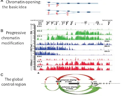
Fig. 2. Chromatin Opening And Transcriptional Regulation. (A)
Chromatin opening: the basic idea. The figure shows two Hox com-plexes. The collinearly ordered Hox genes in each complex are shown (on one DNA strand only as blue ovals). As the chromatin opens and the DNA strands separate, the Hox genes in the opened part transcribe mRNA (red squiggles). (B) Progres-sive chromatin modification in the Hoxd complex in mouse develop-ment.. ESC: Embryonic Stem Cells. E8.5 and E9.5: 8.5 and 9.5 days of mouse development. mRNA (green histograms): messenger RNA transcription. H3K27me3 (blue histograms): A chromatin mark for repression. H3K4me3 (red histograms): A chromatin mark for active transcription. As mouse de-velopment proceeds from day 8.5 to day 9.5, transcription proceeds from 3’ to 5’ through the Hoxd complex. This is accompanied by a 5’ to 3’ decrease in the repression mark. and a 5’ to 3’ increase in the active transcription mark. The embryonic stem cells have a low level of Hox expression, high repression mark and low activation mark. Image from Soshnikova and Duboule (2009). (C)
The global control region (GCR). Action on the posterior Hoxd genes is shown as red and green arrows. There are also inputs to regions outside the Hoxd complex. Image from Spitz et al. (2003). (B,C) reproduced with permission from Science and Cell respectively.
B
to induce temporally collinear Hoxb gene expression (Cambeyron and Bickmore, 2004) (Fig
.
2).There is also evidence that transcriptional control of collinearity might occur across entire Hox clusters. A separate study identified a global control region, situated 200 kb 5’ of the mouse Hoxd complex, that regulates the amplitude of expression of posterior Hoxd genes in the mouse limb bud, posterior gut and posterior CNS (Kmita
et al., 2002, Spitz et al., 2003, 2005).
These findings are generally considered to be strong evidence that chromatin modification and transcriptional control are involved in establishing the spatial and tem-poral collinearity of Hox genes. However, the available evidence comes only from studies in mouse, and is par-ticularly strong for the Hoxd complex. The mouse Hoxa and c complexes and Hox complexes in other bilaterian species have not been investigated. Technical obstacles restrict the possibilities somewhat. Vertebrates other than mouse and human have insufficient genetics for these studies. In invertebrates, Drosophila and Caenorhabditis could certainly have been investigated but have not. There
in which more posterior Hox genes are epistatic to more anterior ones (described already above)(Fig. 3). Work in Drosophila
me-lanogaster shows that this involves transcriptional, but also post
transcriptional and post translational mechanisms.
Working with D. melanogaster, E.B. Lewis showed that loss– of-function mutations in posterior Hox genes drive the segmental phenotype towards that of the more anterior thoracic segment T2, which is determined by the Hox gene Antennapedia (Lewis, 1978, 1995). Struhl used esc- Drosophila embryos, which show constitutive activation of gene expression, in combination with Hox loss of function mutations to elucidate the functional hierarchy of
Drosophila Hox genes (Struhl, 1983). All Drosophila segments were
transformed to the phenotype of the most posterior functional Hox gene expressed. Further experiments showed that transcriptional cross-regulation is not the only driving force of posterior prevalence. Experimentally derived ubiquitous expression of Hox genes under promoters that are known to be transcriptionally irrepressible leads to transformations only in regions anterior to the functional domain of the gene. For example, the thoracic Antennapedia, when ubiq-uitously expressed, suppresses Hox genes of the head, resulting in posterior transformation of head segments towards a thoracic identity while not affecting the abdomen — here, the effect of Antp is phenotypically suppressed by bithorax-complex genes such as
Ubx (Gonzalez-Reyes et al., 1990; Gibson and Gehring, 1988).
Posterior prevalence is now thought to occur via three mechanisms working in parallel: transcriptional control, posttranscriptional con-trol (via micro RNA’s) and posttranslational regulation (involving protein-protein interactions. Posterior prevalence has not been investigated as a mechanism for spatial collinearity because it has generally been assumed that it only occurs once Hox genes have already been expressed (rather than it being involved in the establishment of Hox gene expression), and is most important for generating unique Hox identities in zones in which expression of different Hox genes overlaps. Although it is actually clear that posterior prevalence is mediated by transcriptional repression (Hafen et al., 1984; Struhl and White, 1985; Beachy et al., 1988; Miller et al., 2001), this mechanism is paralleled both by post-transcriptional and by posttranslational regulation, specifically by
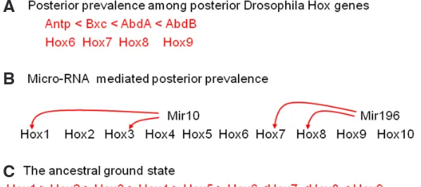
Fig. 3. Posterior Prevalence. (A) Posterior prevalence among posterior Drosophila Hox genes. More posterior genes dominate over more anterior ones. (B) Posterior prevalence mediated by microRNAs. Mir 10 and Mir196 each suppress Hox genes as shown. A single Hox complex is represented and we show which paralogue numbers are affected. The red lines show interactions in vertebrates. (C) Gehring’s proposal for an ancestral ground state is represented by the function of Antennapedia (Hox 6) in segment T2. It is dominated both by anterior and posterior Hox genes.
Box 3. The level of action. All effects above on activation or repression of Hox genes during gastrulation result in more or less Hox mRNA. but not all act on transcription. Recent evidence shows that Hox complex mRNA availability is strongly regulated posttranscription-ally, involving such phenomena as polycistronic transcripts, sense/ antisense transcript interactions and alternative splicing. At least one early vertebrate Hox interaction (downregulation of more 3’ Hox mRNA’s by Hoxb4) is micro RNA mediated (posttranscriptional). We note that the important parameter for collinearity is the sum total of the (activating and repressing) inputs on each Hox gene (there may be many). We think it very significant that posterior prevalence (pp) acts at 3 different levels. If a Hox gene is activated transcriptionally, its mRNA can still be destabilised by pp miRNA action. If the Hox protein is made, it can still be inactivated by pp protein-protein inter-actions. We think that pp is the most important Hox-Hox collinearity interaction and that it needs to be dominant, to ensure the 3’ to 5’ directionality of collinearity.
B
C
A
is no obstacle to investigating the mouse Hox a and c complexes. The main molecular evidence for involvement of global transcrip-tional control and chromatin opening in collinearity is as summarised above. As explained below, we think that this mechanism cannot explain all instances of collinearity in the vertebrate embryo. There is also an evolutionary objection against this mechanism (above) in that collinear properties persist in organisms with dispersed Hox genes (Duboule, 2007) and it is also found that moving a mouse
Hox gene out of a Hox complex does not destroy its normal axial
expression pattern (Krumlauf, 1994).
Interactions between Hox genes
Posterior prevalence: exception or rule?A second possible explanation for a part of the phenomenon of
microRNA-mediated translational control and by protein–protein interactions (see also Box 3) (Plaza et al., 2008; Yekta et al., 2008; Woltering and Durston, 2008). Recent exciting findings implicate the
Hox-associated miRNAs in regulating the translation and stability
of Hox gene mRNAs. These include the Hox4-associated Mir10 in vertebrates, Drosophila and Caenorhabditis, the posterior Mir196 in vertebrates and the posterior iab4 in Drosophila (Yekta et al., 2004, 2008; Woltering and Durston, 2008; Ronshaugen et al., 2005). Therefore, in flies, posterior prevalence mediates functional collinearity via a variety of mechanisms. It is worth noting that any spatial collinearity mechanism is redundant for early Hox gene expression in Drosophila, where expression of Hox genes is turned on by the non collinear segmentation gene hierarchy (Nuesslein-Volhard, 1995). A phenomenon similar to posterior prevalence is also involved in regulating the expression of homeobox-containing genes outside the Hox complexes: these genes are expressed in the head anteriorly to the Hox gene expression domain and are not contained in the Hox complexes (Fig. 3).
Is posterior prevalence the exception or the rule? Posterior prevalence was discovered in Drosophila. We know of much evi-dence (summarised below) that it and other Hox-Hox interactions are equally important in vertebrate embryos as in Drosophila and invertebrates.
Evolution of posterior prevalence in flies and vertebrates It has been reported relatively recently by Gehring et al., (2009) that not all Drosphila Hox interactions show posterior prevalence. The four 3’ Hox genes that are expressed anteriorly to Antennapedia (Lab, Pbx, Def, Scr) are apparently dominant to Antp. and appear to show anterior prevalence among themselves. Loss of function mutations for these genes leads to posterior transformations and gain of function lead to anterior transformations. Gehring has argued that Antennapedia does indeed represent the ancestral
ur-Hox gene and that both more anterior and more posterior Hox
genes are derived from this ancestral state by tandem duplication and evolutionary modification as above.This is a beautiful idea that seems very logical and is supported by solid data, but the following points should be considered.
1) Most Drosophila axial patterning genes clearly show posterior prevalence. This is definitely true of all of the 5’ posterior Drosophila
Hox genes: Antennapedia, Ultrabithorax, Abd A, Abd. B, which are
posteriorly prevalent among themselves. It is also true of a number of Drosophila non Hox homeobox genes: Ey, Toy, Otx, Ems, that are early pattering genes in the head. These are all dominated by
Antennapedia and other Hox genes (refs in Gehring et al., 2009).
2) There is a reason why Drosophila Hox genes might show aberrant collinearity. It is generally accepted that Hox collinearity is in process of disintegration and not fully functional in Drosophila, which has a Hox complex that is split into two. The two halves of the Drosophila Hox complex (Antennapedia and Bithorax com-plexes) are both greatly expanded, compared to the vertebrate
Hox complexes and their Hox genes are very large. Coordinated
regulation of their Hox genes will be hindered by this. The anterior
Antennapedia complex is more degenerate than the posterior Bi-thorax complex. It contains 2 Hox genes (Zen, Ftz) that have been
modified in Drosophila to mediate different (non Hox) functions but whose orthologues are normal functional Hox genes in other phyla (Terol et al., 1995; Krause et al., 1988). The Drosophila Hox genes are also actually turned on individually during early Drosophila
development by a mechanism (the segmentation gene hierarchy: Nuesslein-Volhard, 1995) that is not related to collinearity.
Dro-sophila also has no obvious temporal collinearity (Duboule, 2007).
3) Finally, we should consider whether the Hox interactions in
Drosophila reflect an ancestral Hox mechanism that is also
con-served in vertebrates. Findings in vertebrates show that Hoxb4 and the micro RNA Mir10 act synergistically to repress more anterior Hox genes, instead of more posterior Hox genes, as with the Hoxb4 orthologue Dfd=Deformed in Drosophila (Gehring et
al., 2009; Woltering and Durston 2008; Hooiveld et al., 1999).
Also, that vertebrate Hox1 paralogues are required to activate expression of more posterior Hox genes back to Hox number (paralogue group 6) (McNulty et al., 2005) instead of suppressing these genes, as with labial in Drosophila. These findings contrast with the situation in Porcellio (a Crustacean arthropod), in which the Dfd associated Mir 10 suppresses function of a more poste-rior Hox gene (Scr), similarly as would be expected in Drosophila (Abzhanov and Kaufman, 1999). These findings are not extensive but they open up the possibility that there is a difference between Vertebrates and Arthropods.
We tentatively conclude that the Hox interactions in Drosophila follow an Arthropod strategy that possibly diverges from the ances-tral mechanism in parallel with the disintegration of arthropod Hox collinearity and that vertebrates, which have strongly collinear Hox complexes, follow a different strategy associated with functional
Hox collinearity. This may be the ancestral strategy, but the very
high degree of collinearity seen in vertebrates is however unique in the animal kingdom and may be associated with a new mecha-nism. We note that vertebrate Hox collinearity, unlike Drosophila
Hox collinearity features temporal collinearity and we argue below
that temporal collinearity requires collinear Hox interactions.
A new model: interactions between Hox complexes
Temporal collinearity in the vertebrate gastrula mesodermTo examine the importance of Hox interactions in collinearity, we consider the mechanism underlying Hox temporal collinearity in a vertebrate embryo. The example we choose is Hox expression in the non-organiser mesoderm of the Xenopus laevis gastrula, where
Hox genes are first expressed in the embryo and are expressed
with temporal collinearity. This mesoderm manifests a sharply timed temporally collinear sequence of Hox gene expression that is translated in time and space to generate a spatially collinear pattern of Hox gene expression along the main body axis of the organism (Box 1, Fig. 4).
The 4 Hox gene complexes present in most vertebrates arose through 2 rounds of genome duplication during evolution.
Xeno-pus laevis and teleost fishes have 8 Hox complexes because of 3
genome duplications. A striking feature of the Xenopus gastrula’s temporally collinear Hox expression sequence is that expression of Hox genes from different Hox complexes is integrated into the same perfectly temporally collinear sequence (Fig. 4). The temporal collinearity of the different Hox complexes is therefore synchronised (Wacker et al., 2004a; Durston et al., 2010). The different Hox paralogues (i.e. the different copies of each different Hox gene type, produced by the vertebrate genome duplications) in the dif-ferent complexes are on difdif-ferent chromosomes, ruling out that
Hox collinearity simply reflects cis-localised progressive opening
are clearly needed to synchronise the different Hox complexes (these are presumably needed for chromatin opening in any case) and, since we are dealing with a cell mass rather than a single cell, intercellular signals are also required. We note that these
trans-acting factors and intercellular signals must be very sharply
timed to enable synchronisation of the different Hox complexes and are probably timed to trigger expression of different Hox genes at different times. This conclusion was not a complete surprise. It is known that trans acting factors must mediate collinearity in organ-isms with dispersed Hox genes. This is, however, the first evidence that temporal collinearity is also mediated by trans acting factors. The X. laevis example was chosen because the data are most complete for this system; however, the conclusions are strongly supported by many findings in other vertebrates (zebrafish, chicken and mouse) (Gaunt and Strachan, 1996; Alexandre et al., 1996; Deschamps et al., 1999).This example illustrates that Hox collin-earity cannot depend solely on the collinear opening of chromatin. Because the Hox complexes are synchronised, trans-acting factors and intercellular signals must be involved — trans-acting factors would be necessary for coordinating the sequential 3’ to 5’ activation of Hox genes in and between Hox clusters, and intercellular signals would enable the coordinated initiation of Hox gene expression between cells in a tissue. An alternative explanation is that only
gastrulation itself (although Wnt 8 expression does increase and reach its maximum after the beginning of gastrulation). As such, they could regulate the initiation of Hox complex expression or the timed 3’ to 5’ progression from one Hox gene to the next, or both. These signals must be very sharply timed. A fourth regulator, X Delta2, is relevant, as discussed below. It is not ruled out that there are other relevant pathways.
Ideal regulators for this function are the Hox genes themselves. These are first expressed in the
X. laevis gastrula mesoderm at the right times, in
a temporally collinear sequence. If each Hox gene activated its 5’ neighbour and its own paralogues, that could cause a temporally collinear sequence.
Hox genes do, in fact, regulate themselves and
each other; they also regulate intercellular signal-ling, as discussed below.
A potential mechanism for Hox-mediated trans-interactions
What criteria need to be met for Hox genes to regulate temporal collinearity? First, 3’ Hox genes should activate more 5’ Hox genes. Since activation needs to be sequential according to 3’ to 5’ position in a Hox complex, it is presumably necessary that multiple Hox genes, at different 3’-5’ positions, do this sequentially. As each Hox gene is activated, it should sequentially activate its immediate 5’ neigh-bour and its own paralogues, or the paralogues of its 5’ neighbour, or both; second, this activation needs to travel from cell to cell and third, there needs to be a non-Hox dependent signal that synchronises the initiation of expression of the Hox complexes, presumably by directly regulating the expression of the most 3’ Hox genes, labial (Hox1).
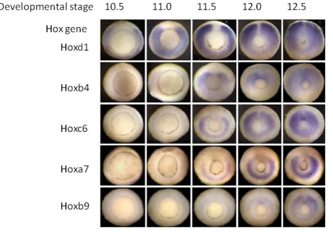
Fig. 4. Temporal collinearity In the Xenopus gastrula.The figure shows Hox expression patterns at sequential stages during gastrulation in Xenopus. The embryos are seen from underneath, where a ring (the blastopore) shows the position where mesoderm tissue invaginates during gastrulation. This ring gets smaller as gastrulation proceeds and the up-per tissues in the embryo spread out and cover the lower part of the embryo (epiboly).The expression of several different Hox genes, seen as blue colour by in situ hybridisation, is in each case initially in the gastrula mesoderm in the zone above (outside) the ring. Hox expres-sion is thus seen as a blue ring, and since it is initially only in part of the mesoderm, the ring is initially broken. The ring of Hox expression gets smaller as the blatopore ring gets smaller and mesoderm invaginates into the embryo.The figure shows expression of a sequence of Hox genes with different paralogue numbers, from 1 to 9. It will be seen that the Hox gene with the lowest paralogue number starts expression first and later numbers start sequentially later. It will also be seen that the Hox genes in this time sequence include members of all of the 4 primary vertebrate paralogue groups (a,b,c,d).
the most 3’ Hox genes (Hox1) transactivate, and the remaining timing is provided by synchronised opening of the Hox complexes. The different structures of the 4 primary vertebrate Hox complexes (with different Hox paralogues missing from each) would, however, make it difficult for progressive opening of different Hox complexes to stay synchronous. Since the gastrula mesoderm is a cell mass, not a single cell, trans-activation needs to be accompanied by intercellular signalling.
Requirement for extracellular signals and trans-acting factors Which candidate molecules could mediate the trans-acting and intercellular signalling effects described above? Three extracellular signals and one intracellular regulator are known to regulate Hox gene expression in the Xenopus gastrula mesoderm; however, only one or possibly two of these have the required properties to be involved in triggering the timed and collinear expression of
Hox-genes during gastrulation. BMP4, Brachyury and Wnt 8 are
Evidence concerning the mechanism
Below we discuss the available evidence supporting the ideal requirements set out in the three points above.
Trans activation
There is evidence from vertebrates that Hox genes can activate their 5’ neighbours, and thus meet the first criterion listed above.
Hox genes auto- and cross-activate in early Drosophila and
ver-tebrate embryos (e.g. McNulty et al., 2005; Hooiveld et al., 1999; Woltering and Durston, 2008; Le Pabic et al., 2010; Lobe, 1995; Maconochie et al., 1997; Gould et al., 1997; Bergson and McGin-nis,1990; Miller et al., 2001).Ectopic expression of at least two
Hox genes (Hoxb4 and Hoxa7) caused net activation of their own
expression and of more 5’ Hox genes in the Xenopus gastrula and in excised gastrula tissues from this organism (Hooiveld et al., 1999) We expect, from the sequential nature of temporal collinearity, that these genes would only cross activate 5’ neighbours. Indeed, in
Xenopus, Hoxb5 was the only directly activated target of Hoxb4,
detected so far, apart from Hoxb4 itself; more 5’ Hox genes were activated indirectly. Cross activation of other Hox genes by a Hox gene occurs in another vertebrate embryo (mouse) and in murine embryocarcinoma cells and Drosophila (Lobe, 1995; LePabic et
al., 2010; Gould et al., 1997; Maconochie et al., 1997; Miller et al.,
2001). Expression of 3’ Hox genes (Hox1 genes) is also required for more 5’ Hox gene expression during early Xenopus develop-ment (McNulty et al., 2006) (Fig. 4).
Intercellular signalling
Besides activation of 5’ neighbouring Hox genes, intercellular signalling is required, to allow Hox activation to be transmitted from cell to cell (criterion 2). Much evidence shows indeed that
Hox genes induce signalling (Bloch- Gallego et al., 1993; Chatelin et al., 1996; Graba et al., 1995; Bruhl, 2004; Manak et al., 1994;
Michaut et al., 2011; Morsi el Kadi et al., 2002; Pearson et al., 2005). Known signalling pathways are Hox targets in Drosophila and vertebrates (eg. Graba et al., 1995; Bruhl, 2004; Manak et al., 1994; Michaut et al., 2011; Morsi el Kadi et al., 2002; Pearson et
al., 2005) and Prochiantz and colleagues have also demonstrated
that the Hox proteins themselves are unexpectedly translocated from cell to cell, acting as unorthodox intercellular signals (Bloch- Gallego et al., 1993; Chatelin et al., 1996). Furthermore, in the
Xenopus gastrula, activation of Hox genes by Hoxb4 is non cell
autonomous (Hooiveld et al., 1999).
A signal for initiation
There is evidence for a non Hox dependent signal that induces expression of iabial Hox genes directly in the gastrula. Wnt 8 in-duces labial Hox genes directly and other Hox genes indirectly (In der Rieden et al., 2010). It may not be not the only signal involved in Hox complex initiation because it is available from before gas-trulation (although its amplitude does increase markedly during gastrulation) and therefore may possibly not initiate the sharply synchronised Hox complex expression during gastrulation.
Posterior prevalence
Posterior prevalence occurs in vertebrates (Yekta et al., 2004, 2008; Hooiveld et al., 1999; Woltering and Durston, 2008; Wellik and Capecci, 2003; Carapuco et al., 2005; Duboule, 2007) and is an extremely important Hox interaction. It is evident in the Xenopus
gastrula. Expression of more 3’ Hox genes is downregulated in the Xenopus gastrula by early ectopic expression of Hoxb4 and
Hoxa7. This is classical posterior prevalence as in Drosophila and
is entirely logical here. 3’ Hox genes are expressed earlier than 5’ Hox genes during temporal collinearity, so their expression is already stabilised by the time 5’ Hox genes are activated. Therefore they are not expected to be evidently repressed in vivo. Expression of 3’ and 5’ Hox genes can overlap as is observed. Repression of 3’ by 5’ Hox genes is presumably required to prevent secondary retrograde activation of 3’ genes, which would destroy temporal collinearity. It is especially important to ensure that if a Hox gene receives a combination of activating and repressing signals, the repressing signals dominate (see section on Posterior Prevalence and Box 3). Downregulation of more 3’ genes by Hoxb4 has been shown in two early vertebrate embryos: Xenopus and zebrafish (Hooiveld et al., 1999; Woltering and Durston, 2008). Hoxb4 acts in synergy with Mir10. Posterior prevalence is clearly important in all vertebrates, including the mouse as well as in Drosophila. It is probably the most important Hox interaction. We think that it is the key collinearity property because it ensures directionality in net Hox interactions. Net 3’ interactions in gastrula mesoderm in
vivo should be negative. Net 5’ interactions can be positive. The
reason posterior prevalence acts at 3 levels may be to ensure that it is always the dominant interaction.
Regulation of Hox collinearity by the somitogenesis clock What is needed to regulate early collinearity is one or more signals that are turned on at specific times during gastrulation. These could regulate initiation of expression of Hox complexes or 3’ to 5’ progression of expression from one Hox gene to the next, or both. They need to be sharply timed. The possibilities are: 1) they come on as a step function; the signal is first off, then sharply on; 2) they are expressed as a pulse; the signal comes on sharply, then disappears. Pulsatile signals are typically oscil-latory (i.e. you get periodic pulses). In addition to regulation by interactions among the Hox genes themselves, there might be a need for other sharply timed signals. The third intercellular signal known to regulate Xenopus gastrula Hox expression is actually an oscillatory signal. This is Xdelta2, an intercellular signal mediating somitogenesis (i.e. mesoderm segmentation) (Peres et al., 2006 and below).
Vertebrate somitogenesis (segmentation of axial mesoderm) works via a mechanism where an oscillating system of gene expres-sion generates a spatial pattern by time–space translation, just as in genesis of the vertebrate axial Hox pattern (see above and Box 1). The temporal oscillation in gene expression (somitogenesis clock) generates spatially periodic segments in the axial mesoderm: the somites (Palmeirim et al., 1997). This dynamic process is known to start during gastrulation in chicken and Xenopus (Peres et al., 2006; Jouve et al., 2002) and is closely linked to collinear Hox ex-pression. Hox spatial expression boundaries coincide with somite/ segment boundaries and several vertebrate somitogenesis genes are known to regulate Hox expression (Peres et al., 2006; Dubrulle
et al., 2001; Dubrulle and Pourquie, 2004; Zakany et al., 2001). Xdelta2 is a Xenopus oscillating somitogenesis gene (Jen et al.,
collinear expression of the Hox complexes. It could do so either by regulating only initiation of expression of Hox complexes (via labial Hox genes) or by driving initiation and 3’ to 5’ progression, (repeatedly inducing expression of different Hox genes). We note that XDelta2 drives expression of at least 3 different Hox paralog groups including labial). If delta drives progression as well as initiation, a repeated periodic pulsatile signal is required. The idea that the somitogenesis clock drives Hox temporal collinearity is very attractive because both of these timers are known to operate already in the gastrula and because of the evidence linking Hox patterning and segmentation (above). Such a signalling pathway might act separately from the Hox genes or be downstream of them. XDelta 2 is indeed downstream of Hox genes as well as upstream. There is a positive feedback loop (McNulty et al., 2005; Peres et al., 2006). XDelta 2 may thus mediate Hox induced signalling.
Conclusion: a new hypothesis
Vertebrate and Drosophila Hox genes undergo trans-interac-tions in early embryos. These putatively mediate the synchronised temporal collinearity of the Hox complexes in the vertebrate gastrula stage. These interactions include posterior prevalence, autoactivation and cross activation. Posterior prevalence is a key interaction because it can ensure 3’ to 5’ directionality in the net
Hox interactions and can thus generate collinearity. These Hox
interactions are not necessarily always direct. Besides trans-interactions, Hox dependent cell interactions are also required.
Hox proteins activate many signalling pathways and are also
signalling molecules themselves. These cell interactions are needed to mediate non-cell autonomous Hox interactions. One of the signalling pathways involved in Xenopus is the somitogenesis
related Delta-Notch pathway. XDelta2 is a timed signalling molecule downstream
of Hox genes that activates different Hox
genes during gastrulation. Hox chromatin opening may also be involved in early
Hox collinearity but this mechanism does
not require it.
Our ideas about Hox interactions and the somitogenesis clock are illustrated in Fig. 5.
Concluding remarks:
relation-ships between different aspects
of collinearity
Hox colinearity, which mediates axial patterning in some or all bilateria, is a spectacular phenomenon that has attracted much interest. It is presently generally assumed that its mechanism is progressive opening for transcription of
Hox complexes. This is presumably
im-portant. However, we develop a different mechanistic hypothesis: that collinearity is mediated by Hox gene interactions. This idea was already indicated by in-vestigations of posterior prevalence. We review new evidence that trans-acting
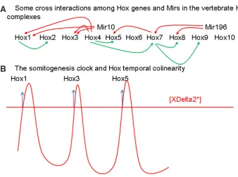
Fig. 5. Hox-Hox Interactions And Somitogenesis Timing.(A) Some cross interactions between Hox genes and Mirs in the vertebrate Hox complexes during gastrulation. Red: repression. Green: activa-tion.(B) The somitogenesis clock and Hox temporal collinearity. We show an oscillating concentration of XDelta2. Sequential peaks of XDelta2 activate expression of different Hox genes. [XDelta2*]; The threshold concentration of XDelta2 at which Hox expression is activated.
B
A
factors and intercellular signals mediate vertebrate Hox collinear-ity; that these include interactions among Hox genes, including posterior prevalence, as well as somitogenesis signals. We pro-pose that these Hox interactions have a role in generating Hox temporal and spatial collinearity, as well as functional collinearity. We note also that an evolutionary explanation for collinearity ac-tually probably obviates any requirement for a dedicated integral collinearity mechanism. Our conclusions open new perspectives for research into the mechanisms underlying collinearity. Testing this model will require a much more extensive investigation and description of early vertebrate Hox temporal collinearity.
Acknowledgements
References
ABZHANOV A. and KAUFMAN T.C., (1999). Novel regulation of the homeotic gene Scr associated with a crustacean leg-tomaxilliped appendage transformation. Development 126: 1121–1128.
ALEXANDRE D, CLARKE J.D., OTOXBY E., YAN Y.L., JOWETT T. and HOLDER N. (1996). Ectopic expression of Hoxa-1 in the zebrafish alters the fate of the mandibular arch neural crest and phenocopies a retinoic acid-induced phenotype. Development 122: 735-746.
BEACHY P.A., KRASNOW M.A., GAVIS E.R. and HOGNESS D.S.(1988). An Ultrabi-thorax protein binds sequences near its own and the Antennapedia P1 promoters. Cell 155: 1069-1081.
BERGSON C. and MCGINNIS W. (1990). An autoregulatory enhancer element of the Drosophila homeotic gene Deformed EMBO J 13: 4287-4297.
BLOCH-GALLEGO E., LE ROUX I., JOLIOT A.H., VOLOVITCH M., HENDERSON C.E., PROCHIANTZ A. (1993). Antennapedia homeobox peptide enhances growth and branching of embryonic chicken motoneurons in vitro. J Cell Biol 120: 485-492. BRUHL T, URBICH, C., AICHER, D. and ACKER-PALMER, A. (2004). Homeobox
A9 transcriptionally regulates the EphB4 receptor to modulate endothelial cell migration and tube formation. Circ Res 94: 743–751.
CAMBEYRON S. and BICKMORE W.A. (2004). Chromatin decondensation and nuclear reorganization of the HoxB locus upon induction of transcription. Genes Dev 18: 1119–1130.
CARAPUCO M., NOVOA A., BOBOLA N. and MALLO M. (2005). Hox genes specify vertebral types in the presomitic mesoderm. Genes Dev 19: 2116-2121. CARRASCO A.E., MCGINNIS W., GEHRING W.J. and DE ROBERTIS E.M. (1984).
Cloning of an X. laevis gene expressed during early embryogenesis coding for a peptide region homologous to Drosophila homeotic genes. Cell 37: 409-414. CHATELIN J., VOLOVITCH M., JOLIOT A.H., PEREZ F. and PROCHIANTZ A. (1996).
Transcription factor hoxa-5 is taken up by cells in culture and conveyed to their nuclei. Mech Dev 55: 111-117.
DE ROBERTIS E.M. (2008). Evo-devo: variations on ancestral themes. Cell 132: 185-195.
DESCHAMPS J., VAN DEN AKKER E., FORLANI S., DE GRAAFF W., OOSTERVEEN T., ROELEN B. and ROELFSEMA J. (1999). Initiation, establishment and mainte-(1999). Initiation, establishment and mainte-Initiation, establishment and mainte-nance of Hox gene expression patterns in the mouse. Int J Dev Biol 43: 635-650. DUBOULE, D. (1994). Temporal colinearity and the phylotypic progression: a basis
for the stability of a vertebrate Bauplan and the evolution of morphologies through heterochrony. Development 135-142.
DUBOULE, D. (2007). The rise and fall of Hox gene clusters. Development 134: 2549-2460.
DUBRULLE, J., MCGREW, M.J. and POURQUIÉ, O. (2001). FGF Signaling Con-trols Somite Boundary Position and Regulates Segmentation Clock Control of Spatiotemporal Hox Gene Activation. Cell 106: 219-232.
DUBRULLE J. and POURQUIE O. (2004). Coupling segmentation to axis formation. Development 131: 5783-5793.
DURSTON A., JANSEN H.J. and WACKER SA (2010). Time-Space Translation Regulates Trunk Axial Patterning In The Early Vertebrate Embryo. Genomics 95: 250-255.
GAUNT S.J. and STRACHAN L. (1996). Temporal colinearity in expression of anterior Hox genes in developing chick embryos. Dev Dyn 207: 270-280.
GEHRING W.J., KLOTER U. and SUGA H. (2009). Evolution of the Hox Gene Complex from an Evolutionary Ground State. Curr. Top. Dev. Biol. 88: 35-61.
GIBSON G. and GEHRING W. (1988). Head and thoracic transformations caused by ectopic expression of Antennapedia during Drosophila development. Develop-ment 102: 657–675.
GONZALEZ-REYES URQUIA N., GEHRING W.J., STRUHL G. and MORATA G. (1990). Are cross-regulatory interactions between homoeotic genes functionally significant? Nature 344: 78–80.
GOODMAN, F.R. (2003). Congenital abnormalities of body patterning: embryology revisited. Lancet. 362: 651-62.
GOULD A., MORRISON A., SPROAT G., WHITE R.A. and KRUMLAUF R. (1997). Positive cross-regulation and enhancer sharing: two mechanisms for specifying overlapping Hox expression patterns. Genes Dev 11: 900-913.
GRABA, Y., GIESELER, K., ARAGNOL, D., LAURENTI, P., MARIOL, M.C., BERENGER, H., SAGNIER, T. and PRADEL, J. (1995). DWnt-4, a novel Dro-sophila Wnt gene acts downstream of homeotic complex genes in the visceral mesoderm. Development 121: 209–218.
GRIER D.G., THOMPSON A., KWASNIEWSKA A., McGONIGLE G.J. and HAL-LIDAY H.L. (2005). The pathophysiology of HOX genes and their role in cancer. J Pathol 205: 154–171.
IN DER RIEDEN P.M.J., LLORET VILASPASA F. and Durston A.J. (2010). Xwnt8 directly initiates expression of labial Hox genes. Dev. Dynamics 29: 226-239. HAFEN E., LEVINE M. and GEHRING W.J.(1984). Regulation of Antennapedia
transcript distribution by the bithorax complex in Drosophila. Nature 307: 287-289. HOOIVELD M., MORGAN R., IN DER RIEDEN P., HOUTZAGER E., PANNESE M., DAMEN K., BONCINELLI E. and DURSTON A., (1999). Novel colinear interactions between vertebrate Hox genes. Int. J. Dev. Biol. 43: 665-674.
JEN W.C., WETTSTEIN D., TURNER D., CHITNIS A. and KINTNER C. (1997). The Notch ligand, X-Delta-2, mediates segmentation of the paraxial mesoderm in Xenopus embryos. Development 124: 1169-1178.
JEN W.C., GAWANTKA V., POLLET N., NIEHRS C. and KINTNER C. (1999). Peri-odic repression of Notch pathway genes governs the segmentation of Xenopus embryos. Genes Dev 13: 1486-1499.
JOUVE C., IIMURA T. and POURQUIE O. (2002). Onset of the segmentation clock in the chick embryo: evidence for oscillations in the somite precursors in the primitive streak. Development 129: 1107-1111.
KMITA M., VAN DER HOEVEN F., ZAKANY J., KRUMLAUF R. and DUBOULE D., (2000). Mechanisms of Hox gene colinearity: transposition of the anterior Hoxb1 gene into the posterior HoxD complex. Genes Dev 14: 198-211.
KMITA, M., FRAUDEAU, N., HE´ RAULT, Y. and DUBOULE, D. (2002). Serial dele-tions and duplicadele-tions suggest a mechanism for the collinearity of Hoxd genes in limbs. Nature 420: 145.
KMITA M. and DUBOULE D. (2003). Organizing axes in time and space; 25 years of colinear tinkering. Science 301: 331-333..
KRAUSE H.M., KLEMENZ R. and GEHRING W.J. (1988). Expression, modification, and localization of the fushi tarazu protein in Drosophila embryos. Genes Dev 2: 1021-1036.
KRUMLAUF R. (1994) Hox genes in vertebrate development. Cell 78: 191-201. LE PABIC P., SCEMAMA J.L. and STELLWAG E.J. (2010). Role of Hox PG2 genes in
Nile tilapia pharyngeal arch specification: implications for gnathostome pharyngeal arch evolution. Evol. Dev. 12: 45-60.
LEWIS E.B., (1978). A Gene Complex Controlling Segmentation in Drosophila. Nature 276: 565-568.
LEWIS E.B., (1995) The bithorax complex: the first fifty years. (Nobel lecture). In Genes, Development and Cancer. The Life and Work of Edward B. Lewis, (Ed. H. Lifshitz). Kluwer Academic Publishers, Norwell, MA.
LOBE C.G. (1995). Activation of Hox gene expression by Hoxa-5. DNA Cell Biol 14: 817-823.
MACONOCHIE, M.K., NONCHEV, S., STUDER, M., CHAN, S.K., POPPERL, H., SHAM, M.H., MANN, R.S. and KRUMLAUF R. (1997). Cross-regulation in the mouse HoxB complex: the expression of Hoxb2 in rhombomere 4 is regulated by Hoxb1. Genes Dev 11: 1885–1895.
MAINGUY G., KOSTER J., WOLTERING J., JANSEN H. and DURSTON A., (2007). Extensive polycistronism and antisense transcription in the Mammalian Hox clusters. PLoS ONE 2: e356.
MANAK J.R., MATHIES L.D. and SCOTT M.P. (1994). Regulation of a decapentaplegic midgut enhancer by homeotic proteins. Development 120: 3605–3612. MCNULTY C., PERES J., VAN DEN AKKER W., BARDINE N. and DURSTON A.
(2005). Knockdown of the complete Hox paralogous group 1 leads to dramatic hindbrain and neural crest defects. Development 132: 2861-2871.
MICHAUT L., JANSEN H., BARDINE N., DURSTON A. and GEHRING W.J. (2011). Analysing the function of a hox gene: an evolutionary approach. Dev. Growth Differ. 53: 982-93.
MILLER D.F., ROGERS B.T., KALKBRENNER A., HAMILTON B., HOLTZMAN S.L. and KAUFMAN T. (2001). Cross-regulation of Hox genes in the Drosophila Me-lanogaster embryo. Mech Dev 102: 3-16.
regulation. Mech Dev 113: 131-139.
NUESSLEIN-VOLHARD, C. (1995). The identification of genes controlling develop-ment in flies and fishes. Nobel Lectures, Physiology or Medicine (1991-1995). (Ed. Nils Ringertz). World Scientific Publishing Co., Singapore.
PALMEIRIM I., HENRIQUE D., ISH-HOROWICZ D. and POURQUIE O. (1997). Avian hairy gene expression identifies a molecular clock linked to vertebrate segmenta-tion and somitogenesis. Cell 91: 639-648.
PEARSON J.C., LEMONS D. and MCGINNIS W. (2005). Modulating Hox Gene Functions During Animal Body Patterning. Nature Rev. Genet. 6: 893. PERES J., MCNULTY C. and DURSTON A. (2006). Interaction between X-Delta-2
and Hox genes regulates segmentation and patterning of the anteroposterior axis. Mech Dev 123: 321-333.
PLAZA S., PRINCE F., ADACHI Y., PUNZO C., CRIBBS D.L. and GEHRING W.J. (2008). Cross-regulatory protein-protein interactions between Hox and Pax tran-scription factors. Proc Natl Acad Sci USA 105: 13439-13444.
RONSHAUGEN M., BIEMAR F., PIEL J., LEVINE M. and LAI E.C. (2005).The Dro-sophila microRNA iab-4 causes a dominant homeotic transformation of halteres to wings. Genes Dev 19: 2947-2952.
SEO H.C., EDVARDSEN R.B., MAELAND A.D., BJORDAL M., JENSEN M.F., HAN-SEN A., FLAAT M., WEISHAN-SENBACH J., LEHRACH H. and WINCKER P. (2004). Hox cluster disintegration with persistent anteroposterior order of expression in Oikopleura dioica. Nature 431: 67 -71.
SOSHNIKOVA N. and DUBOULE D. (2009). Epigenetic Temporal Control of Mouse Hox Genes in vivo. Science 324: 1320-1323.
SPITZ F., GONZALEZ F. and DUBOULE D.(2003). A global control region defines a chromosomal regulatory landscape containing the HoxD cluster. Cell 113: 405-417. SPITZ F., HERKENNE C., MORRIS M.A. and DUBOULE D. (2005). Inversion-induced disruption of the Hoxd cluster leads to the partition of regulatory landscapes. Nat
Genet 37: 889-893.
STRUHL G. (1983). Role of the esc+ gene product in ensuring the selective ex-pression of segment-specific homeotic genes in Drosophila. J. Embryol. Exp. Morphol. 76: 297–331.
STRUHL G. and WHITE R.A. (1985). Regulation of the Ultrabithorax gene of Dro-sophila by other bithorax complex genes. Cell 43: 507-519.
TEROL J., PEREZ-ALONSO M. and DE FRUTOS R. (1995). Molecular character-ization of the zerknüllt region of the Antennapedia complex of D. subobscura. Chromosoma 103: 613-624.
VAN DER HOEVEN F., ZAKANY J. and DUBOULE D. (1996). Transpositions in the HoxD complex reveal a hierarchy of regulatory controls. Cell 85: 1025-1035. WACKER S.A., JANSEN H.J., MCNULTY C.L., HOUTZAGER E. and DURSTON
A.J. (2004a). Timed interactions between the Hox expressing non-organiser mesoderm and the Spemann organiser generate positional information during vertebrate gastrulation. Dev Biol 268: 207-219.
WACKER SA, MCNULTY CL, DURSTON AJ. (2004b). The initiation of Hox gene expression in Xenopus laevis is controlled by Brachyury and BMP-4. Dev Biol 266: 123-137.
WELLIK D.M. and CAPECCHI M.R., (2003). Hox10 and Hox11 genes are required to globally pattern the mammalian skeleton. Science 301: 363–367.
WOLTERING J.M., and DURSTON A., (2008). MiR10 represses HoxB1a and HoxB3a in Zebrafish. PLoS ONE 3: e1396.
YEKTA S., SHIH H. and BARTEL D.P. (2004). MicroRNA-directed cleavage of HOXB8. mRNA. Science 304: 594–596.
Further Related Reading, published previously in the Int. J. Dev. Biol.
Retinoid signalling is required for information transfer from mesoderm to neuroectoderm during gastrulation
Ferran Lloret-Vilaspasa, Hans J. Jansen, Koen de Roos, Rosh A.S. Chandraratna, Maija H. Zile, Claudio D. Stern and Antony J. Durston Int. J. Dev. Biol. (2010) 54: 599-608
Identification of hoxb1b downstream genes: hoxb1b as a regulatory factor controlling transcriptional networks and cell movement during zebrafish gastrulation
Willem M.R. van den Akker, Antony J. Durston and Herman P. Spaink Int. J. Dev. Biol. (2010) 54: 55-62
The evolution and maintenance of Hox gene clusters in vertebrates and the teleost-specific genome duplication Shigehiro Kuraku and Axel Meyer
Int. J. Dev. Biol. (2009) 53: 765-773
Hox and ParaHox genes in Nemertodermatida, a basal bilaterian clade Eva Jiménez-Guri, Jordi Paps, Jordi García-Fernández and Emili Saló Int. J. Dev. Biol. (2006) 50: 675-679
Head-tail patterning of the vertebrate embryo: one, two or many unresolved problems?
Claudio D. Stern, Jeroen Charité, Jacqueline Deschamps, Denis Duboule, Anthony J. Durston, Marie Kmita, Jean-François Nicolas, Isabel Palmeirim, Jim C. Smith and Lewis Wolpert
Int. J. Dev. Biol. (2006) 50: 3-15
Evolution of the Hox/ParaHox gene clusters. David E K Ferrier and Carolina Minguillón Int. J. Dev. Biol. (2003) 47: 605-611