A systematic survey of HOX and TALE expression profiling
in human cancers
YUNLONG JIA, FRANÇOISE BLEICHER and SAMIR MERABET*
Institut de Génomique Fonctionnelle de Lyon, Université de Lyon, Université Lyon 1, CNRS,Ecole Normale Supérieure de Lyon, Lyon, France
ABSTRACT HOX and TALE genes encode homeodomain (HD)-containing transcription factors that act in concert in different tissues to coordinate cell fates and morphogenesis throughout embryonic development. These two evolutionary conserved families contain several members that form differ-ent types of protein complexes on DNA. Mutations affecting the expression of HOX or TALE genes have been reported in a number of cancers, but whether and how the two gene families could be perturbed together has never been explored systematically. As a consequence, the putative collab-orative role between HOX and TALE members for promoting or inhibiting oncogenesis remains to be established in most cancer contexts. Here, we address this issue by considering HOX and TALE expression profiling in normal and cancer adult tissues, using normalized RNA-sequencing expres-sion data deriving from The Cancer Genome Atlas (TCGA) and Genotype-Tissue Expresexpres-sion (GTEx) research projects. Information was extracted from 28 cancer types originating from 21 different tissues, constituting a unique comparative analysis of HOX and TALE expression profiles between normal and cancer contexts in human. We present the general and specific rules that could be de-duced from this large-scale comparative analysis. Overall this work provides a precious annotated support to better understand the role of specific HOX/TALE combinatorial codes in human cancers.
KEY WORDS:
HOX, TALE, homeodomain, cancer
Introduction
HOX proteins are homeodomain (HD)-containing transcription factors (TFs) that control various developmental processes during embryogenesis, including axis patterning (Pearson et al., 2005), limb formation (Zakany and Duboule, 2007) or organ differentia-tion (Chojnowski et al., 2014; Gligorov et al., 2013; Wellik, 2011). HOX proteins are also required in the adult, in particular for the homeostasis of stem cell lineages in different tissues (Lebert-Ghali et al., 2016; Rux et al., 2016; Sugimura et al., 2017; Xin et al., 2017). These various and specific functions are thought to rely on the partnership with diverse types of cofactors that remain to be identified for most of them (Merabet and Dard, 2014).
The best-characterized class of HOX cofactors are the PBC (Pre-B cell complex) proteins, which belong to the TALE (Three Amino acids Loop Extension) family of HD-containing TFs (Bürglin, 1997). PBC cofactors interact with the large majority of HOX pro-teins on DNA, forming protein complexes with higher DNA-binding
www.intjdevbiol.com
*Address correspondence to: Samir Merabet. ENSL, IGFL, 46 Allée d’Italie, 69346 LYON cedex 07-France. Tel:+33 (0)426731317. E-mail: [email protected] https://orcid.org/0000-0001-7629-703X
Supplementary Material (three tables) for this paper is available at: https://doi.org/10.1387/ijdb.180286fb
Submitted: 30 August, 2018; Accepted: 13 September, 2018.
ISSN: Online 1696-3547, Print 0214-6282 © 2018 UPV/EHU Press
Printed in Spain
Abbreviations used in this paper: ACC, adrenocortical carcinoma; BLCA, bladder
specificity and affinity (Mann et al., 2009). Two other classes of the TALE family, the MEIS (Myeloid Ecotropic viral Integration Site) and PREP (Pbx-regulating protein, also called PKNOX) classes, also participate in HOX functions. MEIS and PREP originate from a common ancestor called MEINOX (Bürglin, 1998) and display intricate regulatory relationships with HOX and PBC proteins. For example, MEIS and PREP compete to interact with PBC and this interaction is necessary for the nuclear translocation hence activity of each TALE component. Competition between MEIS and PREP also occurs at the level of DNA-binding for target gene regulation on the genome (Dardaei et al., 2014). In addition, the role of MEIS and PREP as direct HOX-binding partners on DNA without PBC proteins remains to be unequivocally determined. Along the same line, genome-wide binding analysis of PBC, MEIS and PREP in mice showed preferential and distinct combinations with HOX proteins. In particular, PBC/PREP DNA-binding sites appear to be enriched in promoters and nearby regions, while HOX/PBC/MEIS DNA-binding sites are preferentially found in inter- and intra-genic regions (Penkov et al., 2013). Finally, MEIS but not PREP induces strong HOX-PBC interaction remodeling, revealing the role of vari-ous and specific HOX protein motifs for trimeric complex formation (Dard et al., 2018). This interaction plasticity has been proposed to be important for diversifying and specifying HOX-TALE functions during development and evolution (Merabet and Mann, 2016).
What about HOX and TALE in cancer? In fact, the role of these families in cancer was historically identified from mutations affecting the protein function. PBC and MEIS were first identified in vertebrates from chromosomal translocations and viral inser-tions that respectively led to Pre-B acute lymphoblastic leukemia in children and acute myeloid leukemia in mouse (Kamps et al., 1990; Moskow et al., 1995; Nourse et al., 1990; Steelman et al., 1997). HOXA9 was described in 1996 as being responsible for acute and chronic leukemia, due to a chromosomal translocation leading to a fusion with the nucleoporin NUP98 (Borrow et al., 1996; Nakamura et al., 1996). More recently, mutations in HOXB13 cod-ing sequences have also been associated with familial or high-risk prostate cancers (Karlsson et al., 2014; Saunders et al., 2014). The large majority of HOX and TALE mutations in cancer are how-ever not affecting the protein function but the expression profile. A number of studies have reported aberrant expression profiles of HOX, and to lesser extent, TALE genes, in solid cancers and leukemia. Moreover, the correlation with poor prognosis along with functional validations upon artificial expression in cancer-derived cell lines has been demonstrated in several instances. Interestingly, HOX genes are described to be up- or down-regulated depending on the cancer type, acting as pro- or anti-oncogenes. The same HOX gene can even have opposite functions in different cancer types (see (Argiropoulos and Humphries, 2007; Bhatlekar et al., 2014; Eklund, 2011; Sitwala et al., 2008) for excellent compiling reviews on this subject). With regard to TALE proteins, PBC and MEIS members are generally described as oncoproteins, while PREP is more frequently associated with a tumor suppressor function, due to its competitive role against MEIS (see (Blasi et al., 2017) for review). The cooperative role of HOX and TALE in cancer is best established during leukemogenesis, where it has been demonstrated that PBX3 and MEIS1 are important cofactors for the transformation/immortalization activity of HOX proteins (in particular HOXA9) in hematopoietic stem cells (Li et al., 2013; Rozovskaia et al., 2001). Such cooperative role has rarely been
shown in solid cancers (see for example (Fernandez et al., 2008)) and is principally deduced from indirect studies using a HOX-PBC interaction inhibitory peptide in cancer cell lines (see for example (Morgan et al., 2012). Moreover, the role of PBC, MEIS and PREP was analyzed with one member of each subfamily in most cancer studies (PBX1, MEIS1 and PREP1), which asks for the role of the other TALE members (PBX2-4, MEIS2-3 and PREP2).
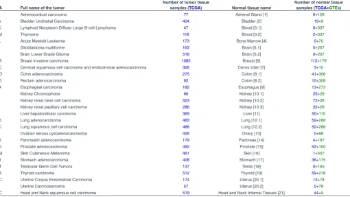
Here we present a systematic analysis of the expression profile of the 39 human HOX members and their associated TALE cofactors (PBX1-4, MEIS1-3 and PREP1-2) in 28 cancer types deriving from 21 different tissues, comparing the normal and oncogenic context in each case. Raw RNA sequencing expression was extracted from the TCGA (The Cancer Genome Atlas: https://cancergenome. nih.gov/) and GTEx (Genotype Tissue Expression: https://www. gtexportal.org/home/) projects and encompasses 5526 normal and 9018 cancer patient samples in total (Table 1, see also methods). Our analysis provides a global picture of enriched HOX and TALE expression profiles in normal and cancer tissues, allowing iden-tifying most significant regulatory changes associated with tumor progression. This information could serve as a molecular support for future therapeutic strategies aiming at targeting specific HOX/ TALE complexes for developing anti-cancer agents or biomarkers.
Results
Expression of HOX and TALE genes in normal tissues
The 39 human HOX genes are distributed in four genomic clus-ters (A, B, C and D) that are located on different chromosomes. Each genomic cluster has a different number of HOX genes, due to gene loss events during evolution. Overall, human HOX genes are organized into 13 paralog groups (PGs) that are defined as anterior (PG1-3), central (PG4-8) or posterior (PG9-13), based on the expression profile along the anterior-posterior axis in the early embryo. HOX genes from anterior PGs are, for example, expressed earlier and in more anterior parts of the vertebrate embryo than HOX genes from central and posterior PGs. In addition, HOX genes from the same PG usually display a highly similar if not identical expression profile along the AP axis, although the underlying cis-regulatory logic could be different (Kmita and Duboule, 2003). The nine TALE cofactor-encoding genes under study (PBX1-4, MEIS1-3 and PREP1-2) are also distributed on different chromosomes and display overlapping as well as distinct expression domains throughout development. Importantly, genetic and expression analyses are consistent with the role of TALE proteins as generic HOX cofactors in vertebrates (Moens and Selleri, 2006).
The role of TALE proteins as HOX cofactors is generally less documented in the adult, except in the context of the hematopoietic stem cell lineage (Alharbi et al., 2013). Given that the role of HOX and TALE genes in cancer could directly be linked to a change in the adult expression profile, we have considered the TCGA and GTEx database to annotate their expression profiles in 28 cancer types derived from 21 different tissues (Table 1).
of a significant enriched expression level (hereafter defined as the global HOX mean value, TPM=4,51, Tables S1 and S2). This choice allowed us to normalize each HOX or TALE expression level in the different tissues (see also Methods). Of note, the same rational has previously been applied for establishing the tissue-specific atlas of the human proteome, although in this study authors arbitrarily decided to consider a fold change of at least 5 as a significant enrichment (Uhlén et al., 2015). Here, we voluntary did not apply the same threshold to consider weakly expressed HOX and TALE genes that could be of functional relevance between the normal and cancer contexts.
Each individual HOX or TALE expression level was reported as a ratio over this global HOX mean value and annotated as a log2 fold change. Expression levels below the global HOX mean corre-spond to negative log2 values and were not considered (light-grey cases in the heatmap of the Fig. 1). Expression levels that were equal or superior to the global HOX mean correspond to positive log2 values and the fold change (log2FC≥0) was illustrated by a
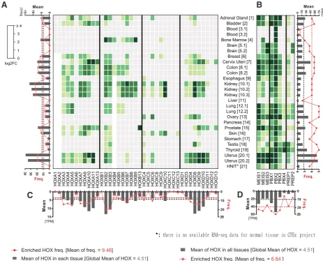
light-to-dark green gradient color code in the heatmap (Fig. 1). Below we summarized the main conclusions that could be deduced from the analysis of normal and tissue-specific HOX and TALE expression profiles in the adult (Fig. 1):
(1) - HOX genes are widely expressed in adult tissues, with only five tissues that show a significant HOX-poor expression profile (blood, brain, liver, pancreas and HINT (internal head and neck tissue)). Four tissues have few enriched HOX genes (less than
six: esophagus, stomach, testis, thyroid) while five tissues harbor enriched HOX genes from the four HOX genomic clusters (Adrenal gland, breast, kidney, skin and uterus). The other tissues have enriched HOX genes belonging to one or two different genomic clusters. In all those cases, tissues express HOX genes that cover anterior, central and posterior PGs.
(2) - Several HOX genes show no or very low frequent expres-sion profiles in adult tissues, as noticed for HOXA1-2, HOXA6, HOXB1, HOXB13, HOXC5, HOXC8, HOXC11-13, HOXD1 and HOXD12-13 (bottom graph). HOX genes of the HOXC genomic cluster are also less widely expressed in general than HOX genes of the three other clusters. In contrast, HOXA9-11, HOXB2-3 and HOXD8-10 show a global high enrichment of expression when compared to the other HOX genes. Together with the point men-tioned in (1), these observations highlight that the HOX expression profile is quite diverse but also specific in adult tissues, which is also illustrated by a weak global mean frequency of expression (value=9,46, which accounts for 1/4 of all HOX genes).
(3) – The five tissues without enriched HOX (blood, brain, liver, pancreas and head neck) express at least one TALE member, il-lustrating potential HOX-independent functions of TALE members in those tissues. Such independent roles have previously been characterized for craniofacial and spinal cord motor neuron de-velopment (Ferretti et al., 2011; Hanley et al., 2016). In the case of blood and liver samples, only PBX members are significantly enriched, which could thus represent non-functional contexts given
TCGA Full name of the tumor Number of tumor tissue samples (TCGA) Normal tissue name Number of normal tissue samples (TCGA+GTEx)
ACC Adrenocortical carcinoma 77 Adrenal Gland [1] 0+128
BLCA Bladder Urothelial Carcinoma 404 Bladder [2] 19+9
DLBC Lymphoid Neoplasm Diffuse Large B-cell Lymphoma 47 Blood [3.1] 0+337
THYM Thymoma 118 Blood [3.2] 2+337
LAML Acute Myeloid Leukemia 173 Bone Marrow [4] 0+70
GBM Glioblastoma multiforme 163 Brain [5.1] 0+207
LGG Brain Lower Grade Glioma 518 Brain [5.2] 0+207
BRCA Breast invasive carcinoma 1085 Breast [6] 112+179
CESC Cervical squamous cell carcinoma and endocervical adenocarcinoma 306 Cervix Uteri [7] 3+10
COAD Colon adenocarcinoma 275 Colon [8.1] 41+308
READ Rectum adenocarcinoma 92 Colon [8.2] 10+308
ESCA Esophageal carcinoma 182 Esophagus [9] 13+273
KICH Kidney Chromophobe 66 Kidney [10.1] 25+28
KIRC Kidney renal clear cell carcinoma 523 Kidney [10.2] 72+28
KIRP Kidney renal papillary cell carcinoma 286 Kidney [10.3] 32+28
LIHC Liver hepatocellular carcinoma 369 Liver [11] 50+110
LUAD Lung adenocarcinoma 483 Lung [12.1] 59+288
LUSC Lung squamous cell carcinoma 486 Lung [12.2] 50+288
OV Ovarian serous cystadenocarcinoma 426 Ovary [13] 0+88
PAAD Pancreatic adenocarcinoma 179 Pancreas [14] 4+167
PRAD Prostate adenocarcinoma 492 Prostate [15] 52+100
SKCM Skin Cutaneous Melanoma 461 Skin [16] 1+557
STAD Stomach adenocarcinoma 408 Stomach [17] 36+175
TGCT Testicular Germ Cell Tumors 137 Testis [18] 0+165
THCA Thyroid carcinoma 512 Thyroid [19] 59+278
UCEC Uterine Corpus Endometrial Carcinoma 174 Uterus [20.1] 13+78
UCS Uterine Carcinosarcoma 57 Uterus [20.2] 0+78
HNSC Head and Neck squamous cell carcinoma 519 Head and Neck Internal Tissues [21] 44+0
TABLE 1
NUMBER OF SAMPLES USED FOR RNA SEQUENCING IN NORMAL AND CANCER TISSUES
that PBX proteins normally need MEIS or PREP partners to trans-locate into the nucleus.
(4) - PBX (with the exception of PBX4) and MEIS members are generally more widely and strongly expressed than HOX members in adult tissues. PBX4 is particularly enriched in testis and thyroid tissues. Specific enrichment of PBX4 in testis was also previously reported in mouse (Wagner et al., 2001). PREP members are less strongly and less frequently expressed than PBX or MEIS, which recapitulates previous observations (Longobardi et al., 2014). Overall, the global mean expression level of TALE genes in adult tissues is five times higher than the global HOX mean expression level, with a global high mean frequency value (corresponding to 3/5 of all TALE genes).
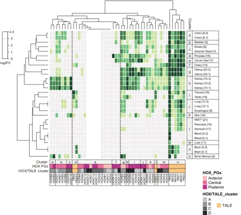
We next performed a clustering analysis to assess whether HOX and TALE genes could be preferentially organized in specific ensembles based on their normal expression profile in adult tissues (Fig. 2). This analysis confirms the distinct expression properties of PBX (except PBX4) and MEIS members, which form a unique ensemble of highly and widely expressed genes. In contrast, PREP1 and PBX4/PREP2 form two independent and isolated branches within ensembles of highly- or poorly-expressed HOX genes, re-spectively. This observation suggests that PREP1, but not PBX4 and PREP2, could participate to HOX functions in normal tissues.
HOX members form three main ensembles that basically regroup high, medium and low expression levels. Interestingly, ensembles corresponding to high and medium expression levels can be divided
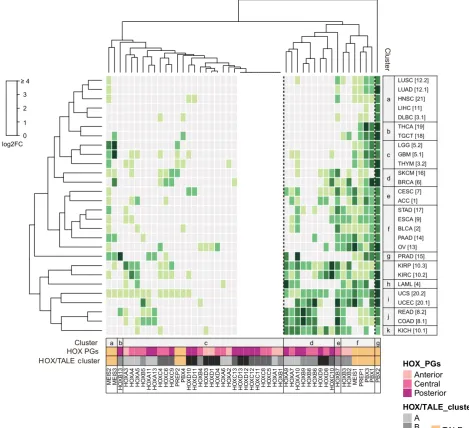
Fig. 1. HOX and TALE expression profiles in normal tissues. Expression profile is obtained from RNA-sequencing data performed in 21 different tissue types (see Table 1 for the full nomenclature), using TCGA and GTEx portals. Each value (see Table S1) was reported to the global HOX mean (TPM = 4.51), followed by log2 fold change (FC) conversion. Heatmap colors represent HOX and TALE enrichment as indicated in the color key. Varying shades of green indicate enrichment level (from 0 to ≥4), while light gray represents non-significant expression levels, tacking the global HOX mean expression level in all tissues as the reference value (TPM=4,51). Histograms around the heatmap indicate mean expression levels (dark gray bars) and frequency (red connected dots). Gray and red dotted lines note the global mean of expression level and frequency, respectively. (A) HOX mean expression and enriched HOX frequency in each normal tissue; (B) TALE mean expression and enriched TALE frequency in each normal tissue; (C) all normal tissues’ mean expression and enrichment frequency for each HOX member; (D) all normal tissues’ mean expression and enrichment frequency for each TALE member. Cervix Uteri [7]Breast [6]
Brain [5.2] Brain [5.1] Bone Marrow [4]Blood [3.2]
Blood [3.1]
MEIS1 MEIS2 MEIS3 PBX1 PBX2 PBX3 PBX4 PREP1 PREP2
Enriched TALE freq. [Mean of freq. = 6 ]
into several sub-ensembles that are quite homogenous in terms of the genomic cluster (sub-ensembles a, c, g or k), grouped into consistent anterior/central/posterior PG identity (sub-ensembles b, f, I and j). The ensemble regrouping weakly and non-significantly expressed HOX genes is more disparate although continuous HOX genes are present in small homogenous groups.
Tissue clustering confirms that different samples providing from the same tissue are quite homogenous, with highly similar HOX and TALE expression profiles. The clustering also revealed similarities between different tissues, as noticed for the breast and adrenal gland, thyroid and testis, or cluster composed by HNIT, pancreas and stomach. Other tissues appear more distinct, like the ovary, skin and bone marrow.
Overall the clustering analysis showed that the majority of HOX genes formed homogenous ensembles of two to five members
based on their expression profile in adult tissues. This organization follows the PG affiliation (anterior, central or posterior) and/or the genomic cluster identity, highlighting that various cis-regulatory rules could be responsible for the expression of specific combina-tions of HOX genes in different tissues. Moreover, tissue-specific HOX combinatorial codes are systematically associated with a high expression level of several PBX and MEIS members. This observation suggests that the information provided by each specific combination of HOX genes in normal adult tissues is dependent on a general partnership with PBX and MEIS members.
Expression of HOX and TALE genes in oncogenic tissues
The expression profile of HOX and TALE genes in cancer was analyzed in 28 cancer types deriving from the same 21 different tissues, using data samples from TCGA (Tables 1 and S2). For
H
OXC6 PBX2 MEIS3 MEIS2 PBX1 MEIS1 PBX3
Bone Marrow [4]
better comparison with the normal context, each log2 value was calculated by considering individual HOX and TALE cancer level over the same global HOX mean value previously defined in normal tissues. The deduced log2 values were given in a heatmap with the same color code as in the Fig. 1 to represent non-enriched (grey) or enriched (green) expression levels (Fig. 3).
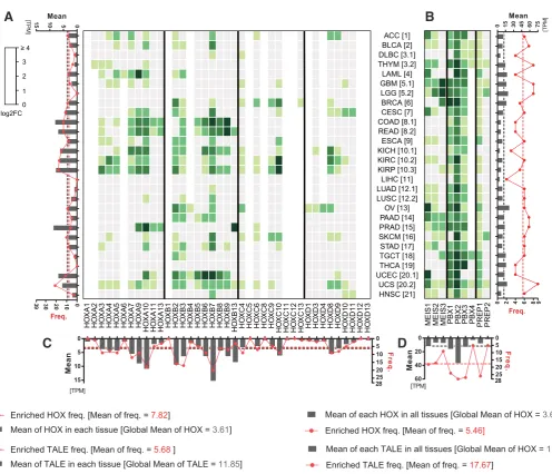
The heatmap shows that HOX genes are globally less frequently and less strongly expressed in cancer tissues (see the global mean values and graphs in the Fig. 3). The decrease of expression level is most apparent for tissues or HOX genes that displayed high enrichments in the normal condition (compare for example kidney, uterus or the HOXD cluster between Fig. 1 and 2). Still, there are also novel HOX expression profiles, particularly in cancers derived from tissues that were negative in normal condition (lymphoid neoplasma, thymoma, glioblastoma, pancreatic adenocarcinoma and HNIT: Fig. 3). In contrast, only one HOX gene among the
nine that were classified as non-enriched in the normal condition becomes positive in one cancer tissue (HOXA2 in thymoma: Fig. 3). This observation highlights that most HOX genes that were not enriched in normal tissues remain refractory to up-regulatory mechanisms in cancer. The expression level of TALE factors in cancer tissues is also significantly diminished, with a two folds decrease on average when compared to normal tissues (Fig. 3). Still, the TALE frequency pattern is comparable between cancer and normal tissues, and the expression level remains more than three times higher than the global HOX mean level. In conclusion, the cancer heatmap shows that a number of HOX and TALE genes have a modified expression profile that principally results from a reduced expression level.
We next performed a clustering analysis to assess whether expression changes in cancer could modify the overall distribution of HOX and TALE members when compared to the normal tissues.
Fig. 3. HOX and TALE expression profiles in cancer tissues. Expression profile is obtained from RNA-sequencing data performed in 28 cancer types deriving from the 21 different tissues used in the Figure 1. Numbers between brackets refer to each respective normal tissue. Values (see Table S2) were reported to the global HOX mean value in normal tissues (TPM=4,51) and given as a log2 fold change (FC). Heatmap color code and histograms are as in Fig. 1.
MEIS1 MEIS2 MEIS3 PBX1 PBX2 PBX3 PBX4 PREP1 PREP2 0
Enriched TALE freq. [Mean of freq. = 5.68 ]
Mean of TALE in each tissue [Global Mean of TALE =11.85] Enriched HOX freq. [Mean of freq. = 7.82]
Mean of HOX in each tissue [Global Mean of HOX = 3.61] Enriched HOX freq. [Mean of freq. = 5.46]
Mean of each HOX in all tissues [Global Mean of HOX = 3.61]
Enriched TALE freq. [Mean of freq. = 17.67]
Mean of each TALE in all tissues [Global Mean of HOX = 11.85]
Results show that cancer types remain clustered in function of their tissue origin, showing that the same logic of deregulation applies for different cancers deriving from the same tissue (Fig. 4). The overall organization of HOX and TALE members is however drasti-cally remodeled when compared to normal tissues, with basidrasti-cally two main ensembles of strongly or weakly expressed genes, and an independent branch corresponding to the unique wide and strong expression pattern of PBX2 (as already noticed in normal tissues). The two main ensembles are disparate in their HOX formula, although they contain groups of two or three continuous HOX genes of the same genomic cluster several times. Among the TALE members, PREP1 became closer to PBX1 and PBX3 than MEIS1, showing a potentially interesting role of PREP1 in place of MEIS1 in cancer tissues. Interestingly, MEIS1 becomes closer to HOXB2 and HOXB3 in the same ensemble, suggesting that a specific and general relationship could exist between these three factors in cancer. All other HOX genes are quite distantly related
to the TALE factors, with HOXB7 and HOXB13 lying apart in inde-pendent branches. Finally, two tandems belong to the ensemble of weakly expressed HOX genes: PBX4/PREP2 and MEIS2/MEIS3. The novel distribution of the MEIS2/MEIS3 tandem highlights that these two TALE members are more generally affected in cancers than the other TALE members. Overall the clustering analysis shows that the HOX expression profile is more sensitive to deregulatory changes in cancer than the TALE expression profile, which is ex-plained by their respective moderate and high expression levels in general in normal tissues.
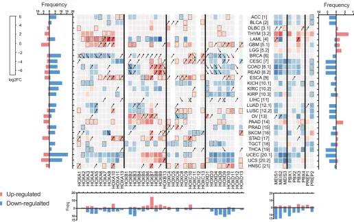
To get more insights into the HOX and TALE expression changes in cancer contexts, we generated a third cancer heatmap based on the fold change enrichment or loss of individual HOX and TALE members when compared to their respective expression level in the normal tissue (Table S3 and Fig. 5). Fold changes were calculated as a log2 value and considered as significant when they were at least two times superior (red cases in the Fig. 5) or inferior (blue
PBX2
cases in the Fig. 5) to the normal value. The magnitude of positive or negative change was represented in each case with a graded red or blue color code, respectively. Values corresponding to a non-significant change are represented by light-grey cases. Of note, contexts with a significant change but with values that were below the threshold defined for an enriched expression level in the normal tissue are considered apart (corresponding to boxes surrounded by a dotted line in the Fig. 5).
This novel representation confirmed that the large majority of TALE, and to some extent HOX genes, are down-regulated in the cancer context when compared to the normal tissue, as il-lustrated with the number of blue cases (Fig. 5). For example, 15 different cancer contexts show a decreased expression level of TALE members while the reverse is only observed in three cases (thymoma (THYM), brain lower grade glioma (LGG) and pancre-atic adenocarcinoma (PAAD)). Along the same line, members of the HOXA and HOXD clusters are most often down-regulated, while members of the HOXB and HOXC clusters show a more balanced pattern, with up-regulation events that are slightly more frequent than down-regulation events (Fig. 5). In total, all TALE members and 33/39 HOX genes show a significant change in the expression level when comparing cancer and normal tissues.
Ac-cordingly, only one (liver hepatocellular carcinoma) or two (brain lower grade glioma and liver hepatocellular carcinoma) cancer types show no significant changes in TALE or HOX expression profiles, respectively (Fig. 5).
In several cases, HOX expression changes in cancer do not systematically correspond to a down-regulation but also to up-regulations in few instances, showing that the same HOX gene could be repressed or activated depending on the cancer type. This fluctuated pattern is observed for most members of the HOXA-C clusters (exceptions are for HOX genes that are mono-deregulated in a few cancers, as noticed for HOXA6, A11, A13 and B13). In contrast, all but two members of the HOXD cluster show a more uniform deregulation, with a large majority of down regulations (bottom graph in the Fig. 5), especially in breast (BRCA), cervi-cal (CESC), colon (COAD), rectum (READ), and kidney (KICH, KIRC, KIRP) and uterus (UCEC, UCS) cancers. This observation suggests that HOXD members could have a more general anti-tumorigenic action.
The majority of cancers have a homogenous distribution of up- or down-regulation in the HOX gene expression profile (graph on the right in the Fig. 5). For example, large B-cell lymphoma (DLBC), thymoma (THYM), glioblastoma (GBM), esophageal carcinoma
H
MEIS1 MEIS2 MEIS3 PBX1 PBX2 PBX3 PBX4 PREP1 PREP2
Down-regulalted
(ESCA), pancreatic/stomach adenocarcinoma (PAAD/STAD) and head/neck squamous carcinoma (HNSC) show only up-regulation of HOX genes, while the reverse is observed for adrenocortical carcinoma (ACC), bladder urothelial carcinoma (BLCA), kidney cancers (KICH, KIRC, KIRP), lung adenocarcinoma (LUAD), tes-ticular germ cell tumors (TGCT) and thyroid carcinoma (THCA). Notably, cancers with a balanced pattern of both strong up- and down-regulation of HOX genes are less frequent, and relate to acute myeloid leukemia (LAML), colon (COAD), rectum (READ) and uterine (UCEC/UCS) adenocarcinoma. Interestingly, these versatile distributions can be HOX cluster specific (LAML, UCEC and UCS), with antagonistic distribution between HOX members of the same PG (like HOXA9 and HOXB9 in LAML for example), or occur between members of the same HOX cluster (as observed for the HOXB complex in colon (COAD) and rectum adenocarcinoma (READ)). Cancer tissues have also a uniform distribution of TALE down-regulations, except for thymoma (THYM) and pancreatic adenocarcinoma (PAAD), where TALE factors are systematically up-regulated (right graph in the Fig. 5). Acute myeloid leukemia (LAML) and glioblastoma (GBM) are the only cancer contexts with a more balanced pattern of up- and down-regulations (Fig. 5).
This heatmap also shows that HOX and TALE expression pro-files follow the same type of remodeling in the majority of cancers. Cancers with a major decrease of HOX expression levels have an associated decrease of TALE enrichment. Interestingly, the two cancer contexts with a strong and homogenous enrichment of TALE expression coincide with a uniform up-regulation of HOX genes (thymoma and pancreatic adenocarcinoma: Fig. 5). Finally, one of the rare contexts with a strong opposite enrichment between two different TALE members (MEIS2 and PBX3) corresponds to a cancer context with the same kind of strong opposite enrichment between HOXA and HOXB members (LAML: Fig. 5).
Altogether, these observations highlight that HOX and TALE factors are deregulated in a coordinated manner in the majority of cancers, suggesting that the two families are not acting indepen-dently of each other for cancer progression or arrest.
A comparison with the literature
Our systematic analysis of the 39 human HOX members and their associated TALE cofactors (PBX1-4, MEIS1-3 and PREP1-2) provides a global picture of HOX and TALE expression profiles in normal and cancer tissues.
To our knowledge, this analysis is the first attempt to couple the aberrant expression profile of HOX and TALE genes in a vast number of different cancer cell types. Data were extracted from RNA-sequencing experiments deposited in the TGCA and GTEx portals and we arbitrarily decided to assign the global HOX mean expression level in the 21 normal tissues under study as the mini-mal reference value for a significant enriched expression level. Under this condition, weakly expressed HOX genes could not be considered. In any case, given that all measures were reported to this minimal reference value, this choice has no influence on the log2 fold change that could be find between the normal and the corresponding cancer tissue. In addition, we considered significant fold changes from values that were below the global HOX mean value, considering that these modifications could still be not neutral in the cancer context (see below).
A large number of samples were considered for each cancer type (from 47 to 1085 cancer samples and 13 to 558 normal tissue
samples), making the data collection highly heterogeneous. This collection of cancers likely comprises tumors of various natures, with different aggressive behaviors, at different stages, with or without metastases. By comparison, most of the analyses of HOX expression in normal (Takahashi et al., 2004) or cancer (Abdel-Fattah et al., 2006; Buccoliero et al., 2009; Hur et al., 2014; Kanai et al., 2010; Kelly et al., 2016; Makiyama et al., 2005; Miller et al., 2003; Plowright et al., 2009; Yamashita et al., 2006) tissues are based on RT-PCR or QPCR in cell lines or from more restricted and more homogenous tumor samples (using approximately 4 to 20 tumor samples). These differences might explain that we did not systematically reproduce observations from previous work.
samples and cell lines highlighted that only two (HOXC10 and HOXC12) of the 39 human HOX genes are reported as being not affected in a solid tumors (Bhatlekar et al., 2014). Here we noticed that HOXC10 is either up- or down-regulated in different cancer contexts, including glioblastoma (GBM), breast BRCA), cervical (CESC), esophageal (ESCA), kidney (KICH), skin (SKCM) and stomach (STAD) cancers (Fig. 5). In fact, more recent studies
have also described a role of HOXC10 in gastric (Guo et al., 2017),
breast (Sadik et al., 2016), and cervix (Zhai et al., 2007) cancers. More generally, our analysis reveals most significant variations of HOX gene expression in eight novel cancer types (adrenocortical carcinoma (ACC), Lymphoid Neoplasm (DLBC), Thymoma (THYM), Kidney chromophobe (KICH), Kidney renal papillary cell carcinoma (KIRC), Pancreatic adenocarcinoma (PAAD), Testicular germ cell tumor (TGCT) and Uterus corpus endometrial carcinoma (UCEC)). It also validates and expands previous important conclusions, in-cluding i) the specific pattern of change in HOX gene expression depending on the cancer type, ii) the more frequent up-regulation of posterior HOX genes in solid tumors, and iii) a cluster-specific enrichment, with different preferential patterns depending on the cancer type.
TALE factors are generally less studied than HOX genes in cancers, and our survey is therefore informative with this regard. Among the few studies on TALE in cancer, one reported that PBX1 was up regulated in mouse immortalized hepatoblast cells screened for transposon genomic insertions that led to mesenchymal liver tumor upon transplantation into nude mice (Kodama et al., 2016). We did not find a particularly high level of PBX1 expression in either wild type or oncogenic liver tissues (covering 160 and 369 different normal or cancer tissue samples, respectively: Table 1). Results obtained from this study are however difficult to compare with our datasets since the cell contexts are quite different. Another study reported that high level of PBX1 correlated with shorter survival in post-chemotherapy ovarian cancer patients, and that silencing PBX1 reduced stem-like properties of ovarian tumor cells (Jung et al., 2016). We noticed that PBX1 was highly expressed in both wild type and tumor ovarian tissues (Figs. 1 and 3), explaining why it was not captured as an enriched gene in ovarian cancer in our analysis. Similarly, high level of PREP1 was reported as being important for triggering epithelial-mesenchymal transisition (EMT), invasion and metastasis in lung adenocarcinoma cells (Risolino et al., 2014). Our study revealed that PREP1 is strongly expressed in the normal and cancer lung tissues (Figs. 1 and 3). These observations suggest that the same TALE factor could have opposite functions between the normal and cancer cell context, being strongly expressed in both contexts for promoting differen-tiation or proliferation, respectively. Such antagonistic functional switch could potentially be linked to a variation of the HOX formula during cancer progression.
Our analysis reveals that TALE members are generally down regulated in cancer although PBX and MEIS are usually considered as oncoproteins. In fact, the role of PBX and MEIS as oncoproteins results principally from studies in leukemia, while conclusions in solid cancers are deduced from few classical expression analy-ses (considered as high with no systematic comparison with the normal tissue) and overexpression experiments in few cancer cell lines (see (Blasi et al., 2017) for review). Interestingly, we found a systematic enrichment of TALE factors in three cancer types (Thymus (THYM), Brain lower grade glioma (LGG), and
Pancreatic adenocarcinoma (PAAD)), two of them correlating to increased expression of a specific HOX cluster (THYM and PAAD). Our analysis thus revealed that THYM and PAAD constitute the best examples of a putative pro-oncogenic and collaborative role between HOX and TALE proteins in solid cancers.
Finally, our heatmaps show that TALE members are five times more enriched on average in normal tissues than HOX genes. Although this enrichment is strongly diminished (from five to two times on average, except for PBX2) in cancer tissues, it is main-tained, which could explain why PBX and MEIS are considered as enriched factors in previous studies. In addition, PREP members remain systematically much less expressed than MEIS members in cancer, which is in accordance with the general tumor suppressor function of PREP due to its competitive role against MEIS.
Conclusion
A striking aspect of our large-scale analysis is the global strong expression level of TALE members, especially PBX and MEIS, when compared to the HOX family members (three times stronger on average). As a consequence, the HOX expression profile is much more sensitive to subtle deregulations in cancers than the TALE expression profile. Accordingly, several cancer contexts express only a significant level of TALE members and no HOX, while the reverse is never observed. Thus, a hallmark of tumor cells is the expression of PBX and MEIS in a HOX-low or free state. Surpris-ingly, most of the studies have focused on HOX genes in cancer while the role of TALE factors remains to be determined in many different cancers.
Several cancers have a mixture of down- and up-regulation of HOX genes, highlighting the importance of the cell context for HOX function. In those cases, up-regulated HOX members could potenti-ate the effect of TALE members. This effect is often cluster-specific, as observed for HOXB members in uterus, ovarian and pancreatic cancers, or HOXA members in thymoma and glioblastoma. What could dictate the pro- or anti-oncogenic activity of HOX proteins with PBX and MEIS is clearly a key issue to understand the HOX/ TALE molecular code in cancer. The dose of each HOX and TALE molecule is certainly not neutral, as is the role of the different TALE members in association with the HOX family. In any case, given the general high expression level of PBX and MEIS members in cancer, one promising avenue for future therapeutic strategies could be to alter the activity of TALE products specifically in neoplastic cells. This could be achieved by overexpressing PREP, to block the activity of MEIS, or targeting MEIS, to make PBX non-functional (cytoplasmic). Interestingly, a dominant negative form of MEIS is also described in the literature (Jaw et al., 2000) and could be easily tested as a potential and general inhibitory peptide in many different cancer cell lines.
Materials and Methods
Data collection
different tissues that have more than 10 different normal control samples [from TCGA and/or GTEx, Table 1).
Data preprocessing
A mean TPM (Transcripts Per Kilobase Million) value was calculated for each HOX and TALE member in each tissue type (Tables S1 and S2). Values that were inferior to the global HOX mean value in all tissues (TPM= 4,51) were considered as not significantly enriched (gray cases in Tables S1 and S2). We noticed that HOX genes that have no known functions in a given tissue could have a TPM value closed to the global HOX mean TPM value, as noticed for example for HOXC4 in breast (TPM=4,12) or HOXA6 in adrenal gland (TPM=3,73). In contrast, known HOX functions correspond to higher TPM values, from 5.32 (HOXA3 in breast) to 49,19 (HOXA10 in cervix uteri). This observation suggests that the global HOX mean TPM value constitutes a good threshold for discriminating enriched expression profiles linked to a putative function in vivo. All HOX and TALE expression levels were then normalized over the global HOX mean TPM value and
log2 processed to do the heatmap (Figs. 1 and 3). This normalization allowed comparing HOX and TALE expression levels between different tissues. Hierarchical clustering is according to the Euclidean distance based on log2 fold change (FC) values, using complete linkage method.
Identification of differentially expressed genes between normal and cancer tissues
Differentially expressed gene (DEG) analysis was performed using GE-PIA, which was assessed by the R package limma using linear model and empirical Bayes method, with adjusted p-value (Benjamini and Hochberg FDR). Significantly modified HOX and TALE expression profiles meet the following conditions (Tables S3):
1) Enrichment: log2FC ≥1, and q-value ≤ 0.01. 2) Depletion: log2FC ≤-1, and q-value ≤ 0.01. All data are displayed as log2 transformed in heatmap.
Data visualisation
All heatmaps were performed in the statistical programming environment R (version 3.3.0) using functions available from Bioconductor (Huber et al., 2015), and histograms were performed using GraphPad Prism 7 software.
Acknowledgments
We thank LS Shashidhara for comments on the manuscript. Research in the author’s laboratory is supported by the CNRS, ENS-Lyon and grants from Cefipra, FRM, ARC, Ligue Contre le Cancer and China Scholarship Council (grant #201708070003 to Y. Jia).
References
ABDEL-FATTAH R, XIAO A, BOMGARDNER D, PEASE CS, LOPES MBS, HUSSAINI IM (2006). Differential expression of HOX genes in neoplastic and non-neoplastic human astrocytes. J Pathol 209: 15–24.
ALHARBI RA, PETTENGELL R, PANDHA HS, MORGAN R (2013). The role of HOX genes in normal hematopoiesis and acute leukemia. Leukemia 27: 1000–1008. ARGIROPOULOS B, HUMPHRIES RK (2007). Hox genes in hematopoiesis and
leukemogenesis. Oncogene 26: 6766–6776.
BHATLEKAR S, FIELDS JZ, BOMAN BM (2014). HOX genes and their role in the development of human cancers. J Mol Med Berl Ger 92: 811–823.
BLASI F, BRUCKMANN C, PENKOV D, DARDAEI L (2017). A tale of TALE, PREP1, PBX1, and MEIS1: Interconnections and competition in cancer. BioEssays 39: : 1600245. doi: 10.1002/bies.201600245
BORROW J, SHEARMAN AM, STANTON VP, BECHER R, COLLINS T, WILLIAMS AJ, DUBÉ I, KATZ F, KWONG YL, MORRIS C, OHYASHIKI K, TOYAMA K, ROWLEY J, HOUSMAN DE (1996). The t(7;11)(p15;p15) translocation in acute myeloid leukaemia fuses the genes for nucleoporin NUP98 and class I homeoprotein HOXA9. Nat Genet 12: 159–167.
BUCCOLIERO AM, CASTIGLIONE F, DEGL’INNOCENTI DR, AMMANATI F, GIORDANO F, SANZO M, MUSSA F, GENITORI L, TADDEI GL (2009). Hox-D
genes expression in pediatric low-grade gliomas: Real-time-PCR study. Cell Mol Neurobiol 29: 1–6.
BÜRGLIN TR (1997). Analysis of TALE superclass homeobox genes (MEIS, PBC, KNOX, Iroquois, TGIF) reveals a novel domain conserved between plants and animals. Nucleic Acids Res 25: 4173–4180.
BÜRGLIN TR (1998). The PBC domain contains a MEINOX domain: Coevolution of Hox and TALE homeobox genes? Dev Genes Evol 208: 113–116.
CANTILE M, PETTINATO G, PROCINO A, FELICIELLO I, CINDOLO L, CILLO C (2003). In vivo expression of the whole HOX gene network in human breast cancer. Eur J Cancer 39: 257–264.
CHOJNOWSKI JL, MASUDA K, TRAU HA, THOMAS K, CAPECCHI M, MANLEY NR (2014). Multiple roles for HOXA3 in regulating thymus and parathyroid differentia-tion and morphogenesis in mouse. Development 141: 3697–3708.
DARDAEI L, LONGOBARDI E, BLASI F (2014). Prep1 and Meis1 competition for Pbx1 binding regulates protein stability and tumorigenesis. Proc Natl Acad Sci USA 111: E896–905.
DARD A, REBOULET J, JIA Y, BLEICHER F, DUFFRAISSE M, VANACKER JM, FORCET C, MERABET S (2018). Human HOX Proteins Use Diverse and Context-Dependent Motifs to Interact with TALE Class Cofactors. Cell Rep 22: 2809–2817. EKLUND E (2011). The role of Hox proteins in leukemogenesis: insights into key
regulatory events in hematopoiesis. Crit Rev Oncog 16: 65–76.
FERNANDEZ LC, ERRICO MC, BOTTERO L, PENKOV D, RESNATI M, BLASI F, CARÉ A (2008). Oncogenic HoxB7 requires TALE cofactors and is inactivated by a dominant-negative Pbx1 mutant in a cell-specific manner. Cancer Lett 266: 144–155. FERRETTI E, LI B, ZEWDU R, WELLS V, HEBERT JM, KARNER C, ANDERSON MJ,
WILLIAMS T, DIXON J, DIXON MJ, DEPEW MJ, SELLERI L (2011). A Conserved Pbx-Wnt-p63-Irf6 Regulatory Module Controls Face Morphogenesis by Promoting Epithelial Apoptosis. Dev Cell 21: 627–641.
GLIGOROV D, SITNIK JL, MAEDA RK, WOLFNER MF, KARCH F (2013). A Novel Function for the Hox Gene Abd-B in the Male Accessory Gland Regulates the Long-Term Female Post-Mating Response in Drosophila. PLoS Genet 9: e1003395. GOLDMAN M, CRAFT B, ZHU J, HAUSSLER D (2017). Abstract 2584: The UCSC
Xena system for cancer genomics data visualization and interpretation. Cancer Res 77: 2584–2584.
GUO C, HOU J, AO S, DENG X, LYU G (2017). HOXC10 up-regulation promotes gastric cancer cell proliferation and metastasis through MAPK pathway. Chin J Cancer Res Chung-Kuo Yen Cheng Yen Chiu 29: 572–580.
HANLEY O, ZEWDU R, COHEN LJ, JUNG H, LACOMBE J, PHILIPPIDOU P, LEE DH, SELLERI L, DASEN JS (2016). Parallel Pbx-Dependent Pathways Govern the Coalescence and Fate of Motor Columns. Neuron 91: 1005–1020. HUBER W, CAREY VJ, GENTLEMAN R, ANDERS S, CARLSON M, CARVALHO
BS, BRAVO HC, DAVIS S, GATTO L, GIRKE T, et al., (2015). Orchestrating high-throughput genomic analysis with Bioconductor. Nat Methods 12: 115–121. HUR H, LEE JY, YUN HJ, PARK BW, KIM MH (2014). Analysis of HOX Gene
Expres-sion Patterns in Human Breast Cancer. Mol Biotechnol 56: 64–71.
JAW TJ, YOU L, KNOEP PS, YAO L, PAI C, TANG C, CHANG L, BERTHELSEN J, BLASI F, KAMPS MP, SUN YH (2000). Direct interaction of two homeoproteins, Homothorax and Extradenticle, is essential for EXD nuclear localization and function. Mech Dev 91: 279–291.
JUNG JG, SHIH IM, PARK JT, GERRY E, KIM TH, AYHAN A, HANDSCHUH K, DAVIDSON B, FADER AN, SELLERI L, WANG TL (2016). Ovarian cancer che-moresistance relies on the stem cell reprogramming factor PBX1. Cancer Res 76: 6351–6361.
KAMPS MP, MURRE C, SUN X, BALTIMORE D (1990). A new homeobox gene contributes the DNA binding domain of the t(1;19) translocation protein in pre-B all. Cell 60: 547–555.
KANAI M, HAMADA J-I, TAKADA M, ASANO T, MURAKAWA K, TAKAHASHI Y, MURAI T, TADA M, MIYAMOTO M, KONDO S, MORIUCHI T (2010). Aberrant expressions of HOX genes in colorectal and hepatocellular carcinomas. Oncol Rep 23: 843–851.
KARLSSON R, ALY M, CLEMENTS M, ZHENG L, ADOLFSSON J, XU J, GRÖNBERG H, WIKLUND F (2014). A population-based assessment of germline HOXB13 G84E mutation and prostate cancer risk. Eur Urol 65: 169–176.
cancer. Int J Cancer 139: 1608–1617.
KMITA M, DUBOULE D (2003). Organizing axes in time and space; 25 years of colinear tinkering. Science 301: 331–333.
KODAMA T, NEWBERG JY, KODAMA M, RANGEL R, YOSHIHARA K, TIEN JC, PARSONS PH, WU H, FINEGOLD MJ, COPELAND NG, JENKINS NA (2016). Transposon mutagenesis identifies genes and cellular processes driving epithelial-mesenchymal transition in hepatocellular carcinoma. Proc Natl Acad Sci USA 113: E3384–E3393.
LEBERT-GHALI CE, FOURNIER M, KETTYLE L, THOMPSON A, SAUVAGEAU G, BIJL JJ (2016). Hoxa cluster genes determine the proliferative activity of adult mouse hematopoietic stem and progenitor cells. Blood 127: 87–90.
LI Z, ZHANG Z, LI Y, ARNOVITZ S, CHEN P, HUANG H, JIANG X, HONG GM, KUNJAMMA RB, REN H, et al., (2013). PBX3 is an important cofactor of HOXA9 in leukemogenesis. Blood 121: 1422–1431.
LONGOBARDI E, PENKOV D, MATEOS D, DE FLORIAN G, TORRES M, BLASI F (2014). Biochemistry of the tale transcription factors PREP, MEIS, and PBX in vertebrates. Dev Dyn 243: 59–75.
MAKIYAMA K, HAMADA JI, TAKADA M, MURAKAWA K, TAKAHASHI Y, TADA M, TAMOTO E, SHINDO G, MATSUNAGA A, TERAMOTO KI, KOMURO K, KONDO S, KATOH H, KOIKE T, MORIUCHI T (2005). Aberrant expression of HOX genes in human invasive breast carcinoma. Oncol Rep 13: 673–679.
MANN RS, LELLI KM, JOSHI R (2009). Chapter 3 Hox Specificity. Unique Roles for Cofactors and Collaborators. Curr Top Dev Biol 88: 63–101.
MERABET S, DARD A (2014). Tracking context-specific transcription factors regulat-ing hox activity. Dev Dyn 243: 16–23.
MERABET S, MANN RS (2016). To Be Specific or Not: The Critical Relationship Between Hox And TALE Proteins. Trends Genet 32: 334–347.
MILLER GJ, MILLER HL, VAN BOKHOVEN A, LAMBERT JR, WERAHERA PN, SCHIRRIPA O, LUCIA MS, NORDEEN SK (2003). Aberrant HOXC expression ac-companies the malignant phenotype in human prostate. Cancer Res 63: 5879–5888. MOENS CB, SELLERI L (2006). Hox cofactors in vertebrate development. Dev Dyn
291: 193–206.
MORGAN R, BOXALL A, HARRINGTON KJ, SIMPSON GR, GILLETT C, MICHAEL A, PANDHA HS (2012). Targeting the HOX/PBX dimer in breast cancer. Breast Cancer Res Treat 136: 389–398.
MOSKOW JJ, BULLRICH F, HUEBNER K, DAAR IO, BUCHBERG AM (1995). Meis1, a PBX1-related homeobox gene involved in myeloid leukemia in BXH-2 mice. Mol Cell Biol 15: 5434–5443.
NAKAMURA T, LARGAESPADA DA, LEE MP, JOHNSON LA, OHYASHIKI K, TOYAMA K, CHEN SJ, WILLMAN CL, CHEN IM, FEINBERG AP, JENKINS NA, COPELAND NG, SHAUGHNESSY JD (1996). Fusion of the nucleoporin gene NUP98 to HOXA9 by the chromosome translocation t(7;11)(p15;p15) in human myeloid leukaemia. Nat Genet 12: 154–158.
NOURSE J, MELLENTIN JD, GALILI N, WILKINSON J, STANBRIDGE E, SMITH SD, CLEARY ML (1990). Chromosomal translocation t(1;19) results in synthesis of a homeobox fusion mRNA that codes for a potential chimeric transcription factor. Cell 60: 535–545.
PEARSON JC, LEMONS D, MCGINNIS W (2005). Modulating Hox gene functions during animal body patterning. Nat Rev Genet 6: 893–904.
PENKOV D, SANMARTÍN D, FERNANDEZ-DÍAZ L, ROSSELLÓ C, TORROJA C, SÁNCHEZ-CABO F, WARNATZ HJ, SULTAN M, YASPO M, GABRIELI A, TKACHUK V, BRENDOLAN A, BLASI F, TORRES M (2013). Analysis of the DNA-Binding Profile and Function of TALE Homeoproteins Reveals Their Specialization and Specific Interactions with Hox Genes/Proteins. Cell Rep 3: 1321–1333. PLOWRIGHT L, HARRINGTON KJ, PANDHA HS, MORGAN R (2009). HOX
tran-scription factors are potential therapeutic targets in non-small-cell lung cancer (targeting HOX genes in lung cancer). Br J Cancer 100: 470–475.
RISOLINO M, MANDIA N, IAVARONE F, DARDAEI L, LONGOBARDI E, FERNANDEZ S, TALOTTA F, BIANCHI F, PISATI F, SPAGGIARI L, HARTER PN, MITTEL-BRONN M, SCHULTE D, INCORONATO M, DI FIORE PP, BLASI F, VERDE P (2014). Transcription factor PREP1 induces EMT and metastasis by controlling the TGF- -SMAD3 pathway in non-small cell lung adenocarcinoma. Proc Natl Acad Sci USA 111: E3775–E3784.
ROZOVSKAIA T, FEINSTEIN E, MOR O, FOA R, BLECHMAN J, NAKAMURA T, CROCE CM, CIMINO G, CANAANI E (2001). Upregulation of Meis1 and HoxA9 in acute lymphocytic leukemias with the t(4 : 11) abnormality. Oncogene 20: 874–878. RUX DR, SONG JY, SWINEHART IT, PINEAULT KM, SCHLIENTZ AJ, TRULIK KG,
GOLDSTEIN SA, KOZLOFF KM, LUCAS D, WELLIK DM (2016). Regionally Restricted Hox Function in Adult Bone Marrow Multipotent Mesenchymal Stem/ Stromal Cells. Dev Cell 39: 653–666.
SADIK H, KORANGATH P, NGUYEN NK, GYORFFY B, KUMAR R, HEDAYATI M, TEO WW, PARK S, PANDAY H, MUNOZ TG, MENYHART O, SHAH N, PANDITA RK, CHANG JC, DEWEESE T, CHANG HY, PANDITA TK, SUKUMAR S (2016). HOXC10 Expression Supports the Development of Chemotherapy Resistance by Fine Tuning DNA Repair in Breast Cancer Cells. Cancer Res 76: 4443–4456. SAUNDERS EJ, DADAEV T, LEONGAMORNLERT DA, JUGURNAUTH-LITTLE
S, TYMRAKIEWICZ M, WIKLUND F, AL OLAMA AA, BENLLOCH S, NEAL DE, HAMDY FC, et al., (2014). Fine-Mapping the HOXB Region Detects Common Variants Tagging a Rare Coding Allele: Evidence for Synthetic Association in Prostate Cancer. PLoS Genet 10: e1004129. (doi: 10.1371/journal.pgen.1004129) SITWALA K V, DANDEKAR MN, HESS JL (2008). HOX proteins and leukemia. Int
J Clin Exp Pathol 1: 461–474.
STEELMAN S, MOSKOW JJ, MUZYNSKI K, NORTH C, DRUCK T, MONTGOMERY JC, HUEBNER K, DAAR IO, BUCHBERG AM (1997). Identification of a conserved family of Meis1-related homeobox genes. Genome Res 7: 142–156.
SUGIMURA R, JHA DK, HAN A, SORIA-VALLES C, DA ROCHA EL, LU YF, GOETTEL JA, SERRAO E, ROWE RG, MALLESHAIAH M, WONG I, SOUSA P, ZHU TN, DITADI A, KELLER G, ENGELMAN AN, SNAPPER SB, DOULATOV S, DALEY GQ (2017). Haematopoietic stem and progenitor cells from human pluripotent stem cells. Nature 545: 432–438.
TAKAHASHI Y, HAMADA J, MURAKAWA K, TAKADA M, TADA M, NOGAMI I, HAYASHI N, NAKAMORI S, MONDEN M, MIYAMOTO M, KATOH H, MORIUCHI T (2004). Expression profiles of 39 HOX genes in normal human adult organs and anaplastic thyroid cancer cell lines by quantitative real-time RT-PCR system. Exp Cell Res 293: 144–153.
TAMINIAU A, DRAIME A, TYS J, LAMBERT B, VANDEPUTTE J, NGUYEN N, RE-NARD P, GEERTS D, REZSÖHAZY R (2016). HOXA1 binds RBCK1/HOIL-1 and TRAF2 and modulates the TNF/NF-κB pathway in a transcription-independent manner. Nucleic Acids Res 44: 7331–7349.
TANG Z, LI C, KANG B, GAO G, LI C, ZHANG Z (2017). GEPIA: A web server for cancer and normal gene expression profiling and interactive analyses. Nucleic Acids Res 45: W98–W102.
UHLÉN M, FAGERBERG L, HALLSTRÖM BM, LINDSKOG C, OKSVOLD P, MARDI-NOGLU A, SIVERTSSON Å, KAMPF C, SJÖSTEDT E, ASPLUND A, et al., (2015). Proteomics. Tissue-based map of the human proteome. Science 347: 1260419. WAGNER K, MINCHEVA A, KORN B, LICHTER P, PÖPPERL H (2001). Pbx4, a new
Pbx family member on mouse chromosome 8, is expressed during spermatogen-esis. Mech Dev 103: 127–131.
WELLIK DM (2011). Hox genes and kidney development. Pediatr Nephrol Berl Ger 26: 1559–1565.
XIN X, YAN L, GUANGFA Z, YAN H, KENG L, CHUNTING W (2017). Mesenchymal Stem Cells Promoted Lung Wound Repair through Hox A9 during Endotoxemia-Induced Acute Lung Injury. Stem Cells Int 2017: :3648020. (doi: 10.1155/2017/3648020) YAMASHITA T, TAZAWA S, YAWEI Z, KATAYAMA H, KATO Y, NISHIWAKI K, YO-KOHAMA Y, ISHIKAWA M (2006). Suppression of invasive characteristics by antisense introduction of overexpressed HOX genes in ovarian cancer cells. Int J Oncol 28: 931–938.
ZAKANY J, DUBOULE D (2007). The role of Hox genes during vertebrate limb development. Curr Opin Genet Dev 17: 359–366.
ZHAI Y, KUICK R, NAN B, OTA I, WEISS SJ, TRIMBLE CL, FEARON ER, CHO KR (2007). Gene Expression Analysis of Preinvasive and Invasive Cervical Squamous Cell Carcinomas Identifies HOXC10 as a Key Mediator of Invasion. Cancer Res 67: 10163–10172.
Gdf11/Smad signalling and Cdx proteins cooperate to activate the Hoxc8 early enhancer in HepG2 cells Stephen J. Gaunt
Int. J. Dev. Biol. (2017) 61: 427-432 https://doi.org/10.1387/ijdb.170066sg
ADHFe1: a novel enzyme involved in retinoic acid-dependent Hox activation Yehuda Shabtai, Natalie Shukrun and Abraham Fainsod
Int. J. Dev. Biol. (2017) 61: 303-310 https://doi.org/10.1387/ijdb.160252af
The significance of Hox gene collinearity Stephen J. Gaunt
Int. J. Dev. Biol. (2015) 59: 159-170 https://doi.org/10.1387/ijdb.150223sg
Direct regulation of siamois by VegT is required for axis formation in Xenopus embryo
5 yr ISI Impact Factor (2016) = 2.421
Hong-Yan Li, Warif El Yakoubi and De-Li Shi Int. J. Dev. Biol. (2015) 59: 443-451 https://doi.org/10.1387/ijdb.150040ds
Synergistic action in P19 pluripotential cells of retinoic acid and Wnt3a on Cdx1 en-hancer elements
Stephen J. Gaunt and Yu-Lee Paul Int. J. Dev. Biol. (2014) 58: 307-314 https://doi.org/10.1387/ijdb.140003sg
The Parahox gene Pdx1 is required to maintain positional identity in the adult foregut Andrew M. Holland, Sonia Garcia, Gaetano Naselli, Raymond J. MacDonald and Leonard C. Harrison
Int. J. Dev. Biol. (2013) 57: 391-398 https://doi.org/10.1387/ijdb.120048ah
A possible role of Reproductive homeobox 6 in primordial germ cell differentiation Chang Liu, Paichi Tsai, Ana-Marie García, Brandon Logeman and Tetsuya S. Tanaka Int. J. Dev. Biol. (2011) 55: 909-916