Expression of serotonergic system components
during early Xenopus embryogenesis
DENIS A. NIKISHIN
1, STANISLAV V. KREMNYOV
2, VICTORIA V. KONDUKTOROVA
3and YURI B. SHMUKLER*
,11Koltzov Institute of Developmental Biology, Russian Academy of Sciences, Group of Embryophysiology, 2Lomonosov Moscow State University, Faculty of Biology, Department of Embryology, Laboratory of Developmental
Biophysics and 3Engelhardt Institute of Molecular Biology, Russian Academy of Sciences, Laboratory of Molecular
Biology of Development, Moscow, Russia
ABSTRACT Despite abundant research studies on the physiological and biochemical nature of embryonic neurotransmitter function, little is known about the molecular genetic mechanisms involved. The expression of the main components of the serotonergic system during early Xenopus embryogenesis was investigated using RT-PCR, real time PCR and in situ hybridization. Transcripts encoding the serotonin receptors HTR2C and HTR7, as well as the vesicular monoamine transporter VMAT2, the serotonin transporter (SERT) and the serotonin synthesis enzymes tryptophan hydroxy-lase (TPH2) and aromatic amino acid decarboxyhydroxy-lase (AAAD) were found to be expressed during the cleavage division stages, whereas the degradation enzyme monoamine oxidase A (MAOA) was absent. The main components of the serotonergic system were found to be expressed during the earliest stages of embryonic development. The embryonic transmitter mechanism, its complexity, and its variability among various species are discussed.
KEY WORDS:
serotonin, receptor, transporter, embryo, Xenopus
The presence and functional activity of the substances known as classic neurotransmitters (serotonin, acetylcholine, adrenaline and dopamine) during embryogenesis were first investigated by Buznikov and his collaborators (see Buznikov, 1967). Since then, it has become clear that the physiological functions of these substances are not limited to synaptic transmission and persist throughout ontogenesis, including the very early stages (see Shmukler, Buznikov, 1998; Buznikov, 2007).
The serotonergic system is one of the most intensively studied transmitter systems in early embryogenesis. It was demonstrated that serotonin (5-HT) is involved in cell cycle control, the regulation of the cytoskeleton state, and blastomere interactions during cleav-age divisions (Shmukler, Buznikov, 1998). During the later stcleav-ages of development, serotonin is involved in the regulation of ciliary motility, gastrulation, the establishment of left-right asymmetry, morphogenetic processes, metamorphosis, and organogenesis (Buznikov, 1990; Fukumoto et al., 2005b). However, there is sparse molecular genetic data on the prenervous serotonin mechanisms (Vesela et al., 2003; Dube, Amireault, 2007; see also Table 1).
The aim of the present work is to comprehensively examine the expression of serotonergic system components in X. laevis embryos. The presence of serotonin and its functional activity
www.intjdevbiol.com
*Address correspondence to: Yuri B. Shmukler. N.K. Koltzov Institute of Developmental Biology, Russian Academy of Sciences, 26 Vavilov St., Moscow, 119334,
Russia. Fax: +7-499-135-8012. Tel: +7-499-135-0052. e-mail: [email protected] - web: idbras.comcor.ru/indexr.HTM
Accepted: 16 March 2012. Final, author-corrected PDF published online: 26 June 2012.
ISSN: Online 1696-3547, Print 0214-6282 © 2012 UBC Press
Printed in Spain
Abbreviations used in this paper: 5-HT, serotonin; AAAD, aromatic aminoacid
decar-boxylase; AC, animal cap; EST, expressed sequence tag; HTR, serotonin receptor; ISH, in situ hybridization; MAOA, monoamine oxidase A; MMR, Marc’s modified Ringer’s; MZ, marginal zone; nAChR, nicotinic acetylcholine receptor; ODC, ornithine decarboxylase; RT-PCR, reverse transcription polymerase chain reaction; SERT, serotonin transporter; TPH, tryptophan hydroxylase; VMAT, vesicular monoamine transporter; VP, vegetal pole fragment; b-AdR, b-adrenoreceptor. (Fukumoto et al., 2005b) in Xenopus embryos allows for the investigation of the expression of serotonin receptors HTR1A, HTR2B, HTR2C, HTR3A, HTR4, HTR7. Our analysis of the se-rotonergic system also encompasses the study of monoamine vesicular transporters VMAT1 and VMAT2, sodium-dependent serotonin transporter (SERT), enzymes involved in serotonin synthesis (tryptophan hydroxylases TPH1 and ТРН2, and aromatic aminoacid decarboxylase (AAAD)), and degradation (monoamine oxidase A (MAOA)).
Results
Serotonin receptors
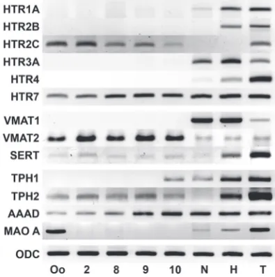
To detect the serotonin receptors involved during early Xeno-pus laevis embryogenesis, we performed temporal expression analysis of six types of these receptors using the known mRNA sequences. The total RNA was isolated from the embryos at dif-ferent developmental stages and used for RT-PCR. The results are presented in Fig. 1. The expression of HTR2C and HTR7 was detected at the stages of oocyte, 2 blastomeres (stage 2),
and midblastula, i.e., before the beginning of zygotic genome expression. The sequence of the PCR products from the oocyte stage corresponded to the X. laevis HTR2C and HTR7 mRNAs (data not shown). The expression of these two genes was also detected during midblastula (stage 8), late blastula (stage 9), and early gastrula (stage 10) after Xenopus zygotic genome activation. During the neurula stage, the HTR7 transcripts persisted, and the expression of HTR1A, HTR3A, and HTR4 was also detected; however, HTR2C mRNA was absent at this stage. All serotonin receptors were expressed after hatching except HTR2C. All 6 serotonin receptors were expressed during the tadpole stage.
The transcripts of at least two serotonin receptors were pres-ent within the maternal mRNA pool during the earliest stages of development. Quantitative real-time PCR was used to evaluate the levels and dynamics of their expression. The expression levels were normalized to the expression level of ornithine decarboxylase (ODC) at stage 2, which is the stage when the maternal genes are expressed exclusively. The expression level of HTR2C during stage 2 was approximately 0.002% of that of ODC (Fig. 2). HTR2C expression reached a maximum during the two-blastomere stage and then steadily decreased (Fig. 3) to 57.8% during stage 8, 36.9% during stage 9 and 11.3% dur-ing stage 10. HTR2C expression was not detectable durdur-ing the neurula and hatching stages. During stage 2, the expression of HTR7 mRNA was approximately 4.9% compared with ODC (Fig. 2). HTR7 was expressed without interruption (Fig. 3) at 96.8% during stage 8, 78.2% during stage 9, 72.6% during stage 10, 78.0%, during the neurula stage and decreasing to 32.2% during hatching, compared with the expression level during stage 2.
Samples of the animal caps (AC), marginal zones (MZ), and vegetal pole fragments (VP) during stage 9 were isolated to determine the relative expression of HTR2C and HTR7along the animal-vegetal axis. The real-time PCR results using these fragments are shown in Fig. 4. Compared with the AC, the HTR2C expression levels in the MZ and VC samples were 83.0% and 64.7%, respectively, whereas those of HTR7 were 119.3% and 90.6%, respectively. Thus, the relative levels of serotonin receptor expression along the animal-vegetal axis either did not differ (in
Fig. 1. Temporal expression of serotonergic system components dur-ing Xenopus laevis development. The serotonin receptors HTR2C and HTR7, vesicular transporter VMAT2, sodium-dependent transporter SERT, enzymes of synthesis TPH2 and AAAD are expressed during the early stages of development. The MAOA expression detected in oocytes is due to the jelly coats (see Fig. 8 and Discussion). Abbreviations: Oo, oocyte; 2, 2-cell embryo (stage 2); 8, midblastula (stage 8); 9, late blastula (stage 9); 10, early gastrula (stage 10); N, neurula (stage 15); H, hatched larvae (stage 33-35) and T, tadpoles (stage 57). ODC is the endogenous control.
0 20 40 60 80 100 120 140
2 8 9 10 N H 2 8 9 10 N H 2 8 9 10 N H
HTR2C HTR7 VMAT2
RQ, %
Fig. 2 (left). Relative expression levels of HTR2C, HTR7 and VMAT2 compared with ODC at the two-cell embryonic stage. Real-time PCR was performed using different primers for the same probe. The Ct values were calculated via equal threshold levels. The relative expression levels (RQ) were calculated via the efficiency-corrected ΔCt method (Bookout et al., 2005). The ODC expression level was considered to be 100%. The RQ for VMAT2 was approximately 43.9%, HTR7 approximately 4.9%, and HTR2C 0.002%.
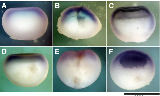
the case of HTR7) or the difference was statistically insignificant (in case the of HTR2C). These data are supported by the results of the in situ hybridization of HTR7 transcripts that were expressed during early embryogenesis at relatively high levels (Fig. 5). The HTR7 transcripts in the zygotes and cleavage-stage embryos were localized to the periphery, mainly in the animal area. Such localization persisted also during the blastula stage; the signal was visible in both the animal and vegetal zones but was much more intense in the AC.
Serotonin transporters
The RT-PCR analysis of the vesicular transporters during em-bryogenesis (Fig. 1) demonstrated that VMAT1 was expressed during neurulation, hatching and tadpole stage, whereas it was absent at the earlier developmental stages. VMAT2 was expressed throughout embryonic development but at very low levels after stage 10. The VMAT2 PCR product obtained from the samples of oocytes was isolated and sequenced. The sequence obtained
0
Fig. 4. The spatial distribution of the mRNA expression of HTR2C, HTR7 and VMAT2 within the
Xenopus blastula-stage embryo. The relative expression levels (RQ) were normalized to ODC of the AC sample (± SD). VMAT2 expression has pronounced animal-vegetal gradient, whereas HTR2C and HTR7 expression have not. AC, animal cap; MZ, marginal zone; VP, vegetal portion of the embryo.
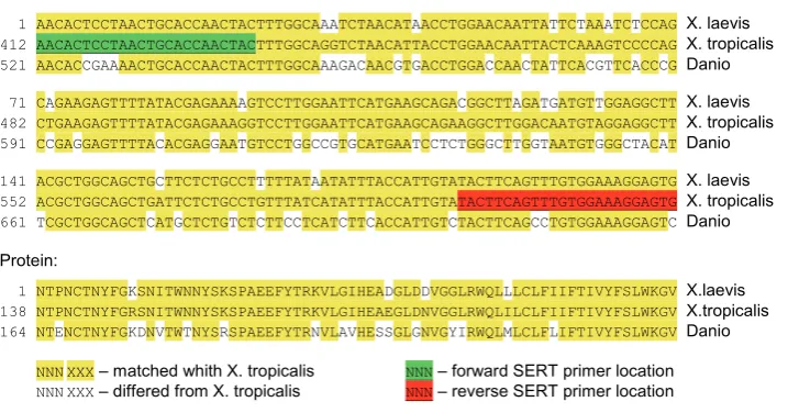
analyzed the SERT mRNA sequences of X. tropicalis and Danio and selected prim-ers for the regions of X. tropicalis SERT mRNA that were maximally homologous to Danio SERT mRNA (see Fig. 6). RТ-PCR using these primers produced a poorly expressed product in all samples obtained before the hatching stage (Fig. 1). The PCR product was isolated from the oocytes and sequenced (GeneBank ID: JQ001861) to demonstrate that SERT was expressed during the early devel-opmental stages. The sequence of the PCR product and its alignment with the sequence of that of X. tropicalis is shown in Fig 6. The comparison indicates that there are 17 nucleotide substitutions, 4 of which were non-synonymous and only one of which was non-conservative. Thus,
Fig. 5. Pattern of expression of HTR7 and VMAT2 mRNA during early
Xeno-pus embryogenesis.In situ hybridization for (A-C) HTR7 and (D-F) VMAT2 to oocytes (A,D), cleavage embryos (B,E) and blastula (C,F). The animal-vegetal gradient of distribution was detected both for the VMAT2 and HTR7 transcripts. During the blastula stage, the VMAT2 transcripts were localized mainly to the animal hemisphere, whereas the HTR7 transcripts were also present in the vegetal hemisphere.
B
C
D
E
F
A
corresponded to that of X. laevis VMAT2 mRNA.We examined the gene expression profiles of VMAT2 during X. laevis development using real-time PCR. The VMAT2 expres-sion level during stage 2 was approximately 43.9% compared with that of ODC (Fig. 2). Until gastrulation, VMAT2 expression remained relatively constant: 115.9% during the midblastula stage and 87.3% during the late blastula stage compared with stage 2, and then it decreased to 54.9% during early gastrula-tion and decreased sharply to 3.5% during neurulagastrula-tion, and to 2.7% during the post-hatching stage (Fig. 3).
Real-time PCR using the AC, MZ, and VC samples was performed to study the animal-vegetal distribution of VMAT2 expression (Fig. 4). The VMAT2 expression level in the MZ and VP samples achieved 69.7% and 20.6%, respectively, compared with that of the AC. Thus, VMAT2 expression had a pronounced animal-vegetal gradient. We performed an in situ hybridization of VMAT2 (Fig. 5) and obtained a more pronounced expression within the AC of the blastula as well as within the animal area of the zygote and cleavage-stage embryo.
The research would not be complete without a similar analysis of the serotonin transporter SERT. However, the SERT sequence of X. laevis was unknown. Therefore, we
the expression of SERT was confirmed during the early stages of Xenopus embryogenesis.
Enzymes of serotonin synthesis and degradation
Unlike AAAD and TPH1, the sequence of TPH2 mRNA of X. laevis is unknown. We analyzed the X. laevis EST database and located the sequence derived from the adult eye (CF547701), which was homologous to TPH2. The expression of these genes was analyzed during embryogenesis using RT-PCR (Fig. 1). TPH1 was not expressed during early embryogenesis and only appeared during stage 10. TPH2 was expressed during all of the stages studied, although the levels of expression were low from the oocyte to hatching stage. AAAD was expressed throughout development.
were expressed within the ovary (both types of TPH and AAAD). To determine the source of embryonic serotonin, the expression of the aforementioned enzymes was investigated in the samples from the follicle envelope and immature oocytes (stages I – III) (Fig. 7). AAAD was expressed in the immature oocytes as well as in the follicle envelope. TPH1 was detected in the follicle envelope but not in the oocyte, whereas TPH2 was expressed in the oocytes but not in the follicle envelope.
Finally, we performed a temporal expression analysis of MAOA (Fig. 1). This gene was distinctly expressed in mature oocytes, but its transcripts could not be detected between stage 2 and stage 10. MAOA was then expressed during the neurula stage and also during the hatching and tadpole stages. This difference in expression between the oocyte and 2-blastomere stage embryo required additional analysis. Samples of the oocytes for these experiments were obtained directly from a female frog without avoiding the jelly coat (most likely contaminated with some cells from the genital tract), in contrast to other samples. Therefore, the samples of RNA were obtained from dejellied oocytes and jelly coat material and used for additional RT-PCR analysis (Fig. 8). MAOA expression was detected in jelly coat material but was absent in dejellied oocytes. Hence, we concluded that MAOA was not expressed during the early stages of X. laevis embryogenesis.
Discussion
The present research showed a com-plex expression pattern of the of serotonin receptors during Xenopus laevis embryonic and larval development. According to Xe-nbase (www.xeXe-nbase.org), there are 15 main types of serotonin receptors in the Xenopus tropicalis genome. However, for X. laevis, the mRNA sequences of only 6 serotonin receptors are known.
We have shown that two of these recep-tors, HTR2C and HTR7, are expressed during the earliest stages of X. laevis embryogenesis. Interestingly, the expres-sion levels of these receptors differ quan-titatively; there are 2500-fold more HTR7 transcripts than those of HTR2C. It is known that transcription process is absent until Xenopus midblastula stage, and protein synthesis occurs using the maternal mRNA accumulated during oogenesis (Heasman, 2006). Thus, the transcripts of these two serotonin receptors are a part of the pool of maternal mRNA. It is logical to suggest that the prenervous functions of serotonin
mRNA:
1 AACACTCCTAACTGCACCAACTACTTTGGCAAATCTAACATAACCTGGAACAATTATTCTAAATCTCCAG X. laevis
412 AACACTCCTAACTGCACCAACTACTTTGGCAGGTCTAACATTACCTGGAACAATTACTCAAAGTCCCCAG X. tropicalis
521 AACACCGAAAACTGCACCAACTACTTTGGCAAAGACAACGTGACCTGGACCAACTATTCACGTTCACCCG Danio
71 CAGAAGAGTTTTATACGAGAAAAGTCCTTGGAATTCATGAAGCAGACGGCTTAGATGATGTTGGAGGCTT X. laevis
482 CTGAAGAGTTTTATACGAGAAAGGTCCTTGGAATTCATGAAGCAGAAGGCTTGGACAATGTAGGAGGCTT X. tropicalis
591 CCGAGGAGTTTTACACGAGGAATGTCCTGGCCGTGCATGAATCCTCTGGGCTTGGTAATGTGGGCTACAT Danio
141 ACGCTGGCAGCTGCTTCTCTGCCTTTTTATAATATTTACCATTGTATACTTCAGTTTGTGGAAAGGAGTG X. laevis
552 ACGCTGGCAGCTGATTCTCTGCCTGTTTATCATATTTACCATTGTATACTTCAGTTTGTGGAAAGGAGTG X. tropicalis
661 TCGCTGGCAGCTCATGCTCTGTCTCTTCCTCATCTTCACCATTGTCTACTTCAGCCTGTGGAAAGGAGTC Danio
Protein:
1 NTPNCTNYFGKSNITWNNYSKSPAEEFYTRKVLGIHEADGLDDVGGLRWQLLLCLFIIFTIVYFSLWKGV X.laevis
138 NTPNCTNYFGRSNITWNNYSKSPAEEFYTRKVLGIHEAEGLDNVGGLRWQLILCLFIIFTIVYFSLWKGV X.tropicalis
164 NTENCTNYFGKDNVTWTNYSRSPAEEFYTRNVLAVHESSGLGNVGYIRWQLMLCLFLIFTIVYFSLWKGV Danio
NNNXXX – matched whith X. tropicalis
NNNXXX – differed from X. tropicalis NNN NNN – forward SERT primer location – reverse SERT primer location
Fig. 6. Alignment of SERT mRNA and the appropriate protein sequences. The PCR product of X. laevis SERT (JQ001861) and the deduced protein sequence were aligned via the Clustal W method with X. tropicalis SERT mRNA (XM_002932084) and protein (XP_002932130) as well as the Danio SERT mRNA (NM_001177459) and protein (NP_001170930). The primers were designed to X. tropicalis SERT mRNA regions that were the most similar to that of Danio. 17 nucleotides differed between the nucleotide sequences of X. laevis and X. tropicalis SERT is, but the protein sequence had only 4 substitutions.
are mediated by receptors 5-HT2C and 5-HT7 during early X. laevis embryogenesis. On the other hand, the maternal mRNA may also be translated at later stages, even after the midblastula transition (activation of the zygotic genome). Of course, mRNA expression does not guarantee the production of the corresponding protein during the early stages of Xenopus embryogenesis. However, the dynamics of RNA expression can provide insight regarding the function of the corresponding gene.
We demonstrated that the expression dynamics of these re-ceptors differed greatly. HTR7 expression remained at an even level until hatching, whereas the expression of HTR2C constantly decreased and disappeared by the neurula stage. Therefore, it is possible that the 5-HT2C receptor is involved in the early prenervous processes whereas the 5-HT7 receptor can be functional throughout development. This possiblility is supported by the ability of 5-HT2A and 5-HT2C antagonist ritanserine to induce the germinal vesicle breakdown in the immature Xenopus oocytes (Sheng et al., 2005). It is important that 5-HT2C and 5-HT7 receptors are coupled to different systems of second messengers; the 5-HT2C receptor activates the phosphatidylinositol signal pathway, whereas the 5-HT7 receptor activates the adenylate cyclase signaling pathway (Peroutka, 1997). The activation of these signal cascades has various consequences, such as changing the state of the cytoskeleton (Larsson, 2005)
Fig. 7 (left). Expression of serotonin synthesis enzymes in
Xeno-pus ovaries. I-III, immature oocytes (stages I-III); AAAD, aromatic aminoacid decarboxylase; F, follicular envelope; Ov, total ovary; TPH, tryptophan hydroxylase.
and setting up the axial complex (Kim, Han, 1999).
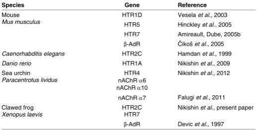
Other serotonin receptors that have been studied are not expressed during the early stages of development. HTR1A is detectable beginning at late blastula stage, HTR3A and HTR4 beginning at the neurula stage, and HTR2B after hatching. The entire set of serotonin receptors is expressed at the tadpole stage only. Pharmacological and immunohistochemical data support the possible participation of 5-HT3 and 5-HT4 receptors in establishing the left-right asymmetry in Xenopus (Fukumoto et al., 2005b). The 5-HT3 receptor is a ligand-dependent ionic channel whose functional activity is associated with the presence of HTR3A-subunits (Boyd et al., 2002). Therefore, the absence of HTR3A expression during early embryogenesis results in the absence of de novo formation of the functional 5-HT3 receptors at this stage. The controversy between our data and those reported by Fukumoto et al., (2005b) can be explained by the difference in the sensitivity between mam-malian and amphibian serotonin receptors to the same ligands. Fukumoto et al., (2005b) also reported the presence of HTR4 transcripts during the early stages of the development using in situ hybridization (ISH). Taking into account the higher specificity and sensitivity of RT-PCR compared with ISH, a false-positive result of the ISH is more probable than a false-negative RT-PCR result. It is intriguing that we found a wide diversity of serotonin recep-tor types that are expressed during early development (Table 1) when summarizing both our data and the literature data, with a few coincidences – the 5-HT2C in Xenopus and C. elegans, and the 5-HT7 in Xenopus and mouse. It is possible to reliably predict that serotonin receptors are also expressed in the embryos of species not yet studied in this regard, similarly to serotonin and other transmitters that were discovered in embryos of all species investigated. However, the types of serotonin receptor evidently are not strictly determined, although their mechanisms would be highly conserved. Furthermore, serotonin receptors do not encom-pass possible transmitter mechanisms in embryo because, both in Xenopus (Devic et al., 1997) and mouse (Čikoš et al., 2005), the b-adrenoreceptors (b-AdR) and serotonin receptors are expressed simultaneously (Table 1). A similar situation occurs during the early development of sea urchins where homologs of HTR4 and the a6- and a10-subunits of nicotinic acetylcholine receptor (nAChR) are simultaneously expressed (Nikishin et al., 2012).
Transporters are equally important components of the
serotoner-gic system, although there are few data on their presence during early embryogenesis. The expression of the vesicular monoamine transporter during the early developmental stages was first shown in the present work. VMAT2 transcripts are present in the maternal mRNA pool where their amount is half of that of the house-keeping gene ODC. The more pronounced VMAT2 expression in the ani-mal part of the embryo, where the most active cleavage divisions and morphogenetic processes occur, suggests the involvement of vesicular transport in these processes. A number of vesicular structures, where serotonin and other biogenic monoamines can be accumulated, are present in the embryonic cells.
Furthermore, we have shown that SERT is expressed during entire embryogenesis, although at low levels until hatching. SERT is expressed in the mammalian preimplantation embryos and is capable of serotonin uptake from the intercellular space into the cytosol (Amireault, Dube, 2005a; Basu et al., 2008). This may take place in Xenopus and is supported by the pharmacological and molecular-genetic experiments demonstrating the participa-tion of VMAT and SERT in establishing the left-right asymmetry (Fukumoto et al., 2005a).
Serotonin biosynthesis includes two stages involving tryptophan hydroxylase (TPH) and the aromatic aminoacid decarboxylase (AAAD). The dynamics of the serotonin concentration during Xeno-pus embryogenesis suggest that the level of serotonin is maximal in the zygote and then falls to a minimum at hatching (Fukumoto et al., 2005b). We have shown that AAAD is expressed at all stages; however, ТРН2 is expressed weakly during early embryogenesis and ТРН1 only after hatching. This result indicates that embry-onic serotonin can be synthesized during early embryogenesis, although at low levels.
We have shown in the present work that the synthesis of sero-tonin, accumulated in the oocyte, may occur in the follicle envelope with the participation of TPH1. Oocytes and follicle cells interact during oogenesis and are coupled via gap junctions that allow the exchange of substances of a low molecular weight (Zhang, Levin, 2009). Besides, blood vessels and nervous terminals may be an additional source of exogenous serotonin. We have shown that SERT is expressed in oocytes, i.e., the accumulation of serotonin may occur as a result of its reuptake from the intercellular space.
It is known that the main pathway of serotonin degradation is its enzymatic oxidation by monoamine oxidase A (Shih et al., 1999). The MAOA mRNA expression was not detected in dejellied oocytes, whereas it was detected in the intact oocytes and in the material obtained upon their dejelling. Therefore, it is possible that the source of MAOA mRNA in the samples of intact oocytes may be cells from the female genital tract. MAOA is not expressed during early embryogenesis until the neurula stage. This is in agreement with the earlier data on low levels of MAO activity during early development (Baker, Quay, 1969). It is important that another mechanism of serotonin inactivation exists in the embryonic cells to transport the transmitter from the cytosol to the extracellular medium (see Buznikov, 1990).
Therefore, a question arises regarding the physiological meaning of the set of transporters expressed during early embryogenesis. On one hand, these transporters are usually required for the canonical serotonergic mechanism that may also participate in the cellular interactions during early embryogenesis (Shmukler, Buznikov, 1998). On the other hand, transporters can participate in the embryonic functions of serotonin (regulation of the cell cycle
Species Gene Reference
Mouse
Mus musculus HTR1D HTR5 Vesela Hinckley et al., 2003 et al., 2005
HTR7 Amireault, Dube, 2005b β-AdR Čikoš et al., 2005 Caenorhabditis elegans HTR2C Hamdan et al., 1999 Danio rerio HTR1A Nikishin et al., 2009
Sea urchin
Paracentrotus lividus nAChR HTR4 α6 nAChR α10
Nikishin et al., 2012
nAChR α7 Falugi et al., 2011
Clawed frog
Xenopus laevis HTR2C HTR7 Nikishin et al., present paper β-AdR Devic et al., 1997
TABLE 1
and the state of cytoskeleton) where the embryonic transmitter itself and its receptor are both localized intracellularly (Buznikov, 1967, 1990). In the absence of enzymatic transmitter degradation, the transporters, whose mRNA expression were demonstrated in the present work, can inactivate the transmitter via its removal from the cytosol. Transmitter molecules that leaks into the extracellular medium can be further reutilized for the blastomere interaction.
The components of the definitive serotonergic process, such as the receptors, transporters, and enzymes of serotonin synthesis, are expressed during the early stages of Xenopus development long before the formation of nerve cells and their precursors. This system is not “the supply for future” but a part of the multifunctional system that controls a number of early developmental processes (Shmukler, Buznikov, 1998). Based on the expression pattern obtained, we suggest that the serotonergic system with certain modifications persists throughout the ontogenesis of Xenopus. Moreover, embryonic mRNA of the 5-HT receptors are identical to those of the adult. Together with the expression of the neuronal forms of the transporters and the enzymes of serotonin synthesis, we believe that the embryonic transmitter mechanism is the onto-genetic precursor of neuronal transmitter mechanism.
Materials and Methods
Probe preparation and RNA isolation
Xenopus laevis embryos were obtained by in vitro fertilization and staged according to the tables of normal development by Nieuwkoop and Faber. The embryos were dejellied in 2% cysteine, pH 8 and then cultured in 0.1X MMR. For temporal expression analysis, unfertilized oocytes were obtained and fixed immediately, whereas for MAOA expression analysis, the oocytes were activated and dejellied. The jelly coat samples were obtained via washout centrifugation for 5 minutes at 5000 g. Immature oocytes were dissected from the ovary, and the follicular envelopes were removed by using crude collagenase (Sigma-Aldrich) at 2 mg/ml for 1 h at 37ºC. The samples were fixed in a ten-fold volume of RNAlater (Ambion), and the total RNA was isolated using TRI Reagent (Sigma), according to the manufacturer’s instructions, and treated with DNase I (Fermentas) to remove the genomic DNA.
Reverse transcription, PCR and quantitative real-time PCR
1 mg of RNA was used for cDNA synthesis by M-MLV reverse transcrip-tase (Silex) and random hexanucleotides (Silex). The PCR was performed on an amplificator MJ Mini (BioRad) using ColoredTaq-polymerase (Silex) and specific oligonucleotides (Lytech) using the parameters that were selected considering the sequence of the primers and the length of the
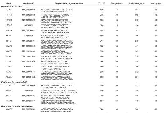
Gene GenBank ID Sequences of oligonucleotides Tann, ºC Elongation, s Product length, bp N of cycles
(A) Primers for RT-PCR
ODC NM_001086698 GCCATTGTGAAGACTCTCTCCATTC
TTCGGGTGATTCCTTGCCAC 60.0 15 221 40
HTR1A NM_001085830 TTATCTCCTTGGACGTGTTGTGTT
AGCGGGCTGCCTTGAATA 56.8 30 336 40
HTR2B NM_001089275 GAAATGCTGGCTGGCTCTGC
CTGCGGTGGCTTGTTCTTGATTA 56.0 45 616 40
HTR2C AJ549931 TTGCCAGTTATATCCGTTGTC
AGTCCCAGGTTGCATTTCTAT 50.4 15 194 40
HTR3A NM_001086077 ACGGCCAAATGTCCCTAACT
TGCCCAAACAATAATAAGAATA 50.8 30 391 40
HTR4 AY583535 CAGCCTCCATCCTTCATCTTTG
GGCATAGGGCTTGTTCACCAT 54.0 30 266 40
HTR7 NM_001085784 GACAAGCTCCCGCCTAAGAAGAAT
GTAGCCGAGCCACAGAAGAGTCCT 57.4 30 270 40
VMAT1 NM_001085945 ATCCATTTGGCTGCACATCTCATA
CCCACTCGCACATTCTCTTCATTT 55.2 30 441 40
VMAT2 NM_001086569 AGAGACGGCCTTTCCAGA
CACTCCAGCCCCTATACATTACAT 51.0 30 365 40
SERT XM_002932084 AACACTCCTAACTGCACCAACTAC
CACTCCTTTCCACAAACTGAAGTA 52.0 15 210 45
TPH1 NM_001087454 GGCCGGGCTGCTTTCTTCTA
ACCCCGAGCTGCTTGTTCATC 54.4 30 339 40
TPH2 CF547701 AATGTATTATCACCGACTTTCAG
TGTTTTAGATCACTCCTTA 46.0 15 205 45
AAAD NM_001110741 TCTTCGGACCAGGCACACTCAT
CTACCAGCATAAGCCGCATCAATA 55.0 30 272 40
MAOA NM_001094885 AACTGCCATCAATAAGGAACCA
CAGGAGGGAAATATGCAGTATAAC 53.0 30 384 40
(B) Primers for real-time PCR
ODC NM_001086698 GCCATTGTGAAGACTCTCTCCATTC
TTCGGGTGATTCCTTGCCAC 60.0 60 221 40
HTR2C AJ549931 AGAGCATTTGCCAGTTATATCCGTTGTC
CATTTCTATTGCTTCCTCTGTTTCATCCA 60.0 60 189 40
HTR7 NM_001085784 GGACCTGGTTGGGGGAGAATG
CGGGATACGTTAAAGGGCGAGTT 60.0 60 152 40
VMAT2 NM_001086569 ACGGAGTGCTACGAGGGGAAGGA
GGCAAAGGCAAACATGATAGTGGAGA 60.0 60 195 40
(C) Primers for in situ hybridization
VMAT2 NM_001086569 ACAGAGTATGGGAAAGGAAACACA
AACAAACTCATACATGGCAGAACC 54.8 60 546 40
TABLE 2
product (see Table 2). To exclude false-positive results, negative controls were included (PCR without reverse transcription and PCR without cDNA). The PCR products were analyzed by 1.5% agarose gel electrophoresis with ethidium bromide (0.5 mg/ml). Real-time PCR was performed using a
7500 Real-Time PCR System (Applied Biosystems) and EVA-Green/ROX reaction mixture (Syntol). The relative expression levels were calculated using the efficiency-corrected ΔΔCt method (Bookout et al., 2005). The
primers were designed by Lasergene PrimerSelect (DNASTAR) using sequences from the NCBI GeneBank databases (Table 2 A,B). The primer sequences for ODC that was used as endogenous control were obtained from Nandadasa et al., (2009).
In situ hybridization
The full-length coding sequence of HTR7 was amplificated using the primer sequences from Sheng et al., (2005). The PCR product (1290 bp) was cloned into the pGEM-T vector (Promega). A 3’ fragment length of 780 bp was then deleted using the SalI endonuclease, and the pGEM-HTR7-5’ vector was circularized using T4 ligase. The partial coding sequence of VMAT2 (546 bp) was amplified (see primers in Table 2 C). pGEM-VMAT2 was obtained by cloning this PCR product into T (Promega). pGEM-HTR7-5’ and pGEM-VMAT2 were used for synthesizing digoxygenin-labeled riboprobes. The hybridization was performed as previously described by Harland (1991), including negative control with sense riboprobe.
Sequencing
Sequencing was performed using the same primers as for PCR in both directions using the ABI PRIZM 3100 with the BigDye v1.1 kit (Applied Biosystems) and followed by the analysis and alignment of the sequences using the bioinformation software Lasergene (DNASTAR).
Acknowledgements
The authors are grateful to Dr. N.S.Mugue for his invaluable help in the organization of sequencing and to Dr. A.S.Mikaelyan for his advice on the molecular biology methods and generous support. This work was supported by the Russ. Fund for Basic Research grant 11-04-01469-а for D.N. and Y.S.
References
ALUIGI MG, DIASPRO A, RAMOINO P, RUSSO P, FALUGI C. (2012). The sea urchin, Paracentrotus lividus, as a model to investigate the onset of molecules immunologically related to the a-7 subunit of nicotinic receptors during embryonic
and larval development. Curr Drug Targets. 13: 587-593.
AMIREAULT P, DUBÉ F. (2005a). Serotonin and its antidepressant-sensitive transport in mouse cumulus-oocyte complexes and early embryos. Biol Reprod 73: 358-365. AMIREAULT P, DUBÉ F. (2005b). Intracellular cAMP and calcium signaling by
serotonin in mouse cumulus-oocyte complexes. Mol Pharmacol 68: 1678-1687. BAKER PC, QUAY WB. (1969). 5-hydroxytryptamine metabolism in early embryo-genesis, and the development of brain and retinal tissues. Brain Res 12: 272-295. BASU B, DESAI R, BALAJI J, CHAERKADY R, SRIRAM V, MAITI S, PANICKER
MM. (2008). Serotonin in pre-implantation mouse embryos is localized to the mi-tochondria and can modulate mimi-tochondrial potential. Reproduction 135: 657-669. BOOKOUT AL, CUMMINS CL, MANGELSDORF DJ. (2005). High-throughput real-time quantitative reverse transcription PCR. In Current protocols in molecular
biology (Eds. Ausubel, F.A., Brent, R., Kingston, R.E., Moore, D.D., Seidman,
J.G., Smith, J.A., and Struhl, K.) John Wiley & Sons, New York pp. 15.8.1-15.8.21. BOYD GW, LOW P, DUNLOP JI, ROBERTSON LA, VARDY A, LAMBERT JJ, PETERS JA, CONNOLLY CN. (2002) Assembly and cell surface expression of homomeric and heteromeric 5-HT3 receptors: the role of oligomerization and chaperone proteins. Mol Cell Neurosci 21: 38-50.
BUZNIKOV GA. (1967). Low-molecular regulators of embryonic development. Sci-ence, Moscow. (in Russian). 265 pp.
BUZNIKOV GA. (1990). Neurotransmitters in Embryogenesis. Harwood Academic Publ., Chur.
BUZNIKOV G.A. (2007). Preneural transmitters as regulators of embryogenesis. Current state of the problem. Russ. J. Dev. Biol 38: 213-220.
ČIKOŠ Š, VESELÁ J, IL’KOVA G, REHÁK P, CZIKKOVÁ S, KOPPEL J. (2005). Expression of beta adrenergic receptors in mouse oocytes and preimplantation embryos. Mol Reprod Dev 71: 145-153.
DEVIC E, PAQUEREAU L, STEINBERG R, CAPUT D, AUDIGIER Y. (1997). Early expression of a beta1-adrenergic receptor and catecholamines in Xenopus oocytes and embryos. FEBS Lett 417: 184-190.
FUKUMOTO T, BLAKELY R, LEVIN M. (2005a). Serotonin transporter function is an early step in left-right patterning in chick and frog embryos. Dev Neurosci 27: 349-363.
FUKUMOTO T, KEMA IP, LEVIN M. (2005b). Serotonin signaling is a very early step in patterning of the left-right axis in chick and frog embryos. Curr Biol 15: 794-803. HAMDAN FF, UNGRIN MD, ABRAMOVITZ M, RIBEIRO P. (1999). Characterization of a novel serotonin receptor from Caenorhabditis elegans: cloning and expression of two splice variants. J Neurochem 72: 1372-1383.
HARLAND RM.(1991). In situ hybridization: an improved whole-mount method for
Xenopus embryos. Methods Cell Biol 36: 685-695.
HEASMAN J. (2006). Patterning the early Xenopus embryo. Development 133: 1205-1217.
HINCKLEY M, VACCARI S, HORNER K, CHEN R, CONTI M. (2005). The G-protein-coupled receptors GPR3 and GPR12 are involved in cAMP signaling and main-tenance of meiotic arrest in rodent oocytes. Dev Biol 287: 249-261.
KIM MJ, HAN JK. (1999). The involvement of cAMP signaling pathway in axis speci-fication in Xenopus embryos. Mech Dev 89: 55-64.
LARSSON C. (2005). Protein kinase C and the regulation of the actin cytoskeleton.
Cell Signal 18: 276-284.
NANDADASA S, TAO Q, MENON NR, HEASMAN J, WYLIE C. (2009). N- and E-cadherins in Xenopus are specifically required in the neural and non-neural ectoderm, respectively, for F-actin assembly and morphogenetic movements.
Development 136: 1327-1338.
NIKISHIN DA, IVASHKIN EG, MIKAELYAN AS, SHMUKLER YB. (2009). Expression of serotonin receptors during early embryogenesis. Simpler Nervous Systems,
IX East European Conference of the International Society for Invertebrate Neu-robiology pp. 70 (Abstr).
NIKISHIN DA, SEMENOVA MN, SHMUKLER YB. (2012). Expression of transmitters receptors during early development of sea urchin Paracentrotus lividus. Rus
J.Dev Biol 43: 181-184
PEROUTKA SJ. (1997) 5-Hydroxytryptamine receptor subtypes. In Serotonin
Re-ceptors and their Ligands (Eds. B. Olivier, I. van Wijngaarden and W. Soudijn)
Elsevier Science B.V., pp. 3-13.
SCHUFF M, RÖSSNER A, DONOW C, KNÖCHEL W. (2006). Temporal and spatial expression patterns of FoxN genes in Xenopus laevis embryos. Int J Dev Biol 50: 429-434.
SHENG Y, WANG L, LIU XS, MONTPLAISIR V, TIBERI M, BALTZ JM, LIU XJ. (2005). A serotonin receptor antagonist induces oocyte maturation in both frogs and mice: evidence that the same G protein-coupled receptor is responsible for maintaining meiosis arrest in both species. J Cell Physiol 202: 777-786.
SHIH JC, CHEN K, RIDD MJ. (1999) Role of MAO A and B in neurotransmitter metabolism and behavior. Pol J Pharmacol 51: 25-29.
SHMUKLER YB, BUZNIKOV GA. (1998). Functional coupling of neurotransmitters with second messengers during cleavage divisions: facts and hypotheses. Perspect
Dev Neurobiol 5: 469-480.
VESELÁ J, REHÁK P, MIHALIK J, CZIKKOVÁ S, POKORNÝ J, KOPPEL J. (2003). Expression of serotonin receptors in mouse oocytes and preimplantation embryos.
Physiol Res 52: 223-228.
Increased cellular turnover in response to fluoxetine in neuronal precursors derived from human embryonic stem cells
Eun-Ah Chang, Zeki Beyhan, Myung-Sik Yoo, Kannika Siripattarapravat, Tak Ko, Keith J. Lookingland, Burra V. Madhukar and Jose B. Cibelli Int. J. Dev. Biol. (2010) 54: 707-715
5 yr ISI Impact Factor (2010) = 2.961
Spatio-temporal expression of a Netrin homolog in the sea urchin Hemicentrotus pul-cherrimus (HpNetrin) during serotonergic axon extension
Hideki Katow
Int. J. Dev. Biol. (2008) 52: 1077-1088
Serotonin involvement in the metamorphosis of the hydroid Eudendrium racemosum Giuliana Zega, Roberta Pennati, Arianna Fanzago and Fiorenza De Bernardi
Int. J. Dev. Biol. (2007) 51: 307-313
Action of serotonin antagonists on cytoplasmic calcium levels in early embryos of sea urchin Lytechinus pictus
Y B Shmukler, G A Buznikov and M J Whitaker Int. J. Dev. Biol. (1999) 43: 179-182
Activation of an 85 kDa ribosomal S6 kinase during serotonin-induced oocyte maturation Y Durocher and P Guerrier