Modulation of mitochondrial biogenesis and bioenergetic
metabolism upon in vitro and in vivo differentiation of
human ES and iPS cells
ALESSANDRO PRIGIONE and JAMES ADJAYE*
Department of Vertebrate Genomics, Molecular Embryology and Aging Group, Max Planck Institute for Molecular Genetics, Berlin, Germany
ABSTRACT Reprogramming somatic cells to induced pluripotent stem (iPS) cells transforms differentiated cells to an embryonic stem (ES) cell-like state characterized by the acquisition of pluripotency and self-renewal capabilities. We recently demonstrated that human ES and iPS cells share similar mitochondrial properties and bioenergetic metabolism, which are distinct from those of fibroblasts. In the present study, we have applied a global transcriptome profiling approach to compare the mitochondrial-related transcriptional signature upon the loss of self renewal and pluripotency in human ES and iPS cells. This was achieved by inducing in vitro and in vivo spontaneous differentiation. ES and iPS cells showed a similar degree of correlation both in the undifferentiated state and in all the stages of differentiation analyzed, suggesting that their transcriptional similarities are retained upon differentiation. Moreover, comparable induction of transcripts involved in epithelial to mesenchymal transition was observed in both cell types. Analysis of mitochondrial-related nuclear transcripts revealed consensual regulation of genes involved in mitochondrial biogenesis and bioenergetic metabolism upon in vitro differentiation of human ES and iPS cells, while specific differences were identified within in vivo differentiated cells. Significant changes were not detected for antioxidant-related genes. Finally, we formulate a "metabolic state hypothesis" linking mitochondrial state and cellular metabolism to the stage of differentiation. Overall, our data unveil differences and similarities between human ES and iPS cells during spontaneous differentiation and suggest that the study of mitochondrial and metabolic remodeling may reveal key mechanisms underlying the acquisition, maintenance and exit of a self-renewing pluripotent state.
KEY WORDS:
iPS cells, reprogramming, differentiation, mitochondria, metabolism
Introduction
Somatic cells have been found able to acquire a self-renewing pluripotent state upon the ectopic expression of a set of genes commonly present in embryonic stem (ES) cells (Takahashi et al., 2007; Yu et al., 2007; Park et al., 2008; Singh et al., 2009). The somatic cell-derived induced pluripotent stem (iPS) cells share multiple features with ES cells, including cellular morphology and cell cycle structure, transcriptional and epigenetic signatures, unlimited propagation without senescence and most importantly developmental potential. In addition, we recently demonstrated that human ES and iPS cells are characterized by similar
bioen-BIOLOGY
www.intjdevbiol.com*Address correspondence to: James Adjaye. Ihnestrasse 73, D-14195 Berlin, Germany. Fax: +49-30-8413-1128. e-mail: adjaye@molgen.mpg.de web: http://www.molgen.mpg.de/~molemb/
Supplementary Material (3 figures + 1 table) for this paper is available at: http://dx.doi.org/10.1387/ijdb.103198ap Final author corrected PDF published online: 23 December 2010.
ISSN: Online 1696-3547, Print 0214-6282
© 2010 UBC Press Printed in Spain
Abbreviations used in this paper: EB, embryoid body; EMT, epithelial-mesenchymal transition; ES, embryonic stem; ETC, electron transport chain; iPS, induced pluripotent stem; OXPHOS, oxidative phosphorylation; ROS, reactive oxygen species.
ergetic metabolic profiles, redox status and mitochondrial func-tion (Prigione et al. 2010).
major source of endogenous reactive oxygen species (ROS), common by-products of oxidative phosphorylation (OXPHOS), which can in turn generate oxidative damage to DNA, proteins, and lipids (Balaban et al., 2005). Moreover, mitochondria possess their own DNA, which encodes 13 ETC peptides. Due to the presence of ROS and the lack of efficient repairing mechanisms, mutations within the mitochondrial DNA (mtDNA) can accumulate over time (Cortopassi et al., 1992; Wallace, 1994) causing multiple cellular dysfunctions (Alemi et al., 2007; Prigione et al., 2007).
Approximately 1500 mitochondrial-related genes reside within the nuclear genome, including key regulators of mitochondrial replication (Kelly et al., 2004; Scarpulla, 2008). Transcription of mtDNA requires a small number of nucleus-encodedproteins comprising the mitochondrial-specific DNA polymerase gamma, which is composed of one catalytic subunit (POLG) and two accessory subunits (POLG2), and the mitochondrial transcription factor A (TFAM) (Clayton, 1998). Additional regulating elements include the transcription factor NRF-1 and members of the PGC-1family (PGC1-a and PGC1-b). In particular, PGC1a appears as
a master regulator of mitochondrial biogenesis due to its ability to activate of TFAM through direct interaction with NRF1 (Goffart et al., 2003).
Under physiological normoxic conditions, human somatic cells are characterized by active mitochondria and aerobic ETC-based metabolism. On the other hand, solid tumor cells shift to glycolysis-based metabolism as a result of inefficient vascolarization, an event known since the early 1930s under the name of “Warburg effect”(Warburg, 1956). Glycolysis might be advantageous in comparison to mitochondrial respiration. Indeed, it can provide quick energy supplies and avoids toxic ROS generation. In addi-tion, by promoting the flux through the pentose phosphate path-way, it can support antioxidant defenses by inducing the production of NADPH, which is required for the generation of reduced glu-tathione (GSH), a key antioxidant enzyme (Pfeiffer et al., 2001; Hsu
et al., 2008). Early-stage mammalian embryos and in vivo stem cell niches are also exposed to extremely low oxygen conditions (Fischer et al., 1993). Thus, hypoxic environment and anaerobic metabolism appear to favorably support self-renewal and pluripo-tency. Accordingly, hypoxic culture and mitochondrial inhibition have been associated with reduced ES cell differentiation and more recently with improved generation of iPS cells (Ezashi et al., 2005; Varum et al., 2009; Yoshida et al., 2009).
For these reasons, mitochondrial structure and function have been sug-gested as indicators of stem cell com-petence (Chen et al.; Lonergan et al., 2006; Siggins et al., 2008; Parker et al., 2009). In particular, relative un-der-developed mitochondrial network and low mitochondrial activity emerge as common features of ´stemness‘, as reported in primordial germ cells, inner cell mass, early embryos, and murine and human ES cells (Lonergan et al., 2007; Shoubridge
et al., 2007; Facucho-Oliveira et al., 2009; Ramalho-Santos et al., 2009; Van Blerkom, 2009). Recent data suggested that the self-renewal state of human iPS cells may also be sup-ported by glycolysis metabolism (Prigione et al. 2010) and by mito-chondrial properties similar to those of ES cells, including low mtDNA copy number, immature organelle shape with under-developed cristae, and low levels of oxidative stress (Armstrong et al. 2010; Prigione et al.
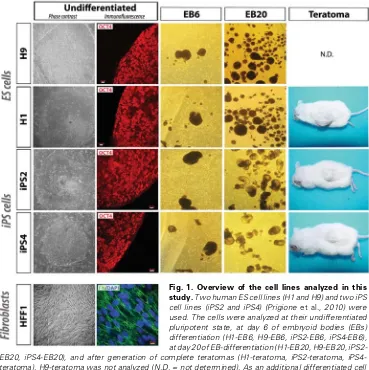
2010). Hence, it appears that so-matic cells undergo a metabolic shift in order to acquire ES cell-like fea-tures. On the other hand, functionally active mitochondria are necessary for successful differentiation of ES cells, which requires an opposite Fig. 1. Overview of the cell lines analyzed in this
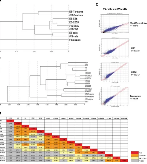
Fig. 2. Transcriptional profiling analysis. (A) Hierarchical clustering of the samples; different cell lines were pooled together to provide an overview of the global correlations between undifferentiated cells and differentiated cells at various stages. (B) Hierarchical clustering of all the samples used in the study. The two ES cell lines and the two iPS cell lines cluster together in all the different cell states (undifferentiation state, EB6, EB20, and teratomas). (C) Scatter plot graphs showing the degree of correlation between ES and iPS cells at distinct stages of differentiation. Blue dots highlight genes whose detection p-value is lower than 0.01. (D) Table showing all the correlation coefficient values between the single samples analyzed. For color coding, four distinct degrees of correlation are represented (red for r=1; orange for 1<r<0.9; yellow for 0.9<r<0.85; and grey for r<0.85).
B
C
switch in energy metabolism from anaerobic glycolysis to aerobic OXPHOS (Cho et al., 2006; Facucho-Oliveira et al., 2007).
In the present study, we aimed at comparing mitochondrial-related transcriptional signatures of human ES and iPS cells during
in vitro and in vivo differentiation. Modulation of these pathways may be essential in the regulation of cellular bioenergetic metabo-lism and might thus reveal important mechanisms for acquiring, maintaining and exiting a self-renewing pluripotent state in human cells.
Results
Transcriptional similarities between human ES and iPS cells are retained upon differentiation
In this study, we used two human ES cell lines (H1 and H9) and two iPS cell lines (iPS2 and iPS4), previously derived by retroviral transduction of OCT4, KLF4, SOX2 and c-MYC (Takahashi et al., 2007) into neonatal foreskin fibroblasts (HFF1) (Prigione et al.
2010). The cells were analyzed at their undifferentiated pluripotent
state and at different stages of differentiation. For in vitro differen-tiation, we employed embryoid body (EB)-based differentiation and chose two time-points for mRNA isolation (EB6 at day 6 and EB20 at day 20). The choice of EB stages was based on our previously published work on EB-based differentiation of human ES cells (Fathi et al., 2009). For in vivo differentiation, dissected teratomas derived from the two iPS cell lines and from one ES cell line (H1) were employed. In addition, we included in the analysis the parental HFF1 fibroblasts from which the two iPS cell lines (iPS2 and iPS4) were derived. An overview of the cells utilized for the transcriptome profiling is shown in Fig. 1.
Global hierarchical clustering of the samples revealed that ES and iPS cell lines share similar transcriptomes, both in the undiffer-entiated state and in all stages of differentiation analysed (Fig. 2 A,B). ES and iPS cells showed a similar degree of correlation in the undifferentiated state (r=0.9045) and in different stages of differ-entiated cell states (EB6 r=0.8778, EB20 r=0.9312, and terato-mas r=0.8216) (Fig. 2C). On the other hand, undifferentiated ES and iPS cells poorly correlated with somatic HFF1 fibroblasts
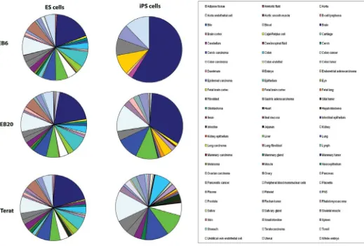
Fig. 3. Increased complexity of tissue-association patterns upon differentiation of ES and iPS cells. For each differentiation stage of ES and iPS cells, a gene list was generated by comparing significantly up-regulated genes (fold change > 1.5, detection p value 0.01, and differential p value
(correlation coefficient r around 6-7) as we and others have previously shown (Takahashi et al., 2007; Prigione et al. 2010) (Fig. 2D). Overall, the results suggested that human ES and iPS cells retain their transcriptional similarities even upon differentia-tion analyzed in vivo and in vitro.
Increased degree of complexity of tissue expression commit-ment upon differentiation of human ES and iPS cells
The ability of human ES cells to mimic the embryonic process of
germ layer formation in vitro can be utilized as a tool to understand the mechanisms underlying human embryogenesis at the mo-lecular and cellular level (Nishikawa et al., 2007). Indeed, previ-ous transcriptome-based analysis employed in vitro differentiated human ES cells to dissect the complex molecular signature of early embryogenesis (Ivanova et al., 2002; Bhattacharya et al., 2005; Fathi et al., 2009).
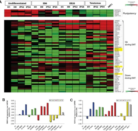
ES and iPS cells displayed a consensual up-regulation of key markers of germ layer commitment during spontaneous differen-Fig. 4. Activation of epithelial-mesenchymal transition (EMT)-related transcripts upon differentiation. (A) Upper panel, heatmap depicting the regulation of expression of genes related to pluripotency upon differentiation. Lower panel, modulation of Epithelial to Mesenchymal Transition (EMT)-related genes during spontaneous differentiation. EMT transcripts were divided into two categories “Up in EMT” and “Down in EMT”, according to the literature (Nakaya et al., 2008) and to the SA bioscience PRC array list (see material and methods for details). Values represent the ratio of the array average signal for the given gene divided by the average signal of H1 ES cell line (fold change 1.5, detection p value 0.01, and differential p value 0.01). Down-regulated genes are shown in green, up-regulated genes in red. Highlighted in yellow are four key EMT genes: SNAI1, SLUG, VIM,
and CDH1. The first three were induced in differentiated cells in comparison to undifferentiated cells, while the opposite was found for the epithelial marker CDH1. (B,C) Q-PCR analysis of SNAIL1 (SNAI1) and SNAIL 2 (SLUG), two driver transcription factors of the EMT response, performed in all analyzed samples confirming the array data.
B
C
B
A
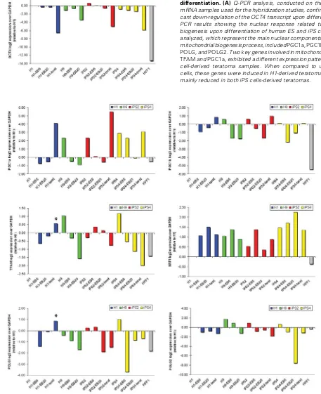
Fig. 5. Mitochondrial biogenesis response during in vitro and in vivodifferentiation. (A) Q-PCR analysis, conducted on the same original mRNA samples used for the hybridization studies, confirmed the signifi-cant down-regulation of the OCT4 transcript upon differentiation. (B) Q-PCR results showing the nuclear response related to mitochondrial biogenesis upon differentiation of human ES and iPS cells. The genes analyzed, which represent the main nuclear components involved in the mitochondrial biogenesis process, included PGC1a, PGC1b, TFAM, NRF1, POLG, and POLG2. Two key genes involved in mitochondrial biogenesis,
TFAM and PGC1a, exhibited a different expression pattern in ES and iPS cell-derived teratoma samples. When compared to undifferentiated cells, these genes were induced in H1-derived teratoma (asterisks) and mainly reduced in both iPS cells-derived teratomas.
tiation (Supp. Fig. 1). Moreover, by assigning tissue-specific annotations to genes up-regulated at different differentiation stages in comparison to undifferentiated cells, we observed that the complexity of tissue generation increased over time in differ-entiated ES and iPS cells (Fig. 3). The number of tissue-related transcripts rose significantly upon differentiation in a similar
fashion in ES and iPS cells, including genes associated with adipose tissue, colon, fibroblasts, kidney, liver, lung, muscle, pancreas, plasma, platelets, small intestine, and spleen. The tissue categories called embryo and whole embryo, which com-prise genes related to gastrulation and development (such as
PLXNA3, TAPT1, TSPAN9, and VIM), showed a similar induction in both ES and iPS cells. Interestingly, however, brain-related transcripts exhibited a distinct expression pattern in the two pluripotent stem cells. In ES cells, the number of brain-related genes gradually increased upon differentiation while it ap-peared reduced over time in iPS cells (Fig. 3). These results may reflect distinct properties of the specific iPS cell lines used in this study and further analysis conducted on several iPS cell lines will be needed to substantiate this observation.
drivers such as SNAIL1 (SNAI1), SNAIL 2 (SLUG) and VIMENTIN (VIM) was induced in differentiated cells in compari-son to undifferentiated cells, while the epithelial markers E-CADHERIN (CDH1), DSP and KRT19 were down-regulated. Q-PCR analysis of SNAI1 and SLUG confirmed that two factors, known to repress transcription of the epithelial specifying gene
CDH1 (Ullmann et al., 2007; Thiery et al., 2009), were gradually activated upon differentiation in both human ES and iPS cells (Fig. 4 B,C).
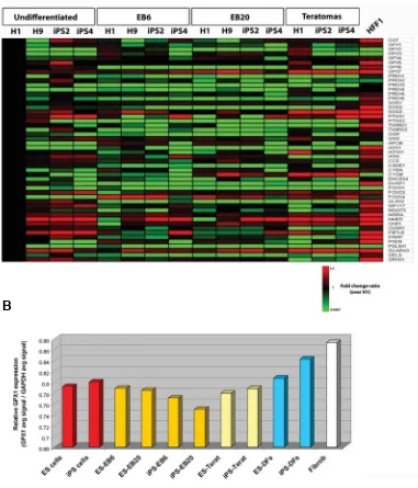
Fig. 6. Expression analysis of genes involved in the response to oxidative stress. (A) Heatmap figure showing a panel of gene transcripts related to oxidative stress and antioxidant defense. Values represent the ratio of the array average signal of the given gene divided by the average signal of H1 ES cell line (fold change 1.5, p value 0.01, and differential p value 0.01). Down-regulated genes are shown in green, up-regulated genes in red. (B) GPX1 gene expression measured as the ratio of GPX1 average array signal over GAPDH average array signal. The expression values within the analyzed samples were compared to the values previously detected by microarray in derived fibroblasts (DFs) obtained from ES and iPS cells (Prigione
et al., 2010).
Comparable induction of EMT-related transcripts during spon-taneous differentiation of hu-man ES and iPS cells
During the developmental pro-cess of gastrulation, a critical role is played by epithelial to mesen-chymal transition (EMT) (Nakaya
et al., 2008). By analyzing numer-ous published transcriptome datasets related to human ES and iPS cells, we previously observed that genes involved in the pro-gression of EMT were mainly down-regulated in undifferenti-ated cells compared to somatic fibroblasts (Wang et al., 2010). Inhibition of EMT and activation of its reciprocal process MET, likely occurring through the modu-lation of the TGF- pathway, might thus represent a key aspect for efficient reprogramming to pluri-potency of cells of mesenchymal phenotype, such as somatic fi-broblasts.
To further validate this obser-vation, we analyzed the transcrip-tional regulation of EMT-related genes during differentiation of hu-man ES and iPS cells. Upon dif-ferentiation, pluripotency-associ-ated genes were significantly down-regulated, while EMT-re-lated genes showed a trend of up-regulation (Fig. 4A). In HFF1 fi-broblasts, EMT genes were strongly up-regulated in compari-son to undifferentiated ES and iPS cells. Gradual activation of EMT-related genes could be ob-served in spontaneously differen-tiated ES and iPS cells. In particu-lar, the induction of EMT genes was detected in the latest in vitro
differentiation stage (EB20) and even more evidently within the teratoma samples (Fig. 4A). Over-all, the expression of key EMT
Modulation of mitochondrial biogenesis upon spontaneous differentiation of human ES and iPS cells
In order to compare the mitochondrial-related signatures upon differentiation of human ES and iPS cells, we analyzed by Q-PCR the expression of key genes involved in mitochondrial biogenesis (Fig. 5). The Q-PCR data showed consensual down-regulation of OCT4 expression during differentiation of ES and iPS cells, which was in accordance with the array analysis (Fig. 4A and 5A). In agreement with previous reports (Armstrong et al. 2010; Prigione
et al., 2010), undifferentiated human ES and iPS cells presented similar expression levels for most of the genes involved in mito-chondrial biogenesis (PGC1a, PGC1b, TFAM, NRF1, POLG, and
POLG2), which appeared up-regulated in both ES and iPS cells in comparison to somatic HFF1 fibroblasts (Fig. 5B).
During spontaneous in vitro differentiation of ES and iPS cells, we detected a consensual down-regulation of nuclear genes responsible for regulating mitochondrial biogenesis, as earlier demonstrated in human ES cells [27]. At the EB6 stage, the expression of mitochondrial biogenesis genes (with the exception of NRF1) showed decreased gradually in all four pluripotent stem cells (Fig. 5B). At the latest stage of in vitro differentiation (EB20), both ES and iPS cell-derived EBs showed consistent down-regulation of genes related to mitochondrial biogenesis in com-parison to undifferentiated H1, still with the exception of NRF1
(Fig. 5B).
On the other hand, in fully-differentiated teratoma samples some mitochondrial biogenesis factors appeared up-regulated compared to undifferentiated cells. These included PGC1a and
PGC1b, TFAM and POLG. Most of the genes were consensually expressed in ES and iPS cell-derived teratomas. When compared to undifferentiated stem cells, PGC1a and PGC1b were up-regulated, NRF1 was unchanged, and POLG2 was down-regu-lated (Fig. 5B). However, although ES and iPS cell-derived teratomas showed quite similar global transcriptional signatures (Fig. 2), two key genes involved in mitochondrial biogenesis,
TFAM and PGC1a, were differentially expressed in ES and iPS
cell-derived teratoma samples. Their expression level was in-duced in H1-derived teratoma compared to undifferentiated cells (Fig. 5B, asterisks); while it was mainly decreased in teratomas obtained fromthe two iPS cell lines.
Transcriptional regulation of bioenergetic metabolism upon spontaneous differentiation of ES and iPS cells
There is a paucity of data available on the regulation of genes involved in bioenergetic metabolism during stem cell differentia-tion. Employing global transcriptome analysis, we analyzed nuclear-encoded genes involved in mitochondrial energy me-tabolism (ETC genes) and compared their expression within cells at different stages of differentiation in relation to undifferentiated H1 cells.
ETC genes were mainly similarly expressed in both ES and iPS cells compared to somatic fibroblasts (Supp. Fig. 2), in accor-dance to our previous results (Prigione et al., 2010). Interestingly however, one iPS cell line (iPS2) showed higher expression of some of these genes than the other undifferentiated cell lines (Supp. Fig. 2), possibly due to different patterns of viral integration within different iPS cell lines.
Within the EB6 and EB20 stages, both ES and iPS cell-derived samples showed down-regulation of ETC genes compared to undifferentiated cells -although less evident in the case of Com-plex III- suggesting a possible occurrence of a metabolic shift at these differentiation stages (Supp. Fig. 2). The expression of ETC-related genes was not identical within in vivo differentiated ES and iPS cells, in a similar fashion to transcripts involved in mitochondrial biogenesis (Fig. 5 and Supp. Fig. 2). ETC tran-scripts within teratoma samples show a trend of up-regulation compared to undifferentiated cells to a level more similar to somatic fibroblast. However, the up-regulation of transcripts re-lated to Complex I, III, IV, and V was more evident in H1-teratoma rather than in iPS2 or iPS4-teratomas (Supp. Fig. 2). This may imply further differences related to mitochondrial activity between the two pluripotent cell types at this stage of differentiation,
despite the detected global transcriptional similarities.
Lack of transcriptional modulation of antioxidant genes within spontaneously differentiated ES and iPS cells
Finally, we aimed to analyze the expression level of genes related to the response to oxidative stress in undifferentiated and spontaneously differentiated stem cells (Fig. 6). We earlier de-tected a different redox status between undifferentiated and fully-differentiated fibroblast cells (Prigione et al., 2010). In accor-dance, HFF1 somatic fibroblasts presented a consistent up-regulation of most of antioxidant genes in comparison to undiffer-entiated stem cells (Fig. 6A). However, these genes did not show transcriptional induction within in vitro and in vivo differentiated ES and iPS cells. A mild up-regulation could be identified only in teratoma samples in comparison to undifferentiated stem cells; however, the expression level was not as high as in HFF1 fibroblasts (Fig. 6A).
To address whether this lack of transcriptional change of antioxidant genes upon differentiation was specific to spontane-ously differentiated EBs and teratomas, we compared the array expression level of GPX1, a key antioxidant gene, within cells at all different stages of differentiation and in derived fibroblasts (DFs), fibroblast-like cells previously obtained from ES cells (H1 and H9) and iPS cells (iPS2 and iPS4) (Prigione et al., 2010).
GPX1 expression was significantly increased in somatic fibro-blasts and in iPS-DFs compared to undifferentiated cells, in accordance with our previous immunoblot data (Prigione et al., 2010), but mostly unchanged in EBs and teratomas (Fig. 6B). Accordingly, DFs clustered together with somatic HFF1 fibro-blasts and far more distant from ES and iPS cells in either undifferentiated or spontaneously differentiated states (Supp. Fig. 3A). The correlation values between undifferentiated cells and DFs (r=0.7216 for ES cells and r=0.6646 for iPS cells) were similar to those between undifferentiated cells and somatic fibro-blasts (r=0.6979 for ES cells and r=0.6782 for iPS cells) (Fig. 2D and Supp. Fig. 3B).
Overall, the results could imply that differentiation towards a specific lineage may generate transcriptional changes in the response to oxidative stress in a fashion more similar to the representative somatic tissue. On the other hand, the EB and teratoma-based spontaneous differentiation models might be masking the specific changes occurring within cells due to the simultaneous generation of distinct cell types representing the three germ layers. Future studies would have to confirm these findings in a larger number of samples and determine whether spontaneously differentiated cells might exhibit a different redox status compared to cells committed to a lineage-specific differen-tiated state.
Discussion
Human iPS cells have enormous potential for regenerative medicine, both as an isogenic source for cellular replacement therapy and as an in vitro tool for studying complex human disorders and discovering new drug approaches (Saha et al., 2009; Deng 2010; Kiskinis et al. 2010). The capability to achieve these invaluable goals is linked to the ability of iPS cells to efficiently differentiate into relevant functional cells. Nonetheless, recent findings suggest that differentiated cells derived from
human iPS cells might show premature senescence and reduced efficiency and increased variability in comparison to those ob-tained from ES cells (Feng et al. 2010; Hu et al. 2010). This raised concerns over the real potential of the somatic-derived repro-grammed cells. Thus, in depth comparisons of the differentiation competence of human ES and iPS cells are needed to help determine the extent of these possibly relevant differences.
By analyzing the global transcriptional changes accompanying the spontaneous differentiation of human ES and iPS cells, we observed that the similarities between the two types of pluripotent cells are retained upon differentiation suggesting that ES and iPS cells might share analogous molecular mechanisms both for the maintenance of the self-renewal state and for the exiting from pluripotency due to spontaneous differentiation. Interestingly, the number of tissue-associated genes increased over time in both spontaneously differentiated ES and iPS cells but attained a different degree of complexity in the two pluripotent stem cells. These results might imply a distinct potential in embryonic and somatic cell-derived stem cells. Future studies conducted on several iPS cell lines are warranted to investigate whether this is a trait common to all iPS cells or restricted to specific iPS cell lines obtained from a definite tissue.
Spontaneous stem cell differentiation can mimic gastrulation and have potential applications for the study of early embryogen-esis at the molecular level (Ivanova et al., 2002; Bhattacharya et al., 2005; Fathi et al., 2009). In particular, a critical component of these developmental processes is considered to be the epithelial to mesenchymal transition (EMT) (Nakaya et al., 2008). In accor-dance, differentiated cells derived from both human ES and iPS cells showed transcriptional changes consensual with an EMT response. These data could also be of specific interest in the context of cellular reprogramming, as a reciprocal process (mes-enchymal to epithelial transition, MET) has been detected upon full reprogramming of human fibroblasts to iPS cells, whereas partially reprogrammed cells were characterized by incomplete MET (Wang et al., 2010). Thus, manipulation of the EMT-MET responses may prove to be useful for enhancing or opposing the two inverse processes of reprogramming to pluripotency (espe-cially for mesenchymal-derived cells, such as fibroblasts) and differentiation. Interestingly, during the course of revision of this manuscript, new findings demonstrated that MET is indeed play-ing a critical role in initiatplay-ing cellular reprogrammplay-ing events in fibroblast cells (Li et al. 2010; Samavarchi-Tehrani et al. 2010).
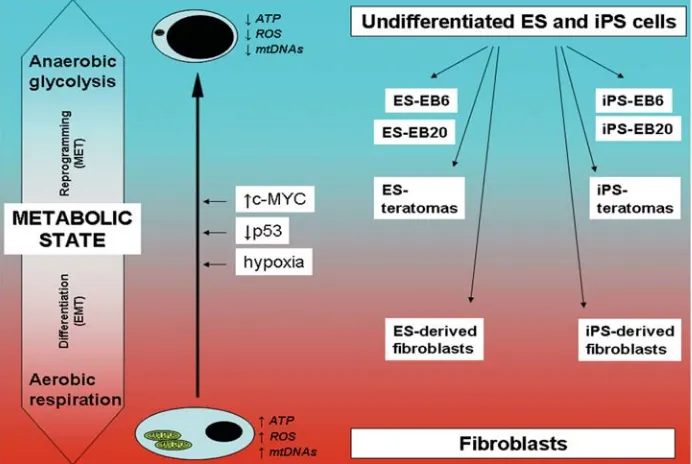
Applying the "metabolic state hypothesis" to reprogram-ming, self-renewal and differentiation
high rate of glucose uptake. On the other hand, an opposite switch from anaerobic to aerobic metabolism can occur when undifferen-tiated cells exit self-renewal and start differentiating (Chen et al.
2010).
In order to better understand how this latter transition may be regulated at the molecular level, we focused on the nuclear response of genes involved in mitochondrial biogenesis, bioener-getic metabolism and oxidative stress in both human ES and iPS cells. Based on our previously published findings (Prigione et al., 2010) and the data reported here, we have formulated a possible hypothesis linking the cellular metabolic state to the degree of differentiation. The hypothesis, named ´metabolic state hypoth-esis‘, is schematically represented in Fig. 7. At the two extremes of the metabolic arrows are undifferentiated ES cells/ iPS cells and somatic fibroblasts, respectively.
Nuclear-encoded genes involved in mitochondrial biogenesis were mainly up-regulated in undifferentiated ES and iPS cells compared to somatic fibroblasts. This may represent a nuclear response to decreased content of mitochondrial DNA (mtDNA) (Lloyd et al., 2006), which was found reduced in undifferentiated human ES and iPS cells (Armstrong et al. 2010; Prigione et al., 2010). Indeed, cells depleted of mtDNA have been shown to induce the expression of mitochondrial biogenesis factors (Holmuhamedov et al., 2003) and POLG expression was up-regulated in cells harboring large deletions of mtDNA (Alemi et al., 2007). Although one iPS cell line showed mild up-regulation of some ETC genes compared to somatic fibroblasts, most of the nuclear encoded ETC components were similarly expressed in both undifferentiated pluripotent cells and somatic fibroblasts, suggesting that the metabolic shift occurring upon reprogram-ming may not be strictly dependent on transcriptional regulation of ETC.
A similar effect has been described for cancer cells, in which the reduced dependence on mitochondrial respiration is not generally due to defects in OXPHOS or altered expression of ETC components, but rather to upstream mechanisms involving fac-tors associated with oncogenesis able to promote glycolysis such as c-MYC, the hypoxia-related factor HIF1a, and p53 (Vousden et al., 2009). Changes in bioenergetics may then induce modifica-tions of mtDNA copy number and modulate the expression of genes involved in mitochondrial biogenesis (Leary et al., 1998; Lebedeva et al., 2009).
Some of these factors and associated mechanisms may also be in place during cellular reprogramming. c-MYC, whose up-regulation has been associated with elevated lactate dehydroge-nase-a expression and glycolytic activity (Shim et al., 1997) and with induction of gene expression programs favoring mitochon-drial biogenesis (Li et al., 2005), is one of the four factors comprising the Yamanaka reprogramming cocktail (Takahashi et al., 2007). Hypoxia-related pathways, which have been found to change metabolic homeostasis by inducing glycolysis (Carmeliet
et al., 1998; Silvan et al., 2009), may also play a role during cellular reprogramming, as hypoxic conditions have been recently dem-onstrated to enhance iPS cell generation (Yoshida et al., 2009). Finally, p53 can modulate cellular metabolism by reducing the flux through the glycolytic pathway and inducing OXPHOS (Hu et al.; Bensaad et al., 2006; Matoba et al., 2006). Interestingly, p53 activation leads to rapid ES cell differentiation (Maimets et al., 2008), while its abrogation can improve the derivation of human
and mouse iPS cells (Hong et al., 2009; Marion et al., 2009; Utikal
et al., 2009). Moreover, reduced mtDNA levels, which have been detected within undifferentiated human ES and iPS cells (Armstrong et al. 2010; Prigione et al. 2010), have been linked to loss of p53 (Lebedeva et al., 2009). Hence, p53 might also represent a barrier for cellular reprogramming due to its meta-bolic-related function of opposing the “Warburg effect”.
Overall, although the ectopic introduction of c-MYC, hypoxia and p53 inhibition might not be essential for reprogramming, as functional iPS cells can be derived even in their absence, they all appear to play a critical role in enhancing the process (Nakagawa
et al., 2008; Hong et al., 2009; Marion et al., 2009; Utikal et al., 2009; Yoshida et al., 2009). Thus, it is temping to speculate that the same mechanisms may be already in place during cellular reprogramming and modulation of the common downstream metabolic pathways might represent an important step for effi-cient generation of iPS cells (Fig. 7).
During spontaneous in vitro differentiation of ES and iPS cells, we detected a consensual down-regulation of nuclear genes responsible for regulating mitochondrial biogenesis and mito-chondrial bioenergetic metabolism (ETC genes). Although rela-tive little data exist on the regulation of genes involved in bioen-ergetic metabolism during stem cell differentiation, earlier works on mitochondrial biogenesis in human ES cells, showed a similar down-regulation of these genes during differentiation (St John et al., 2005; Cho et al., 2006; Armstrong et al. 2010; Prigione et al., 2010). However, the same genes have been found up-regulated upon differentiation of human mesenchymal stem cells (Chen et al., 2008) and mouse ES cells (Facucho-Oliveira et al., 2007). The most likely explanation of these conflicting results may be that the nuclear response is depending on the mtDNA content (Mercy et al., 2005; Lloyd et al., 2006) which may vary within different cell types or different time points. Interestingly, within teratoma samples, some genes involved in both mitochondrial biogenesis and bioenergetic metabolism were up-regulated compared to undifferentiated cells, suggesting a different mitochondrial re-sponse during in vitro and in vivo spontaneous differentiation of human pluripotent cells, possibly due to the tumorigenic nature of teratomas. Moreover, specific differences could be detected between teratoma samples derived from ES and iPS cells, de-spite a high level of global transcriptional similarities. These results might imply a potential distinct mode of mitochondrial regulation between in vivo differentiated human ES and iPS cells and may possibly reflect random viral integration effects occurring within viral-derived iPS cells. Alternatively, they could reflect specific properties of the original somatic cell lines.
addressing the mitochondrial-related changes upon differentia-tion should focus on cell-type specific differentiadifferentia-tion protocols in order to better dissect the modulation of mitochondrial function in the context of a selected cell type.
Summary
We have reported the mitochondrial-related transcriptional signatures of spontaneously differentiated human ES and iPS cells and suggested that mitochondrial and metabolic remodeling may play a key role during the acquisition of pluripotency upon cellular reprogramming and loss of pluripotency due to differen-tiation.
Interestingly, malignant transformation and cellular reprogram-ming might share a similar metabolic shift. Since proposed anti-cancer agents include glycolytic inhibitors (Pelicano et al., 2006), future studies are warranted to distinguish the mitochondrial and metabolic profiles of stem cells to that of transformed cells (Chen
et al. 2010; Yanes et al. 2010) in order to prevent a possible elimination of stem cells by treatments aimed against tumorigenic cells.
In conclusion, understanding the mechanisms underlying the modulation of mitochondrial and bioenergetic metabolism may eventually shed some light on the processes of acquisition, maintenance and exit of a self-renewal state in pluripotent human cells.
Materials and Methods
Cell lines and culture conditions
Human ES cell lines (H1 and H9) were obtained from the WiCell Institute (Madison, WI, USA). iPS cell lines iPS2 and iPS4 were generated by retroviral transduction of the Yamanka cocktail (OCT4, KLF4, SOX2 and c-MYC) from neonatal foreskin fibroblasts HFF1 (ATCC, #1041, Manassa, VA, USA) and fully characterized (Prigione et al., 2010). ES and iPS cells were cultured in human ES media containing knockout DMEM supplemented with 20% knockout serum replacement (SR), nonessential amino acids, L-glutamine, penicillin/streptomycin, sodium pyruvate, 0.1mM
-mercaptoethanol (all from Invitrogen, Carlsbad, CA), and 8 ng/ml bFGF (Prepotech, Rocky Hill, NJ). Cultures were maintained on mitomycin-inactivated mouse embryonic fibroblast (MEFs) and manually passaged using a cut-and-paste technique (Babaie et al., 2007). Prior to RNA isolation, cells were grown under feeder-free conditions on Matrigel (BD Bioscience, San Diego) - coated dishes as previously described (Greber
et al., 2007).
In vitro and in vivo differentiation
For in vitro differentiation, embryoid bodies (EBs) were generated as previously described (Prigione et al., 2010). Briefly, ES and iPS cells were seeded onto low-attachment dishes in differentiating medium (human ES medium without bFGF supplementation). Two time-points (day 6 and day 20) were chosen on the basis of previous results (Fathi et al., 2009) and EBs were harvested for RNA isolation. In vivo differentiation experiments were performed by EPO-Berlin Gmbh using NOD scid gamma mice as previously described (Prigione et al., 2010). Teratomas obtained from ES and iPS collected 50-70 days after injection, dissected and fast-frozen for RNA isolation.
Microarray-based global gene expression analysis
Total messenger RNA (mRNA) was isolated using the RNeasy mini kit (Qiagen, USA) and quality-checked by Nanodrop analysis (Nanodrop Technologies, Wilmington, DE, USA). 400ng of mRNA was used as input
for generating biotin-labelled cRNA (Ambion, Austin, TX, United States). cRNA samples were then hybridized onto Illumina human-8 BeadChips version 3. Hybridizations, washing, Cy3-streptavidin staining and scan-ning were performed on the Illumina BeadStation 500 platform (Illumina, San Diego, CA, USA), according to the manufacturer’s instruction. The following samples were hybridized in duplicate: HFF1, H1, H9, iPS2, iPS4, H1-EB6, H1-EB20, H9-EB6, H9-EB20, iPS2-EB6, iPS2-EB20, iPS4-EB6, iPS4-EB20, H1-teratoma, iPS2-teratoma, iPS4-teratoma. Expression data analysis was carried out using the BeadStudio software 3.0 (Illumina, San Diego, CA, USA). Raw data were background-sub-tracted, normalized using the “rank invariant” algorithm, and filtered for significant expression on the basis of negative control beads. Genes were considered significantly expressed with detection p values 0.01. Differ-ential expression analysis was performed using the illumina custom method; the following parameters were set to identify statistical signifi-cance: differential p values 0.01, fold change ratio > 1.5. Pathway analysis and tissue expression annotation were performed using DAVID Bioinformatics Resources 6.7 (http://david.abcc.ncifcrf.gov). Heatmaps were generated using Microarray Software Suite TM4 (TMEV.bat). Se-lected gene lists were derived from SA Biosciences PCR arrays: EMT PCR array, Mitochondrial Energy Metabolism PCR Array, and Oxidative stress and Antioxidant defense PCR array (www.sabiosciences.com).
Quantitative real-time polymerase chain reaction (Q-PCR)
Real-Time PCR was performed in 384 Well Optical Reaction Plates (Applied Biosystems, Foster City, CA, United States) using SYBR®Green PCR Master Mix (Applied Biosystems). Reactions were carried out on the ABI PRISM 7900HT Sequence Detection System (Applied Biosystems) using the following program: 50C for 2 min, 95C for 10 min, 95C for 15 s and 60C for 1 min, 95C for 15 s, 60C for 15 s and 95C for 15 s for a total of 40 cycles. Triplicate amplifications were carried out for each target gene with three wells serving as negative controls. Quantification was performed using the comparative Ct method (ABI instruction manual), normalized with the housekeeping gene GAPDH and presented as a percentage of the expression of ES cell line H1. All primer sequences are provided in Supplementary Table 1.
Immunofluorescence and microscopy
For immunocytochemistry, cells were fixed with 4% paraformaldehyde for 20min at RT, washed two times with PBS and blocked with 10% chicken serum (Vector Laboratories) and 0,1% Triton X-100 (Sigma). Primary antibodies included OCT4 (1:100 Santa Cruz #sc-5279) and Fibronectin (1:100, Sigma #F3648). Secondary antibodies used were conjugated with either Alexa 488 or Alexa 594 (Invitrogen). Nuclei were counter-stained with DAPI (200ng/ml, Molecular Probes, # D-1306). Coverslips were mounted using Dako fluorescent mounting medium (Dako #S3023) and visualized using a confocal microscope LSM 510 (Zeiss). Phase-contrast pictures were taken using a digital camera (Canon).
Acknowledgements
The authors would like to thank all the colleagues in the lab for useful discussion, declare no competing financial or commercial interests, and acknowledge support from the BMBF (01GN0807) and the Max Planck Society.
References
ALEMI, M., PRIGIONE, A., WONG, A., SCHOENFELD, R., DIMAURO, S., HIRANO, M., TARONI, F., CORTOPASSI, G. (2007). Mitochondrial DNA deletions inhibit proteasomal activity and stimulate an autophagic transcript. Free Radic Biol Med 42: 32-43.
regulation similar to those of human embryonic stem cells. Stem Cells 28: 661-673.
BABAIE, Y., HERWIG, R., GREBER, B., BRINK, T. C., WRUCK, W., GROTH, D., LEHRACH, H., BURDON, T., ADJAYE, J. (2007). Analysis of Oct4-dependent transcriptional networks regulating self-renewal and pluripotency in human embryonic stem cells. Stem Cells 25: 500-510.
BALABAN, R. S., NEMOTO, S., FINKEL, T. (2005). Mitochondria, oxidants, and aging. Cell 120: 483-495.
BENSAAD, K., TSURUTA, A., SELAK, M. A., VIDAL, M. N., NAKANO, K., BARTRONS, R., GOTTLIEB, E., VOUSDEN, K. H. (2006). TIGAR, a p53-inducible regulator of glycolysis and apoptosis. Cell 126: 107-120.
BHATTACHARYA, B., CAI, J., LUO, Y., MIURA, T., MEJIDO, J., BRIMBLE, S. N., ZENG, X., SCHULZ, T. C., RAO, M. S., PURI, R. K. (2005). Comparison of the gene expression profile of undifferentiated human embryonic stem cell lines and differentiating embryoid bodies. BMC Dev Biol 5: 22.
CARMELIET, P., DOR, Y., HERBERT, J. M., FUKUMURA, D., BRUSSELMANS, K., DEWERCHIN, M., NEEMAN, M., BONO, F., ABRAMOVITCH, R., MAX-WELL, P., KOCH, C. J., RATCLIFFE, P., MOONS, L., JAIN, R. K., COLLEN, D., KESHERT, E. (1998). Role of HIF-1alpha in hypoxia-mediated apoptosis, cell proliferation and tumour angiogenesis. Nature 394: 485-490.
CHEN, C. T., HSU, S. H., WEI, Y. H. (2010). Upregulation of mitochondrial function and antioxidant defense in the differentiation of stem cells. Biochim Biophys Acta 1800: 257-263.
CHEN, C. T., SHIH, Y. R., KUO, T. K., LEE, O. K., WEI, Y. H. (2008). Coordinated changes of mitochondrial biogenesis and antioxidant enzymes during osteo-genic differentiation of human mesenchymal stem cells. Stem Cells 26: 960-968.
CHO, Y. M., KWON, S., PAK, Y. K., SEOL, H. W., CHOI, Y. M., PARK DO, J., PARK, K. S., LEE, H. K. (2006). Dynamic changes in mitochondrial biogenesis and antioxidant enzymes during the spontaneous differentiation of human embry-onic stem cells. Biochem Biophys Res Commun 348: 1472-1478.
CLAYTON, D. A. (1998). Nuclear-mitochondrial intergenomic communication. Biofactors 7: 203-205.
CORTOPASSI, G. A., SHIBATA, D., SOONG, N. W., ARNHEIM, N. (1992). A pattern of accumulation of a somatic deletion of mitochondrial DNA in aging human tissues. Proc Natl Acad Sci USA 89: 7370-7374.
DENG, W. (2010). Induced pluripotent stem cells: paths to new medicines. A catalyst for disease modelling, drug discovery and regenerative therapy. EMBO Rep 11: 161-165.
DYALL, S. D., BROWN, M. T., JOHNSON, P. J. (2004). Ancient invasions: from endosymbionts to organelles. Science 304: 253-257.
EZASHI, T., DAS, P., ROBERTS, R. M. (2005). Low O2 tensions and the prevention of differentiation of hES cells. Proc Natl Acad Sci USA 102: 4783-4788. FACUCHO-OLIVEIRA, J. M., ALDERSON, J., SPIKINGS, E. C., EGGINTON, S.,
ST JOHN, J. C. (2007). Mitochondrial DNA replication during differentiation of murine embryonic stem cells. J Cell Sci 120: 4025-4034.
FACUCHO-OLIVEIRA, J. M., ST JOHN, J. C. (2009). The relationship between pluripotency and mitochondrial DNA proliferation during early embryo develop-ment and embryonic stem cell differentiation. Stem Cell Rev Rep 5: 140-158. FATHI, A., PAKZAD, M., TAEI, A., BRINK, T. C., PIRHAJI, L., RUIZ, G., SHARIF TABE BORDBAR, M., GOURABI, H., ADJAYE, J., BAHARVAND, H., SALEKDEH, G. H. (2009). Comparative proteome and transcriptome analyses of embryonic stem cells during embryoid body-based differentiation. Proteomics 9: 4859-4870.
FENG, Q., LU, S. J., KLIMANSKAYA, I., GOMES, I., KIM, D., CHUNG, Y., HONIG, G. R., KIM, K. S., LANZA, R. (2010). Hemangioblastic derivatives from human induced pluripotent stem cells exhibit limited expansion and early senescence. Stem Cells 28: 704-712.
FISCHER, B., BAVISTER, B. D. (1993). Oxygen tension in the oviduct and uterus of rhesus monkeys, hamsters and rabbits. J Reprod Fertil 99: 673-679. GOFFART, S., WIESNER, R. J. (2003). Regulation and co-ordination of nuclear
gene expression during mitochondrial biogenesis. Exp Physiol 88: 33-40. GREBER, B., LEHRACH, H., ADJAYE, J. (2007). Fibroblast growth factor 2
modulates transforming growth factor beta signaling in mouse embryonic fibroblasts and human ESCs (hESCs) to support hESC self-renewal. Stem Cells 25: 455-464.
HOLMUHAMEDOV, E., JAHANGIR, A., BIENENGRAEBER, M., LEWIS, L. D., TERZIC, A. (2003). Deletion of mtDNA disrupts mitochondrial function and structure, but not biogenesis. Mitochondrion 3: 13-19.
HONG, H., TAKAHASHI, K., ICHISAKA, T., AOI, T., KANAGAWA, O., NAKAGAWA, M., OKITA, K., YAMANAKA, S. (2009). Suppression of induced pluripotent stem cell generation by the p53-p21 pathway. Nature 460: 1132-1135.
HSU, P. P., SABATINI, D. M. (2008). Cancer cell metabolism: Warburg and beyond. Cell. 134: 703-7.
HU, B. Y., WEICK, J. P., YU, J., MA, L. X., ZHANG, X. Q., THOMSON, J. A., ZHANG, S. C. (2010). Neural differentiation of human induced pluripotent stem cells follows developmental principles but with variable potency. Proc Natl Acad Sci USA 107: 4335-4340.
HU, W., ZHANG, C., WU, R., SUN, Y., LEVINE, A., FENG, Z. (2010). Glutaminase 2, a novel p53 target gene regulating energy metabolism and antioxidant function. Proc Natl Acad Sci USA 107: 7455-7460.
IVANOVA, N. B., DIMOS, J. T., SCHANIEL, C., HACKNEY, J. A., MOORE, K. A., LEMISCHKA, I. R. (2002). A stem cell molecular signature. Science 298: 601-604.
KELLY, D. P., SCARPULLA, R. C. (2004). Transcriptional regulatory circuits controlling mitochondrial biogenesis and function. Genes Dev 18: 357-368. KISKINIS, E., EGGAN, K. (2010) Progress toward the clinical application of
patient-specific pluripotent stem cells. J Clin Invest 120: 51-59.
LEARY, S. C., BATTERSBY, B. J., HANSFORD, R. G., MOYES, C. D. (1998). Interactions between bioenergetics and mitochondrial biogenesis. Biochim Biophys Acta 1365: 522-530.
LEBEDEVA, M. A., EATON, J. S., SHADEL, G. S. (2009). Loss of p53 causes mitochondrial DNA depletion and altered mitochondrial reactive oxygen species homeostasis. Biochim Biophys Acta 1787: 328-334.
LI, F., WANG, Y., ZELLER, K. I., POTTER, J. J., WONSEY, D. R., O’DONNELL, K. A., KIM, J. W., YUSTEIN, J. T., LEE, L. A., DANG, C. V. (2005). Myc stimulates nuclearly encoded mitochondrial genes and mitochondrial biogenesis. Mol Cell Biol 25: 6225-6234.
LI, R., LIANG, J., NI, S., ZHOU, T., QING, X., LI, H., HE, W., CHEN, J., LI, F., ZHUANG, Q., QIN, B., XU, J., LI, W., YANG, J., GAN, Y., QIN, D., FENG, S., SONG, H., YANG, D., ZHANG, B., ZENG, L., LAI, L., ESTEBAN, M. A., PEI, D. (2010). A mesenchymal-to-epithelial transition initiates and is required for the nuclear reprogramming of mouse fibroblasts. Cell Stem Cell 7: 51-63. LLOYD, R. E., LEE, J. H., ALBERIO, R., BOWLES, E. J., RAMALHO-SANTOS, J.,
CAMPBELL, K. H., ST JOHN, J. C. (2006). Aberrant nucleo-cytoplasmic cross-talk results in donor cell mtDNA persistence in cloned embryos. Genetics 172: 2515-27.
LONERGAN, T., BAVISTER, B., BRENNER, C. (2007). Mitochondria in stem cells. Mitochondrion 7: 289-296.
LONERGAN, T., BRENNER, C., BAVISTER, B. (2006). Differentiation-related changes in mitochondrial properties as indicators of stem cell competence. J Cell Physiol 208: 149-153.
MAIMETS, T., NEGANOVA, I., ARMSTRONG, L., LAKO, M. (2008). Activation of p53 by nutlin leads to rapid differentiation of human embryonic stem cells. Oncogene 27: 5277-5287.
MARION, R. M., STRATI, K., LI, H., MURGA, M., BLANCO, R., ORTEGA, S., FERNANDEZ-CAPETILLO, O., SERRANO, M., BLASCO, M. A. (2009). A p53-mediated DNA damage response limits reprogramming to ensure iPS cell genomic integrity. Nature 460: 1149-1153.
MATOBA, S., KANG, J. G., PATINO, W. D., WRAGG, A., BOEHM, M., GAVRILOVA, O., HURLEY, P. J., BUNZ, F., HWANG, P. M. (2006). p53 regulates mitochon-drial respiration. Science 312: 1650-1653.
MERCY, L., PAUW, A., PAYEN, L., TEJERINA, S., HOUBION, A., DEMAZY, C., RAES, M., RENARD, P., ARNOULD, T. (2005). Mitochondrial biogenesis in mtDNA-depleted cells involves a Ca2+-dependent pathway and a reduced mitochondrial protein import. FEBS J 272: 5031-5055.
NAKAGAWA, M., KOYANAGI, M., TANABE, K., TAKAHASHI, K., ICHISAKA, T., AOI, T., OKITA, K., MOCHIDUKI, Y., TAKIZAWA, N., YAMANAKA, S. (2008). Generation of induced pluripotent stem cells without Myc from mouse and human fibroblasts. Nat Biotechnol 26: 101-106.
NISHIKAWA, S., JAKT, L. M., ERA, T. (2007). Embryonic stem-cell culture as a tool for developmental cell biology. Nat Rev Mol Cell Biol 8: 502-507.
PARK, I. H., ZHAO, R., WEST, J. A., YABUUCHI, A., HUO, H., INCE, T. A., LEROU, P. H., LENSCH, M. W., DALEY, G. Q. (2008). Reprogramming of human somatic cells to pluripotency with defined factors. Nature 451: 141-146. PARKER, G. C., ACSADI, G., BRENNER, C. A. (2009). Mitochondria: determinants
of stem cell fate? Stem Cells Dev 18: 803-806.
PELICANO, H., MARTIN, D. S., XU, R. H., HUANG, P. (2006). Glycolysis inhibition for anticancer treatment. Oncogene 25: 4633-4646.
PFEIFFER, T., SCHUSTER, S., BONHOEFFER, S. (2001). Cooperation and competition in the evolution of ATP-producing pathways. Science 292: 504-507. PRIGIONE, A., CORTOPASSI, G. (2007). Mitochondrial DNA deletions induce the adenosine monophosphate-activated protein kinase energy stress pathway and result in decreased secretion of some proteins. Aging Cell 6: 619-30. PRIGIONE, A., FAULER, B., LURZ, R., LEHRACH, H., ADJAYE, J. (2010). The
Senescence-Related Mitochondrial/Oxidative Stress Pathway is Repressed in Human Induced Pluripotent Stem Cells. Stem Cells 28: 721-733
RAMALHO-SANTOS, J., VARUM, S., AMARAL, S., MOTA, P. C., SOUSA, A. P., AMARAL, A. (2009). Mitochondrial functionality in reproduction: from gonads and gametes to embryos and embryonic stem cells. Hum Reprod Update 15: 553-572.
SAHA, K., JAENISCH, R. (2009). Technical challenges in using human induced pluripotent stem cells to model disease. Cell Stem Cell 5: 584-595. SAMAVARCHI-TEHRANI, P., GOLIPOUR, A., DAVID, L., SUNG, H. K., BEYER, T.
A., DATTI, A., WOLTJEN, K., NAGY, A., WRANA, J. (2010). L. Functional genomics reveals a BMP-driven mesenchymal-to-epithelial transition in the initiation of somatic cell reprogramming. Cell Stem Cell 7: 64-77.
SCARPULLA, R. C. (2008). Transcriptional paradigms in mammalian mitochondrial biogenesis and function. Physiol Rev 88: 611-638.
SHIM, H., DOLDE, C., LEWIS, B. C., WU, C. S., DANG, G., JUNGMANN, R. A., DALLA-FAVERA, R., DANG, C. V. (1997). c-Myc transactivation of LDH-A: implications for tumor metabolism and growth. Proc Natl Acad Sci USA 94: 6658-6663.
SHOUBRIDGE, E. A., WAI, T. (2007). Mitochondrial DNA and the mammalian oocyte. Curr Top Dev Biol 77: 87-111.
SIGGINS, R. W., ZHANG, P., WELSH, D., LECAPITAINE, N. J., NELSON, S. (2008). Stem cells, phenotypic inversion, and differentiation. Int J Clin Exp Med 1: 2-21.
SILVAN, U., DIEZ-TORRE, A., ARLUZEA, J., ANDRADE, R., SILIO, M., ARECHAGA, J. (2009). Hypoxia and pluripotency in embryonic and embryonal carcinoma stem cell biology. Differentiation 78: 159-168.
SINGH, A. M., DALTON, S. (2009). The cell cycle and Myc intersect with mecha-nisms that regulate pluripotency and reprogramming. Cell Stem Cell 5: 141-149.
ST JOHN, J. C., RAMALHO-SANTOS, J., GRAY, H. L., PETROSKO, P., RAWE, V. Y., NAVARA, C. S., SIMERLY, C. R., SCHATTEN, G. P. (2005). The expression of mitochondrial DNA transcription factors during early cardiomyocyte in vitro differentiation from human embryonic stem cells. Cloning Stem Cells 7: 141-153.
TAKAHASHI, K., TANABE, K., OHNUKI, M., NARITA, M., ICHISAKA, T., TOMODA, K., YAMANAKA, S. (2007). Induction of pluripotent stem cells from adult human fibroblasts by defined factors. Cell 131: 861-872.
THIERY, J. P., ACLOQUE, H., HUANG, R. Y., NIETO, M. A. (2009). Epithelial-mesenchymal transitions in development and disease. Cell 139: 871-890. ULLMANN, U., IN’T VELD, P., GILLES, C., SERMON, K., DE RYCKE, M., VAN DE
VELDE, H., VAN STEIRTEGHEM, A., LIEBAERS, I. (2007). Epithelial-mesen-chymal transition process in human embryonic stem cells cultured in feeder-free conditions. Mol Hum Reprod 13: 21-32.
UTIKAL, J., POLO, J. M., STADTFELD, M., MAHERALI, N., KULALERT, W., WALSH, R. M., KHALIL, A., RHEINWALD, J. G., HOCHEDLINGER, K. (2009). Immortalization eliminates a roadblock during cellular reprogramming into iPS cells. Nature 460: 1145-1148.
VAN BLERKOM, J. (2009). Mitochondria in early mammalian development. Semin Cell Dev Biol 20: 354-364.
VARUM, S., MOMCILOVIC, O., CASTRO, C., BEN-YEHUDAH, A., RAMALHO-SANTOS, J., NAVARA, C. S. (2009). Enhancement of human embryonic stem cell pluripotency through inhibition of the mitochondrial respiratory chain. Stem Cell Res 3: 142-156
VOUSDEN, K. H., RYAN, K. M. (2009). p53 and metabolism. Nat Rev Cancer 9: 691-700.
WALLACE, D. C. (1994). Mitochondrial DNA sequence variation in human evolution and disease. Proc Natl Acad Sci USA 91: 8739-8746.
WANG, Y., MAH, N., PRIGIONE, A., WOLFRUM, K., ANDRADE-NAVARRO, M. A., ADJAYE, J. (2010). A Transcriptional Roadmap to the Induction of Pluripotency in Somatic Cells. Stem Cell Rev 6: 282-296.
WARBURG, O. (1956). On the origin of cancer cells. Science 123: 309-314. YANES, O., CLARK, J., WONG, D. M., PATTI, G. J., SANCHEZ-RUIZ, A.,
BENTON, H. P., TRAUGER, S. A., DESPONTS, C., DING, S., SIUZDAK, G. (2010). Metabolic oxidation regulates embryonic stem cell differentiation. Nat Chem Biol 6: 411-417.
YOSHIDA, Y., TAKAHASHI, K., OKITA, K., ICHISAKA, T., YAMANAKA, S. (2009). Hypoxia enhances the generation of induced pluripotent stem cells. Cell Stem Cell 5: 237-241.
Further Related Reading, published previously in the Int. J. Dev. Biol.
See our recent Special Issue Developmental Hematopoiesis
edited by Charles Durand, Tierry Jaffredo and Alexander Medvinsky at: http://www.ijdb.ehu.es/web/contents.php?vol=54&issue=6-7
Feeder- and serum-free establishment and expansion of human induced pluripotent stem cells
Mehdi Totonchi, Adeleh Taei, Ali Seifinejad, Mohammadsharif Tabebordbar, Hassan Rassouli, Ali Farrokhi, Hamid Gourabi, Nasser Aghdami, Ghasem Hosseini-Salekdeh and Hossein Baharvand
Int. J. Dev. Biol. (2010) 54: 877-886
Placental metabolic reprogramming: do changes in the mix of energy-generating substrates modulate fetal growth?
Nicholas P. Illsley, Isabella Caniggia and Stacy Zamudio Int. J. Dev. Biol. (2010) 54: 409-419
Mouse induced pluripotent stem cells
Eamon Geoghegan and Lucy Byrnes Int. J. Dev. Biol. (2008) 52: 1015-1022
Pluripotency and differentiation in embryos and stem cells - Pavia, 17-18 January 2008
James A. Adjaye, Anne G. Byskov, Jose B. Cibelli, Ruggero De Maria, Stephen Minger, Maurilio Sampaolesi, Giuseppe Testa, Catherine Verfaillie, Magdalena Zernicka-Goetz, Hans Schöler, Michele Boiani, Nicola Crosetto and Carlo A. Redi
Int. J. Dev. Biol. (2008) 52: 801-809
Embryonic stem cell differentiation and the analysis of mammalian development
Stephen J Rodda, Steven J Kavanagh, Joy Rathjen and Peter D Rathjen Int. J. Dev. Biol. (2002) 46: 449-458
Mitochondrial DNA content and mitochondrial gene transcriptional activities in the early development of loach and goldfish
G Wang and S Yan
Int. J. Dev. Biol. (1992) 36: 477-482